Embed Size (px)
Citation preview
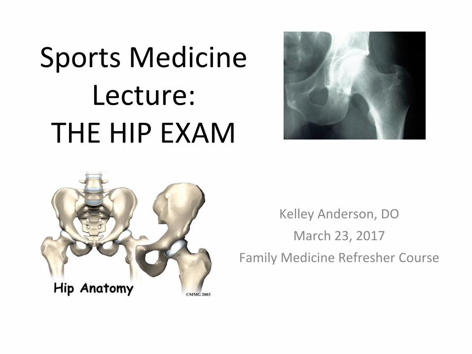
Sports Medicine Lecture:
THE HIP EXAM
Kelley Anderson, DO
March 23, 2017
Family Medicine Refresher Course
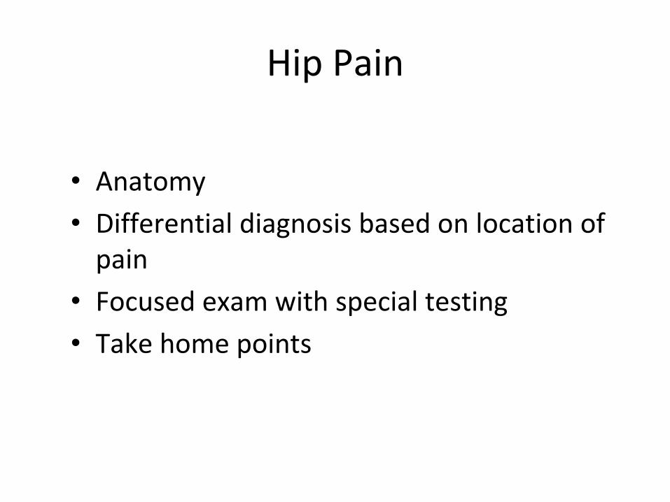
Hip Pain
• Anatomy
• Differential diagnosis based on location of pain
• Focused exam with special testing
• Take home points
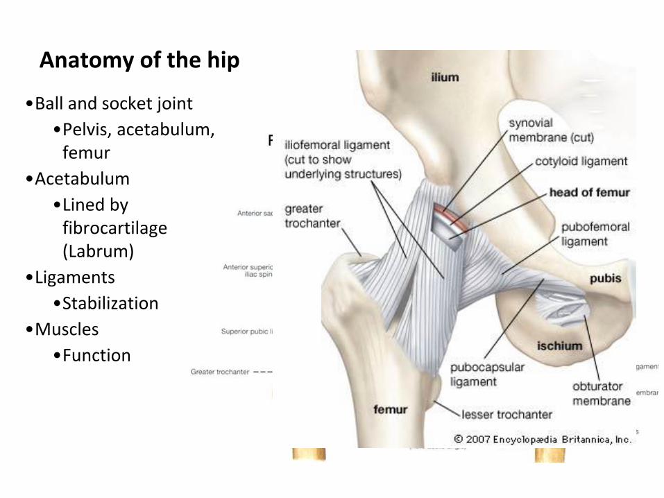
Anatomy of the hip
•Ball and socket joint
•Pelvis, acetabulum, femur
•Acetabulum
•Lined by fibrocartilage (Labrum)
•Ligaments
•Stabilization
•Muscles
•Function
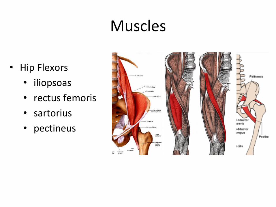
Muscles
• Hip Flexors
• iliopsoas
• rectus femoris
• sartorius
• pectineus
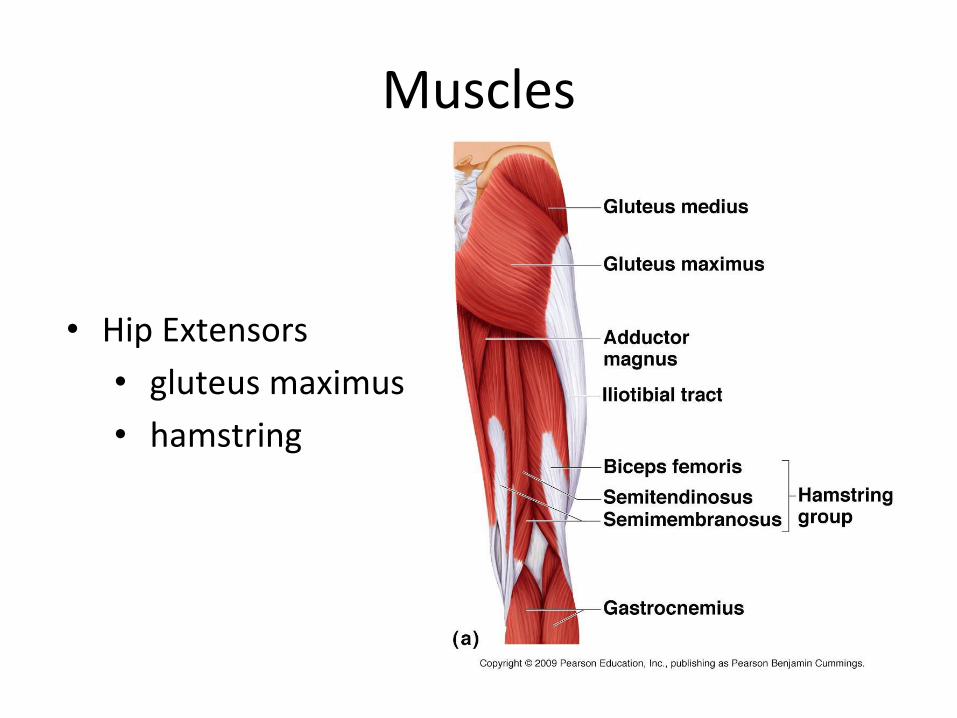
Muscles
• Hip Extensors
• gluteus maximus
• hamstring
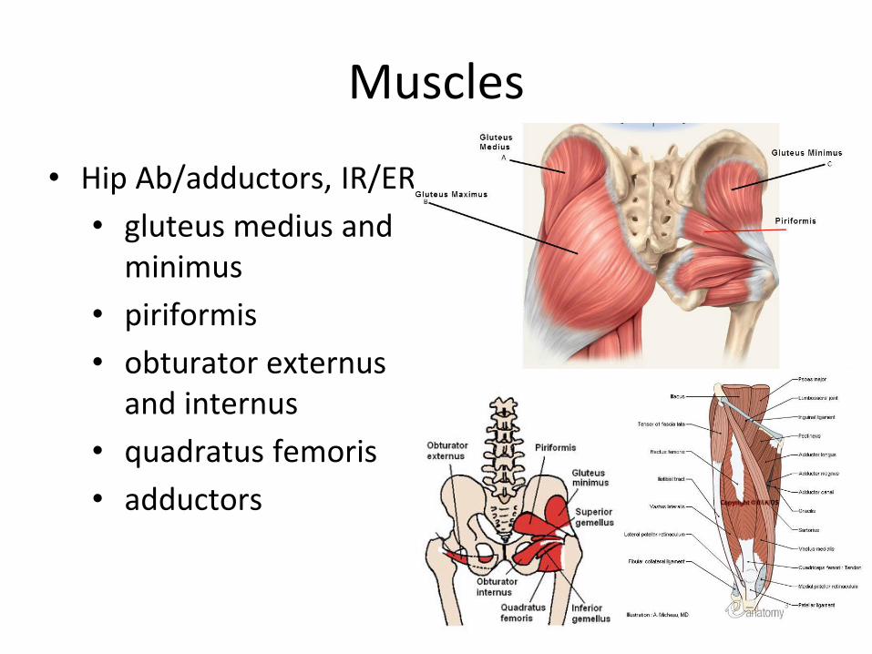
Muscles
• Hip Ab/adductors, IR/ER
• gluteus medius and minimus
• piriformis
• obturator externus and internus
• quadratus femoris
• adductors
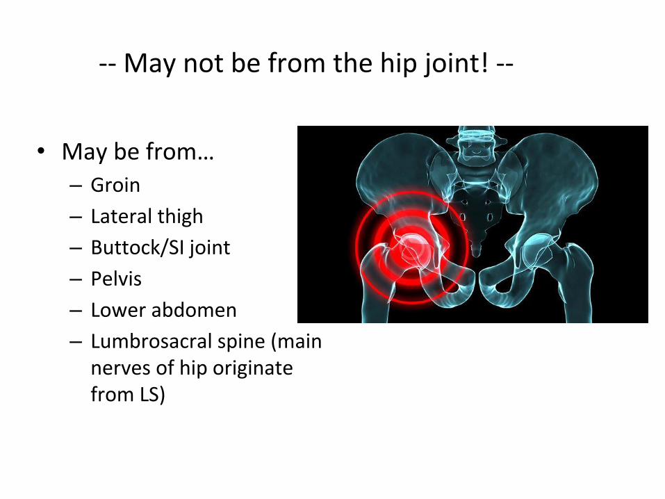
• May be from… – Groin
– Lateral thigh
– Buttock/SI joint
– Pelvis
– Lower abdomen
– Lumbrosacral spine (main nerves of hip originate from LS)
-- May not be from the hip joint! --
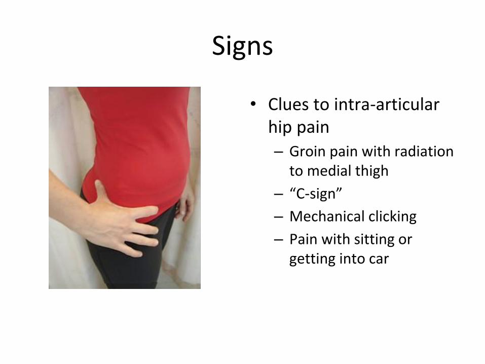
Signs
• Clues to intra-articular hip pain – Groin pain with radiation
to medial thigh
– “C-sign”
– Mechanical clicking
– Pain with sitting or getting into car
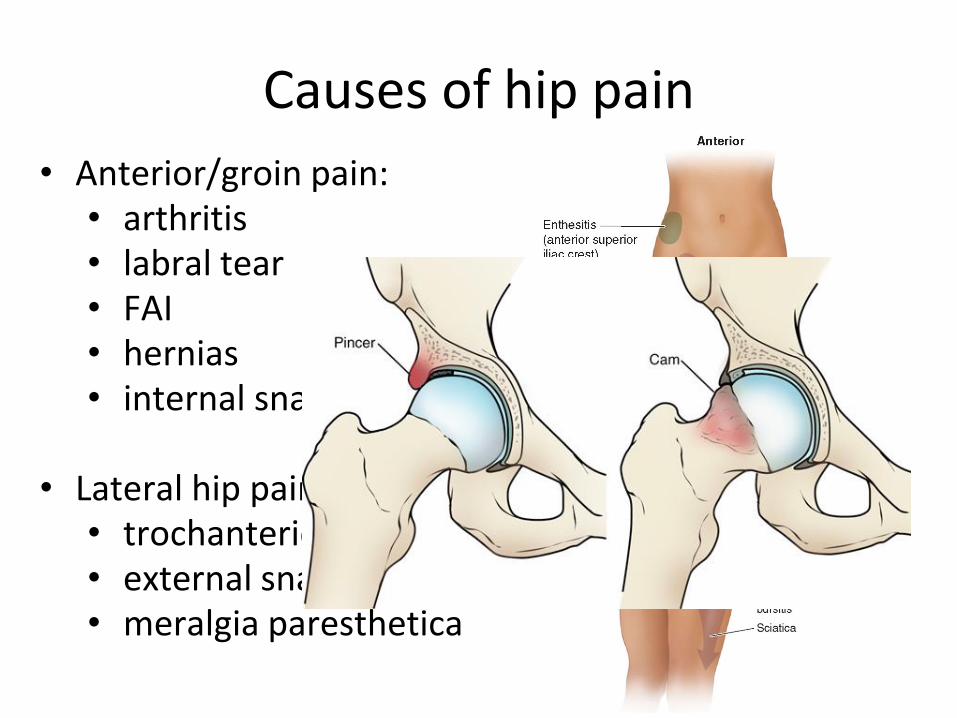
• Anterior/groin pain: • arthritis • labral tear • FAI • hernias • internal snapping hip
• Lateral hip pain:
• trochanteric bursitis • external snapping hip • meralgia paresthetica
Causes of hip pain
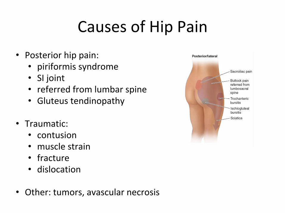
Causes of Hip Pain
• Posterior hip pain: • piriformis syndrome • SI joint • referred from lumbar spine • Gluteus tendinopathy
• Traumatic:
• contusion • muscle strain • fracture • dislocation
• Other: tumors, avascular necrosis
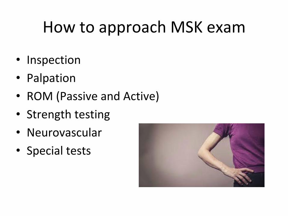
How to approach MSK exam
• Inspection
• Palpation
• ROM (Passive and Active)
• Strength testing
• Neurovascular
• Special tests
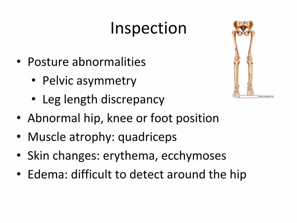
Inspection
• Posture abnormalities
• Pelvic asymmetry
• Leg length discrepancy
• Abnormal hip, knee or foot position
• Muscle atrophy: quadriceps
• Skin changes: erythema, ecchymoses
• Edema: difficult to detect around the hip
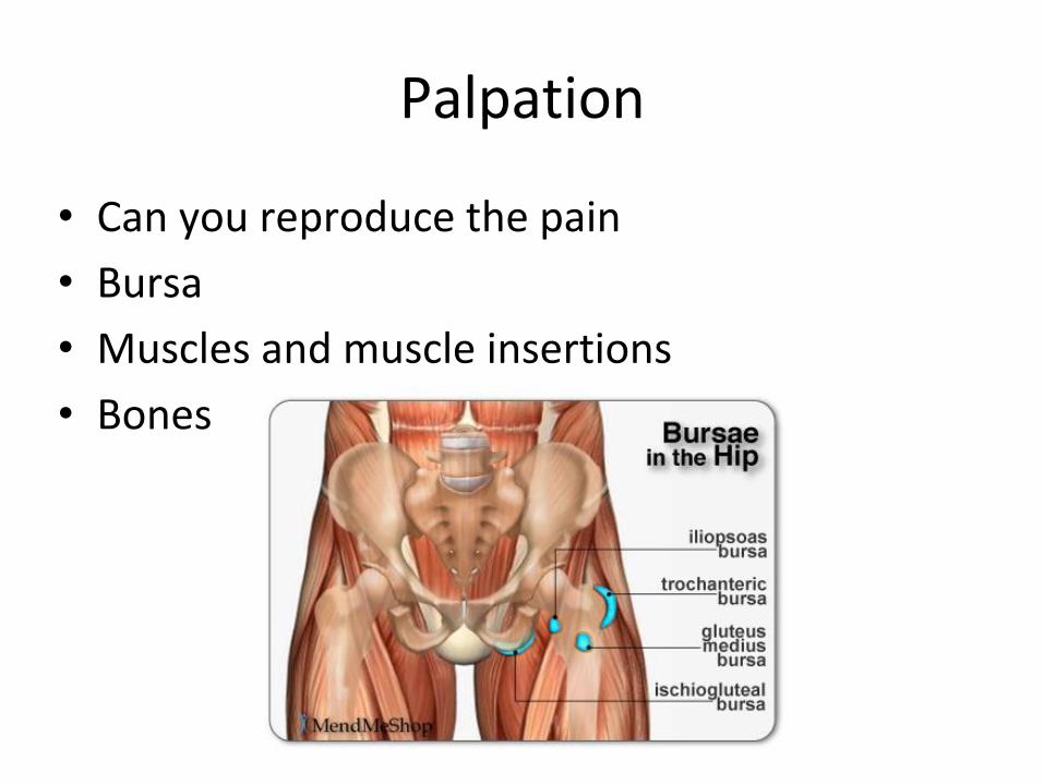
Palpation
• Can you reproduce the pain
• Bursa
• Muscles and muscle insertions
• Bones
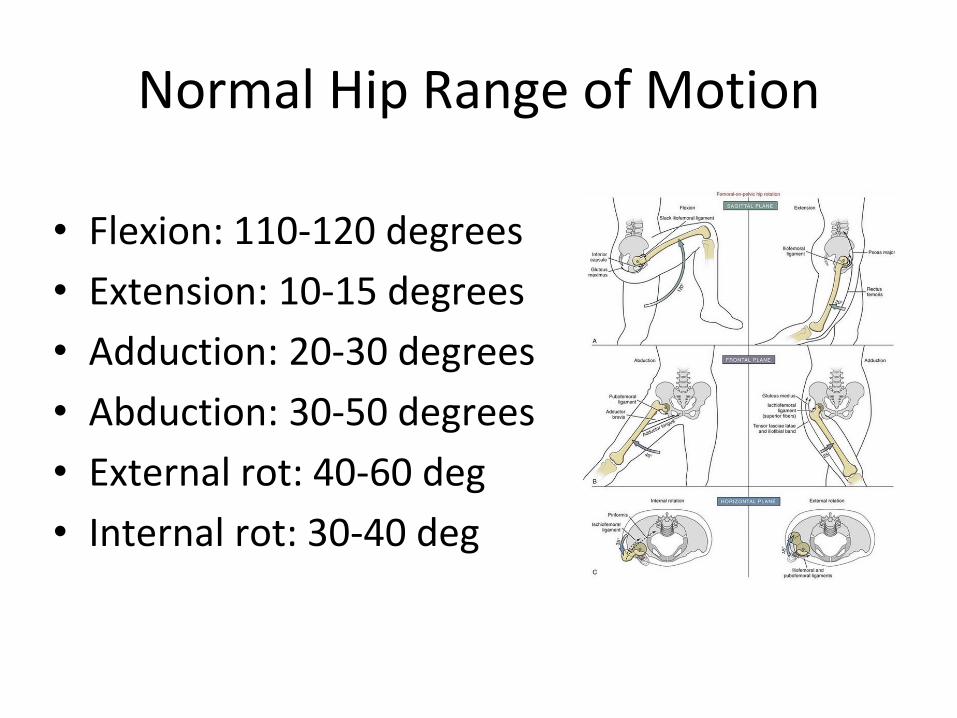
Normal Hip Range of Motion
• Flexion: 110-120 degrees
• Extension: 10-15 degrees
• Adduction: 20-30 degrees
• Abduction: 30-50 degrees
• External rot: 40-60 deg
• Internal rot: 30-40 deg
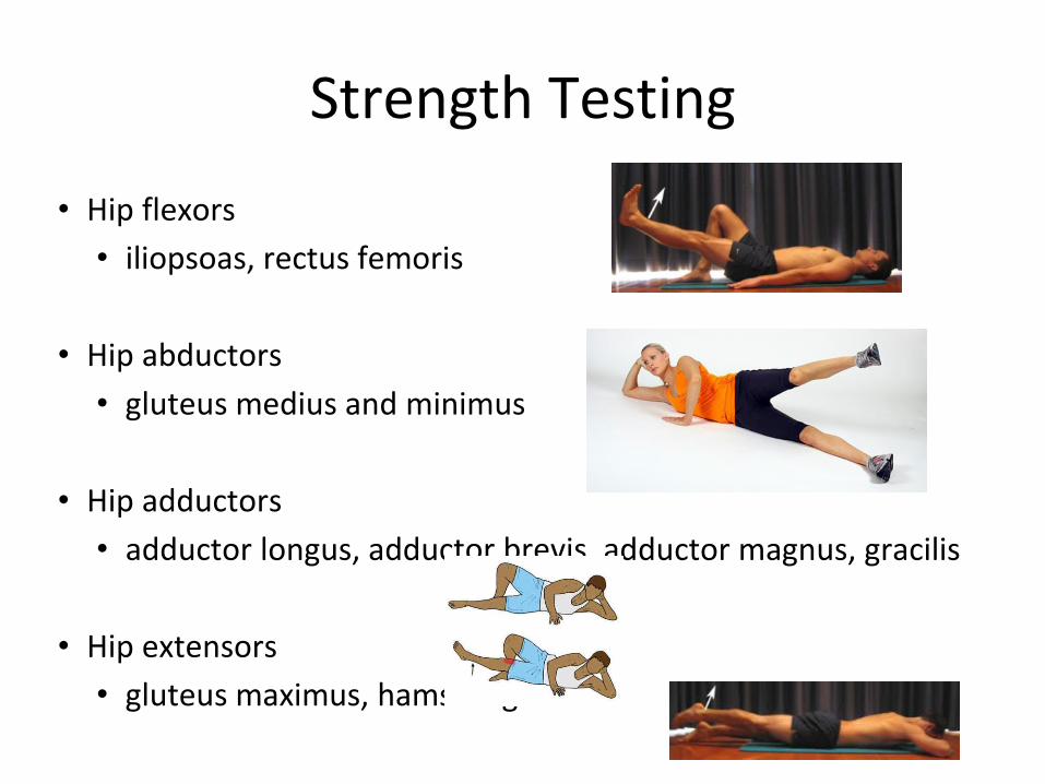
Strength Testing
• Hip flexors
• iliopsoas, rectus femoris
• Hip abductors
• gluteus medius and minimus
• Hip adductors
• adductor longus, adductor brevis, adductor magnus, gracilis
• Hip extensors
• gluteus maximus, hamstring
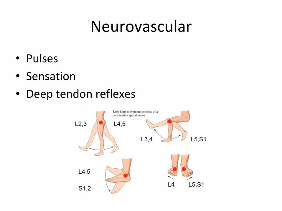
Neurovascular
• Pulses
• Sensation
• Deep tendon reflexes

SPECIAL TESTS!
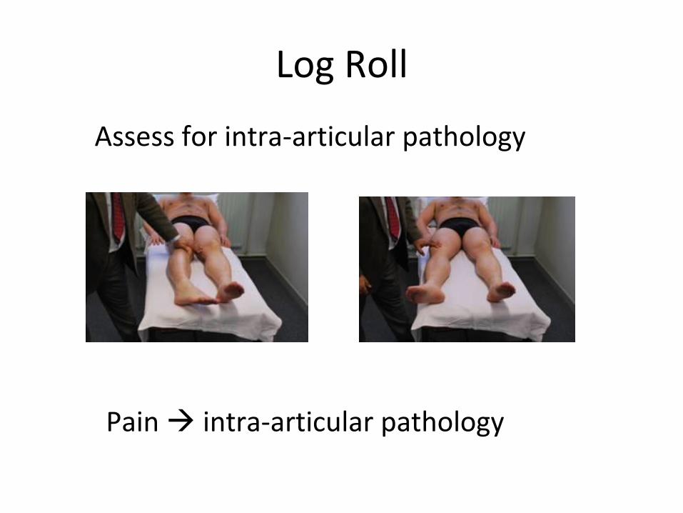
Log Roll
Pain intra-articular pathology
Assess for intra-articular pathology
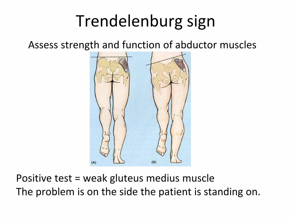
Trendelenburg sign
Positive test = weak gluteus medius muscle The problem is on the side the patient is standing on.
Assess strength and function of abductor muscles
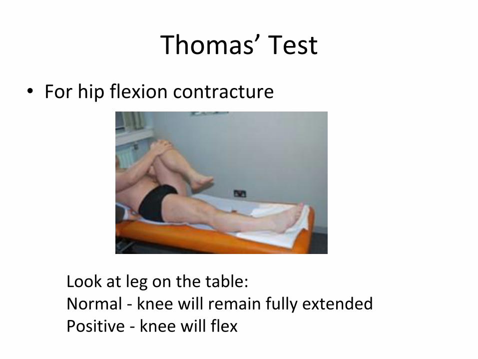
Thomas’ Test
• For hip flexion contracture
Look at leg on the table: Normal - knee will remain fully extended Positive - knee will flex
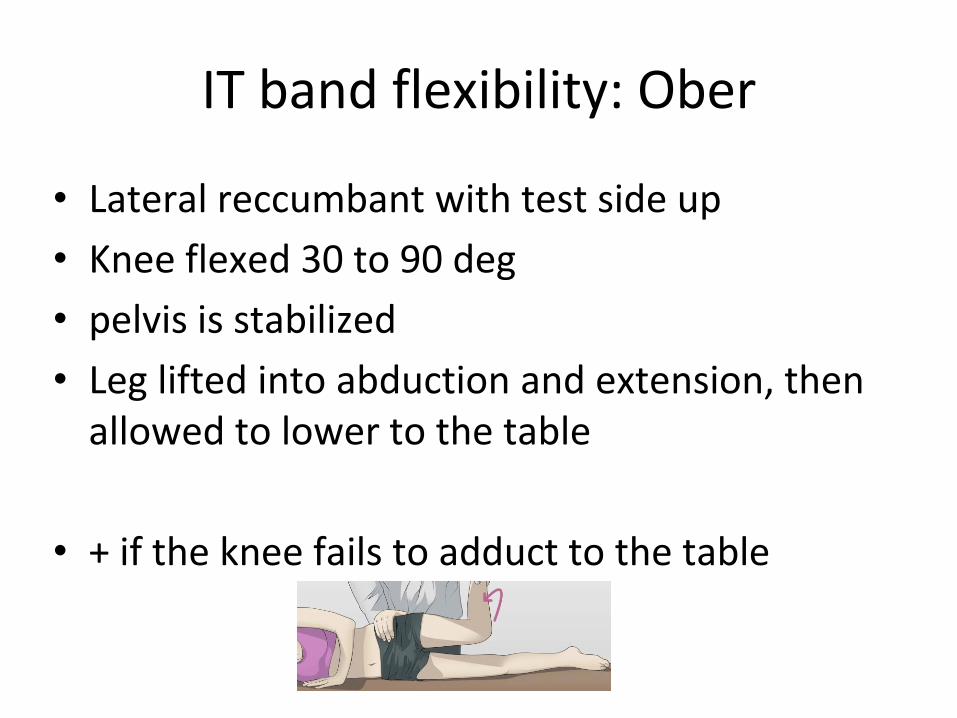
IT band flexibility: Ober
• Lateral reccumbant with test side up
• Knee flexed 30 to 90 deg
• pelvis is stabilized
• Leg lifted into abduction and extension, then allowed to lower to the table
• + if the knee fails to adduct to the table
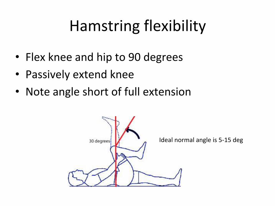
Hamstring flexibility
• Flex knee and hip to 90 degrees
• Passively extend knee
• Note angle short of full extension
Ideal normal angle is 5-15 deg
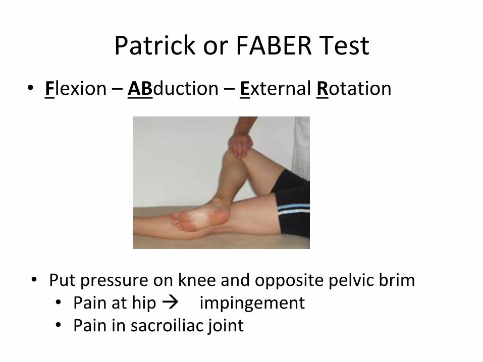
Patrick or FABER Test
• Flexion – ABduction – External Rotation
• Put pressure on knee and opposite pelvic brim • Pain at hip impingement • Pain in sacroiliac joint
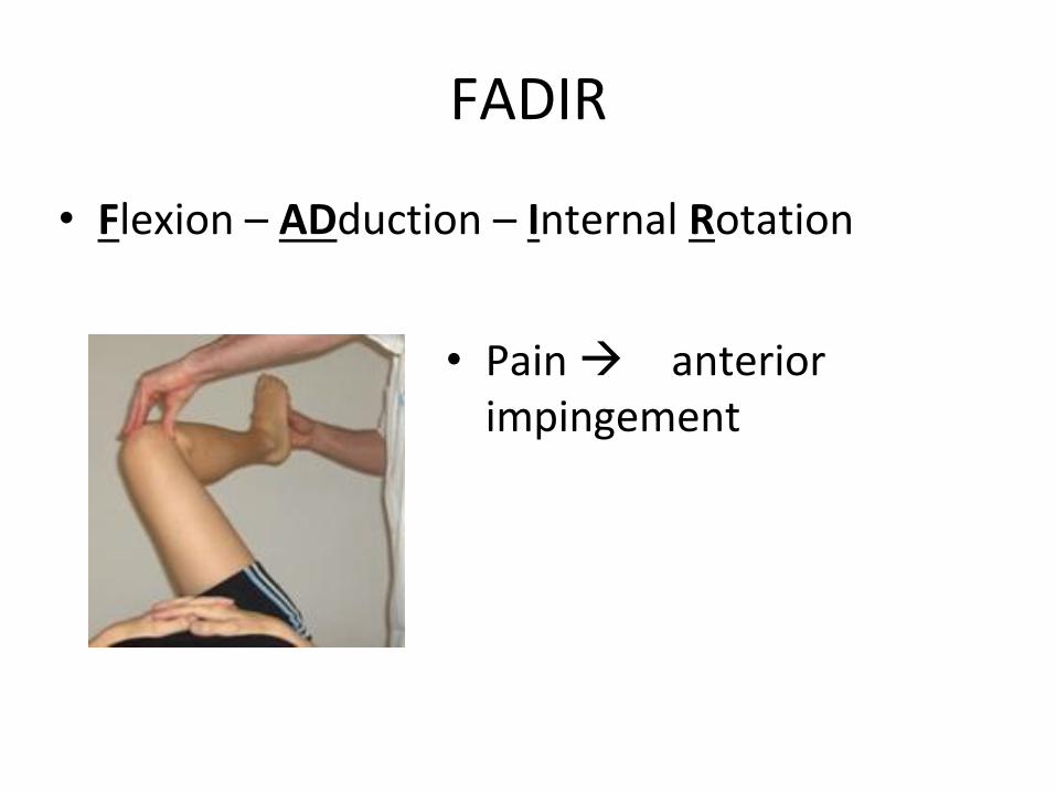
FADIR
• Flexion – ADduction – Internal Rotation
• Pain anterior impingement
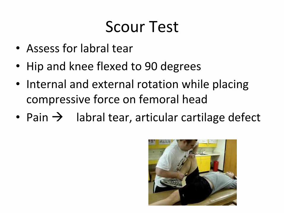
Scour Test • Assess for labral tear
• Hip and knee flexed to 90 degrees
• Internal and external rotation while placing compressive force on femoral head
• Pain labral tear, articular cartilage defect
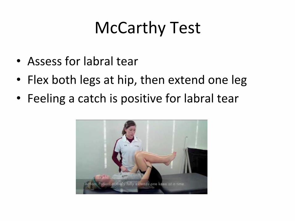
McCarthy Test
• Assess for labral tear
• Flex both legs at hip, then extend one leg
• Feeling a catch is positive for labral tear
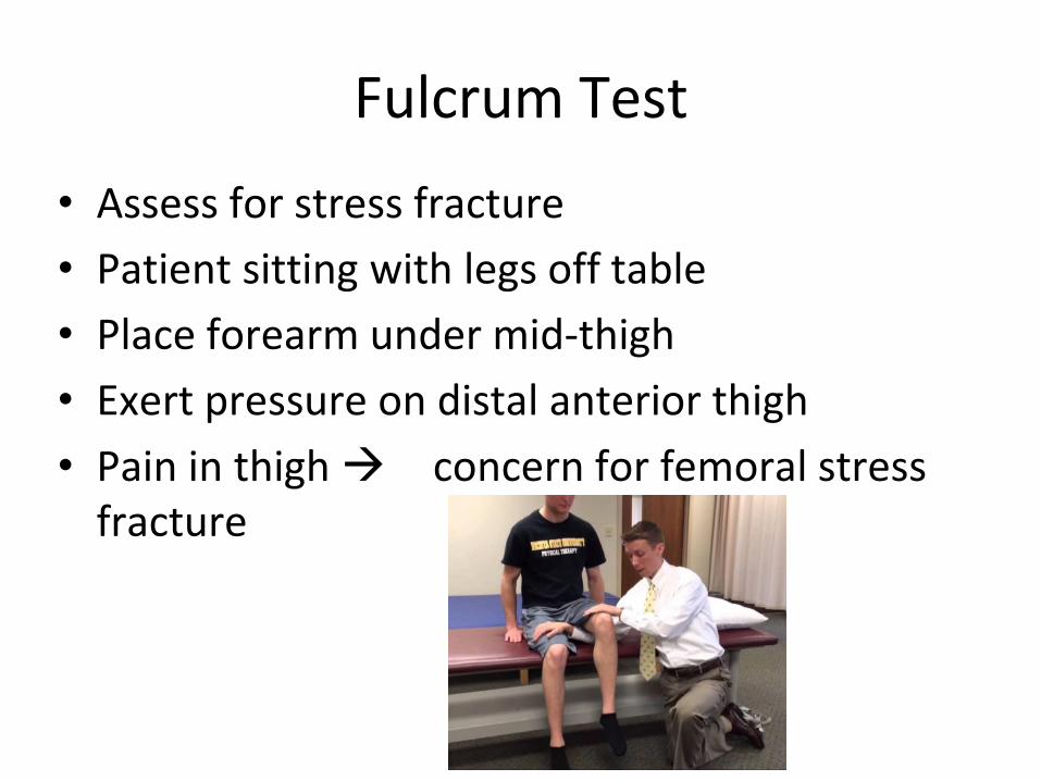
Fulcrum Test
• Assess for stress fracture
• Patient sitting with legs off table
• Place forearm under mid-thigh
• Exert pressure on distal anterior thigh
• Pain in thigh concern for femoral stress fracture
Take Home Points
• Get a good history
• Assess location of pain
• Watch the patient walk
• Inspect
• Palpate
• Strength and flexibility testing
• Special tests
Questions