Embed Size (px)
Citation preview
Classification of Injuries
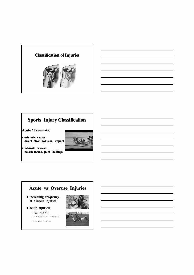
Sports Injury Classification
Acute / Traumatic
• extrinsic causes: direct blow, collision, impact • intrinsic causes: muscle forces, joint loadings
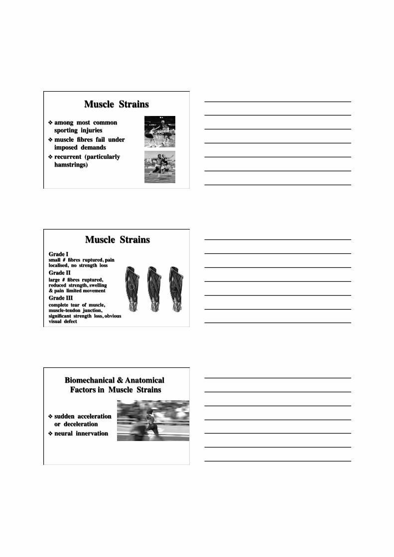
Acute vs Overuse Injuries v increasing frequency
of overuse injuries
v acute injuries: high velocity uncontrolled impacts macro-trauma
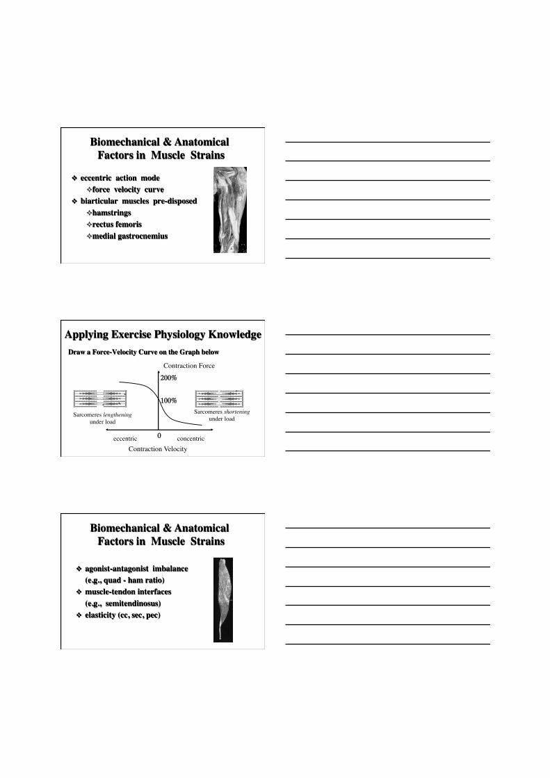
v among most common sporting injuries
v muscle fibres fail under imposed demands
v recurrent (particularly hamstrings)
Muscle Strains
Muscle Strains Grade I small # fibres ruptured, pain localised, no strength loss Grade II large # fibres ruptured, reduced strength, swelling & pain limited movement Grade III complete tear of muscle, muscle-tendon junction, significant strength loss, obvious visual defect
v sudden acceleration or deceleration
v neural innervation
Biomechanical & Anatomical Factors in Muscle Strains
v eccentric action mode ² force velocity curve
v biarticular muscles pre-disposed ² hamstrings ² rectus femoris ² medial gastrocnemius
Biomechanical & Anatomical Factors in Muscle Strains
Applying Exercise Physiology Knowledge Draw a Force-Velocity Curve on the Graph below
0 eccentric concentric
100%
200%
Contraction Velocity
Contraction Force
Sarcomeres lengthening under load
Sarcomeres shortening under load
v agonist-antagonist imbalance (e.g., quad - ham ratio)
v muscle-tendon interfaces (e.g., semitendinosus)
v elasticity (cc, sec, pec)
Biomechanical & Anatomical Factors in Muscle Strains
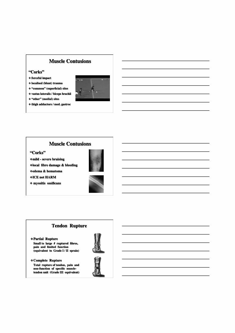
Muscle Contusions
“Corks” v forceful impact v localised (blunt) trauma
v “common” (superficial) sites
v vastus lateralis / biceps brachii v “other” (medial) sites
v thigh adductors / med. gastroc
Muscle Contusions “Corks” v mild - severe bruising v local fibre damage & bleeding
v edema & hematoma
v ICE not HARM v myositis ossificans
Tendon Rupture
v Partial Rupture Small to large # ruptured fibres, pain and limited function (equivalent to Grade I / II sprain)
v Complete Rupture Total rupture of tendon, pain and non-function of specific muscle-tendon unit (Grade III equivalent)
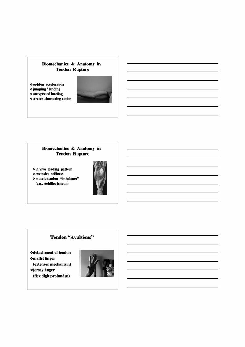
Biomechanics & Anatomy in Tendon Rupture
v sudden acceleration v jumping / landing v unexpected loading v stretch-shortening action
Biomechanics & Anatomy in Tendon Rupture
v in vivo loading pattern v excessive stiffness v muscle-tendon “imbalance” (e.g., Achilles tendon)
Tendon “Avulsions”
v detachment of tendon v mallet finger (extensor mechanism)
v jersey finger (flex digit profundus)
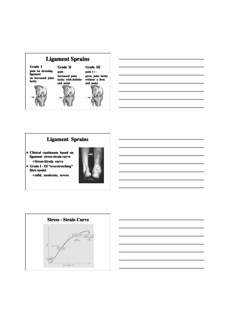
Ligament Sprains Grade I pain on stressing ligament no increased joint laxity
Grade II pain increased joint laxity with definite end point
Grade III pain + / - gross joint laxity without a firm end point
v Clinical continuum based on ligament stress-strain curve ² Stress-Strain curve
v Grade I - III “overstretching” fibre model ² mild, moderate, severe
Ligament Sprains
Stress - Strain Curve
Classic Ankle Inversion Sprain
Rupture of Joint Capsule / Ligaments���(AC Injury Examination under Anesthesia)
v AC Injuries: Type I - VI (Rockwood classification)
v joint capsule v acromioclavicular lig’s v coracoclavicular lig’s trapezoid conoid
v single, debilitating episode ² e.g., Knee ² ACL ² ACL + L/MCL ² ACL + L/MCL + Meniscus ² ACL + PCL
Ligament Sprains
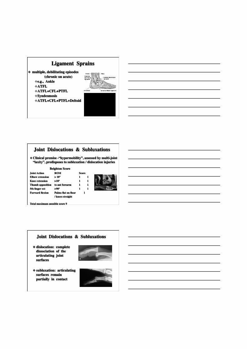
v multiple, debilitating episodes (chronic on acute)
² e.g., Ankle ² ATFL ² ATFL+CFL+PTFL ² Syndesmosis ² ATFL+CFL+PTFL+Deltoid
Ligament Sprains
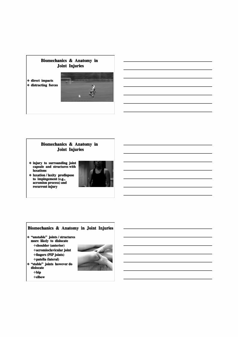
v Clinical premise -“hypermobility”, assessed by multi-joint “laxity”, predisposes to subluxation / dislocation injuries
Joint Dislocations & Subluxations
Joint Action ROM Score Elbow extension > 10° 1 1 Knee extension >10° 1 1 Thumb apposition to ant forearm 1 1 5th finger ext >90° 1 1 Forward flexion Palms flat on floor 1 / knees straight
Total maximum possible score 9
Beighton Score
Joint Dislocations & Subluxations v dislocation: complete
dissociation of the articulating joint surfaces
v subluxation: articulating surfaces remain partially in contact
Biomechanics & Anatomy in Joint Injuries
v direct impacts v distracting forces
Biomechanics & Anatomy in Joint Injuries
v injury to surrounding joint capsule and structures with luxations
v luxation / laxity predispose to impingement (e.g., acromion process) and recurrent injury
Biomechanics & Anatomy in Joint Injuries
v “unstable” joints / structures more likely to dislocate ² shoulder (anterior) ² acromioclavicular joint ² fingers (PIP joints) ² patella (lateral)
v “stable” joints however do dislocate ² hip ² elbow
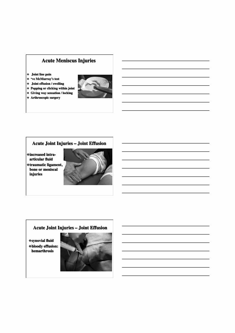
Acute Meniscus Injuries
v Joint line pain v +ve McMurray’s test v Joint effusion / swelling v Popping or clicking within joint v Giving way sensation / locking v Arthroscopic surgery
Acute Joint Injuries – Joint Effusion
v increased intra-articular fluid
v traumatic ligament, bone or meniscal injuries
Acute Joint Injuries – Joint Effusion
v synovial fluid v bloody effusion:
hemarthrosis
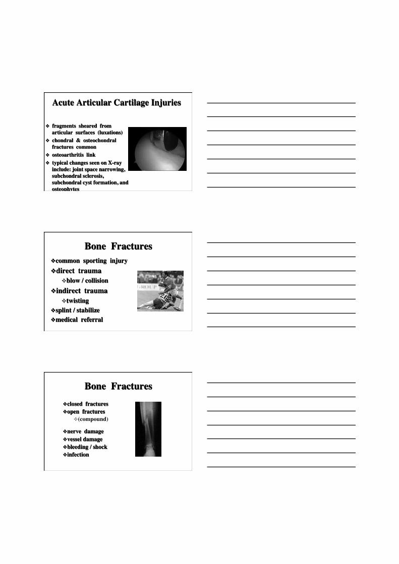
Acute Articular Cartilage Injuries
v fragments sheared from articular surfaces (luxations)
v chondral & osteochondral fractures common
v osteoarthritis link v typical changes seen on X-ray
include: joint space narrowing, subchondral sclerosis, subchondral cyst formation, and osteophytes
v better detection (MRI, CT) v arthroscopic surgery
Bone Fractures v common sporting injury v direct trauma
² blow / collision v indirect trauma
² twisting v splint / stabilize v medical referral
Bone Fractures v closed fractures v open fractures
² (compound)
v nerve damage v vessel damage v bleeding / shock v infection
Simple & Complex Fractures
Peak Fracture Prevalence in Adolescence
Collés Fracture
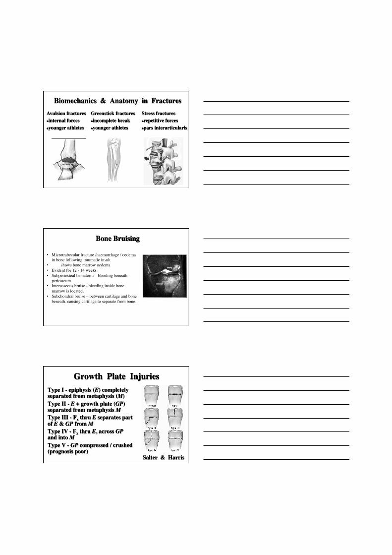
Biomechanics & Anatomy in Fractures Avulsion fractures l internal forces l younger athletes
Stress fractures l repetitive forces l pars interarticularis
Greenstick fractures l incomplete break l younger athletes
Bone Bruising
• Microtrabecular fracture /haemorrhage / oedema in bone following traumatic insult
• MRI shows bone marrow oedema • Evident for 12 - 14 weeks • Subperiosteal hematoma - bleeding beneath
periosteum. • Interosseous bruise - bleeding inside bone
marrow is located. • Subchondral bruise – between cartilage and bone
beneath, causing cartilage to separate from bone.
Growth Plate Injuries
Salter & Harris
Type I - epiphysis (E) completely separated from metaphysis (M) Type II - E + growth plate (GP) separated from metaphysis M Type III - Fx thru E separates part of E & GP from M Type IV - Fx thru E, across GP and into M Type V - GP compressed / crushed (prognosis poor)
Overuse Injuries
v repetitive microtrauma exceeds tissue repair capacity
v ⇑ prostaglandin E2 in tissues - ⇑ collagenase, ⇓ collagen synthesis
v upregulation of genes for cartilage, down-regulation of genes for tendon (rat) ⇒ tendon morphology alters to more cartilaginous.
Overuse Injuries
v important to identify / consider risk factors (RF)
v addressing intrinsic / extrinsic RF may help prevent re-injury
v often recalcitrant to treatment (months to resolve)
Overuse Risk Factors v Extrinsic Factors
² Training Errors ² Technique Errors ² Surfaces ² Shoes ² Equipment
Overuse Risk Factors v Intrinsic Factors
² Previous Injury ² Lack of Flexibility ² Leg Length Discrepancy ² Malalignment tibial torsion / vara genu valgum / varum
Overuse Risk Factors v Intrinsic Factors
² Malalignment patella alta pes planus / cavus
² Muscle Imbalance ² Muscle Weakness
In the News

Tendinopathies (Overuse)
v pathology is tendon (collagen) degeneration (tendinosis) not inflammation
v surrounding structures may
have inflammation (paratendinitis)
Multiple tendons (Supraspinatus, ECRB, Achilles) v Collagen disarray v Absent cells, prolific cells
v Abnormal vessels & nerves
v Abnormal extracellular matrix
Pathological Findings Normal → Abnormal
Dealing with Tendinopathies (Overuse Injuries)
v History (onset, nature, potential causes)
v Examination (anatomical structure, reproduce pain) Diagnosis required!
v Treatment v Relative rest / maintain fitness v Avoid aggravating activities v Pharmacology (NSAIDs??, GTN) v Rehab (eccentric strengthening) Try to find intrinsic / extrinsic causes!
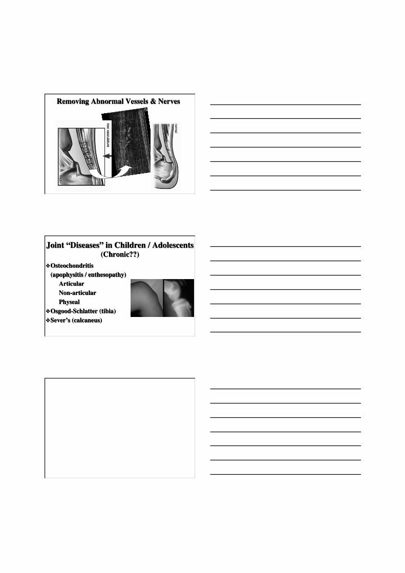
Removing Abnormal Vessels & Nerves
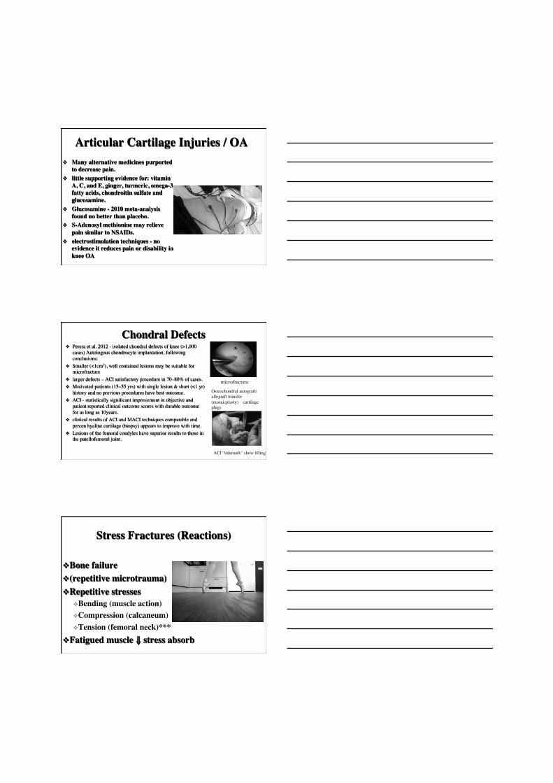
Joint “Diseases” in Children / Adolescents���(Chronic??)
v Osteochondritis (apophysitis / enthesopathy) Articular Non-articular Physeal
v Osgood-Schlatter (tibia) v Sever’s (calcaneus)
Articular Cartilage Injuries / OA v Many alternative medicines purported
to decrease pain. v little supporting evidence for: vitamin
A, C, and E, ginger, turmeric, omega-3 fatty acids, chondroitin sulfate and glucosamine.
v Glucosamine - 2010 meta-analysis found no better than placebo.
v S-Adenosyl methionine may relieve pain similar to NSAIDs.
v electrostimulation techniques - no evidence it reduces pain or disability in knee OA
Chondral Defects v Perera et al. 2012 - isolated chondral defects of knee (>1,000
cases) Autologous chondrocyte implantation, following conclusions:
v Smaller (<1cm2), well contained lesions may be suitable for microfracture
v larger defects – ACI satisfactory procedure in 70–80% of cases. v Motivated patients (15–55 yrs) with single lesion & short (<1 yr)
history and no previous procedures have best outcome. v ACI - statistically significant improvement in objective and
patient reported clinical outcome scores with durable outcome for as long as 10years.
v clinical results of ACI and MACI techniques comparable and percen hyaline cartilage (biopsy) appears to improve with time.
v Lesions of the femoral condyles have superior results to those in the patellofemoral joint.
microfracture
Osteochondral autograft/allograft transfer (mosaicplasty) – cartilage plugs
ACI “tidemark” show filling
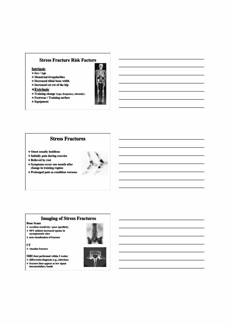
Stress Fractures (Reactions)
v Bone failure v (repetitive microtrauma) v Repetitive stresses
² Bending (muscle action) ² Compression (calcaneum) ² Tension (femoral neck)***
v Fatigued muscle ⇓ stress absorb
Stress Fracture Risk Factors
Intrinsic v Sex / Age v Menstrual irregularities v Decreased tibial bone width v Increased ext rot of the hip v Extrinsic v Training change (type, frequency, intensity) v Footwear / Training surface v Equipment
Stress Fractures
v Onset usually insidious v Initially pain during exercise v Relieved by rest v Symptoms occur one month after
change in training regime v Prolonged pain as condition worsens
Imaging of Stress Fractures Bone Scans v excellent sensitivity / poor specificity v 50% athletes increased uptake in
asymptomatic sites v non-visualization of fracture
CT v visualise fracture
MRI (best performed within 3 weeks) v differential diagnosis (e.g., infection) v fracture lines appear as low signal
intramedullary bands
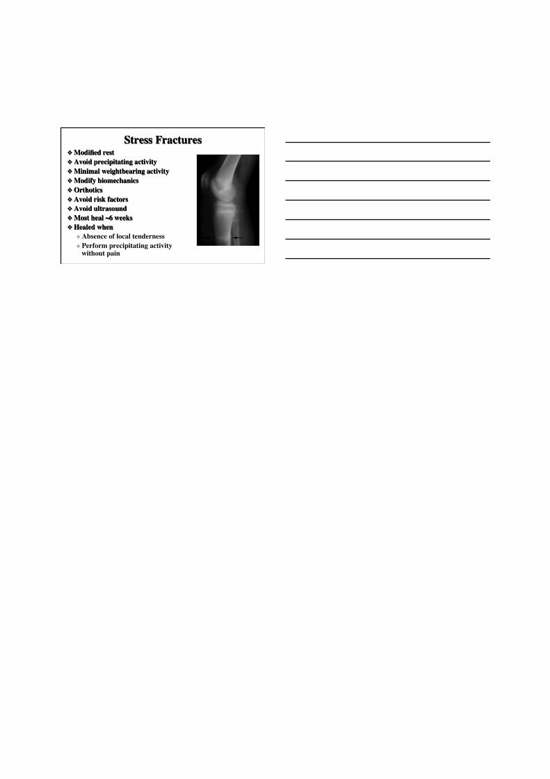
Stress Fractures v Modified rest v Avoid precipitating activity v Minimal weightbearing activity v Modify biomechanics v Orthotics v Avoid risk factors v Avoid ultrasound v Most heal ~6 weeks v Healed when
² Absence of local tenderness ² Perform precipitating activity
without pain