Embed Size (px)
Citation preview

dentistry journal
Review
Sport and Dental Traumatology: Surgical Solutionsand Prevention
Lorenzo Mordini 1,* , Po Lee 1, Ricardo Lazaro 1, Roberto Biagi 2 and Luca Giannetti 3
�����������������
Citation: Mordini, L.; Lee, P.; Lazaro,
R.; Biagi, R.; Giannetti, L. Sport and
Dental Traumatology: Surgical
Solutions and Prevention. Dent. J.
2021, 9, 33. https://doi.org/10.3390/
dj9030033
Academic Editor: Gabriel Krastl
Received: 8 February 2021
Accepted: 17 March 2021
Published: 23 March 2021
Publisher’s Note: MDPI stays neutral
with regard to jurisdictional claims in
published maps and institutional affil-
iations.
Copyright: © 2021 by the authors.
Licensee MDPI, Basel, Switzerland.
This article is an open access article
distributed under the terms and
conditions of the Creative Commons
Attribution (CC BY) license (https://
creativecommons.org/licenses/by/
4.0/).
1 Department of Periodontology, Tufts University School of Dental Medicine, 1 Kneeland Street,Boston, MA 02111, USA; [email protected] (P.L.); [email protected] (R.L.)
2 Department of Biomedical, Surgical and Dental Sciences, School of Dentistry, University of Milan,Via Della Commenda, 10-20122 Milan, Italy; [email protected]
3 Department of Dentistry and Oral Maxillofacial Surgery, University of Modena and Reggio Emilia,Via Del Pozzo n◦, 41-41124 Modena, Italy; [email protected]
* Correspondence: [email protected]
Abstract: Trauma is a worldwide cause of millions of deaths and severe injuries every year, all overthe world. Despite the limited extension of the oral region compared to the whole body, dental andoral injuries account for a fairly high percentage of all body traumas. Among head and neck traumas,dental and facial injuries are highly correlated to sport activities, and their management can be areal challenge for practitioners of any specialty. In case of trauma directed to periodontal structures,restorative and endodontic solutions may not be sufficient to achieve a definitive and long-lastingtreatment. This article aims to illustrate surgical options and appliances to prevent dental injuriesthat may be available to the clinicians treating dental trauma involving oral soft and hard tissues.
Keywords: dental trauma; periodontology; dental implants; facial injury; tooth auto-transplantation
1. Introduction
Every year, trauma is a worldwide cause of more than 5 million deaths for individualsup to the age of 45 years, causing half of all deaths in the age range of 10–24 years [1].
Despite the oral region representing 1% of the human body, injuries occurring inthe oral region reach 5% of total bodily injuries among all ages, as shown by a one-yearlongitudinal prospective Swedish survey [2]. In Swedish newborn children up to 6 yearsof age, injuries to the oral cavity reach 17% of all bodily injuries. Due to their behavioralinclination, the oral region is at a higher risk in children and adolescents, compared toadults and elderly. In fact, maxillofacial trauma represented 33% of all types of trauma, asreported from hospital accident and emergency departments [3].
Dental and facial injuries are highly correlated to sport activities. Unfortunately, thereis a high disparity of dental trauma definitions among scientific literature, which makesit difficult to outline an overall true prevalence [4]. As one could imagine, it has beenreported that trauma as a result of sporting activities represents up to a third of all orofacialinjuries (31%) [5,6]. In particular, 50.1% of those are traumatic dental injuries (TDI) [5,6].Among contact sports athletes, the prevalence of TDI varies between 7.1% and 71.5% [7].Despite the significant variation, studies have indicated that the prevalence of trauma isless than 40%, depending on the type of sport practiced [8]. In other words, the incidencevaries between the different types of sport. The most common TDI occur in the maxillaryincisors, accounting for 80% of all cases [9,10]. Among patients reporting history of injuriesto the oral region, 92% presented with dental trauma, 28% soft tissue injuries and 6% withbone fractures [2]. It was not uncommon to see combinations of the above.
When injuries are restricted to the soft tissues, such as lacerations, abrasions, and con-tusions [11], they create wounds that usually heal without major complications. However,trauma to the dentofacial structures might result in serious injuries that often require tooth
Dent. J. 2021, 9, 33. https://doi.org/10.3390/dj9030033 https://www.mdpi.com/journal/dentistry
Dent. J. 2021, 9, 33 2 of 22
extraction, bone regeneration, and prosthetic replacements [4]. The consequences of theseinjuries can potentially cause severe pain, emotional and psychological impacts, as well aseconomic implications [12]. In fact, a study reported that the mean cost of maxillofacial anddental injuries was more than double that of all other bodily injuries occurred in contactsports [13]. When the dental trauma extends to the supporting periodontal apparatus,more extensive treatment may be required. The intervention of a specialist may be neededto restore not only dental structure but bone and soft tissue that was damaged duringthe injury.
This article aims to illustrate surgical options and the appliances to prevent dentalinjuries caused by sport activities; a review of techniques that may be available to theclinicians treating dental trauma involving soft tissue injuries and alveolar bone willbe presented.
2. Dental Trauma and Injuries2.1. Traumatic Dental Injuries (TDI)
Traumatic forces are considered one of the four most frequent oral diseases [14]. Theynot only affect tooth structure, but they can disrupt the supporting periodontal apparatus,including bone and peripheral soft tissues. TDI related to teeth include crown and/or rootfracture involving the pulp or not. Depending on the magnitude of the injury, teeth canalso experience different degrees of periodontal support alteration, such as concussion,sub-luxation, luxation, and avulsion [14]. Their specific definitions are listed in Table 1.
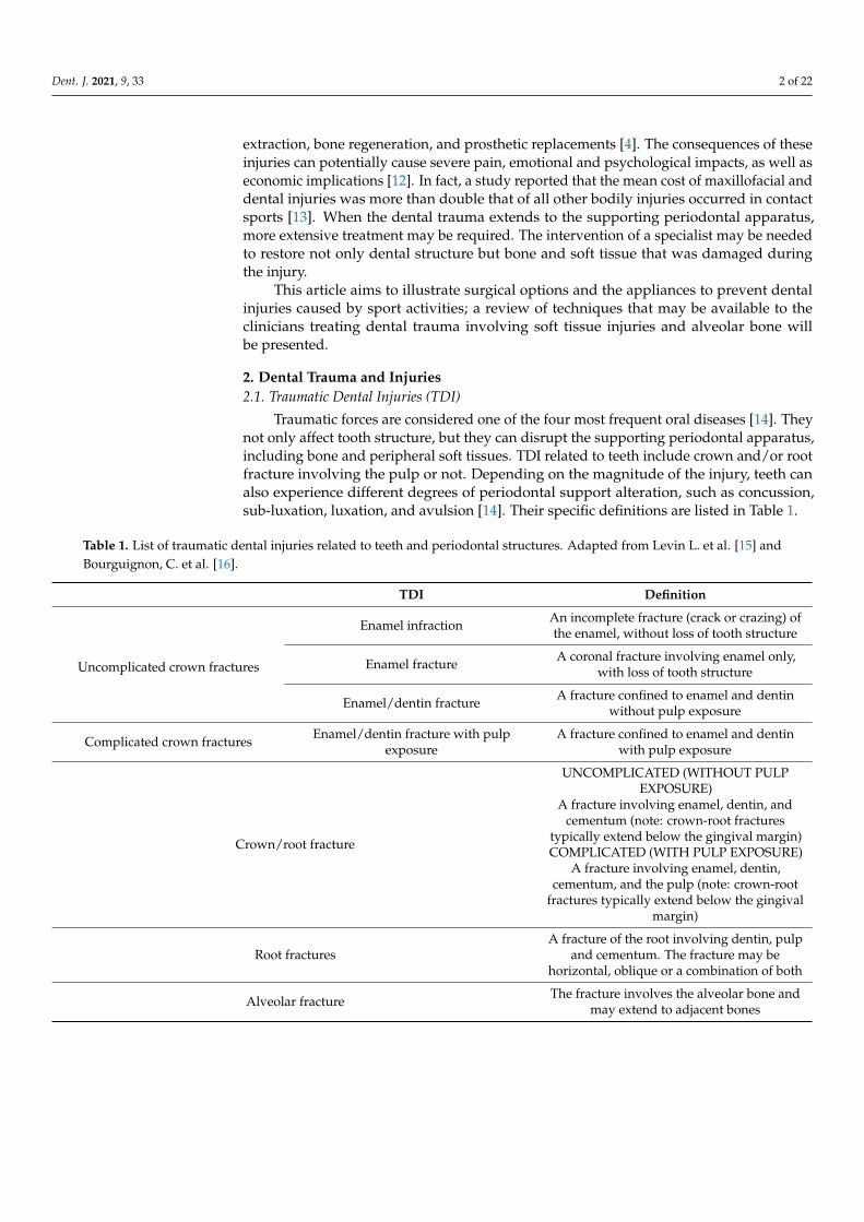
Table 1. List of traumatic dental injuries related to teeth and periodontal structures. Adapted from Levin L. et al. [15] andBourguignon, C. et al. [16].
TDI Definition
Uncomplicated crown fractures
Enamel infraction An incomplete fracture (crack or crazing) ofthe enamel, without loss of tooth structure
Enamel fracture A coronal fracture involving enamel only,with loss of tooth structure
Enamel/dentin fracture A fracture confined to enamel and dentinwithout pulp exposure
Complicated crown fractures Enamel/dentin fracture with pulpexposure
A fracture confined to enamel and dentinwith pulp exposure
Crown/root fracture
UNCOMPLICATED (WITHOUT PULPEXPOSURE)
A fracture involving enamel, dentin, andcementum (note: crown-root fractures
typically extend below the gingival margin)COMPLICATED (WITH PULP EXPOSURE)
A fracture involving enamel, dentin,cementum, and the pulp (note: crown-root
fractures typically extend below the gingivalmargin)
Root fracturesA fracture of the root involving dentin, pulp
and cementum. The fracture may behorizontal, oblique or a combination of both
Alveolar fracture The fracture involves the alveolar bone andmay extend to adjacent bones

Dent. J. 2021, 9, 33 3 of 22
Table 1. Cont.
TDI Definition
Concussion
An injury to the tooth-supporting structureswithout abnormal loosening or displacement
of the tooth, but with marked reaction topercussion
SubluxationAn injury to the tooth-supporting structures
with abnormal loosening, but withoutdisplacement of the tooth
Luxation
Extrusion Displacement of the tooth out of its socket inan incisal/axial direction
Lateral luxation
Displacement of the tooth in any lateraldirection, usually associated with a fractureor compression of the alveolar socket wall or
facial cortical bone
Intrusion Displacement of the tooth in an apicaldirection into the alveolar bone
Avulsion Complete displacement of the tooth out of itssocket
2.2. Prevalence2.2.1. Prevalence Contact Sports
Contact sports are considered activities which aim is to utilize physical contact tolead the team or the individual to win a competition. The physical contact during thesecompetitions is intense and, competitors a have a high risk for dentofacial injuries.
A systematic review and meta-analysis of 17 articles [17] showed a total prevalence ofdentofacial injuries of almost 30%. When single sports were evaluated, rugby presented aprevalence of almost 40%, basketball 27.26%, handball 24.59%, and field hockey 19.07%.Among all injuries, the most common was dental trauma (19.61%). The main limitationof this study was the heterogeneity within the selected studies. The major risk of bias ofthe studies reported in the systematic review was the small sample sizes, not ideal forprevalence studies (≤400). For this reason, it was not possible to generalize the prevalencein the general population. The same study reported that men under 30 who play sports forat least 4 h a week, have the highest risk of injuries related to sport [17].
It is interesting to know that there is no mandatory use of mouthguards for contactsports in USA.
The American Dental Association recommends the use of mouthguards for manysports, including basketball, martial arts, boxing, rugby, football, soccer, hockey, wrestling,lacrosse, and many others [18,19], but only the National Federation of State High SchoolAssociations (NFHS) [20] mandates the use of mouthguards for football, hockey, lacrosse,and wrestling (only if wearing braces).
These high data on prevalence are concerning, and they show the magnitude of dentalinjuries caused by sport activities. The general population can be affected by esthetic andfunctional issues, while professional athletes can see a reduction in their performancesand participation to games and matches. Data showed that ~60% of injuries caused forcedinactivity or occupational disability, damaging athletes’ activities and profession [21].
2.2.2. Prevalence Combat Sports
Many are the sports that can be listed in the “combat” category: boxing, judo, karate,kendo, kung fu, taekwondo, muay thai, sumo, capoeira, fencing, jiu-jitsu, wrestling, andwushu are the most famous around the world (Figure 1).

Dent. J. 2021, 9, 33 4 of 22
Dent. J. 2021, 9, x FOR PEER REVIEW 4 of 23
and functional issues, while professional athletes can see a reduction in their perfor-
mances and participation to games and matches. Data showed that ~60% of injuries
caused forced inactivity or occupational disability, damaging athletes’ activities and pro-
fession [21].
2.2.2. Prevalence Combat Sports
Many are the sports that can be listed in the “combat” category: boxing, judo, karate,
kendo, kung fu, taekwondo, muay thai, sumo, capoeira, fencing, jiu-jitsu, wrestling, and
wushu are the most famous around the world (Figure 1).
Figure 1. This radiograph shows tooth#24 horizontal fracture and radiolucency in the anterior
mandible, as a result of a blow received from the patient playing boxing.
Results from a recent systematic review and meta-analysis suggested a dental injury
pooled prevalence of ~25% and dentofacial injury pooled prevalence of 30.3% [7]. Individ-
ual sport analysis showed jiu-jitsu to have the highest combined prevalence of dentofacial
injuries (52.9%), while judo was the lowest (25%). Among sports that are popular in the
Americas, boxing and wrestling had the highest prevalence of dental injuries, reaching up
to 80.0% in some studies [7]. It has to be noted that the heterogeneity on the estimated
prevalence is high. Some studies obtained data from questionnaires on lifetime past his-
tory while only few studies considered a specific time (from 1 to 15 years) [7]. This sys-
tematic review and meta-analysis included papers with different population background
and skill level, such as competitors, non-, semi- and professionals, and elite athletes. As it
can be expected, professionals and competitive athletes showed higher prevalence of den-
tofacial injuries compared to non-professional ones. In fact, these top competitors reach
higher intensities during competitions and training. A role may be also played by higher
responsibilities and pressure deriving from sponsors and awards. Only one study re-
ported the highest prevalence rate of 41.4% of the African continent. The American conti-
nent ranked second for dentofacial and dental only injuries [7]. Once again, heterogeneity
at various levels represented a limitation. Age groups were highly heterogeneous, and
some athletes may have performed in different combat sport; both these aspects may have
influenced the real total prevalence. Despite lack of description regarding the type of den-
tofacial injuries reported, tooth fracture was the most common (6% to 50%). With regard
to dentofacial trauma, malar bone contusion (0.71–11%) and labial laceration (11–15%)
were the most prevalent [7].
Figure 1. This radiograph shows tooth#24 horizontal fracture and radiolucency in the anteriormandible, as a result of a blow received from the patient playing boxing.
Results from a recent systematic review and meta-analysis suggested a dental injurypooled prevalence of ~25% and dentofacial injury pooled prevalence of 30.3% [7]. Individ-ual sport analysis showed jiu-jitsu to have the highest combined prevalence of dentofacialinjuries (52.9%), while judo was the lowest (25%). Among sports that are popular in theAmericas, boxing and wrestling had the highest prevalence of dental injuries, reaching upto 80.0% in some studies [7]. It has to be noted that the heterogeneity on the estimatedprevalence is high. Some studies obtained data from questionnaires on lifetime past historywhile only few studies considered a specific time (from 1 to 15 years) [7]. This systematicreview and meta-analysis included papers with different population background and skilllevel, such as competitors, non-, semi- and professionals, and elite athletes. As it can beexpected, professionals and competitive athletes showed higher prevalence of dentofacialinjuries compared to non-professional ones. In fact, these top competitors reach higherintensities during competitions and training. A role may be also played by higher respon-sibilities and pressure deriving from sponsors and awards. Only one study reported thehighest prevalence rate of 41.4% of the African continent. The American continent rankedsecond for dentofacial and dental only injuries [7]. Once again, heterogeneity at variouslevels represented a limitation. Age groups were highly heterogeneous, and some athletesmay have performed in different combat sport; both these aspects may have influenced thereal total prevalence. Despite lack of description regarding the type of dentofacial injuriesreported, tooth fracture was the most common (6% to 50%). With regard to dentofacialtrauma, malar bone contusion (0.71–11%) and labial laceration (11–15%) were the mostprevalent [7].
2.3. Epidemiology
In most of the studies, boys experience dental traumas more often compared to girls,to ratios that reach up to 2.5:1. Boys’ permanent teeth are affected almost twice than girls’,most likely because they participate more actively and have more propensity for contactand combat games and sports [14,22,23]. In the latest years this trend has been levelled bygirls’ increased participation and competitive behavior in sports like hockey and soccerthat were once regarded as “boys’ games”, especially modern Western society [14,24]. Datafrom literature [14] demonstrated that most dental trauma occur during childhood andadolescence. It is estimated that up to 90% of all dental injuries sustained in a lifetime occurbefore the age of 19 years. Data showed how traumatic tooth injuries decrease after thetwenties. The higher risk of dental injuries among children that belong to higher socio-economic backgrounds, may be due related to easier access to bicycles, skiing, skateboards,horse-riding, and swimming pools than those from low socio-economic groups [14].

Dent. J. 2021, 9, 33 5 of 22
2.4. Teeth Involved
Not all individuals experiencing sports-related dental trauma are affected in thesame way. Some dental malocclusions (class II) are significant predisposing factors, suchas increased overjet with protrusion of upper incisors and insufficient lip closure [14].The World TDI prevalence in primary dentition, of patients up to 5 years of age, is 23%(17.3–29%); the prevalence on permanent dentition on patients younger than 30 years ofage is 15.5% (13.2–17.9%), with significant decreases afterwards [14].
The preponderance of dental injuries involves the anterior sextants, in particular themaxillary central incisors, while the maxillary lateral and mandibular central incisors areinvolved to a lesser degree. Dental injuries occurring during sport events, can result in asingle or multiple tooth injuries, especially among teenagers [14].
2.5. Etiology
Generally speaking, dental traumas are caused by a collision that can generate a highenergy force leading to an injury. This can derive from an object used for sports (ball,stick, car) or from another athlete’s body part or animal (horse hoofs). Depending on theentity and object of the force, the dental injury can be limited or extended. Physical leisureactivities can be associated to sports on their likelihood of dental injuries [14]. For instance,bicycling and skateboarding are the second most frequent injury cause among adolescents,being responsible for almost 20% of all traumatic events [14]. Sport is responsible forinjuries to permanent teeth, accounting for 13% of all injuries (Table 2).
Table 2. Prevalence of traumatic dental injuries for permanent and primary teeth around the world. On the first column,sport and physical activity are reported as the causes of injuries. The last column indicates the number of studies used forthis data. Adapted from “Textbook and Color Atlas of Traumatic Injuries” [14].
Cause N Subjects Prevalence 95% CI N Studies
Primary and permanent teethSports 13,534 12.5% 8.2%–17.7% 21
Physical activity 10,481 19.45% 12.6%–27.3% 15Permanent teeth
Sports 4811 12.9% 8.3%–18.3% 14Physical activity 2948 20.8% 14.0%–28.6% 8
Primary teethSports 1281 5.8% 3.2%–9.2% 6
Physical activity 1755 11.6% 2.8%–25.4% 9
3. Treatment Options
Depending on the extent, severity and location of the injury, different treatmentoptions may be needed. In the mild traumatic events, the amount of force received leadsto a simple enamel craze lines or a small size enamel-dentin fracture. These cases aretreated with minimal invasive restorative procedures and do not require any surgicalintervention [14]. Nevertheless, dental trauma does not only affect tooth structure but canalter, interrupt or permanently damage the periodontal ligament and attachment apparatus,leading to dental, soft and hard tissues.
Different sports require different healing time. In non-combat sports, the return toplay at a professional level can be as fast as one to two weeks for an isolated fracture,but multiple fractures may require a longer period (Figure 2). Combat athlete may bestopped for longer time in order not to compromise the healing. To date, there is no clearevidence-based guidance [25].

Dent. J. 2021, 9, 33 6 of 22
Dent. J. 2021, 9, x FOR PEER REVIEW 6 of 23
3. Treatment Options
Depending on the extent, severity and location of the injury, different treatment op-
tions may be needed. In the mild traumatic events, the amount of force received leads to
a simple enamel craze lines or a small size enamel-dentin fracture. These cases are treated
with minimal invasive restorative procedures and do not require any surgical interven-
tion [14]. Nevertheless, dental trauma does not only affect tooth structure but can alter,
interrupt or permanently damage the periodontal ligament and attachment apparatus,
leading to dental, soft and hard tissues.
Different sports require different healing time. In non-combat sports, the return to
play at a professional level can be as fast as one to two weeks for an isolated fracture, but
multiple fractures may require a longer period (Figure 2). Combat athlete may be stopped
for longer time in order not to compromise the healing. To date, there is no clear evidence-
based guidance [25].
Figure 2. Ski accident that involved a 26-year-old male. He was treated in the emergency room with a metal retainer
anchored with orthodontic wire around teeth involved in the trauma (a). The diagnosis was non-complicated maxillary
fracture and teeth #7, 8, and 10 concussion and #9 avulsion. Panoramic image of the metal retainer (b). After 1 month of
healing, metal retainer was removed (c) and teeth #7, 8, and 10 diagnosed as necrotic. Root canal treatments were per-
formed (d).
The following injuries are usually treated routinely in a dental office by either the
restorative dentist or surgeon.
3.1. Tooth Avulsion
Avulsion is the most impacting dental trauma event; its emergency treatment is cru-
cial for the fate of the tooth involved. Avulsion can be defined as the complete displace-
ment of the tooth out of its socket. All avulsed permanent and mature teeth eventually
develop pulp necrosis [26]. While revascularization could be achieved in teeth that did
not complete their root maturation, but success rates are not higher than 50% [14,26,27].
Studies have indicated that early replantation is critical for the best chance of success [26–
28] (Table 3).
There are three possible clinical scenarios to treat avulsed permanent teeth: (1) The
tooth has been replanted at the site of injury or before the patient’s arrival at the dental
Figure 2. Ski accident that involved a 26-year-old male. He was treated in the emergency room with a metal retaineranchored with orthodontic wire around teeth involved in the trauma (a). The diagnosis was non-complicated maxillaryfracture and teeth #7, 8, and 10 concussion and #9 avulsion. Panoramic image of the metal retainer (b). After 1 monthof healing, metal retainer was removed (c) and teeth #7, 8, and 10 diagnosed as necrotic. Root canal treatments wereperformed (d).
The following injuries are usually treated routinely in a dental office by either therestorative dentist or surgeon.
3.1. Tooth Avulsion
Avulsion is the most impacting dental trauma event; its emergency treatment is crucialfor the fate of the tooth involved. Avulsion can be defined as the complete displacement ofthe tooth out of its socket. All avulsed permanent and mature teeth eventually developpulp necrosis [26]. While revascularization could be achieved in teeth that did not completetheir root maturation, but success rates are not higher than 50% [14,26,27]. Studies haveindicated that early replantation is critical for the best chance of success [26–28] (Table 3).
There are three possible clinical scenarios to treat avulsed permanent teeth: (1) Thetooth has been replanted at the site of injury or before the patient’s arrival at the dentalclinic. (2) The tooth has been kept in a physiologic storage medium or stored in non-physiologic conditions, with the extra-oral dry time being less than 60 min. (3) The toothhas been in extra-oral dry time longer than 60 min [29]. The 60 min extra oral time hadbeen identified as the threshold after which all periodontal ligament cells undergo necrosisand replacement resorption [30,31]. The reason could be identified in a pulpal bacterialcontamination leading to an inflammatory resorption in association with a periodontalligament damage. The critical time varies between studies but many authors consider anextraoral dry time of 15 min or less to reduce resorption [32].

Dent. J. 2021, 9, 33 7 of 22
Table 3. Treatment of tooth avulsion. Follow up regimens in weeks, months and years are listed, as well as the possible treatment options according to the different scenarios of toothreplanted at the site of injury, dry time of less or more than 60 min. S = SPLINT REMOVAL; R = RADIOGRAPH ADVISED EVEN IF NO CLINICAL SIGNS OR SYMPTOMS; RCT = rootcanal treatment; Adapted from Levin L et al. [15] and Bourguignon, C et al. [16].
PERMANENT DENTITION
Follow-Up Regimens Treatment
Avulsion
TDI 1 W 2 W 4 W 6–8 W 3 M 4 M 6 M 1 Y Yearly (atLeast 5 y)
Tooth replanted at the site of injury orbefore the patient’s arrival at the dental
clinic
• Tooth kept in physiologic solution ornon-physiologic conditions
• extra-oral dry time < 60 min.Extra-oral dry time > 60 min
Common treatment for mature and immature teeth
1. Clean injured area with water,saline, or chlorhexidine.
2. Verify correct position of thereplanted tooth clinically andradiographically.
3. Leave the tooth/teeth in place(except if tooth ismal-positioned.
4. Administer local anesthesia, ifnecessary, and preferably withno vasoconstrictor.
5. If the teeth were replanted in thewrong socket or rotated,consider repositioning thetooth/teeth into the properlocation up to 48 h after thetraumatic incident.
6. Stabilize tooth for 2 weeks usinga passive flexible splint such aswire of a diameter up to 0.016”or 0.4 mm. Keep the compositeand bonding agents away fromthe gingival tissues andproximal areas. Alternatively,nylon fishing line (0.13–0.25 mm)can be used to create a flexiblesplint. Nylon splints are notrecommended for children whenthere are only a few permanentteeth for stabilization. in cases ofassociated alveolar fracture, amore rigid splint is indicatedand should be left in place forabout 4 weeks.
7. Suture gingival lacerations, ifpresent.
8. Administer systemic antibiotics.9. Check tetanus status.
1. If visible contamination, rinse theroot surface with saline orosmolality-balanced media toremove gross debris. Remove anydebris by gently agitating it in thestorage medium.
2. Administer local anesthesia,preferably without a vasoconstrictor.
3. Irrigate the socket with sterile saline.4. If there is a fracture of the socket wall,
reposition the fractured fragmentinto its original position.
5. Removal of the coagulum with asaline stream may allow betterrepositioning of the tooth.
6. Replant the tooth slowly with slightdigital pressure.
7. Verify the correct position of thereplanted tooth both clinically andradiographically.
8. Stabilize tooth for 2 weeks using apassive flexible splint such as wire ofa diameter up to 0.016” or 0.4 mm.Keep the composite and bondingagents away from the gingival tissuesand proximal areas. Alternatively,nylon fishing line (0.13–0.25 mm) canbe used to create a flexible splint.Nylon splints are not recommendedfor children when there are only afew permanent teeth for stabilization.in cases of associated alveolarfracture, a more rigid splint isindicated and should be left in placefor about 4 weeks.
9. Suture gingival lacerations, ifpresent.
10. Administer systemic antibiotics.11. Check tetanus status.
1. Remove loose debris and visiblecontamination by agitating thetooth in physiologic storagemedium, or with gauze soaked insaline.
2. Administer local anesthesia,preferably without vasoconstrictor.
3. Irrigate the socket with sterilesaline.
4. If there is a fracture of the socketwall, reposition the fracturedfragment.
5. Replant the tooth slowly withslight digital pressure. The toothshould not be forced back to place.
6. Verify the correct position of thereplanted tooth both clinically andradiographically.
7. Stabilize tooth for 2 weeks using apassive flexible splint such as wireof a diameter up to 0.016” or 0.4mm. Keep the composite andbonding agents away from thegingival tissues and proximalareas. Alternatively, nylon fishingline (0.13–0.25 mm) can be used tocreate a flexible splint. Nylonsplints are not recommended forchildren when there are only a fewpermanent teeth for stabilization.in cases of associated alveolarfracture, a more rigid splint isindicated and should be left inplace for about 4 weeks.
8. Suture gingival lacerations, ifpresent.
9. Administer systemic antibiotics.10. Check tetanus status.

Dent. J. 2021, 9, 33 8 of 22
Table 3. Cont.
PERMANENT DENTITION
Follow-Up Regimens Treatment
Avulsion(imma-
turetooth)
SR R R R R R R Initiate RCT within 2 weeks after replantation
Avulsion(ma-ture
tooth)
SR R R R R R
Pulp revascularization, which can lead to further root development, is the goal when replanting immature teeth in children.The risk of external root resorption should be weighed against the chances of revascularization.
If spontaneous revascularization does not occur, apexification, pulp revitalization/ revascularization, or root canal treatment shouldbe initiated as soon as pulp necrosis and infection is identified

Dent. J. 2021, 9, 33 9 of 22
1. If the tooth has already been replanted, the injured area should be cleaned with water,saline or chlorhexidine. After, the correct position of the replanted tooth should beverified clinically and radiographically. The tooth should be left in place, except ifthe tooth was positioned in the wrong position. In that case, it should be correctedwith slight finger pressure. Local anesthesia should be administered, if necessary,and preferably with no vasoconstrictor to presence the vascularity. If the teeth werereplanted in the wrong socket or rotated, the tooth should be repositioned up to48 h after the traumatic incident. The tooth should be stabilized for 2 weeks using apassive flexible splint such as wire of a diameter up to 0.4 mm or nylon fishing line.The composite and bonding agents should leave a hygienic space, with some distancefrom from the gingival tissues and interproximal areas. In cases of associated alveolarfracture, a more rigid splint is indicated and should be left in place for about 4 weeks.Finally, gingival lacerations, if present, should be sutured and systemic antibioticsshould be prescribed [29].
2. In the other two scenarios, the root surface should be rinsed with saline or osmolality-balanced media to remove gross debris by gently agitating it in the storage medium.the socket should be irrigated with sterile saline. If there is a fracture of the socketwall, the fractured fragment should be repositioned into its original position. Theremoval of the coagulum with a saline stream may allow better repositioning of thetooth. the tooth should be slowly replanted with slight digital pressure [29].
In all situations, root canal therapy should be initiated within 2 weeks followingreplantation. Clinical and radiographic examination should be carried out at 2 weeks,1 month, 3 months, 6 months, and yearly, at least for 5 years. First-aid actions have to bepromoted among the general population as the prognosis of the tooth is extremely relatedto the actions taken at the place of the accident [15].
When the tooth is immature and presents an open apex, the goal is to achieve pulprevascularization, which can lead to further root development. The risk of external rootresorption should be weighed against the chances of revascularization as the resorption isvery rapid in children. If spontaneous revascularization does not occur, apexification, pulprevitalization/ revascularization, or root canal therapy should be initiated as soon as pulpnecrosis and infection is identified [29].
3.2. Auto-Transplantation
Tooth auto transplantation (TAT) was first proposed in 1970s as a predictable treatmentto restore missing tooth for its long-term success [33]. The potential benefits of this approachwere well-documented in literature. TAT can be performed on growing individuals withopen-apex teeth; it can retain the regenerative potentials for alveolar tissues at recipient site;moreover, the transplanted teeth can further erupt to achieve the harmonic periodontal andocclusal stability [34]. Although the protocol of TAT was initially established for tooth withincomplete root formation (Ideally, 1/2 to 3/4 of expected complete root length), severalstudies have shown the successful long-term outcomes for teeth with both complete andincomplete root formation [34–37]. Specifically, according to meta-analysis by Chung [37],the 1-year and 5-year survival rate of close-apex TAT are 98% and 90.5%, respectively. Onthe other hand, according to meta-analysis by Atala-Acevedo [36], the survival rate ofopen-apex TAT is 98.21% with mean follow-up of 6 years. However, the results shouldbe interpreted with caution since this surgical approach is highly technique-sensitive andrequires strict case selection. Two main possible post-operative complications are reportedas root resorption and ankylosis. They are commonly resulted from the inflammatoryresponse to damage of periodontal ligament on donor teeth and the following tissuerepair mechanism. Although the prevalence of root resorption and ankylosis varies amongstudies, it was concluded as 4% and 4.8% respectively in meta-analysis by Machado [34].
Regarding surgical steps, there are several factors which may influence the clinicaloutcomes and therefore need to be taken into consideration in treatment planning. Accord-ing to meta-analysis by Chung et al. [37], the rate of root resorption is two times higher on

Dent. J. 2021, 9, 33 10 of 22
transplanted with endodontic treatment after 14 days postoperatively than within 14 dayspostoperatively. Secondly, the estimated failure rate is higher in transplanted teeth withsplinting within 14 days postoperatively than after 14 days [37]. The use of systemic antibi-otics shows clinical benefit of reducing failure rate and rate of root resorption. Interestingly,it is shown that molar donor teeth have lower rates of failure and complications. Thereasons could be attributed to the larger surface area of periodontal ligament and higherloading of masticatory functions. However, due to insufficient well-controlled clinicaltrials, data remain inconclusive [37].
Due to the nature of traumatic dental injuries, the prevalence is higher is anteriorteeth. Several clinical studies have shown the high successful rate of TAT from premolarsto maxillary anterior site. Although it is relatively rare to assess patient’s perspective forthe esthetic outcomes, several studies presented high patient-reported satisfaction aftersurgery [35].
From a clinical point of view, the success of TAT is influenced by several factors whichrely on surgeon’s preoperative assessment, surgical skills and experience [38]. Technically,the more attempts of positioning the tooth correspond with more potential damages tothe periodontal attachment apparatus. Nowadays, with the advances of dental cone-beamcomputed tomography (CBCT) systems and 3D printing technology, the surgeon is ableto comprehensively evaluate the donor tooth and recipient extraction socket to plan theircombination. The use of printed replica significant decreased the extraoral time of donortooth during preparation. The prototyping-guided TAT was systematically reviewed [38].The success and survival rates are 80.0–91.1% and 95.5–100% respectively [38], and gener-ally the extraoral time during manipulation can be reduced to less than several minutes(Figure 3).
Auto-transplantations could be utilized where no adhesive prosthetic solutions couldbe delivered, patient’s refusal of mini-implant, and availability or teeth to be transplanted.Unfortunately, not all patients present available teeth to be implanted, and the site oftransplantation may be severely damaged by the injury, impeding the acceptance of thereplacing tooth [14].
3.3. Soft and Hard Tissue Reconstruction
To the best of the author’s knowledge, there are no articles proposing specific protocolsregarding periodontal tissue rehabilitation after sport trauma. The soft and hard tissuedeficiencies following trauma should be re-evaluated after initial healing and stabilization.The deficiencies then can be treated as clinical scenario requiring soft and hard tissueregeneration [39]. One of the consequences after dental traumatic injuries is bone resorption,resulting from either tooth luxation, avulsion or fracture of alveolar process [14]. Severeridge deficiency may require multiple surgical inventions to regain esthetics and functions.
A recent systematic review and meta-analysis on lateral ridge augmentation [40]reported an estimated pooled mean bone gain at the time of regeneration of 3.71 ± 0.24 mm.Taking into account the physiological bone shrinkage, the estimated mean decrease af-ter 6 months was 1.13 ± 0.25 mm. Regardless of the technique used for bone grafting,different degrees of graft resorption should always be expected. To compensate for thisoccurrence, overcorrection of the horizontal defects should be taken into consideration.Mordini et al. [41] showed 5% ± 3.78% resorption rates from 4 to 6 months after guidedbone regeneration in posterior mandible affected by horizontal bone loss.
Elnayef et al. [42] analyzed the efficacy of vertical bone augmentation. The weightedmean was 4.49 ± 0.33 mm with no specific technique showing superiority in terms of im-plant survival and success rates. Guided bone regeneration showed fewest complications.
Dent. J. 2021, 9, 33 11 of 22
Dent. J. 2021, 9, x FOR PEER REVIEW 13 of 23
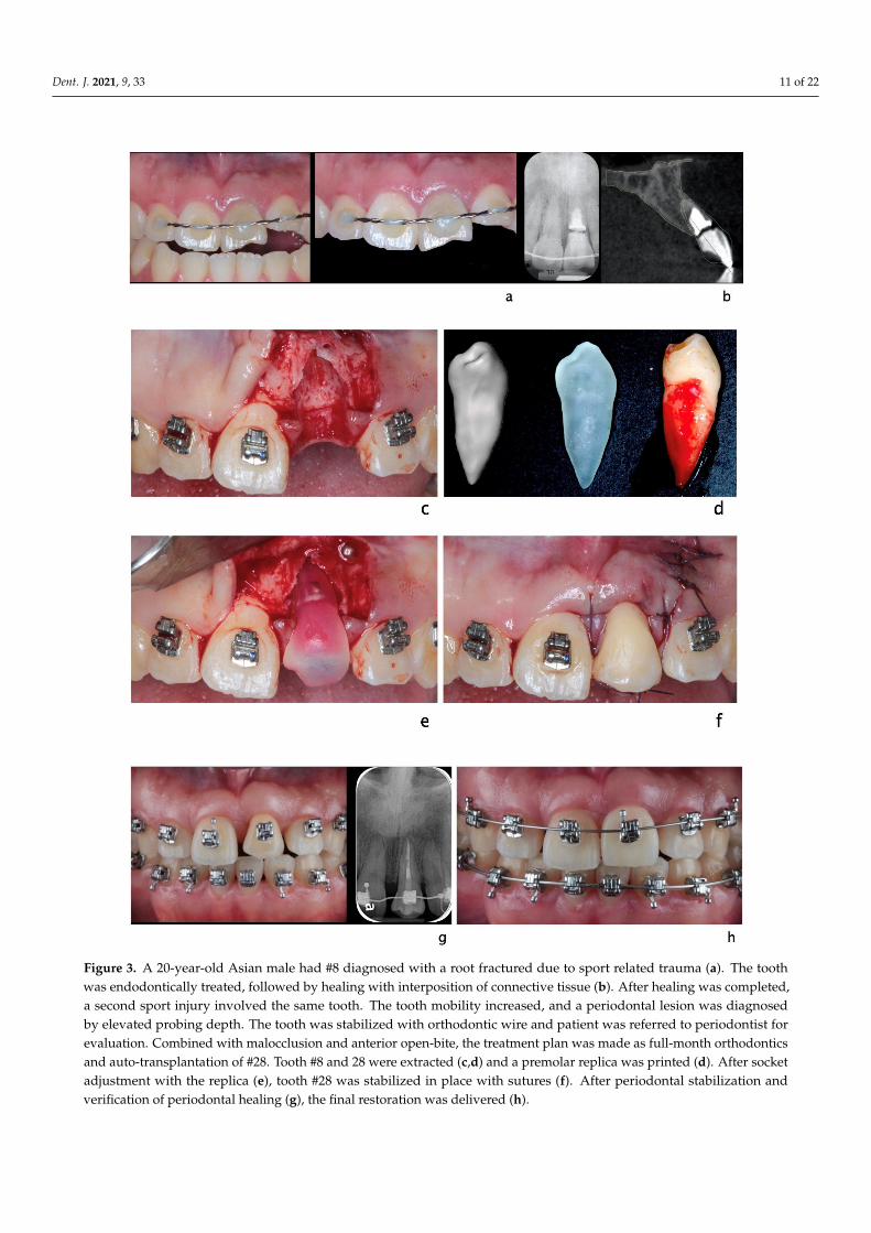
Figure 3. A 20-year-old Asian male had #8 diagnosed with a root fractured due to sport related trauma (a). The tooth was
endodontically treated, followed by healing with interposition of connective tissue (b). After healing was completed, a
second sport injury involved the same tooth. The tooth mobility increased, and a periodontal lesion was diagnosed by
elevated probing depth. The tooth was stabilized with orthodontic wire and patient was referred to periodontist for eval-
uation. Combined with malocclusion and anterior open-bite, the treatment plan was made as full-month orthodontics and
auto-transplantation of #28. Tooth #8 and 28 were extracted (c,d) and a premolar replica was printed (d). After socket
adjustment with the replica (e), tooth #28 was stabilized in place with sutures (f). After periodontal stabilization and ver-
ification of periodontal healing (g), the final restoration was delivered (h).
Figure 3. A 20-year-old Asian male had #8 diagnosed with a root fractured due to sport related trauma (a). The toothwas endodontically treated, followed by healing with interposition of connective tissue (b). After healing was completed,a second sport injury involved the same tooth. The tooth mobility increased, and a periodontal lesion was diagnosedby elevated probing depth. The tooth was stabilized with orthodontic wire and patient was referred to periodontist forevaluation. Combined with malocclusion and anterior open-bite, the treatment plan was made as full-month orthodonticsand auto-transplantation of #28. Tooth #8 and 28 were extracted (c,d) and a premolar replica was printed (d). After socketadjustment with the replica (e), tooth #28 was stabilized in place with sutures (f). After periodontal stabilization andverification of periodontal healing (g), the final restoration was delivered (h).

Dent. J. 2021, 9, 33 12 of 22
Periodontal soft tissue deficiency is also often seen after traumatic dental injuries [43].With extensive damages to the gingival tissues, underlying periosteum and alveolar bone,unfavorable responses to soft tissues may occur. Especially under concomitant bacterialinvasion or severe dysbiosis during wound healing phases, the “pink esthetics” may besignificantly compromised. To enhance the periodontal soft phenotypes, to increase thewidth of keratinized tissues and to gain the root coverage for recession, several surgicalapproaches with different available materials have been purposed. A recent meta-analysisby Barootchi [44] concluded that autogenous tissue grafts seem superior compared tonon-autogenous grafts for all procedures. However, non-autogenous grafts still offer asclinically effective options.
3.4. Dental Implants
TDI may lead to as series of events that include tooth avulsion or need for extraction.Many options are available for tooth replacement and one of these are dental implants.This device, used following tooth extraction, has been proven as a successful approach torestore function and esthetics after traumatic dental injuries. To optimize the outcomesof implant placement, there are several clinical parameters which should be taken intoconsiderations in treatment planning (Figure 4). In clinical scenarios, the timing of implantplacement associate with tooth extraction is generally categorized as [45]:
• Type 1, immediate placement, no later than 24 h after tooth extraction.• Type 2, early placement, typically 4 to 8 weeks after tooth extraction with only healed
soft tissue at extraction site.• Type 3, early placement, typically 12 to 16 weeks after tooth extraction with healed
soft tissue and significant healing of alveolar bone at extraction site.• Type 4, late placement, after 6 months with complete healing at extraction site.
Dent. J. 2021, 9, x FOR PEER REVIEW 15 of 23
tooth outside the socket and the presence of damage on alveolar supporting tissues sig-
nificantly influence the decision regarding timing of implant placement and the need for
adjunctive procedure, i.e., tissue augmentation. In the very rare case scenario of immedi-
ate tooth avulsion with intact alveolar soft and hard tissues, when tooth replantation is
not indicated, type 1 implant placement can be considered. The predictability of successful
immediate implant placement relies on the initial thickness of buccal plate, soft tissue phe-
notype and primary stability provided by bone apical and palatal to the socket. Type 2 or
3 implant placement, referring to early placement with healed soft tissue and partial bone
fill, is more relevant to dental trauma. Additional tissue augmentation is usually indicated
to offer stable three-dimensional environment for ideal implant placement after traumatic
dental injuries. Type 4 implant placement, as a delayed treatment, is indicated when the
treatment plan involves the adjacent teeth for more complex clinical scenarios. Although
there is limited specific clinical trial comparing different protocols of implant placement
following traumatic dental injuries, evidence has shown that, if guidelines are strictly fol-
lowed, different protocols offer similar successful outcomes [46–48].
Figure 4. A 24-year-old female fell from her bike during a race. She hit the tarmac and resulted in losing teeth#9, 10 and
11 (a,b) as well as a portion of the alveolar bone (c) as seen on the 3D print of the maxilla. An incisal chip on tooth #8
completed the damage of the fall. After an analysis of residual hard and soft tissue volumes, a digital wax-up was created
to plan the future implant placement and restorations (d). Guided tissue regeneration was performed, and implants were
placed in a Type 4 timeline (e,f). A provisional fixed partial denture and connective tissue graft were inserted to improve
esthetics and tissue conditioning (g).
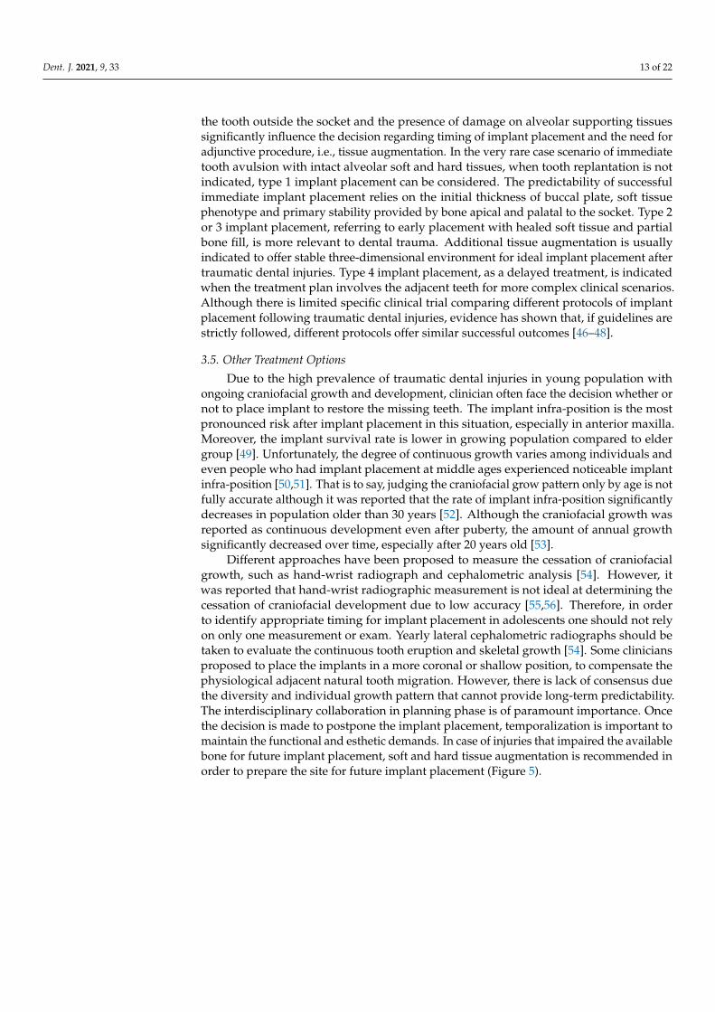
3.5 Other Treatment Options
Due to the high prevalence of traumatic dental injuries in young population with
ongoing craniofacial growth and development, clinician often face the decision whether
or not to place implant to restore the missing teeth. The implant infra-position is the most
pronounced risk after implant placement in this situation, especially in anterior maxilla.
Moreover, the implant survival rate is lower in growing population compared to elder
group [49]. Unfortunately, the degree of continuous growth varies among individuals and
even people who had implant placement at middle ages experienced noticeable implant
infra-position [50,51]. That is to say, judging the craniofacial grow pattern only by age is
not fully accurate although it was reported that the rate of implant infra-position signifi-
cantly decreases in population older than 30 years [52]. Although the craniofacial growth
was reported as continuous development even after puberty, the amount of annual
growth significantly decreased over time, especially after 20 years old [53].
Different approaches have been proposed to measure the cessation of craniofacial
growth, such as hand-wrist radiograph and cephalometric analysis [54]. However, it was
Figure 4. A 24-year-old female fell from her bike during a race. She hit the tarmac and resulted in losing teeth#9, 10 and11 (a,b) as well as a portion of the alveolar bone (c) as seen on the 3D print of the maxilla. An incisal chip on tooth #8completed the damage of the fall. After an analysis of residual hard and soft tissue volumes, a digital wax-up was created toplan the future implant placement and restorations (d). Guided tissue regeneration was performed, and implants wereplaced in a Type 4 timeline (e,f). A provisional fixed partial denture and connective tissue graft were inserted to improveesthetics and tissue conditioning (g).
Following traumatic dental injuries like tooth avulsion, the considerations of implantplacement can be made as for non-trauma related tooth extraction. The time period of
Dent. J. 2021, 9, 33 13 of 22
the tooth outside the socket and the presence of damage on alveolar supporting tissuessignificantly influence the decision regarding timing of implant placement and the need foradjunctive procedure, i.e., tissue augmentation. In the very rare case scenario of immediatetooth avulsion with intact alveolar soft and hard tissues, when tooth replantation is notindicated, type 1 implant placement can be considered. The predictability of successfulimmediate implant placement relies on the initial thickness of buccal plate, soft tissuephenotype and primary stability provided by bone apical and palatal to the socket. Type 2or 3 implant placement, referring to early placement with healed soft tissue and partialbone fill, is more relevant to dental trauma. Additional tissue augmentation is usuallyindicated to offer stable three-dimensional environment for ideal implant placement aftertraumatic dental injuries. Type 4 implant placement, as a delayed treatment, is indicatedwhen the treatment plan involves the adjacent teeth for more complex clinical scenarios.Although there is limited specific clinical trial comparing different protocols of implantplacement following traumatic dental injuries, evidence has shown that, if guidelines arestrictly followed, different protocols offer similar successful outcomes [46–48].
3.5. Other Treatment Options
Due to the high prevalence of traumatic dental injuries in young population withongoing craniofacial growth and development, clinician often face the decision whether ornot to place implant to restore the missing teeth. The implant infra-position is the mostpronounced risk after implant placement in this situation, especially in anterior maxilla.Moreover, the implant survival rate is lower in growing population compared to eldergroup [49]. Unfortunately, the degree of continuous growth varies among individuals andeven people who had implant placement at middle ages experienced noticeable implantinfra-position [50,51]. That is to say, judging the craniofacial grow pattern only by age is notfully accurate although it was reported that the rate of implant infra-position significantlydecreases in population older than 30 years [52]. Although the craniofacial growth wasreported as continuous development even after puberty, the amount of annual growthsignificantly decreased over time, especially after 20 years old [53].
Different approaches have been proposed to measure the cessation of craniofacialgrowth, such as hand-wrist radiograph and cephalometric analysis [54]. However, itwas reported that hand-wrist radiographic measurement is not ideal at determining thecessation of craniofacial development due to low accuracy [55,56]. Therefore, in orderto identify appropriate timing for implant placement in adolescents one should not relyon only one measurement or exam. Yearly lateral cephalometric radiographs should betaken to evaluate the continuous tooth eruption and skeletal growth [54]. Some cliniciansproposed to place the implants in a more coronal or shallow position, to compensate thephysiological adjacent natural tooth migration. However, there is lack of consensus duethe diversity and individual growth pattern that cannot provide long-term predictability.The interdisciplinary collaboration in planning phase is of paramount importance. Oncethe decision is made to postpone the implant placement, temporalization is important tomaintain the functional and esthetic demands. In case of injuries that impaired the availablebone for future implant placement, soft and hard tissue augmentation is recommended inorder to prepare the site for future implant placement (Figure 5).

Dent. J. 2021, 9, 33 14 of 22
Dent. J. 2021, 9, x FOR PEER REVIEW 16 of 23
reported that hand-wrist radiographic measurement is not ideal at determining the cessa-
tion of craniofacial development due to low accuracy [55,56]. Therefore, in order to iden-
tify appropriate timing for implant placement in adolescents one should not rely on only
one measurement or exam. Yearly lateral cephalometric radiographs should be taken to
evaluate the continuous tooth eruption and skeletal growth [54]. Some clinicians proposed
to place the implants in a more coronal or shallow position, to compensate the physiolog-
ical adjacent natural tooth migration. However, there is lack of consensus due the diver-
sity and individual growth pattern that cannot provide long-term predictability. The in-
terdisciplinary collaboration in planning phase is of paramount importance. Once the de-
cision is made to postpone the implant placement, temporalization is important to main-
tain the functional and esthetic demands. In case of injuries that impaired the available
bone for future implant placement, soft and hard tissue augmentation is recommended in
order to prepare the site for future implant placement (Figure 5).
Figure 5. Cont.

Dent. J. 2021, 9, 33 15 of 22Dent. J. 2021, 9, x FOR PEER REVIEW 17 of 23
Figure 5. Clinical scenario of a 17 years-old adolescent hit by a baseball ball in the anterior maxillary region. The boy
presented to the Periodontal Department at Tufts University, Boston USA with crown fracture of left central incisor (#9)
(a,b). Peri-apical radiograph show apical radiolucency, sign of necrosis. After the diagnosis, CaOH2 was applied. The root
canal definitive treatment was completed but after 2 months the patient still presented with a fistula, that was tracked via
a gutta-percha point. A CBCT scan was performed in order to diagnose the extent of the peri-apical lesion (c in sequence).
The extent of the lesion did not suggest an endodontic therapy revision. Exploratory surgery was performed in order to
rule out tooth fracture (d). The apex was resected in order to access the palatal aspect of the tooth. A PA radiograph was
taken in order to verify correct apex resection and endodontic retrograde seal (e). Due to active patient skeletal growth, a
decision was made to enucleate the endodontic cyst and treat the cavity with bone grafting material, in order to preserve
the site for future implant placement (f,g). PA radiograph comparison before and after grafting placement (h,i). The patient
was followed up for 2 months, and a fistula was identified apical to #9 (l). Tooth #10 was diagnosed as necrotic. A root
canal was performed (m) and the apical radiolucency and fistula were resolved at 1 month follow up (n).
Another technique that can be used in adolescents that did not yet complete skeletal
growth, is the use of mini-implants. This technique exploits the advantages of conven-
tional diameter implants and those of reduced diameter implants both during the posi-
tioning phases of the fixtures and during the prosthetic phases. A fixed rehabilitation pro-
vides an obvious psychosocial benefit especially during the complex adolescent age. At
the same time, mini-implants can be replaced by a standard fixture upon completion of
growth and increased esthetic demand from patients. In other words, the use of mini-
implants in growing patients can be both used as a temporary and definitive treatment
option. Therefore, the possibility of carrying out post-traumatic rehabilitation using mini-
implants and cemented prosthetic crowns becomes an interesting alternative for the grow-
ing patient [57–59]. Yet, some concerns regarding the long-term survival rate of these im-
plants still remain. In 2018, the ITI Consensus Report [60] evaluated the influence of im-
plant factors on clinical and radiographic outcomes. One of the analyses regarded the in-
fluence of implant diameter, comparing three categories of narrow implants; category 1
or “Mini-implants” <2.5 mm; category 2: >2.5 mm and <3 mm; category 3: >3 mm and <3.5
mm). The mean survival rates resulted in 94.7% ± 5%, 97.3% ± 5% and 97.7% ± 2.3% for
category 1, 2, and 3, respectively. The authors concluded that implants with diameters of
less than 2.5 mm showed inferior survival rates compared to standard diameter implants.
Similar results were obtained in a systematic review by Bidra et al. [61]. The authors eval-
uated the short-term (1 to 5 years), medium-term (5 to 10 years), and long-term (beyond
10 years) survival rates of mini-implants employed for final prosthetic treatment. The 1st
year survival rate was <95% and the cumulative survival of 9 years was ~92%. Most im-
plants were immediately loaded, and the majority of implant failures happened within
one year of placement. The reported a one-year survival rate was considered acceptable
but many implants analyzed had a minimum follow-up shorter than a year. The authors
concluded that there were insufficient data regarding failures after the first year. It is not
safe to draw conclusions regarding the 5–10-year survival rate of mini-implants. The au-
thors could not find any evidence for the long-term survival of these implants.
Figure 5. Clinical scenario of a 17 years-old adolescent hit by a baseball ball in the anterior maxillary region. The boypresented to the Periodontal Department at Tufts University, Boston USA with crown fracture of left central incisor (#9)(a,b). Peri-apical radiograph show apical radiolucency, sign of necrosis. After the diagnosis, CaOH2 was applied. The rootcanal definitive treatment was completed but after 2 months the patient still presented with a fistula, that was tracked via agutta-percha point. A CBCT scan was performed in order to diagnose the extent of the peri-apical lesion (c in sequence).The extent of the lesion did not suggest an endodontic therapy revision. Exploratory surgery was performed in order to ruleout tooth fracture (d). The apex was resected in order to access the palatal aspect of the tooth. A PA radiograph was taken inorder to verify correct apex resection and endodontic retrograde seal (e). Due to active patient skeletal growth, a decisionwas made to enucleate the endodontic cyst and treat the cavity with bone grafting material, in order to preserve the sitefor future implant placement (f,g). PA radiograph comparison before and after grafting placement (h,i). The patient wasfollowed up for 2 months, and a fistula was identified apical to #9 (l). Tooth #10 was diagnosed as necrotic. A root canal wasperformed (m) and the apical radiolucency and fistula were resolved at 1 month follow up (n).
Another technique that can be used in adolescents that did not yet complete skeletalgrowth, is the use of mini-implants. This technique exploits the advantages of conventionaldiameter implants and those of reduced diameter implants both during the positioningphases of the fixtures and during the prosthetic phases. A fixed rehabilitation provides anobvious psychosocial benefit especially during the complex adolescent age. At the sametime, mini-implants can be replaced by a standard fixture upon completion of growth andincreased esthetic demand from patients. In other words, the use of mini-implants in grow-ing patients can be both used as a temporary and definitive treatment option. Therefore, thepossibility of carrying out post-traumatic rehabilitation using mini-implants and cementedprosthetic crowns becomes an interesting alternative for the growing patient [57–59]. Yet,some concerns regarding the long-term survival rate of these implants still remain. In 2018,the ITI Consensus Report [60] evaluated the influence of implant factors on clinical andradiographic outcomes. One of the analyses regarded the influence of implant diameter,comparing three categories of narrow implants; category 1 or “Mini-implants” <2.5 mm;category 2: >2.5 mm and <3 mm; category 3: >3 mm and <3.5 mm). The mean survival ratesresulted in 94.7% ± 5%, 97.3% ± 5% and 97.7% ± 2.3% for category 1, 2, and 3, respectively.The authors concluded that implants with diameters of less than 2.5 mm showed inferiorsurvival rates compared to standard diameter implants. Similar results were obtainedin a systematic review by Bidra et al. [61]. The authors evaluated the short-term (1 to5 years), medium-term (5 to 10 years), and long-term (beyond 10 years) survival rates ofmini-implants employed for final prosthetic treatment. The 1st year survival rate was <95%and the cumulative survival of 9 years was ~92%. Most implants were immediately loaded,and the majority of implant failures happened within one year of placement. The reporteda one-year survival rate was considered acceptable but many implants analyzed had aminimum follow-up shorter than a year. The authors concluded that there were insufficientdata regarding failures after the first year. It is not safe to draw conclusions regarding the5–10-year survival rate of mini-implants. The authors could not find any evidence for thelong-term survival of these implants.

Dent. J. 2021, 9, 33 16 of 22
4. PreventionAppliances to Prevent Dental Injuries
During sports events, the risk of falling, being hit by an opponent or a ball is high.The sole way of minimizing the number of TDI are to implement preventive approaches.The two main appliances to prevent TDI are faceguards and mouthguards. Prior to themandatory utilization of face and mouthguards in American football, facial, and oraltrauma accounted for 50% of all reported injuries [14]. With the introduction of theseprotective gear, the incidence of oral and facial trauma significantly decreased down to a3.9% [62]. The use of mouthguards decreases 5.55 times the chance of players sufferingdental injuries [63].
Faceguards are prefabricated or custom-made cages made of metal, composite or,more recently, clear polycarbonate plastic which are usually attached to helmets or headstraps. Faceguards and helmets have been effective in practically eliminating all oculofacialinjuries in contact sports. Full face shields have demonstrated to significantly reduce therisk of dento-facial injuries without increasing the risk of neck injuries or concussions [14].
Mouthguards are considered the main appliance for the prevention and reductionof severity of sports-related oral injuries. A mouthguard is defined as a resilient deviceplaced inside the mouth in order to protect the player [64]. They were introduced in the19th century by a dentist named Woolf Krause and their main intention was to protectboxers from soft tissue lacerations [65].
Mouthguards can prevent bruising and lacerations of the perioral and intraoral tis-sues, protect teeth from avulsion, luxation, crown or root fracture, avoid jaw fracturesand dislocations, provide support for the edentulous space, and minimize the severityof condylar dislocation and temporomandibular joint trauma [66]. A systematic reviewreported that athletes wearing mouthguards are 82% to 93% less likely to suffer TDIs [8].In fact, the prevalence of dental trauma ranged from 7.5% to 7.75% among mouthguardwearers compared to 48.31% to 59.98% for non-wearers [8]. Although it was noted thatavulsions and crown fractures were the most frequent injuries [8], overall the number ofreported tooth fractures has been significantly reduced with the use of mouthguards [14].A meta-analysis carried out in 2020, indicated that athletes involved in many differentsports that were not wearing mouthguards showed twice higher risk of suffering orofacialinjuries compared to those wearing one [67]. In a prospective longitudinal study analyzing70,936 athlete exposures, mouthguard users had significantly lower rates of TDI—fractures,luxation and avulsions—(0.6% vs. 2.0%) and lower dental referrals (1.6% vs. 5.8%) com-pared to non-users [68]. Only one study did not find statistically significant differences interms of head, neck, and oral injuries between users and non-users of mouthguards [69].
The main four purposes of mouthguards that have been described in the literatureare: (1) Protecting teeth by absorbing or dissipating the energy of a shock. (2) Preventinglacerations on lips, tongue and gingival tissues. (3) Protecting antagonist teeth fromtraumatic occlusal contact. (4) Providing resilient support to the mandible by absorbingimpacts that could fracture the angle or condyle of the mandible.
A fifth proposed function is the protection against neck and cerebral injuries. However,studies still haven’t been able to demonstrate it [68].
The mechanism in which mouthguards achieve their functions is still not clear. Hy-pothesis have been formulated that the higher the absorption energy, the higher the protec-tion. Nevertheless, this absorbed energy may be transmitted to the underlying dental andperiodontal tissues [70].
There are 3 main types of available mouthguards: 1. Stock (commercially prefabricatedmouthguard sold over the counter with a claimed universal fit and designed to be usedwithout modification) 2. “Boil-and-bite” (commercially available mouthguard that is madeof a thermoplastic material that is softened in hot water and the athlete adapts it to theirown dental arch using finger, tongue, and biting pressure) 3. Custom-made (made by adentist and dental technician using the patient’s cast) [71].

Dent. J. 2021, 9, 33 17 of 22
Both stock and boil-and-bite mouthguards have been reported to lack of properretention and require the user to apply constant occlusal pressure to held them in placemaking them uncomfortable. Custom-made mouthguards are tailor-designed to providebetter fit and comfort as they allow for easier and better breathing and speaking [71–73].Although further comparative studies with larger sample size and longer follow-up timeare need, some studies suggest that custom-made mouthguards offer better protectivecharacteristics [70]. Increased comfort of wear can be experienced if the mouthguard isextended labially to within 2 mm of the vestibular fold as extended as far as the patient cantolerate it and adjusted to allow clearance of frenum [74].
Mouthguards should extend at least up to the distal aspect of the maxillary first molarand should have a labial and occlusal thickness of 3 mm and a palatal thickness of 2 mm.to reduce the effects of impact force on teeth. Occlusion of mouthguards should be bilateraland balanced [75]. The reduction of the palatal extension of mouthguards from 6 to 2 mm.improves the degree of satisfaction of athletes [76].
Custom mouthguard should follow some specific criteria such as being made of aneasily cleaned and disinfected, resilient material, and provide correct retention allowing anadequate occlusal relationship and maximum protection [14]. Furthermore, they should beable to absorb and deflect the energy of an impact by covering the maxillary teeth, excludeany occlusal interference, allow mouth breathing, and protect the soft tissues.
Mouthguards offer a considerable protection against sports-related dental injuries andcustom-made mouthguards provide advantages over the other types. Lately, athletes arebecoming more aware of the importance of using mouthguards as preventive measures toavoid orofacial injuries. In a recent study, 95.7% of a sample of 310 hockey players reportedto have tried a mouthguard while 86.8% and 91.3% used them regularly during trainingsessions and competition, respectively [77]. However, other studies have observed thatthere is still a high number of athletes and sport players that do not use them because ofthe perception of being expensive and the need of at least one visit to the dentist [71]. It isimportant that members of the technical staff encourage the regular use of mouth guardsin athletes who practice risk contact sports [71,78,79].
Dentists should also promote the use of mouthguards among professional and amateurathletes. Nevertheless, the lack of evidence-based guidelines has reflected the differentperspectives and beliefs of orthodontists about recommending the use of mouthguard [80].During the past years, there has been an exponential increase in the number of athletesworldwide and dental trauma has become a significative dental health issue. In 2016, Aprilwas established as the National Facial Protection Month. During this month five differenthealth organizations (American Academy of Pediatric Dentistry, the Academy for SportsDentistry, American Association of Orthodontists and the American Association of Oraland Maxillofacial Surgeons) promote orofacial protection and the use of mouthguards [81].
It has been shown that prevention is the most effective way of decreasing TDIs and,for this reason, sports committees and public authorities should regulate and establishmouthguards as mandatory safety equipment that protect the physical integrity of ath-letes [8].
5. Discussion
As reported in the previous paragraphs, sport injuries can lead to extensive and debil-itating fractures that can involve both facial and dental structures. Many challenges arisefor the clinicians in charge of handling these patients. Many parameters and therapeuticoptions need to be taken into consideration to successfully treat and resolve these compro-mised cases [82]. Patients’ age, their medical history and compliance need to be carefullyreviewed in order to outline the most ideal treatment planning. Nevertheless, even takinginto account all these variables, surgical solutions may be comparable, making the clinicalfinal decision not straight forward [82].
Taking into consideration both patients to the site of injury, some of the technicalconsiderations that need to be evaluated are the extent of the trauma and the age of the

Dent. J. 2021, 9, 33 18 of 22
patient. The clinical scenarios can rage from a minimal deciduous tooth structural loss ona young individual, to a facial bone fracture involving the esthetic area on an adult [14].The treatment approaches would be completely different. The dental trauma on deciduousteeth could be seen as more favorable, first for the extent and, second, because primaryteeth will be replaced by permanent ones. The loss of dentin and enamel could be adjustedwith modern restorative procedures or with the use of temporary crowns [14]. The lossof an element could be replaced with a crown, adhered to neighboring teeth, such asMaryland bridges [14], or with the use of mini implants [57,58]. These options representan increase on invasiveness, starting from a restorative solution to a surgical one. In thismanuscript, the authors listed the surgical techniques that could be employed in medium tosevere dental or facial injuries. If the trauma involves an adolescent, the two main optionscould be tooth transplantation and use of mini-implants. Both these solutions could beconsidered temporary or permanent. In case of auto-transplant, the tooth may still requireroot canal, final crown to match the esthetics and orthodontic treatment. Mini-implants canbe retained for an extended period of time, and they would be considered final solutions.In the majority of cases, they are replaced by standard dental implants, upon adolescentend of growth [61]. In the most advanced cases, where teeth and alveolar bone had beenlost during the sport accident, an extensive bone regeneration could be mandatory beforedental implant therapy [61]. Besides the advantage of natural smile appearance, fixedtype restoration and sparing of dental structure for prosthetic abutments, the standardimplant therapy may not be well accepted because the length of treatment, the discomfortassociated with surgery as well as the economic expense [61].
6. Conclusions
The high prevalence of TDI during sport activities can be a burden for the professionaland amateur athletes. The likelihood of facial and dental injury poses the dental professionon the first line to treat damages that can alter and compromise patient’s function andesthetics. In particular, surgeons would be required in cases of avulsion, tooth, soft andhard tissue loss. In conclusion:
• The timing or tooth replantation after a traumas is crucial. All avulsed permanentand mature teeth eventually develop pulp necrosis. When the tooth is immature andpresents an open apex, further root development can be achieved.
• The advances of dental CBCT and 3D printing allow the surgeon to successfullyplan and execute tooth auto transplantations. The use of printed replica significantlydecreased the surgical time and increased to more than 95% the survival rates.
• The soft and hard tissue deficiencies following trauma should be re-evaluated afterinitial healing and stabilization. The defects then can be treated as clinical scenariorequiring standard soft and hard tissue regeneration.
• If the injured is an adult, tooth/teeth loss, tooth/teeth anxylosis and auto transplanta-tion failure may be treated with dental implants to restore the missing tooth/teeth;
• The diffusion of injuries among adolescents calls for individualized treatment based ongrowth and time that separates the traumatic event to final restorations. Mini-implantscould be used as an interim or final restoration to replace avulsed teeth.
Author Contributions: Conceptualization, L.M.; resources, data curation, writing—original draftpreparation, L.M., P.L., R.L.; writing—review and editing and supervision, L.G., R.B. All authorshave read and agreed to the published version of the manuscript.
Funding: This research received no external funding.
Institutional Review Board Statement: Ethical review and approval were waived for this study, dueto nature of the study. No intervention or no medications or no device were used to treat patients forthis specific study review.
Informed Consent Statement: Informed consent was obtained from all subjects whose clinicalscenarios were included in the study.
Dent. J. 2021, 9, 33 19 of 22
Data Availability Statement: Data sharing not applicable. No new data were created or analyzed inthis study. Data sharing is not applicable to this article.
Acknowledgments: Authors would like to acknowledge and thank Ziad A. Alharbey (Case onFigure 2), Francesco Mintrone (Case on Figure 4), Po-Jan Kuo (Case on Figure 3), Gayathri Shenoy(case on Figure 5), for the wonderful clinical cases utilized for this review.
Conflicts of Interest: Authors report no conflict of interest.
References1. World Health Organization. Injuries and Violence: The Facts 2014; World Health Organization: Geneva, Switzerland, 2014.2. Petersson, E.E.; Andersson, L.; Sörensen, S. Traumatic Oral Vs Non-Oral Injuries. Swed. Dent. J. 1997, 21, 55–68.3. Hayter, J.P.; Ward, A.J.; Smith, E.J. Maxillofacial Trauma in Severely Injured Patients. Br. J. Oral Maxillofac. Surg. 1991, 29, 370–373.
[CrossRef]4. Piccininni, P.; Clough, A.; Padilla, R.; Piccininni, G. Dental and Orofacial Injuries. Clin. Sports Med. 2017, 36, 369–405. [CrossRef]
[PubMed]5. Borssén, E.; Källestål, C.; Holm, A.-K. Treatment time of traumatic dental injuries in a cohort of 16-year-olds in northern Sweden.
Acta Odontologica Scandinavica 2002, 60, 265–270. [CrossRef] [PubMed]6. Keçeci, A.D.; Eroglu, E.; Baydar, M.L. Dental trauma incidence and mouthguard use in elite athletes in Turkey. Dent. Traumatol.
2005, 21, 76–79. [CrossRef]7. Polmann, H.; Melo, G.; Conti Réus, J.; Domingos, F.L.; de Souza, B.D.M.; Padilha, A.C.; Duque, T.M.; Porporatti, A.L.; Flores-
Mir, C.; De Luca Canto, G. Prevalence of Dentofacial Injuries among Combat Sports Practitioners: A Systematic Review andMeta-Analysis. Dent. Traumatol. 2020, 36, 124–140. [CrossRef] [PubMed]
8. Fernandes, L.M.; Neto, J.C.L.; Lima, T.F.; Magno, M.B.; Santiago, B.M.; Cavalcanti, Y.W.; de Almeida, L.D.F.D. The Use ofMouthguards and Prevalence of Dento-Alveolar Trauma among Athletes: A Systematic Review and Meta-Analysis. Dent.Traumatol. 2019, 35, 54–72. [CrossRef]
9. Cavalleri, G.; Zerman, N. Traumatic crown fractures in permanent incisors with immature roots: A follow-up study. Dent.Traumatol. 1995, 11, 294–296. [CrossRef] [PubMed]
10. Kumamoto, D.; Maeda, Y. Global Trends and Epidemiology of Sports Injuries. J. Pediatr. Dent. Care 2005, 11, 15–25.11. Reehal, P. Facial Injury in Sport. Curr. Sports Med. Rep. 2010, 9, 27–34. [CrossRef]12. Cortes, M.I.D.S.; Marcenes, W.; Sheiham, A. Impact of traumatic injuries to the permanent teeth on the oral health-related quality
of life in 12-14-year-old children. Community Dent. Oral Epidemiol. 2002, 30, 193–198. [CrossRef] [PubMed]13. Kaufman, B.R.; Heckler, F.R. Sports-Related Facial Injuries. Clin. Sports Med. 1997, 16, 543–562. [CrossRef]14. Andreasen, J.O.; Andreasen, F.M.; Andersson, L. Textbook and Color. Atlas of Traumatic Injuries to the Teeth; John Wiley & Sons:
Hoboken, NJ, USA, 2019.15. Levin, L.; Day, P.F.; Hicks, L.; O’Connell, A.; Fouad, A.F.; Bourguignon, C.; Abbott, P.V. International Association of Dental
Traumatology Guidelines for the Management of Traumatic Dental Injuries: General Introduction. Dent. Traumatol. 2020, 36,309–313. [CrossRef]
16. Bourguignon, C.; Cohenca, N.; Lauridsen, E.; Flores, M.T.; O’Connell, A.C.; Day, P.F.; Tsilingaridis, G.; Abbott, P.V.; Fouad, A.F.;Hicks, L.; et al. International Association of Dental Traumatology Guidelines for the Management of Traumatic Dental Injuries: 1.Fractures and Luxations. Dent. Traumatol. 2020, 36, 314–330. [CrossRef]
17. Oliveira Werlich, M.; Honnef, L.R.; Silva Bett, J.V.; Domingos, F.L.; Pauletto, P.; Dulcineia Mendes de Souza, B.; Duque, T.M.;Curi Hallal, A.L.; De Luca Canto, G. Prevalence of Dentofacial Injuries in Contact Sports Players: A Systematic Review andMeta-Analysis. Dent. Traumatol. 2020, 36, 477–488. [CrossRef] [PubMed]
18. Association, American Dental. Using Mouthguards to Reduce the Incidence and Severity of Sports-Related Oral Injuries. J. Am.Dent. Assoc. 2006, 137, 1712–1720. [CrossRef]
19. For the Dental Patient. The Importance of Using Mouthguards. Tips for Keeping Your Smile Safe. J. Am. Dent. Assoc. 2004, 135,1061. [CrossRef]
20. NFHS. Position Statement and Recommendations for Mouthguard Use in Sport; National Federation of State High School Associations:Indianapolis, IN, USA, 2018.
21. Schneider, S.; Seither, B.; Tönges, S.; Schmitt, H. Sports Injuries: Population Based Representative Data on Incidence, Diagnosis,Sequelae, and High Risk Groups. Br. J. Sports Med. 2006, 40, 334–339. [CrossRef] [PubMed]
22. Zaleckiene, V.; Peciuliene, V.; Brukiene, V.; Drukteinis, S. Traumatic Dental Injuries: Etiology, Prevalence and Possible Outcomes.Stomatologija 2014, 16, 7–14.
23. Navabazam, A.; Farahani, S.S. Prevalence of Traumatic Injuries to Maxillary Permanent Teeth in 9- to 14-Year-Old School Childrenin Yazd, Iran. Dent. Traumatol. 2010, 26, 154–157. [CrossRef]
24. Traebert, J.; Bittencourt, D.D.; Peres, K.G.; Peres, M.A.; De Lacerda, J.T.; Marcenes, W. Aetiology and Rates of Treatment ofTraumatic Dental Injuries among 12-Year-Old School Children in a Town in Southern Brazil. Dent. Traumatol. 2006, 22, 173–178.[CrossRef]

Dent. J. 2021, 9, 33 20 of 22
25. Scott, N.; Hughes, J.; Forbes-Haley, C.; East, C.; Holmes, S.; Wilson, E.; Ball, S.; Hammond, D.; Drake, D.; Hutchison, I.; et al.Management of Facial Injuries in Elite and Professional Sports—A Consensus Report. Br. J. Oral Maxillofac. Surg. 2020, 58,e254–e259. [CrossRef] [PubMed]
26. Trope, M. Clinical Management of the Avulsed Tooth: Present Strategies and Future Directions. Dent. Traumatol. 2002, 18, 1–11.[CrossRef] [PubMed]
27. Andreasen, J.O.; Borum, M.K.; Jacobsen, H.L.; Andreasen, F.M. Replantation of 400 avulsed permanent incisors. 4. Factors relatedto periodontal ligament healing. Dent. Traumatol. 1995, 11, 76–89. [CrossRef] [PubMed]
28. Kinirons, M.J.; Gregg, T.A.; Welbury, R.R.; Cole, B.O.I. Variations in the Presenting and Treatment Features in ReimplantedPermanent Incisors in Children and Their Effect on the Prevalence of Root Resorption. Br. Dent. J. 2000, 189, 263–266. [CrossRef][PubMed]
29. Fouad, A.F.; Abbott, P.V.; Tsilingaridis, G.; Cohenca, N.; Lauridsen, E.; Bourguignon, C.; O’Connell, A.; Flores, M.T.; Day, P.F.;Hicks, L.; et al. International Association of Dental Traumatology Guidelines for the Management of Traumatic Dental Injuries: 2.Avulsion of Permanent Teeth. Dent. Traumatol. 2020, 36, 331–342. [CrossRef] [PubMed]
30. Lin, S.; Zuckerman, O.; Fuss, Z.; Ashkenazi, M. New Emphasis in the Treatment of Dental Trauma: Avulsion and Luxation. Dent.Traumatol. 2007, 23, 297–303. [CrossRef] [PubMed]
31. Schröder, U.; Granath, L.E. Early Reaction of Intact Human Teeth to Calcium Hydroxide Following Experimental Pulpotomy andIts Significance to the Development of Hard Tissue Barrier. Odont Revy 1971, 22, 379–395.
32. Moule, A.J.; Moule, C.A. The Endodontic Management of Traumatized Permanent Anterior Teeth: A Review. Aust. Dent. J. 2007,52 (Suppl. 1), S122–S137. [CrossRef]
33. Slagsvold, O.; Bjercke, B. Indications for Autotransplantation in Cases of Missing Premolars. Am. J. Orthod. 1978, 74, 241–257.[CrossRef]
34. Machado, L.A.; Do Nascimento, R.R.; Ferreira DM, T.P.; Mattos, C.T.; Vilella, O.V. Long-Term Prognosis of Tooth Autotransplanta-tion: A Systematic Review and Meta-Analysis. Int. J. Oral Maxillofac. Surg. 2016, 45, 610–617. [CrossRef]
35. Akhlef, Y.; Schwartz, O.; Andreasen, J.O.; Jensen, S.S. Autotransplantation of Teeth to the Anterior Maxilla: A Systematic Reviewof Survival and Success, Aesthetic Presentation and Patient-Reported Outcome. Dent. Traumatol. 2018, 34, 20–27. [CrossRef]
36. Atala-Acevedo, C.; Abarca, J.; Martínez-Zapata, M.J.; Díaz, J.; Olate, S.; Zaror, C. Success Rate of Autotransplantation of TeethWith an Open Apex: Systematic Review and Meta-Analysis. J. Oral Maxillofac. Surg. 2017, 75, 35–50. [CrossRef]
37. Chung, W.-C.; Tu, Y.-K.; Lin, Y.-H.; Lu, H.-K. Outcomes of autotransplanted teeth with complete root formation: A systematicreview and meta-analysis. J. Clin. Periodontol. 2014, 41, 412–423. [CrossRef] [PubMed]
38. Verweij, J.; Jongkees, F.; Moin, D.A.; Wismeijer, D.; Van Merkesteyn, J. Autotransplantation of teeth using computer-aided rapidprototyping of a three-dimensional replica of the donor tooth: A systematic literature review. Int. J. Oral Maxillofac. Surg. 2017, 46,1466–1474. [CrossRef] [PubMed]
39. Tan, W.L.; Wong, T.L.T.; Wong, M.C.M.; Lang, N.P. A systematic review of post-extractional alveolar hard and soft tissuedimensional changes in humans. Clin. Oral Implant. Res. 2011, 23, 1–21. [CrossRef]
40. Elnayef, B.; Porta, C.; Del Amo, F.S.-L.; Mordini, L.; Gargallo-Albiol, J.; Hernández-Alfaro, F. The Fate of Lateral RidgeAugmentation: A Systematic Review and Meta-Analysis. Int. J. Oral Maxillofac. Implant. 2018, 33, 622–635. [CrossRef] [PubMed]
41. Mordini, L.; Hur, Y.; Ogata, Y.; Finkelman, M.; Cavani, F.; Steffensen, B. Volumetric Changes Following Lateral Guided BoneRegeneration. Int. J. Oral Maxillofac. Implant. 2020, 35, e77–e85. [CrossRef] [PubMed]
42. Elnayef, B.; Monje, A.; Gargallo-Albiol, J.; Galindo-Moreno, P.; Wang, H.L.; Hernandez-Alfaro, F. Vertical Ridge Augmentation inthe Atrophic Mandible: A Systematic Review and Meta-Analysis. Int. J. Oral Maxillofac. Implant. 2017, 32, 291–312. [CrossRef]
43. Yu, C.Y.; Abbott, P.V. Responses of the pulp, periradicular and soft tissues following trauma to the permanent teeth. Aust. Dent. J.2016, 61 (Suppl. S1), 39–58. [CrossRef] [PubMed]
44. Barootchi, S.; Tavelli, L.; Dds, G.Z.; Giannobile, W.V.; Wang, H. Gingival phenotype modification therapies on natural teeth: Anetwork meta-analysis. J. Periodontol. 2020, 91, 1386–1399. [CrossRef]
45. Hämmerle, C.H.F.; Chen, S.T.; Wilson, T.G. Consensus statements and recommended clinical procedures regarding the placementof implants in extraction sockets. Int. J. Oral Maxillofac. Implant. 2004, 19 (Suppl. S2004), 26–28.
46. Bassir, S.H.; El Kholy, K.; Chen, C.-Y.; Lee, K.H.; Intini, G. Outcome of early dental implant placement versus other dental implantplacement protocols: A systematic review and meta-analysis. J. Periodontol. 2018, 90, 493–506. [CrossRef]
47. Chen, S.T.; Wilson, T.G.; Hämmerle, C.H.F. Immediate or early placement of implants following tooth extraction: Review ofbiologic basis, clinical procedures, and outcomes. Int. J. Oral Maxillofac. Implant. 2004, 19, 12–25.
48. Morton, D.; Chen, S.T.; Martin, W.C.; Levine, R.A.; Buser, D. Consensus Statements and Recommended Clinical ProceduresRegarding Optimizing Esthetic Outcomes in Implant Dentistry. Int. J. Oral Maxillofac. Implant. 2014, 29, 216–220. [CrossRef]
49. Bohner, L.; Hanisch, M.; Kleinheinz, J.; Jung, S. Dental implants in growing patients: A systematic review. Br. J. Oral Maxillofac.Surg. 2019, 57, 397–406. [CrossRef]
50. Bernard, J.P.; Schatz, J.P.; Christou, P.; Belser, U.; Kiliaridis, S. Long-Term Vertical Changes of the Anterior Maxillary TeethAdjacent to Single Implants in Young and Mature Adults. A Retrospective Study. J. Clin. Periodontol. 2004, 31, 1024–1028.[CrossRef] [PubMed]
51. Thilander, B.; Ödman, J.; Lekholm, U. Orthodontic Aspects of the Use of Oral Implants in Adolescents: A 10-Year Follow-upStudy. Eur. J. Orthod. 2001, 23, 715–731. [CrossRef]
Dent. J. 2021, 9, 33 21 of 22
52. Schwartz-Arad, D.; Bichacho, N. Effect of Age on Single Implant Submersion Rate in the Central Maxillary Incisor Region: ALong-Term Retrospective Study. Clin. Implant. Dent. Relat. Res. 2013, 17, 509–514. [CrossRef] [PubMed]
53. Fudalej, P.; Kokich, V.G.; Leroux, B. Determining the Cessation of Vertical Growth of the Craniofacial Structures to FacilitatePlacement of Single-Tooth Implants. Am. J. Orthod. Dentofacial Orthop. 2007, 131, S59–S67. [CrossRef] [PubMed]
54. Jensen, S.S. Timing of Implant Placement after Traumatic Dental Injury. J. Endod. 2019, 45, S52–S56. [CrossRef]55. Verma, D.; Peltomäki, T.; Jäger, A. Reliability of growth prediction with hand-wrist radiographs. Eur. J. Orthod. 2009, 31, 438–442.
[CrossRef]56. Flores-Mir, C.; Nebbe, B.; Major, P.W. Use of Skeletal Maturation Based on Hand-Wrist Radiographic Analysis as a Predictor of
Facial Growth: A Systematic Review. Angle Orthod. 2004, 74, 118–124. [PubMed]57. Giannetti, L.; Apponi, R.; Diago, A.M.D.; Mintrone, F. Rehabilitation of a patient with mini-implants after avulsion of the upper
incisors: A 13-year follow up. Dent. Traumatol. 2021, 37, 354–359. [CrossRef] [PubMed]58. Murri dello Diago, A.; Apponi, R.; Colombini, V.; Mordini, L.; Ideo, F. Complex Implant-Prosthetic Rehabilitation Following
Sports Trauma with 14 Years of Follow-Up: Case Report. Dent. J. 2021, 9, 6. [CrossRef] [PubMed]59. Giannetti, L.; Murri Dello Diago, A.; Vecci, F.; Consolo, U. Mini-Implants in Growing Patients: A Case Report. Pediatr. Dent. 2010,
32, 239–244. [PubMed]60. Jung, R.E.; Al-Nawas, B.; Araujo, M.; Avila-Ortiz, G.; Barter, S.; Brodala, N.; Chappuis, V.; Chen, B.; De Souza, A.; Almeida,
R.F.; et al. Group 1 Iti Consensus Report: The Influence of Implant Length and Design and Medications on Clinical andPatient-Reported Outcomes. Clin. Oral Implants Res. 2018, 29 (Suppl. S16), 69–77. [CrossRef]
61. Bidra, A.S.; Almas, K. Mini implants for definitive prosthodontic treatment: A systematic review. J. Prosthet. Dent. 2013, 109,156–164. [CrossRef]
62. Garon, M.W.; Merkle, A.; Wright, J.T. Mouth protectors and oral trauma: A study of adolescent football players. J. Am. Dent.Assoc. 1986, 112, 663–665. [CrossRef]
63. Bergman, L.; Milardovic Ortolan, S.; Žarkovic, D.; Viskic, J.; Jokic, D.; Mehulic, K. Prevalence of Dental Trauma and Use ofMouthguards in Professional Handball Players. Dent. Traumatol. 2017, 33, 199–204. [CrossRef]
64. Tuna, E.B.; Ozel, E. Factors Affecting Sports-Related Orofacial Injuries and the Importance of Mouthguards. Sports Med. 2014, 44,777–783. [CrossRef]
65. Reed, R.V. Origin and early history of the dental mouthpiece. Br. Dent. J. 1994, 176, 478–480. [CrossRef] [PubMed]66. Biasca, N.; Wirth, S.; Tegner, Y. The avoidability of head and neck injuries in ice hockey: An historical review. Br. J. Sports Med.
2002, 36, 410–427. [CrossRef]67. Knapik, J.J.; Hoedebecke, B.L.; Rogers, G.G.; Sharp, M.A.; Marshall, S.W. Effectiveness of Mouthguards for the Prevention of
Orofacial Injuries and Concussions in Sports: Systematic Review and Meta-Analysis. Sports Med. 2019, 49, 1217–1232. [CrossRef]68. Labella, C.R.; Smith, B.W.; Sigurdsson, A. Effect of Mouthguards on Dental Injuries and Concussions in College Basketball. Med.
Sci. Sports Exerc. 2002, 34, 41–44. [CrossRef]69. Blignaut, J.B.; Carstens, I.L.; Lombard, C.J. Injuries sustained in rugby by wearers and non-wearers of mouthguards. Br. J. Sports
Med. 1987, 21, 5–7. [CrossRef] [PubMed]70. Guerard, S.; Barou, J.-L.; Petit, J.; Poisson, P. Characterization of mouthguards: Impact performance. Dent. Traumatol. 2017, 33,
281–287. [CrossRef]71. Newsome, P.R.H.; Tran, D.C.; Cooke, M.S. The role of the mouthguard in the prevention of sports-related dental injuries: A
review. Int. J. Paediatr. Dent. 2001, 11, 396–404. [CrossRef] [PubMed]72. Shrestha, A.; Takahashi, T.; Kurokawa, K.; Mitsuyama, A.; Hayashi, K.; Ishigami, T.; Shahrin, S.; Chowdhury, R.; Churei, H.; Ueno,
T. Effects of Mouthguards on Electromyographic Activity of Masticatory Muscles. Int. J. Sports Dent. 2016, 9, 27–37.73. Spinas, E.; Mameli, A.; Giannetti, L. Traumatic Dental Injuries Resulting from Sports Activities; Immediate Treatment and Five
Years Follow-Up: An Observational Study. Open Dent. J. 2018, 12, 1–10. [CrossRef]74. McClelland, C.; Kinirons, M.; Geary, L. A preliminary study of patient comfort associated with customised mouthguards. Br. J.
Sports Med. 1999, 33, 186–189. [CrossRef] [PubMed]75. Lloyd, J.D.; Nakamura, W.S.; Maeda, Y.; Takeda, T.; Leesungbok, R.; Lazarchik, D.; Dorney, B.; Gonda, T.; Nakajima, K.; Yasui, T.;
et al. Mouthguards and Their Use in Sports: Report of the 1st International Sports Dentistry Workshop, 2016. Dent. Traumatol.2017, 33, 421–426. [CrossRef]
76. Gómez-Gimeno, À.; Zamora-Olave, C.; Cordobés-Navarro, M.; Willaert, E.; Martinez-Gomis, J. Satisfaction with shortening thepalatal extension of a mouthguard for water polo players: A randomized crossover study. Dent. Traumatol. 2019, 35, 135–141.[CrossRef]
77. Zamora-Olave, C.; Willaert, E.; Parera, L.; Riera-Puñet, N.; Martinez-Gomis, J. Experience with Mouthguards and Prevalence ofOrofacial Injuries among Field Hockey Players in Catalonia. Dent. Traumatol. 2020, 36, 285–290. [CrossRef] [PubMed]
78. Spinas, E.; Aresu, M.; Giannetti, L. Use of Mouth Guard in Basketball: Observational Study of a Group of Teenagers with andwithout Motivational Reinforcement. Eur. J. Paediatr. Dent. 2014, 15, 392–396. [PubMed]
79. Spinas, E.; Giannetti, L.; Mameli, A.; Re, D. Dental Injuries in Young Athletes, a Five-Year Follow-up Study. Eur. J. Paediatr. Dent.2018, 19, 187–193. [PubMed]

Dent. J. 2021, 9, 33 22 of 22
80. Bastian, N.E.; Heaton, L.J.; Capote, R.T.; Wan, Q.; Riedy, C.A.; Ramsay, D.S. Mouthguards During Orthodontic Treatment:Perspectives of Orthodontists and a Survey of Orthodontic Patients Playing School-Sponsored Basketball and football. Am. J.Orthod. Dentofac. Orthop. 2020, 157, 516–525. [CrossRef]
81. American Dental Association. Orofacial Protectors; ADA: New York, NY, USA, 2016; p. 181.82. Bister, D. The SAC classification in implant dentistry (2009). Eur. J. Orthod. 2009, 31, 564. [CrossRef]


![[Forensics] traumatology 1](https://img.pdfslide.us/doc/110x75/55c475bdbb61ebbc228b45ab/forensics-traumatology-1.jpg)
![[Forensics] traumatology 2.ppt](https://img.pdfslide.us/doc/110x75/55ce4f98bb61eb46528b47b2/forensics-traumatology-2ppt.jpg)