Embed Size (px)
Citation preview
Copyright © 2011 Journal of Korean Neurotraumatology Society 43
CASE REPORTJ Kor Neurotraumatol Soc 2011;7:43-46 ISSN 1738-8708
Introduction
Chronic subdural hematoma (CSDH) is a common neu-rologic disease among the elderly. Spontaneous intracere-bral hemorrhage (SICH) associated with CSDH has been reported as a rare complication.1,3,5,11,16) In most cases they develop during or after burr hole drainage, and are related to rapid decompression. Coagulopathy, vascular malforma-tion, aneurysm, and neoplasm have been reported as other possible cautions of SICH development associated with CSDH.3,8,10,14) However, remote SICH that occurs during progression of CSDH has not been previously reported. Here we describe a case of SICH that occurred during the progression of CSDH, and discuss possible mechanisms, with clinical and radiologic findings.
Case Report
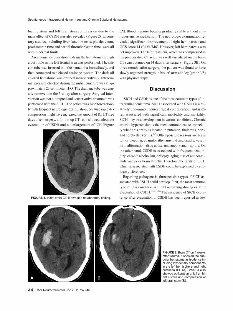
An 87-year-old man was admitted to the emergency de-martment due to vomiting, followed by quadriparesis. The patient was healthy before admission and his medical histo-ry was unremarkable. The patient hit his head after a motor vehicle accident one month prior to admission, and was ad-mitted to another hospital. The initial brain computed to-mography (CT) scan obtained at a local hospital revealed no abnormal findings other than brain atrophy (Figure 1). The patient was managed conservatively for 3 weeks, and dis-charged. He suffered from headache. He was observed at home, until a relapse of headache and quadriparesis, fol-lowed by neurological deterioration, led him to be trans-ferred to our institution four weeks after trauma. On admis-sion, the patient was stuporous with Glasgow coma scale (GCS) score 9 (E2V2M5), and motor examination revealed left hemiparesis (grade 1/5) and right hemiparesis (grade 3/5). Pupils were equal and reactive to light. Blood pressure in the emergency department was 210/130 mmHg. Brain CT scan showed an isodense SDH in the left hemisphere, with a maximum thickness of 1.5 centimeters and acute ICH in the right basal ganglia. Obliteration of the left am-
Received: November 29, 2010 / Revised: February 21, 2011Accepted: April 5, 2011Address for correspondence: Jung-Kil Lee, MDDepartment of Neurosurgery Chonnam National University Hospi-tal & Medical School, 671 Jebong-ro, Dong-gu, Gwangju 501-757, KoreaTel: +82-62-220-6602, Fax: +82-62-224-9865E-mail: [email protected]
Spontaneous Intracerebral Hemorrhage Occurring during Progression of Chronic Subdural Hematoma
Ki-Young Choi, MD1, Jung-Kil Lee, MD1,2, Jae-Won Jang, MD1, Bo-Ra Seo, MD1, Tae-Sun Kim, MD1 and Jae-Hyoo Kim, MD1
1Department of Neurosurgery, Chonnam National University Hospital & Medical School, Gwangju, Korea2The Brain Korea 21 Project, Center for Biomedical Human Resources at Chonnam National University, Gwangju, Korea
Spontaneous intracerbral hemorrhage (SICH) and chronic subdural hematoma (CSDH) represent the most common types of intracranial hematoma. However, SICH during the progression of CSDH is a very rare disease entity due to etiologic differences. The purpose of this report is to alert neurosurgeons to the existence of this condition and to consider possible mechanisms. An 87-year-old man presented with a sudden onset of headache, followed by quadriparesis. Brain computed tomography scan revealed CSDH in the left hemisphere and remote SICH in the right basal ganglia. CSDH was treated with burr hole drainage, however, the deep parenchymal hematoma was treated conservatively. The patient had a progres-sive recovery with a good outcome. We recently experienced a case of SICH that occurred during the progression of CSDH. To our knowledge, this is the first reported case of SICH occurring during the progression of CSDH. (J Kor Neurotraumatol Soc 2011;7:43-46)
KEY WORDS: Chronic subdural hematoma ㆍIntracerebral hemorrhage ㆍProgression.
online © ML Comm
44 J Kor Neurotraumatol Soc 2011;7:43-46
Spontaneous Intracerebral Hemorrhage and Chronic Subdural Hematoma
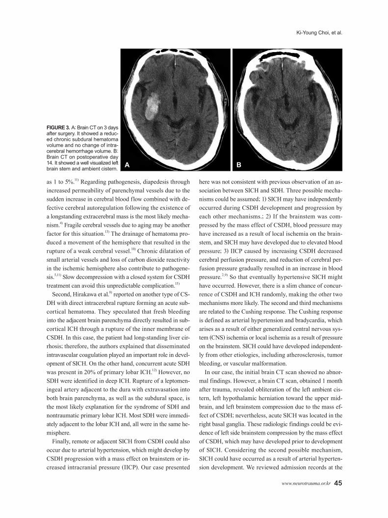
bient cistern and left brainstem compression due to the mass effect of CSDH was also revealed (Figure 2). Labora-tory studies, including liver function tests, platelet count, prothrombin time and partial thromboplastin time, were all within normal limits.
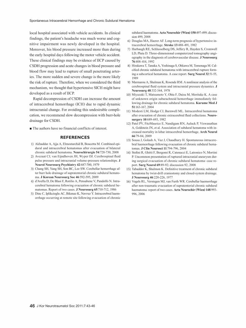
An emergency operation to drain the hematoma through a burr hole in the left frontal area was performed. The sili-con tube was inserted into the hematoma immediately, and then connected to a closed drainage system. The dark-oil colored hematoma was drained intraoperatively. Intracra-nial pressure checked during the initial puncture was at ap-proximately 23 centimeter H2O. The drainage tube was usu-ally removed on the 3rd day after surgery. Surgical inter-vention was not attempted and conservative treatment was performed with the SICH. The patient was monitored close-ly with frequent neurologic examination, because rapid de-compression might have increased the amount of ICH. Three days after surgery, a follow-up CT scan showed adequate evacuation of CSDH and no enlargement of ICH (Figure
3A). Blood pressure became gradually stable without anti-hypertensive medication. The neurologic examination re-vealed significant improvement of right hemiparesis and GCS score 14 (E4V4/M6). However, left hemiparesis was not improved. The left brainstem, which was compressed in the preoperative CT scan, was well visualized on the brain CT scan obtained on 14 days after surgery (Figure 3B). On three months after surgery, the patient was found to have slowly regained strength in his left arm and leg (grade 3/5) with physiotherapy.
Discussion
SICH and CSDH is one of the most common types of in-tracranial hematoma. SICH associated with CSDH is a rel-atively uncommon neurosurgical complication, and is of-ten associated with significant morbidity and mortality. SICH may be a development in various conditions. Chronic arterial hypertension is the most common cause, especial-ly when this entity is located in putamen, thalamus, pons, and cerebellar vermis.6,7) Other possible reasons are brain tumor bleeding, coagulopathy, amyloid angiopathy, vascu-lar malformation, drug abuse, and aneurysmal rupture. On the other hand, CSDH is associated with frequent head in-jury, chronic alcoholism, epilepsy, aging, use of anticoagu-lants, and prior brain atrophy. Therefore, the rarity of SICH which is associated with CSDH could be explained by etio-logic differences.
Regarding pathogenesis, three possible types of SICH as-sociated with CSDH could develop. First, the most common type of this condition is SICH occurring during or after evacuation of CSDH.1,3,5,11,16) The incidence of SICH occur-rence after evacuation of CSDH has been reported as low FIGURE 1. Initial brain CT. It revealed no abnormal finding.
A B
FIGURE 2. Brain CT on 4 weeks after trauma. It showed the sub-dural hematoma as isodense in-cluding low density components in the left hemisphere and right putaminal ICH (A). Brain CT also showed obliteration of left ambi-ent cistern and compression of left brainstem (B).
www.neurotrauma.or.kr 45
Ki-Young Choi, et al.
as 1 to 5%.11) Regarding pathogenesis, diapedesis through increased permeability of parenchymal vessels due to the sudden increase in cerebral blood flow combined with de-fective cerebral autoregulation following the existence of a longstanding extracerebral mass is the most likely mecha-nism.4) Fragile cerebral vessels due to aging may be another factor for this situation.13) The drainage of hematoma pro-duced a movement of the hemisphere that resulted in the rupture of a weak cerebral vessel.10) Chronic dilatation of small arterial vessels and loss of carbon dioxide reactivity in the ischemic hemisphere also contribute to pathogene-sis.5,11) Slow decompression with a closed system for CSDH treatment can avoid this unpredictable complication.15)
Second, Hirakawa et al.8) reported on another type of CS-DH with direct intracerebral rupture forming an acute sub-cortical hematoma. They speculated that fresh bleeding into the adjacent brain parenchyma directly resulted in sub-cortical ICH through a rupture of the inner membrane of CSDH. In this case, the patient had long-standing liver cir-rhosis; therefore, the authors explained that disseminated intravascular coagulation played an important role in devel-opment of SICH. On the other hand, concurrent acute SDH was present in 20% of primary lobar ICH.12) However, no SDH were identified in deep ICH. Rupture of a leptomen-ingeal artery adjacent to the dura with extravasation into both brain parenchyma, as well as the subdural space, is the most likely explanation for the syndrome of SDH and nontraumatic primary lobar ICH. Most SDH were immedi-ately adjacent to the lobar ICH and, all were in the same he-misphere.
Finally, remote or adjacent SICH from CSDH could also occur due to arterial hypertension, which might develop by CSDH progression with a mass effect on brainstem or in-creased intracranial pressure (IICP). Our case presented
here was not consistent with previous observation of an as-sociation between SICH and SDH. Three possible mecha-nisms could be assumed; 1) SICH may have independently occurred during CSDH development and progression by each other mechanisms.; 2) If the brainstem was com-pressed by the mass effect of CSDH, blood pressure may have increased as a result of local ischemia on the brain-stem, and SICH may have developed due to elevated blood pressure; 3) IICP caused by increasing CSDH decreased cerebral perfusion pressure, and reduction of cerebral per-fusion pressure gradually resulted in an increase in blood pressure.2,9) So that eventually hypertensive SICH might have occurred. However, there is a slim chance of concur-rence of CSDH and ICH randomly, making the other two mechanisms more likely. The second and third mechanisms are related to the Cushing response. The Cushing response is defined as arterial hypertension and bradycardia, which arises as a result of either generalized central nervous sys-tem (CNS) ischemia or local ischemia as a result of pressure on the brainstem. SICH could have developed independent-ly from other etiologies, including atherosclerosis, tumor bleeding, or vascular malformation.
In our case, the initial brain CT scan showed no abnor-mal findings. However, a brain CT scan, obtained 1 month after trauma, revealed obliteration of the left ambient cis-tern, left hypothalamic herniation toward the upper mid-brain, and left brainstem compression due to the mass ef-fect of CSDH; nevertheless, acute SICH was located in the right basal ganglia. These radiologic findings could be evi-dence of left side brainstem compression by the mass effect of CSDH, which may have developed prior to development of SICH. Considering the second possible mechanism, SICH could have occurred as a result of arterial hyperten-sion development. We reviewed admission records at the
A B
FIGURE 3. A: Brain CT on 3 days after surgery. It showed a reduc-ed chronic subdural hematoma volume and no change of intra-cerebral hemorrhage volume. B: Brain CT on postoperative day 14. It showed a well visualized left brain stem and ambient cistern.
46 J Kor Neurotraumatol Soc 2011;7:43-46
Spontaneous Intracerebral Hemorrhage and Chronic Subdural Hematoma
local hospital associated with vehicle accidents. In clinical findings, the patient’s headache was much worse and cog-nitive impairment was newly developed in the hospital. Moreover, his blood pressure increased more than during the early hospital days following the motor vehicle accident. These clinical findings may be evidence of IICP caused by CSDH progression and acute changes in blood pressure and blood flow may lead to rupture of small penetrating arter-ies. The more sudden and severe change is the more likely the risk of rupture. Therefore, when we considered the third mechanism, we thought that hypertensive SICH might have developed as a result of IICP.
Rapid decompression of CSDH can increase the amount of intracerebral hemorrhage (ICH) due to rapid dynamic intracranial change. For avoiding this undesirable compli-cation, we recommend slow decompression with burr-hole drainage for CSDH.
■ The authors have no financial conflicts of interest.
REFERENCES1) Akhaddar A, Ajja A, Elmostarchid B, Boucetta M. Combined epi-
dural and intracerebral hematomas after evacuation of bilateral chronic subdural hematoma. Neurochirurgie 54:728-730, 2008
2) Avezaat CJ, van Eijndhoven JH, Wyper DJ. Cerebrospinal fluid pulse pressure and intracranial volume-pressure relationships. J Neurol Neurosurg Psychiatry 42:687-700, 1979
3) Chang SH, Yang SH, Son BC, Lee SW. Cerebellar hemorrhage af-ter burr hole drainage of supratentorial chronic subdural hemato-ma. J Korean Neurosurg Soc 46:592-595, 2009
4) d’Avella D, De Blasi F, Rotilio A, Pensabene V, Pandolfo N. Intra-cerebral hematoma following evacuation of chronic subdural he-matomas. Report of two cases. J Neurosurg 65:710-712, 1986
5) Dinc C, Iplikcioglu AC, Bikmaz K, Navruz Y. Intracerebral haem-orrhage occurring at remote site following evacuation of chronic
subdural haematoma. Acta Neurochir (Wien) 150:497-499; discus-sion 499, 2008
6) Douglas MA, Haerer AF. Long-term prognosis of hypertensive in-tracerebral hemorrhage. Stroke 13:488-491, 1982
7) Harbaugh RE, Schlusselberg DS, Jeffery R, Hayden S, Cromwell LD, Pluta D. Three-dimensional computerized tomography angi-ography in the diagnosis of cerebrovascular disease. J Neurosurg 76:408-414, 1992
8) Hirakawa T, Tanaka A, Yoshinaga S, Ohkawa M, Tomonaga M. Cal-cified chronic subdural hematoma with intracerebral rupture form-ing a subcortical hematoma. A case report. Surg Neurol 32:51-55, 1989
9) Marmarou A, Shulman K, Rosende RM. A nonlinear analysis of the cerebrospinal fluid system and intracranial pressure dynamics. J Neurosurg 48:332-344, 1978
10)Miyazaki T, Matsumoto Y, Ohta F, Daisu M, Moritake K. A case of unknown origin subarachnoid hemorrhage immediately fol-lowing drainage for chronic subdural hematoma. Kurume Med J 51:163-167, 2004
11) Modesti LM, Hodge CJ, Barnwell ML. Intracerebral hematoma after evacuation of chronic extracerebral fluid collections. Neuro-surgery 10:689-693, 1982
12)Patel PV, FitzMaurice E, Nandigam RN, Auluck P, Viswanathan A, Goldstein JN, et al. Association of subdural hematoma with in-creased mortality in lobar intracerebral hemorrhage. Arch Neurol 66:79-84, 2009
13)Sousa J, Golash A, Vaz J, Chaudhary H. Spontaneous intracere-bral haemorrhage following evacuation of chronic subdural hema-tomas. J Clin Neurosci 11:794-796, 2004
14)Stefini R, Ghitti F, Bergomi R, Catenacci E, Latronico N, Mortini P. Uncommon presentation of ruptured intracranial aneurysm dur-ing surgical evacuation of chronic subdural hematoma: case re-port. Surg Neurol 69:89-92; discussion 92, 2008
15)Tabaddor K, Shulmon K. Definitive treatment of chronic subdural hematoma by twist-drill craniostomy and closed-system drainage. J Neurosurg 46:220-226, 1977
16)Vogels RL, Verstegen MJ, van Furth WR. Cerebellar haemorrhage after non-traumatic evacuation of supratentorial chronic subdural haematoma: report of two cases. Acta Neurochir (Wien) 148:993-996, 2006