Embed Size (px)
Citation preview
Korean J Pain 2011 December; Vol. 24, No. 4: 235-238pISSN 2005-9159 eISSN 2093-0569http://dx.doi.org/10.3344/kjp.2011.24.4.235
| Case Report |
Spontaneous Height Restoration of Vertebral Compression Fracture
- A Case Report-
Department of Anesthesiology and Pain Medicine, Seoul National University Hospital, Seoul, *Seoul National University Bundang Hospital, Seongnam, Korea
Young Joo, MD, Pyung Bok Lee, MD*, and Francis Sahngun Nahm, MD*
Vertebral compression fractures result in vertebral height loss and alter sagittal spinal alignment, which in turn can lead to increased morbidity and mortality. Acute osteoporotic vertebral compression fractures are known to increase mobility and instability of the spine. There are limited published data correlating the degree of dynamic mobility and the efficacy of kyphoplasty on vertebral compression fractures. Here we report a 73-year-old female with a severe acute osteoporotic L2 compression fracture who obtained total vertebral height restoration following kyphoplasty, with resolution of back pain. (Korean J Pain 2011; 24: 235-238)
Key Words:
compression fracture, dynamic mobility, kyphoplasty, osteoporosis, spine.
Received August 9, 2011. Revised October 5, 2011. Accepted October 6, 2011.Correspondence to: Francis Sahngun Nahm, MDDepartment of Anesthesiology and Pain Medicine, Seoul National University Bundang Hospital, 166, Gumi-ro, Bundang-gu, Seongnam 463-707, KoreaTel: +82-31-787-7499, Fax: +82-31-787-4063, E-mail: [email protected]
This is an open-access article distributed under the terms of the Creative Commons Attribution Non-Commercial License (http:// creativecommons.org/licenses/by-nc/3.0/), which permits unrestricted non-commercial use, distribution, and reproduction in any medium, provided the original work is properly cited.Copyright ⓒ The Korean Pain Society, 2011
Vertebral compression fractures (VCFs) are primarily
caused by osteoporosis. As the population ages, the in-
cidence of VCFs is also likely to increase [1]. The clinical
characteristics of VCFs are loss of vertebral height and
acute pain. Radiographic images are the standardized pro-
tocol to diagnose VCFs, with changes in vertebral height
revealed on AP and lateral views [2]. However, due to the
increased dynamic mobility (DM) of vertebrae at the site
of a fracture, transient spontaneous restoration of verte-
bral height is also common. Therefore, depending on the
patient’s posture, acute compression fracture or loss of
height may not be visualized on a static image, possibly
leading to inaccurate diagnosis and treatment. Dynamic
mobility of vertebral compression fractures can be meas-
ured by comparing standing and supine images. However,
there is limited information available in the literature on
the extent and frequency of this phenomenon. We describe
a case of acute vertebral compression fracture with a high
degree of dynamic mobility and pain treated with kyph-
oplasty.
CASE REPORT
A seventy-three-year-old woman with a 2-week his-
236 Korean J Pain Vol. 24, No. 4, 2011
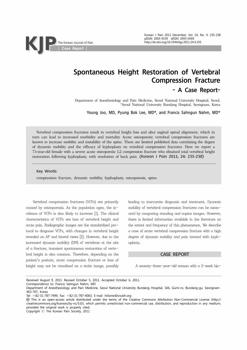
Fig. 1. T1 weighted image of lumbar MRI. There is no height loss except bone marrow edema in 2nd lumbar vertebra.
Fig. 2. Preoperative standing anterior-posterior radiographicstudy showing the anterior height of L2 vertebral was 56.7% compared to that of L3.
Fig. 3. Prone cross-table lateral fluoroscopy was performedand we found the height restoration of L2 vertebral body.
tory of severe back pain after a fall was referred to our
pain clinic. Bed rest and conservative therapies had failed
to provide relief, and the patient rated her pain level as
9 out of 10 cm on the visual analogue scale (VAS). She
had a history of osteoporosis and had been taking bi-
sphosphonates for the past 1 year.
The initial lumbar magnetic resonance imaging (MRI) study revealed marrow edema at the L2 vertebra, but no
definite compression fracture (Fig. 1). Due to the severity
of the patient’s pain, we performed a fluoroscopically-
guided lumbar epidural injection with a mixture of tri-
amcinolone 20 mg and 0.18% levobupivacaine 8 ml. She was
discharged home on UltracetⓇ and nortriptyline. However,
three days later the patient returned to the pain clinic with
progressively worsening back pain. On a repeat lumbar
MRI, an acute vertebral compression fracture was noted
at L2. The anterior height of the L2 vertebral body was
only 54.80% of that of L3 (Fig. 2). Subsequently, the deci-
sion was made to proceed with kyphoplasty of the L2 ver-
tebral body.
The procedure consisted of placing the patient in the
prone position, with bolsters at the upper thorax and pelvis
to allow extension of the spine. The lateral fluoroscopy
view confirmed the total height restoration of the L2 ver-
tebral body (Fig. 3). After sterile preparation of the skin,
kyphoplasty was performed bilaterally under fluoroscopic
guidance.
After the drill was advanced through the access can-
nula, the balloon bone tamp was placed in the cavity. The
cement mixture (polymethyl methacrylate, barium sulfate)
was injected slowly, ensuring no cement leaks from the
borders of the created bone void (Fig. 4). The wounds were
cleaned and dressed, and the patient was monitored over-
night prior to discharge home in satisfactory condition.
There were no complications from the procedure, and the
patient reported a VAS score of 2 out of 10 at the time
of discharge.
Y Joo, et al / Spontaneous Height Restoration of VCF 237
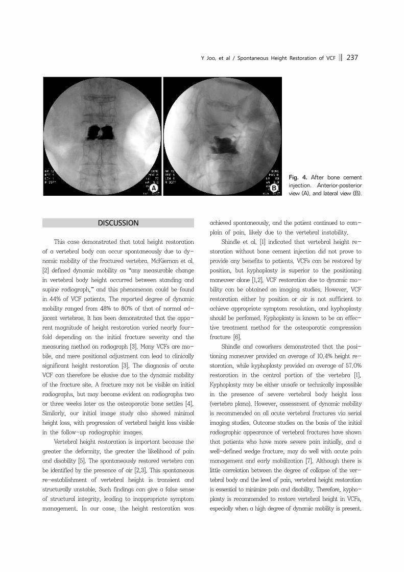
Fig. 4. After bone cement injection. Anterior-posterior view (A), and lateral view (B).
DISCUSSION
This case demonstrated that total height restoration
of a vertebral body can occur spontaneously due to dy-
namic mobility of the fractured vertebra. McKiernan et al.
[2] defined dynamic mobility as “any measurable change
in vertebral body height occurred between standing and
supine radiograph,” and this phenomenon could be found
in 44% of VCF patients. The reported degree of dynamic
mobility ranged from 48% to 80% of that of normal ad-
jacent vertebrae. It has been demonstrated that the appa-
rent magnitude of height restoration varied nearly four-
fold depending on the initial fracture severity and the
measuring method on radiograph [3]. Many VCFs are mo-
bile, and mere positional adjustment can lead to clinically
significant height restoration [3]. The diagnosis of acute
VCF can therefore be elusive due to the dynamic mobility
of the fracture site. A fracture may not be visible on initial
radiographs, but may become evident on radiographs two
or three weeks later as the osteoporotic bone settles [4].
Similarly, our initial image study also showed minimal
height loss, with progression of vertebral height loss visible
in the follow-up radiographic images.
Vertebral height restoration is important because the
greater the deformity, the greater the likelihood of pain
and disability [5]. The spontaneously restored vertebra can
be identified by the presence of air [2,3]. This spontaneous
re-establishment of vertebral height is transient and
structurally unstable. Such findings can give a false sense
of structural integrity, leading to inappropriate symptom
management. In our case, the height restoration was
achieved spontaneously, and the patient continued to com-
plain of pain, likely due to the vertebral instability.
Shindle et al. [1] indicated that vertebral height re-
storation without bone cement injection did not prove to
provide any benefits to patients. VCFs can be restored by
position, but kyphoplasty is superior to the positioning
maneuver alone [1,2]. VCF restoration due to dynamic mo-
bility can be obtained on imaging studies. However, VCF
restoration either by position or air is not sufficient to
achieve appropriate symptom resolution, and kyphoplasty
should be perfomed. Kyphoplasty is known to be an effec-
tive treatment method for the osteoporotic compression
fracture [6].
Shindle and coworkers demonstrated that the posi-
tioning maneuver provided an average of 10.4% height re-
storation, while kyphoplasty provided an average of 57.0%
restoration in the central portion of the vertebra [1].
Kyphoplasty may be either unsafe or technically impossible
in the presence of severe vertebral body height loss
(vertebra plana). However, assessment of dynamic mobility
is recommended on all acute vertebral fractures via serial
imaging studies. Outcome studies on the basis of the initial
radiographic appearance of vertebral fractures have shown
that patients who have more severe pain initially, and a
well-defined wedge fracture, may do well with acute pain
management and early mobilization [7]. Although there is
little correlation between the degree of collapse of the ver-
tebral body and the level of pain, vertebral height restoration
is essential to minimize pain and disability. Therefore, kypho-
plasty is recommended to restore vertebral height in VCFs,
especially when a high degree of dynamic mobility is present.

238 Korean J Pain Vol. 24, No. 4, 2011
REFERENCES
1. Shindle MK, Gardner MJ, Koob J, Bukata S, Cabin JA, Lane JM. Vertebral height restoration in osteoporotic compression fractures: kyphoplasty balloon tamp is superior to postural correction alone. Osteoporos Int 2006; 17: 1815-9.
2. McKiernan F, Jensen R, Faciszewski T. The dynamic mobility of vertebral compression fractures. J Bone Miner Res 2003; 18: 24-9.
3. McKiernan F, Faciszewski T, Jensen R. Reporting height restoration in vertebral compression fractures. Spine (Phila Pa 1976) 2003; 28: 2517-21.
4. Rao RD, Singrakhia MD. Painful osteoporotic vertebral fracture. Pathogenesis, evaluation, and roles of vertebroplasty and kyphoplasty in its management. J Bone Joint Surg Am 2003; 85-A: 2010-22.
5. Silverman SL. The clinical consequences of vertebral com-pression fracture. Bone 1992; 13 Suppl 2: S27-31.
6. Han KR, Kim C, Yang JY, Han ST, Kim YS. Balloon kypho-plasty for the treatment of vertebral compression fractures. Korean J Pain 2006; 19: 56-62.
7. Lyritis GP, Mayasis B, Tsakalakos N, Lambropoulos A, Gazi S, Karachalios T, et al. The natural history of the osteoporotic vertebral fracture. Clin Rheumatol 1989; 8 Suppl 2: 66-9.