Embed Size (px)
Citation preview
SPONTANEOUS CSF LEAKS
DR. ASEEL DOUBI
R5 ORL HNS
OUTLINE:
▸ Physiology of CSF
▸ Types of CSF rhinorrhea
▸ Spontaneous CSF leak approach and diagnosis
▸ Operative options and postoperative care
▸ Lumbar drains current updates

PHYSIOLOGY OF CSF
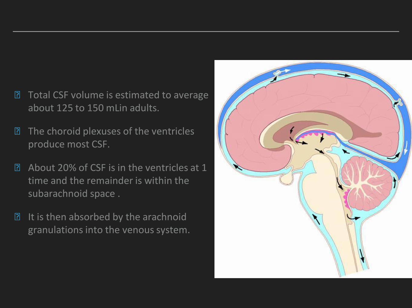
▸ Total CSF volume is estimated to average about 125 to 150 mLin adults.
▸ The choroid plexuses of the ventricles produce most CSF.
▸ About 20% of CSF is in the ventricles at 1 time and the remainder is within the subarachnoid space .
▸ It is then absorbed by the arachnoid granulations into the venous system.

WHAT ARE THE TYPES OF CSF RHINORRHEA?
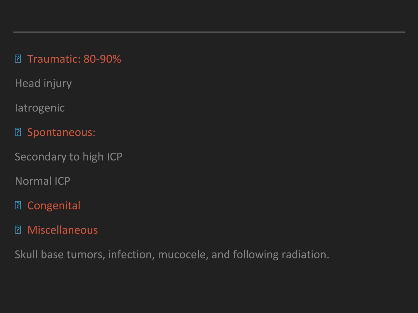
▸ Traumatic: 80-90%
Head injury
Iatrogenic
▸ Spontaneous:
Secondary to high ICP
Normal ICP
▸ Congenital
▸ Miscellaneous
Skull base tumors, infection, mucocele, and following radiation.
HEAD INJURY
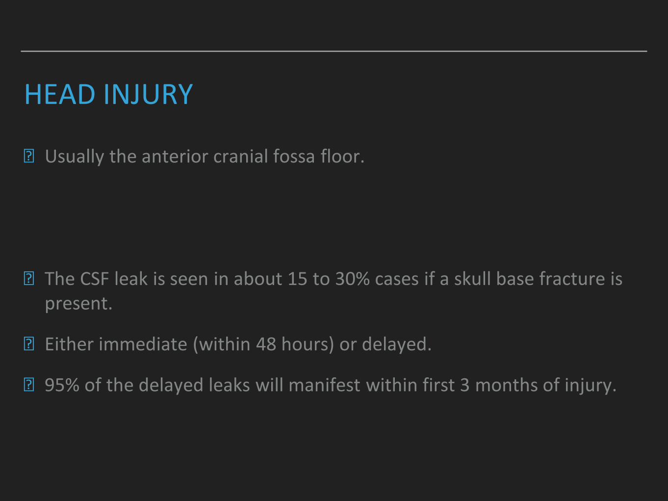
▸ Usually the anterior cranial fossa floor.
▸ The CSF leak is seen in about 15 to 30% cases if a skull base fracture is present.
▸ Either immediate (within 48 hours) or delayed.
▸ 95% of the delayed leaks will manifest within first 3 months of injury.
IATROGENIC
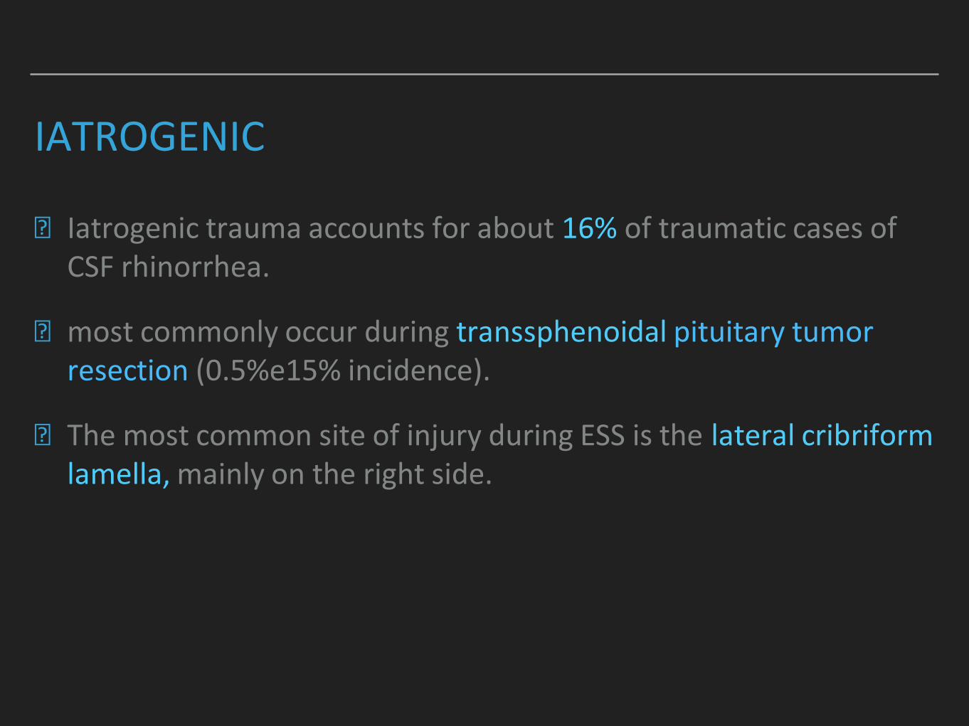
▸ Iatrogenic trauma accounts for about 16% of traumatic cases of CSF rhinorrhea.
▸ most commonly occur during transsphenoidal pituitary tumor resection (0.5%e15% incidence).
▸ The most common site of injury during ESS is the lateral cribriform lamella, mainly on the right side.
CONGENITAL
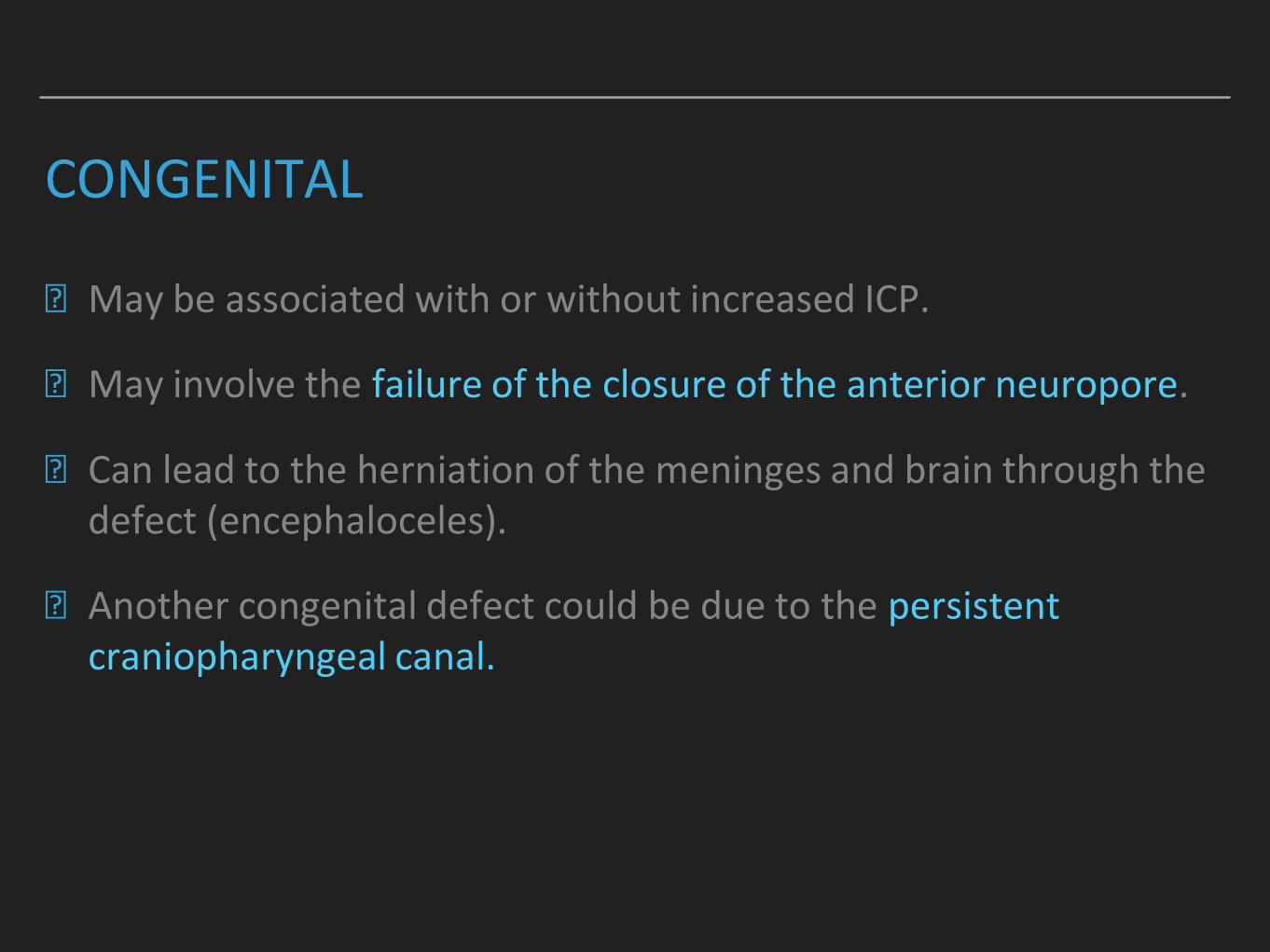
▸ May be associated with or without increased ICP.
▸ May involve the failure of the closure of the anterior neuropore.
▸ Can lead to the herniation of the meninges and brain through the defect (encephaloceles).
▸ Another congenital defect could be due to the persistent craniopharyngeal canal.

SPONTANEOUS
SECONDARY TO HIGH ICP
▸ High pressure leaks could account up to 45% of the non-traumatic CSF rhinorrhea.
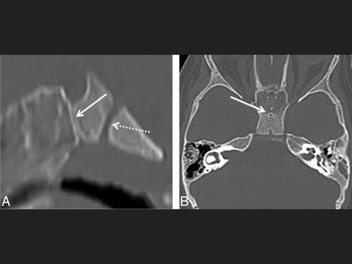
▸ The cribriform plate, craniopharyngeal canal, sella, and spheno-occipital synchondrosis are some of the possible sites
WITH NORMAL ICP
▸ Normal pressure leaks represent 55% of the non-traumatic cases of the CSF rhinorrhea.
▸ Hypothesis is that the spontaneous leak is due to the physiologic alterations in CSF pressure that lead to point erosions in the skull base.
PATHOPHYSIOLOGY OF SPONTANEOUS CSF LEAK
▸ Patients with spontaneous idiopathic CSF leaks are increasingly being recognized as having increased ICP
▸ This increase in ICP is likely due to impaired CSF absorption by the arachnoid villi.
▸ Once the ICP increases, pulsatile forces are exerted on the weakest areas of the skull base, such as the cribriform plate and lateral recesses of hyperpneumatized sphenoid sinuses.
▸ These forces thin the skull base and allow herniation of meninges and brain through the defect, often with resultant CSF leak
LOCATIONS OF CSF LEAKS?
▸ The ethmoid roof and cribriform region are the most commonly involved sites, affected in more than 50% of the cases reviewed.
▸ The most of the leaks are along the course of anterior ethmoid artery followed by the sphenoid sinus in spontaneous leaks.
▸ Defect in the sphenoid sinus could be in the roof, the lateral wall, anterior wall, or the posterior wall.
▸ cribriform plate defects are usually small
▸ Lateral sphenoid defects are larger
Alkis J. Psaltis et al.

DIAGNOSIS?
PRESENTATION:
▸ Spontaneous CSF leaks are highly associated with the female gender and obesity.
▸ In one series of 55 consecutive patients with spontaneous CSF leaks, 70% of patients were women and 46 of 55 patients were obese, with an average body mass index (BMI) of 36.2 kg/m2. Elevated ICP persisted in these patients postoperatively, with an average lumbar drain pressure of 27 cm H2O
Woodworth BA et al. 2008
EXAMINATION:
▸ May identify fluid leaking from the nose as CSF.
▸ Preoperative nasal endoscopy could help in localizing the defect.
▸ Valsalva maneuver and jugular compression could improve detection rate.
LABS:
▸ Beta-2 transferrin can be detected by immunofixation electrophoresis.
▸ With sensitivity of 94% to 100%, and specificity of 98% to 100%
▸ gold standard in detection of CSF leakage.
▸ Beta-2 transferrin is found only in the CSF, vitreous humor of the eye and perilymph
▸ Analysis for beta-2 transferrin requires as little as 0.17 ml of fluid, with results in less than 3 hours
▸ False-positives?
Sarah K. Wise and Rodney J. Schlosser
▸ Beta-trace protein is another marker that is highly sensitive and specific for CSF when detected in rhinorrhea specimens.
▸ Beta-trace protein is secreted into the CSF after being produced in the leptomeninges and choroid plexus.
▸ It is present in other fluids throughout the body, but its concentration elsewhere is significantly lower than that found in CSF
Sarah K. Wise and Rodney J. Schlosser
IMAGING:
▸ Imaging is an important component in the investigation of unilateral watery discharge suspicious of CSF.
▸ It may be difficult to demonstrate the exact site of the leak
CT:
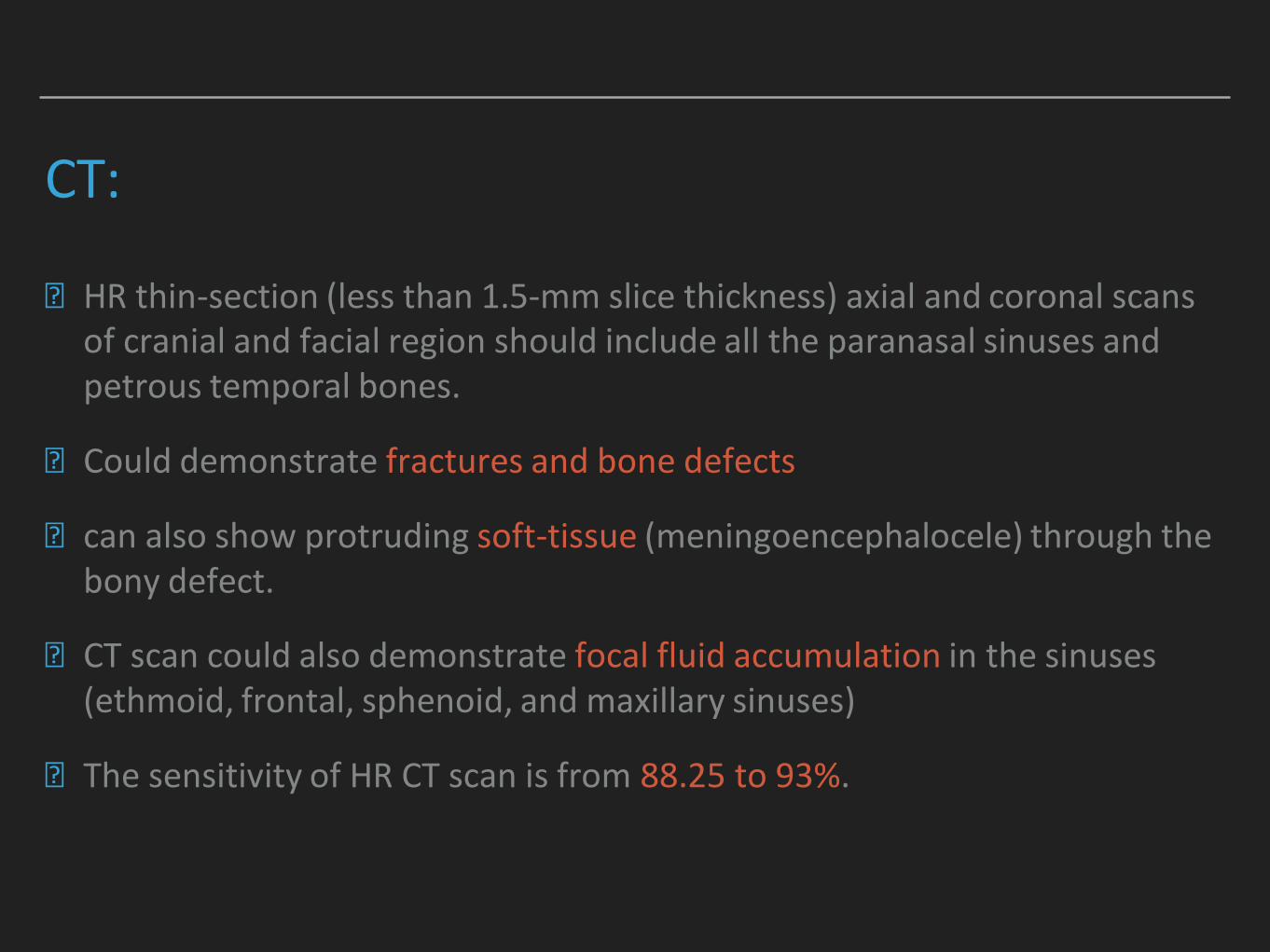
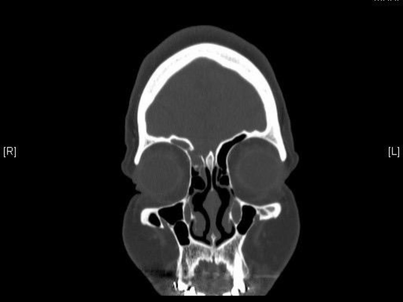
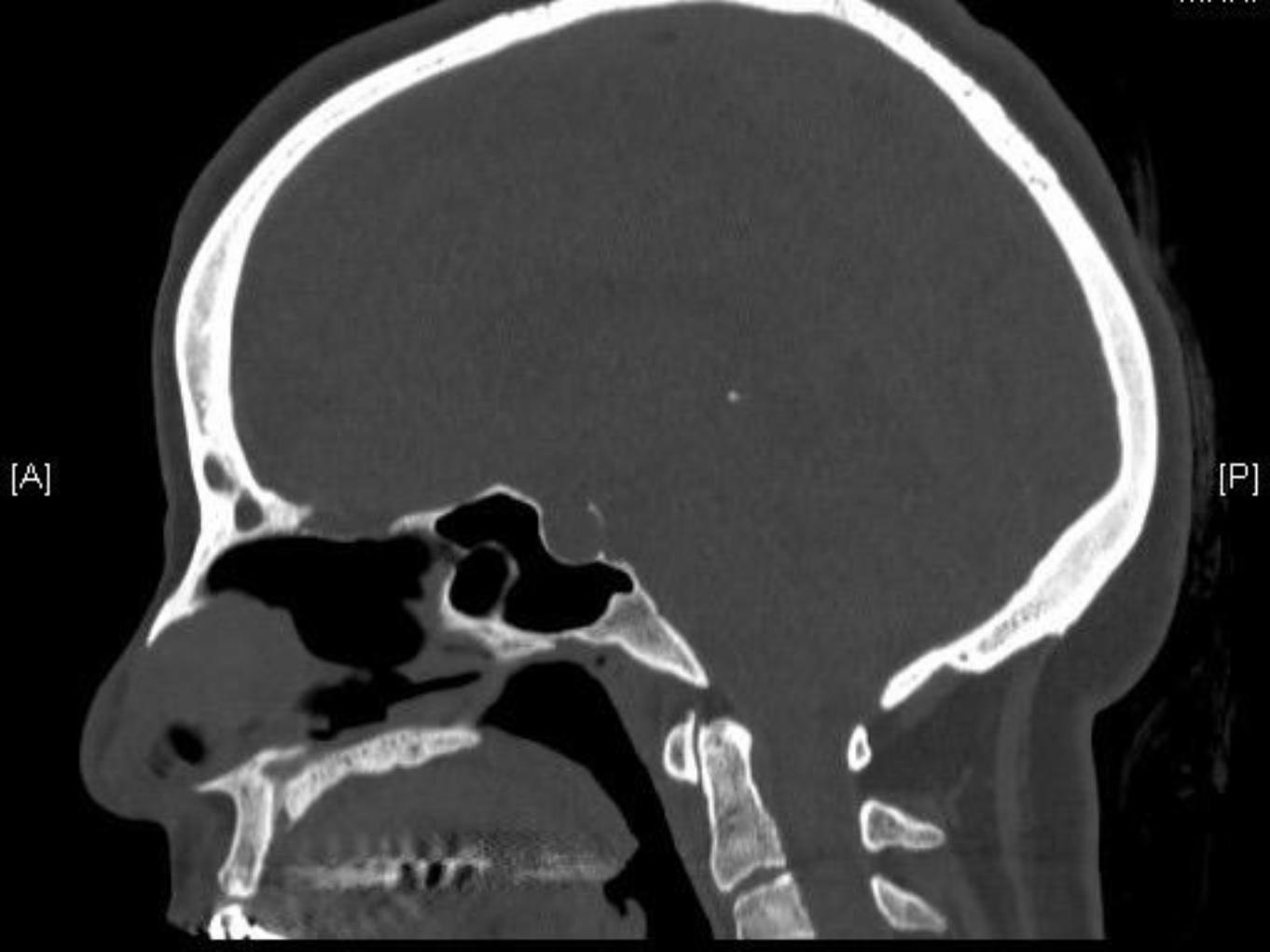
▸ HR thin-section (less than 1.5-mm slice thickness) axial and coronal scans of cranial and facial region should include all the paranasal sinuses and petrous temporal bones.
▸ Could demonstrate fractures and bone defects
▸ can also show protruding soft-tissue (meningoencephalocele) through the bony defect.
▸ CT scan could also demonstrate focal fluid accumulation in the sinuses (ethmoid, frontal, sphenoid, and maxillary sinuses)
▸ The sensitivity of HR CT scan is from 88.25 to 93%.
▸ The ethmoid roof in these patients is dehiscent 14% of the time.
▸ Arachnoid pits, due to bony impressions from arachnoid villi, are identified along the bony skull base in 63% of patients with spontaneous idiopathic CSF leak.
▸ 91% of patients with spontaneous idiopathic CSF leaks were found to have pneumatization of the lateral sphenoid sinus recess, in comparison with similar findings in 23–43% of normal patients. Shetty et al.
Sarah K. Wise and Rodney J. Schlosser
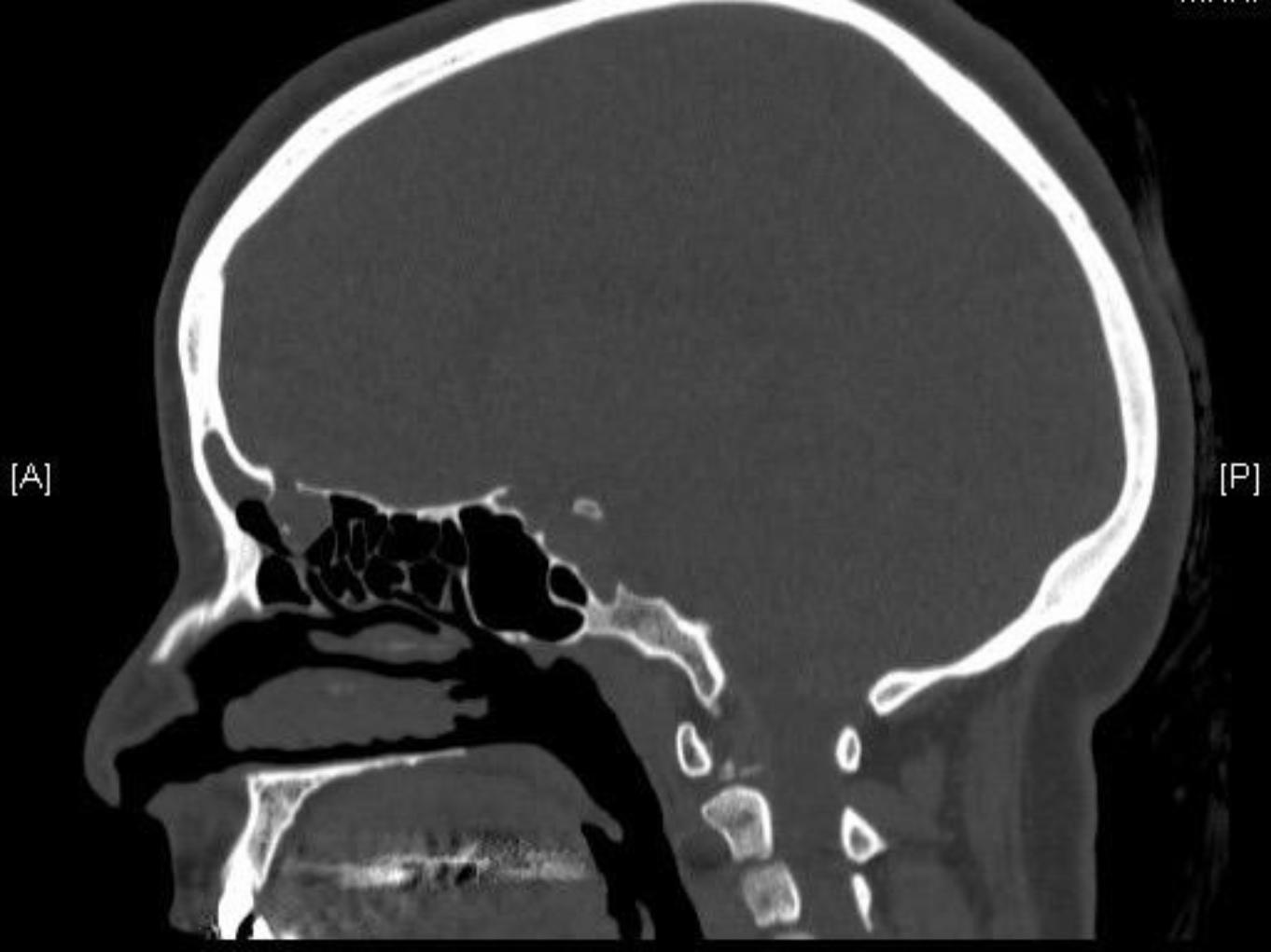
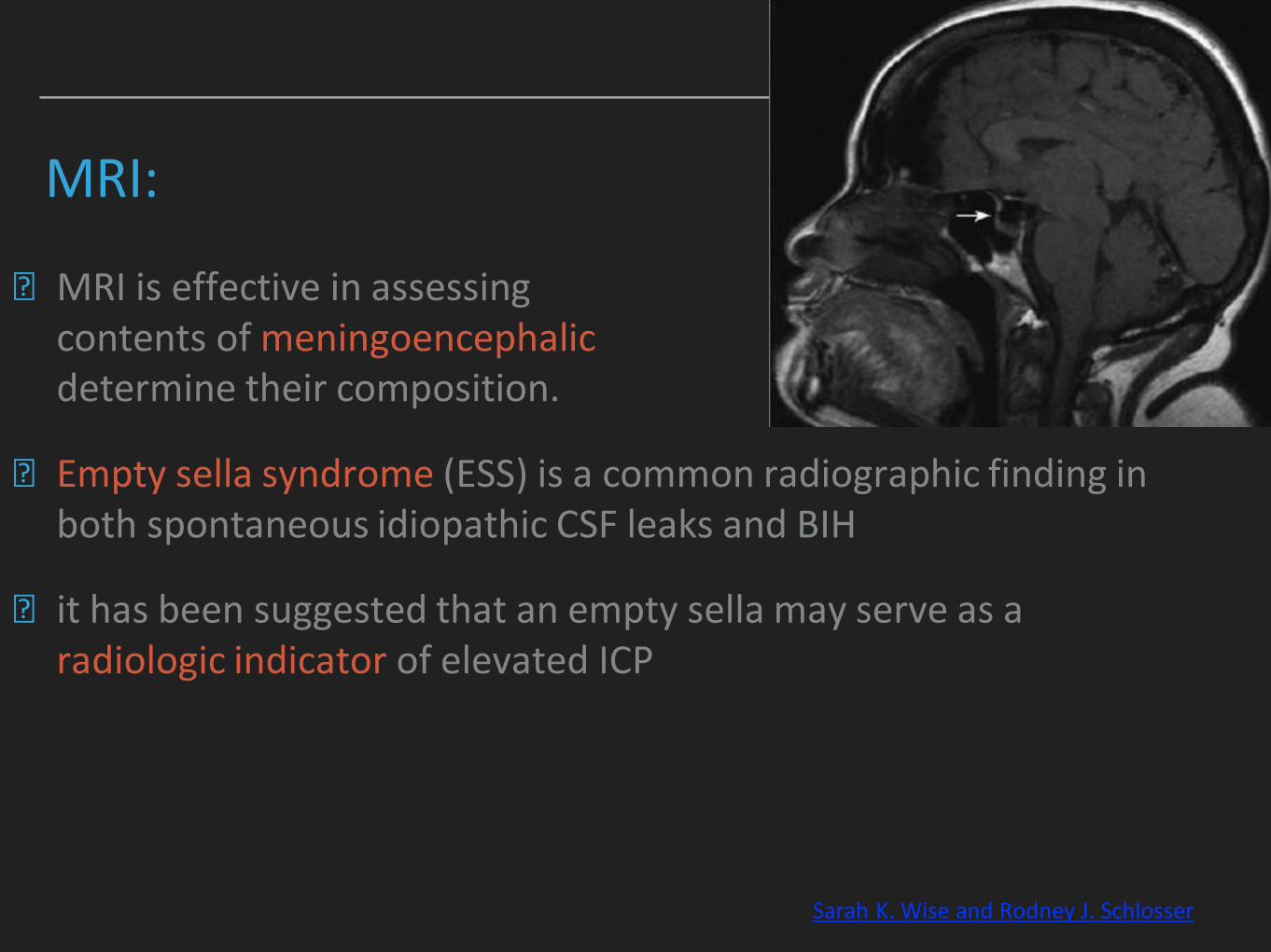
MRI:
▸ MRI is effective in assessing the contents of meningoencephalic sacs to determine their composition.
▸ Empty sella syndrome (ESS) is a common radiographic finding in both spontaneous idiopathic CSF leaks and BIH
▸ it has been suggested that an empty sella may serve as a radiologic indicator of elevated ICP
Sarah K. Wise and Rodney J. Schlosser
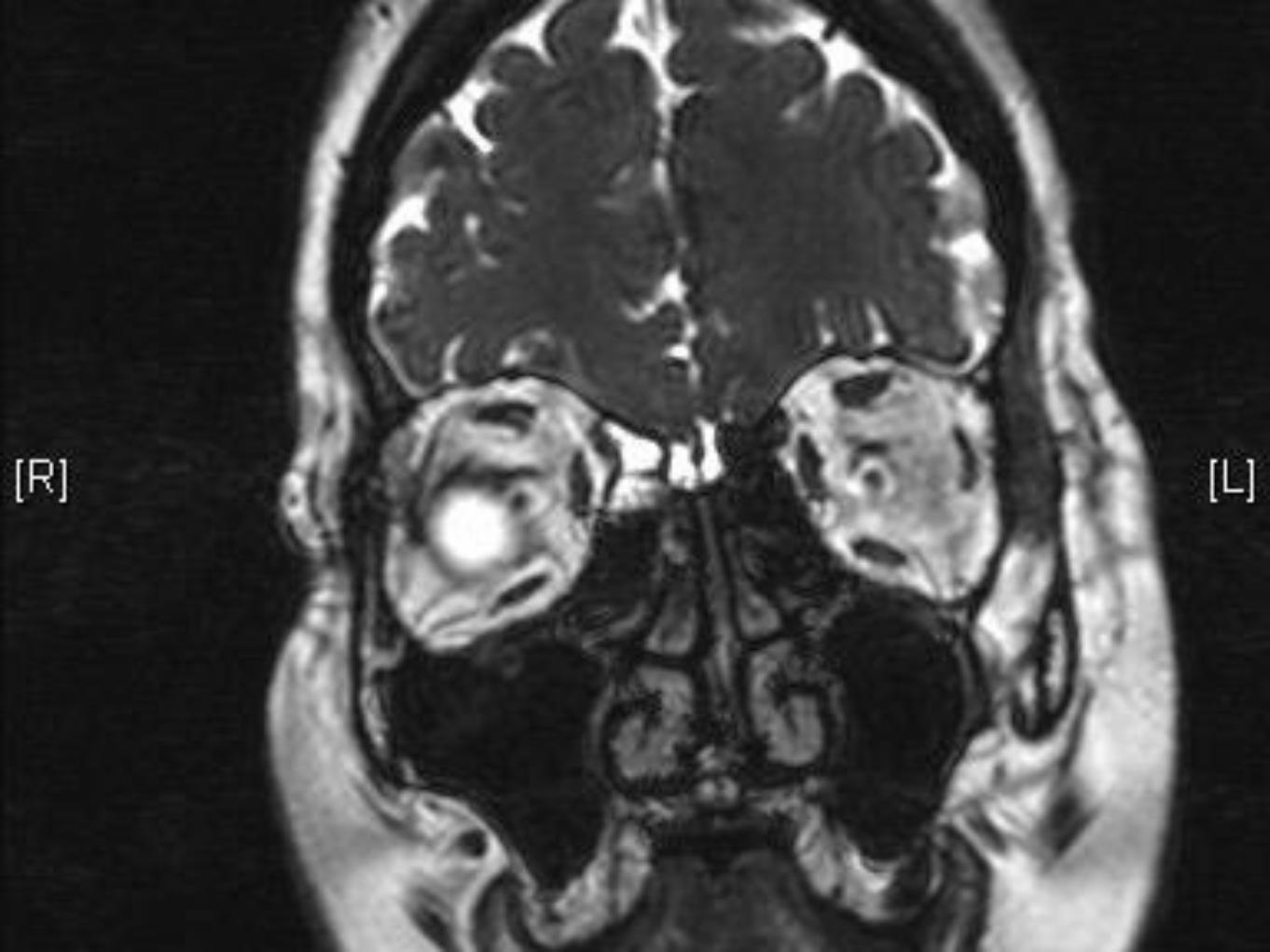
▸ A considerable number of studies reported using both CT and MRI, a practice that has been shown to increase the sensitivity of leak detection to almost 97%.
▸ May also be useful in the detection of multiple leak sites or those with coexistent meningoencephaloceles, which may be present in up to 30% of cases as seen in this study
Alkis J. Psaltis et al.
CT CISTERNOGRAPHY
▸ CT cisternography requires injection of contrast material intrathecally with a LP.
▸ 85% sensitive for detecting active leaks at the time of the study, but its sensitivity varies between 48% and 96% for inactive leaks.
▸ The drawbacks of CT cisternography are its radiation exposure, especially if delayed scans are required, its invasive nature, possible contrast allergic reactions, and rarely intracranial hemorrhage.
JEFFREY C. BEDROSIAN ET AL.
MRI CISTERNOGRAPHY
▸ Advances in MR imaging techniques have improved sensitivity of MR cisternography from 89% to 100% even in inactive leaks.
▸ MR cisternography offers poor spatial and bony resolution.
▸ Algin et al. reported a 100% sensitivity for detection of CSF leakage.
▸ In a larger study of 85 patients, MRI cisternography detected a CSF leak in 100% of patients with clinically evident meningitis or continuous CSF rhinorrhea. Schwartz et al.
▸ Aiden et al. reported no adverse side effects from the 0.5-mL intrathecal gadolinium injection.
INTRATHECAL FLUORESCEIN
▸ Non-ophthalmic solution of 0.1 ml of 10% fluorescein is diluted in 10 ml of CSF and injected into the subarachnoid space over a period of 10 minutes.
▸ Nasal endoscopy is performed approximately 30 minutes after an intrathecal injection.
▸ Fluorescein could be directly observed within the sinonasal cavity using standard xenon light sources in most of the cases.
▸ A blue-light filter (440-490 nm wave length) can help enhance the visualization of fluorescein.
▸ It has been found to be useful in localizing the CSF rhinorrhea. Sensitivity for fluorescein detection varies between 57.7 and 85.6%, while the specificity is 100%. The false-negative rate varies between 15.8 and 43.5%.
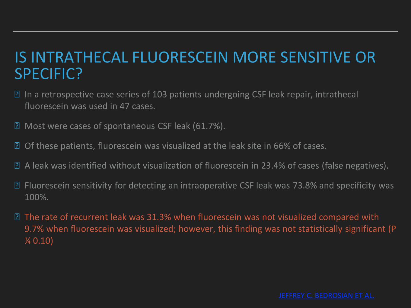
IS INTRATHECAL FLUORESCEIN MORE SENSITIVE OR SPECIFIC?▸ In a retrospective case series of 103 patients undergoing CSF leak repair, intrathecal
fluorescein was used in 47 cases.
▸ Most were cases of spontaneous CSF leak (61.7%).
▸ Of these patients, fluorescein was visualized at the leak site in 66% of cases.
▸ A leak was identified without visualization of fluorescein in 23.4% of cases (false negatives).
▸ Fluorescein sensitivity for detecting an intraoperative CSF leak was 73.8% and specificity was 100%.
▸ The rate of recurrent leak was 31.3% when fluorescein was not visualized compared with 9.7% when fluorescein was visualized; however, this finding was not statistically significant (P ¼ 0.10)
JEFFREY C. BEDROSIAN ET AL.
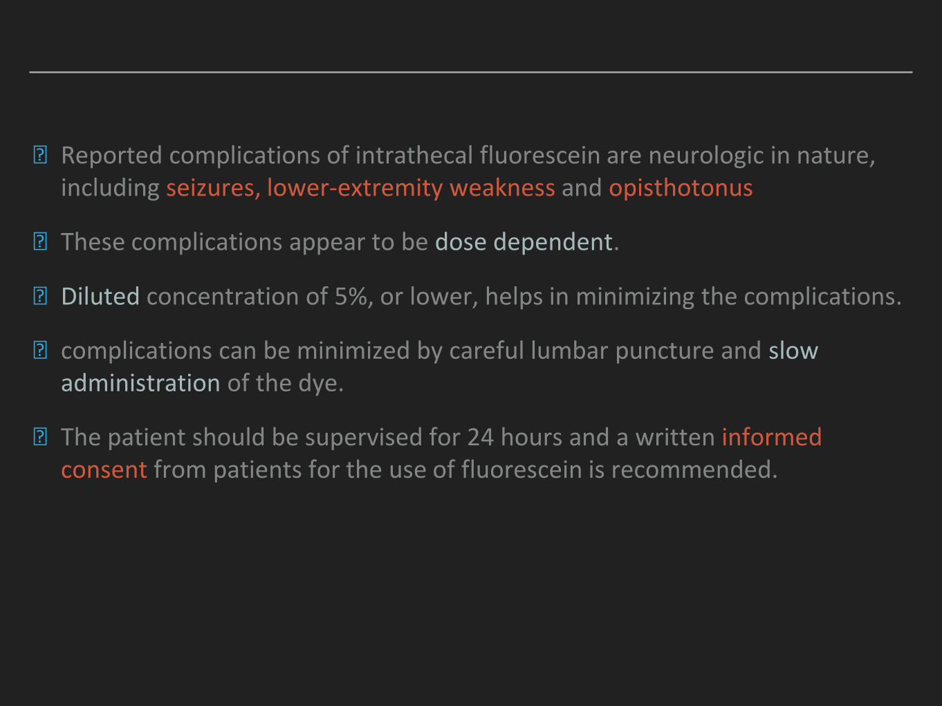
▸ Reported complications of intrathecal fluorescein are neurologic in nature, including seizures, lower-extremity weakness and opisthotonus
▸ These complications appear to be dose dependent.
▸ Diluted concentration of 5%, or lower, helps in minimizing the complications.
▸ complications can be minimized by careful lumbar puncture and slow administration of the dye.
▸ The patient should be supervised for 24 hours and a written informed consent from patients for the use of fluorescein is recommended.

APPROACHES
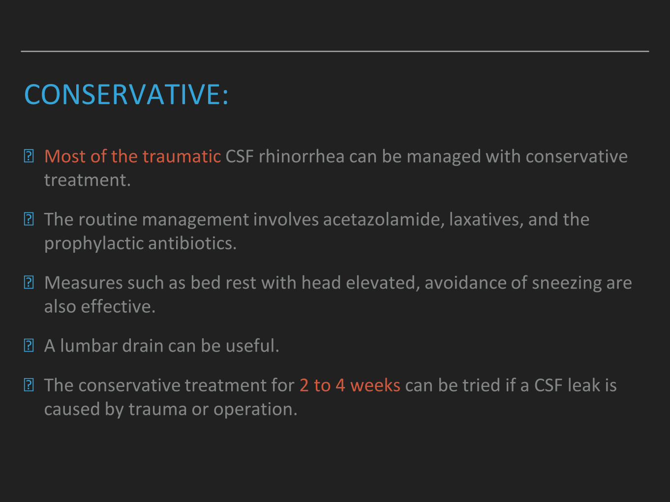
CONSERVATIVE:
▸ Most of the traumatic CSF rhinorrhea can be managed with conservative treatment.
▸ The routine management involves acetazolamide, laxatives, and the prophylactic antibiotics.
▸ Measures such as bed rest with head elevated, avoidance of sneezing are also effective.
▸ A lumbar drain can be useful.
▸ The conservative treatment for 2 to 4 weeks can be tried if a CSF leak is caused by trauma or operation.
SURGICAL:
▸ Intracranial
▸ Extracranial
▸ Endoscopic
▸ Can be classified based on 1) an anatomic target, 2) a cranial base approach, and 3) a nasal corridor.
▸ Four possible corridors:
▸ Transnasal
▸ Transsphenoidal
▸ Transethmoidal
▸ Transmaxillary
ENDOSCOPIC REPAIRS
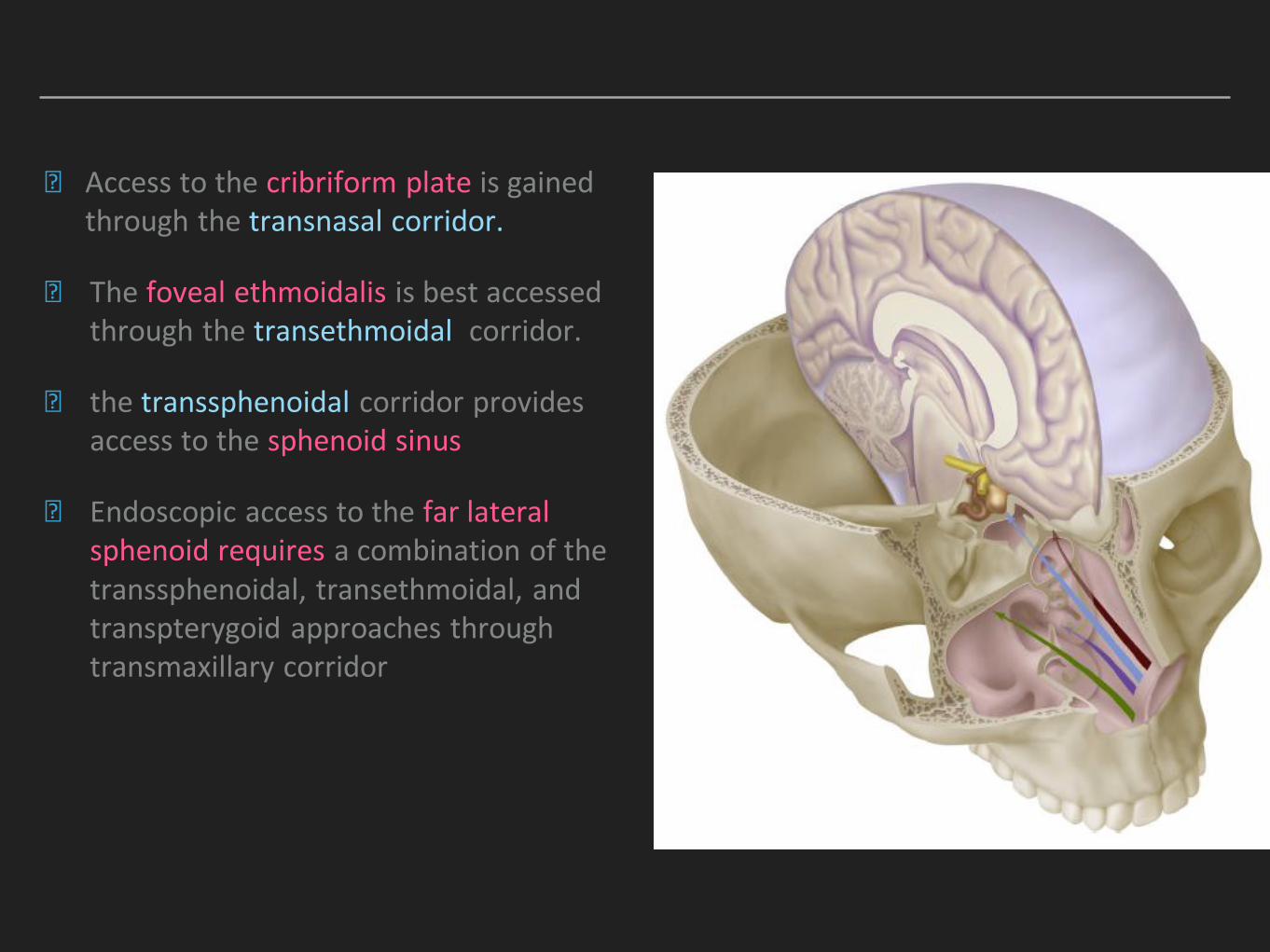
▸ Access to the cribriform plate is gained through the transnasal corridor.
▸ The foveal ethmoidalis is best accessed through the transethmoidal corridor.
▸ the transsphenoidal corridor provides access to the sphenoid sinus
▸ Endoscopic access to the far lateral sphenoid requires a combination of the transsphenoidal, transethmoidal, and transpterygoid approaches through transmaxillary corridor
▸ The frontal sinuses are the most difficult area of the skull base to reach endoscopically.
▸ It may be difficult to visualize and repair CSF leaks of the far lateral frontal sinus through endoscopic means alone.
POST-OPERATIVE CARE
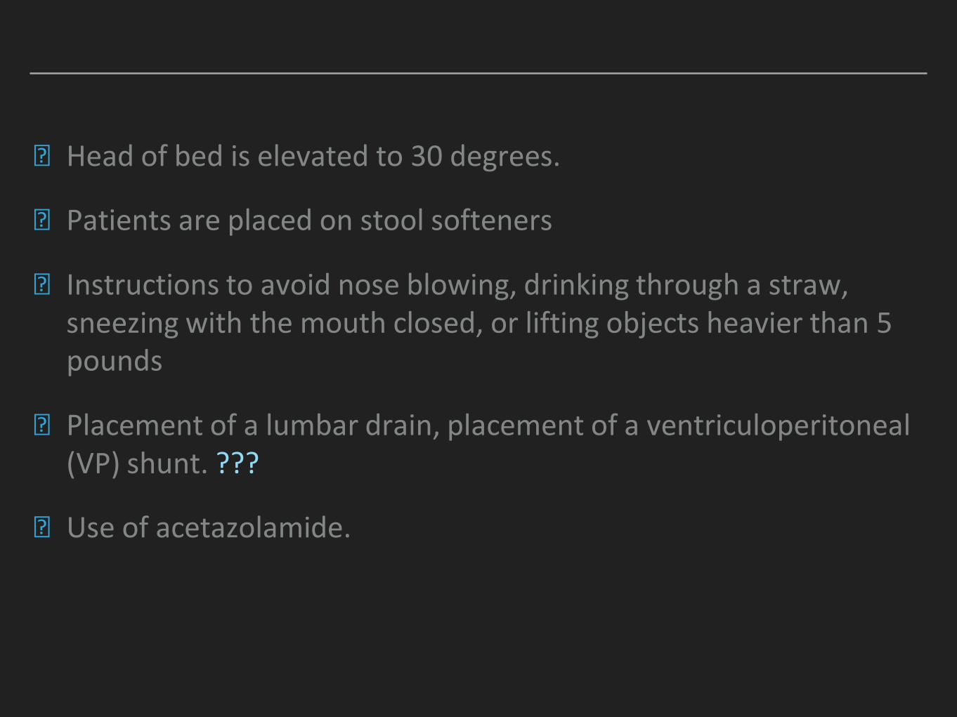
▸ Head of bed is elevated to 30 degrees.
▸ Patients are placed on stool softeners
▸ Instructions to avoid nose blowing, drinking through a straw, sneezing with the mouth closed, or lifting objects heavier than 5 pounds
▸ Placement of a lumbar drain, placement of a ventriculoperitoneal (VP) shunt. ???
▸ Use of acetazolamide.
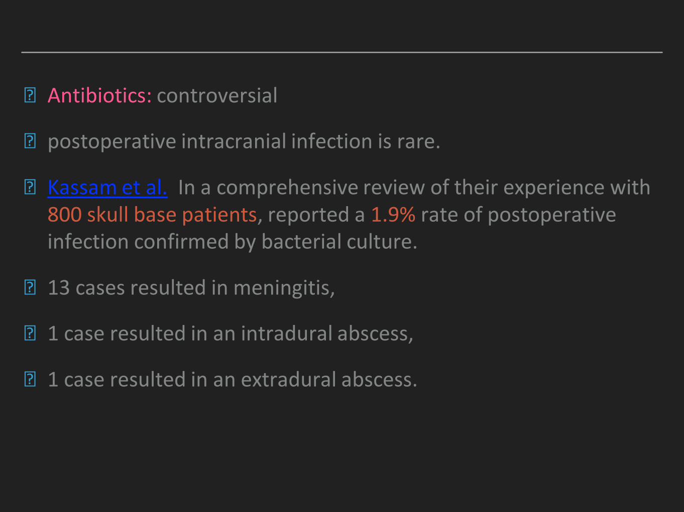
▸ Antibiotics: controversial
▸ postoperative intracranial infection is rare.
▸ Kassam et al. In a comprehensive review of their experience with 800 skull base patients, reported a 1.9% rate of postoperative infection confirmed by bacterial culture.
▸ 13 cases resulted in meningitis,
▸ 1 case resulted in an intradural abscess,
▸ 1 case resulted in an extradural abscess.
OPERATIVE FAILURES AND POST OPERATIVE CSF LEAK RECURRENCES
POST OP CSF LEAK
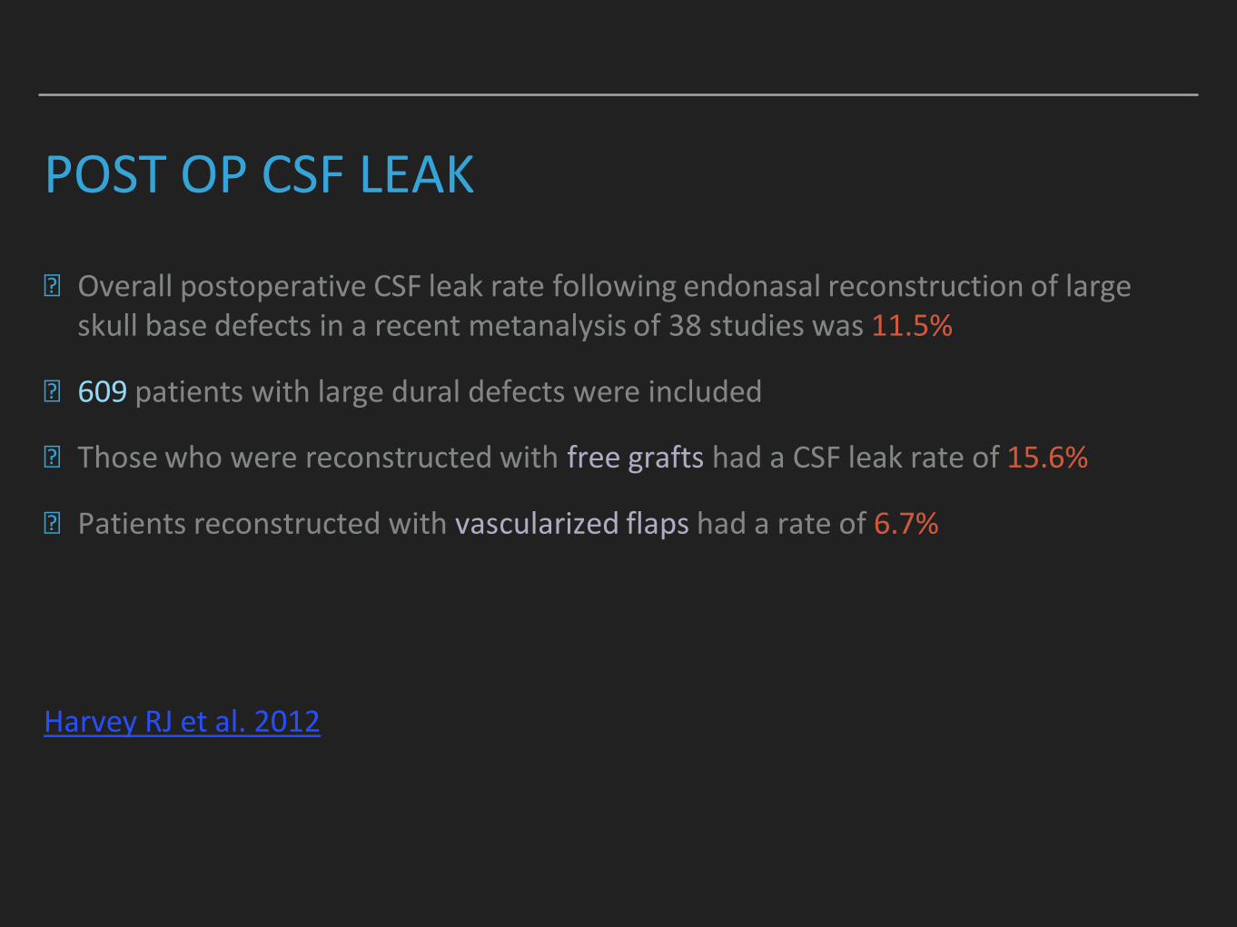
▸ Overall postoperative CSF leak rate following endonasal reconstruction of large skull base defects in a recent metanalysis of 38 studies was 11.5%
▸ 609 patients with large dural defects were included
▸ Those who were reconstructed with free grafts had a CSF leak rate of 15.6%
▸ Patients reconstructed with vascularized flaps had a rate of 6.7%
Harvey RJ et al. 2012
PEDICLED NASOSEPTAL FLAPS?
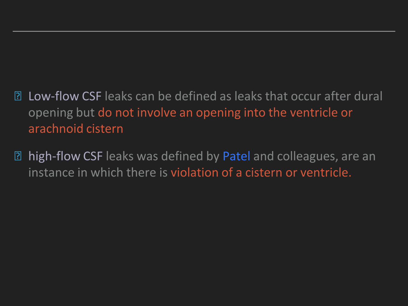
▸ Low-flow CSF leaks can be defined as leaks that occur after dural opening but do not involve an opening into the ventricle or arachnoid cistern
▸ high-flow CSF leaks was defined by Patel and colleagues, are an instance in which there is violation of a cistern or ventricle.
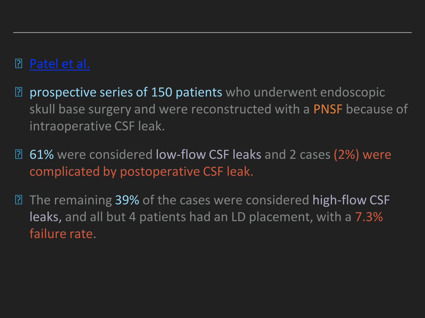
▸ Patel et al.
▸ prospective series of 150 patients who underwent endoscopic skull base surgery and were reconstructed with a PNSF because of intraoperative CSF leak.
▸ 61% were considered low-flow CSF leaks and 2 cases (2%) were complicated by postoperative CSF leak.
▸ The remaining 39% of the cases were considered high-flow CSF leaks, and all but 4 patients had an LD placement, with a 7.3% failure rate.
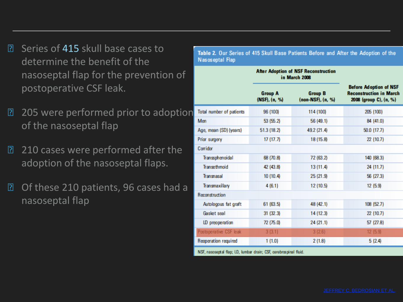
▸ Series of 415 skull base cases to determine the benefit of the nasoseptal flap for the prevention of postoperative CSF leak.
▸ 205 were performed prior to adoption of the nasoseptal flap
▸ 210 cases were performed after the adoption of the nasoseptal flaps.
▸ Of these 210 patients, 96 cases had a nasoseptal flap
JEFFREY C. BEDROSIAN ET AL.

LUMBAR DRAINS ?
LUMBAR DRAINS
▸ lumbar drains are used primarily to reduce stress on the skull base repair.
▸ often kept in place postoperatively to reduce intracranial pressure
▸ to facilitate wound healing and improve the success rate of the reconstruction in obtaining a watertight closure.
▸ Zanation et al.
▸ prospectively evaluated their case series of high-flow intraoperative CSF leak, in which they used a PNSF as part of their skull base reconstruction.
▸ Sixty-five of 70 patients had an LD placed intraoperatively.
▸ This study reported a comparable failure rate for high-flow CSF leak(6.2%)
▸ pediatric patients were at significantly greater risk (P = .002), although a dural opening larger than 2 cm (P = .14) and previous radiation therapy (P 5=.07) trended toward higher failure rates.
AGAINST LUMBAR DRAINS?
▸ Kassamet al.
▸ retrospectively reviewed their initial 800 endoscopic skull base cases
▸ Analyzed the frequency of CSF leak with the degree of difficulty of the resection.
▸ No LDs were placed preoperatively or intraoperatively.
▸ The investigators found an overall CSF leak failure rate of 15.9%.
▸ This important study highlights how CSF diversion is a key tool in managing CSF leak complications
▸ Eloy et al.
▸ retrospective analysis of patients who underwent endoscopic repair of high-flow CSF leaks using a PNSF without CSF diversionover a 3-year period.
▸ A total of 59 defects with leaks considered high flow were repaired, and no postoperative CSF leaks were reported,.
▸ these results support that CSF diversion may not be necessary even in the setting of high-flow CSF leaks.
▸ In an analysis of 9 studies with a collective number of 2049 skull base cases (1961 endoscopic cases):
▸ The overall postoperative CSF leak rate with CSF diversion was 7.5%
▸ and without CSF diversion was 3.4%.
▸ most investigators have advocated placement of an LD in cases of high CSF leak. This finding could explain the discrepancy in failure rates for postoperative CSF leak
Tien et al. 2016
▸ Increasing evidence is suggesting that LDs are not necessary in the setting of endoscopic skull base reconstruction, even when there is a high-flow fistula especially with the increased dependability of vascularized pedicles
▸ The inconsistencies seen in literature makes it difficult to compare outcomes.
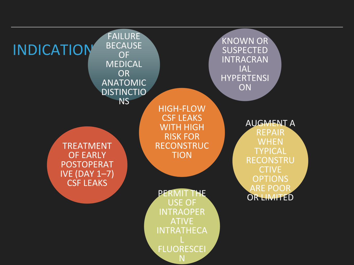
INDICATIONS:KNOWN OR SUSPECTED INTRACRAN
IAL HYPERTENSI
ON
TREATMENT OF EARLY
POSTOPERATIVE (DAY 1–7)
CSF LEAKSPERMIT THE
USE OF INTRAOPER
ATIVE INTRATHECA
L FLUORESCEI
N
AUGMENT A REPAIR WHEN
TYPICAL RECONSTRU
CTIVE OPTIONS
ARE POOR OR LIMITED
HIGH-FLOW CSF LEAKS
WITH HIGH RISK FOR
RECONSTRUCTION
FAILURE BECAUSE
OF MEDICAL
OR ANATOMIC DISTINCTIO
NS
TAKE HOME MESSAGE:
▸ careful selection of cases
▸ High flow high risk cases can benefit from LD placement as well as a peddled flap.
REFERENCES:
▸ Endoscopic management of cerebrospinal fluid rhinorrhea. Yad Ram Yadav, Vijay Parihar, Narayanan Janakiram,1 Sonjay Pande,2 Jitin Bajaj, and Hemant Namdev. Asian J Neurosurg. 2016 Jul-Sep; 11(3): 183–193.
▸ Tension pneumocephalus causing brain herniation after endoscopic sinus surgery. Erhan Çelikoğlu, Jülide Hazneci, and Ali Fatih Ramazanoğlu. Asian journal of neurosurgery,2016(11)3:309-310
▸ Review of the management of pneumocephalus. Carlos B. Dabdoub,Gueider Salas, Elisabeth do N. Silveira, and Carlos F. Dabdoub. Surg Neurol Int. 2015; 6: 155.
▸ Cerebrospinal Fluid Diversion in Endoscopic Skull Base Reconstruction An Evidence-Based Approach to the Use of Lumbar Drains Duc A. Tien, Janalee K. Stokken, Pablo F. Recinos, Troy D. Woodard, Raj Sindwani, Otolaryngol Clin N Am 49 (2016) 119–129.
▸ Evaluation of spontaneous nasal cerebrospinal fluid leaks. Sarah K. Wise and Rodney J. Schlosser, Current Opinion in Otolaryngology & Head and Neck Surgery 2007,15:28–34.
▸ The Endoscopic Endonasal Approach to Repair of Iatrogenic and Noniatrogenic Cerebrospinal Fluid Leaks and Encephaloceles of the Anterior Cranial Fossa Jeffrey C. Bedrosian2, Vijay K. Anand2, Theodore H. Schwartz1,3World Neurosurg.
(2014) 82, 6S:S86-S94.

THANK YOU!


![CSF Leaks - Diagnosis and Management[1]](https://img.pdfslide.us/doc/110x75/577c79721a28abe05492aad0/csf-leaks-diagnosis-and-management1.jpg)