Embed Size (px)
Citation preview
Sponsored by an educational grant from Merck Serono
Dat
e of
Pre
para
tion:
SA
I08
- 00
96 D
ecem
ber
2008
A variety of pituitary casesChairs: Dr Mark Vanderpump, Mr Michael Powell, London
1. Cyclical Cushing’s syndrome due to ectopic Adrenocorticotrophic Hormone secretion bya bronchial carcinoid Senthil Rajasekaran, Niki Karavitaki, John A.H. Wass (Oxford)
IntroductionCyclical Cushing’s syndrome(CS), was first described in 1971 by Bailey in a patient withbronchial carcinoid.The Diagnosis of Cyclical CS is often very difficult resulting in delayedtreatment. In a series of 65 patients with cyclical CS, Cushing’s disease was the underlyingcause in 54%, ectopic CS in 26% and primary adrenal pathology in 11%. We report a rarecase of cyclical CS due to bronchial carcinoid and discuss the diagnostic difficulties encountered.
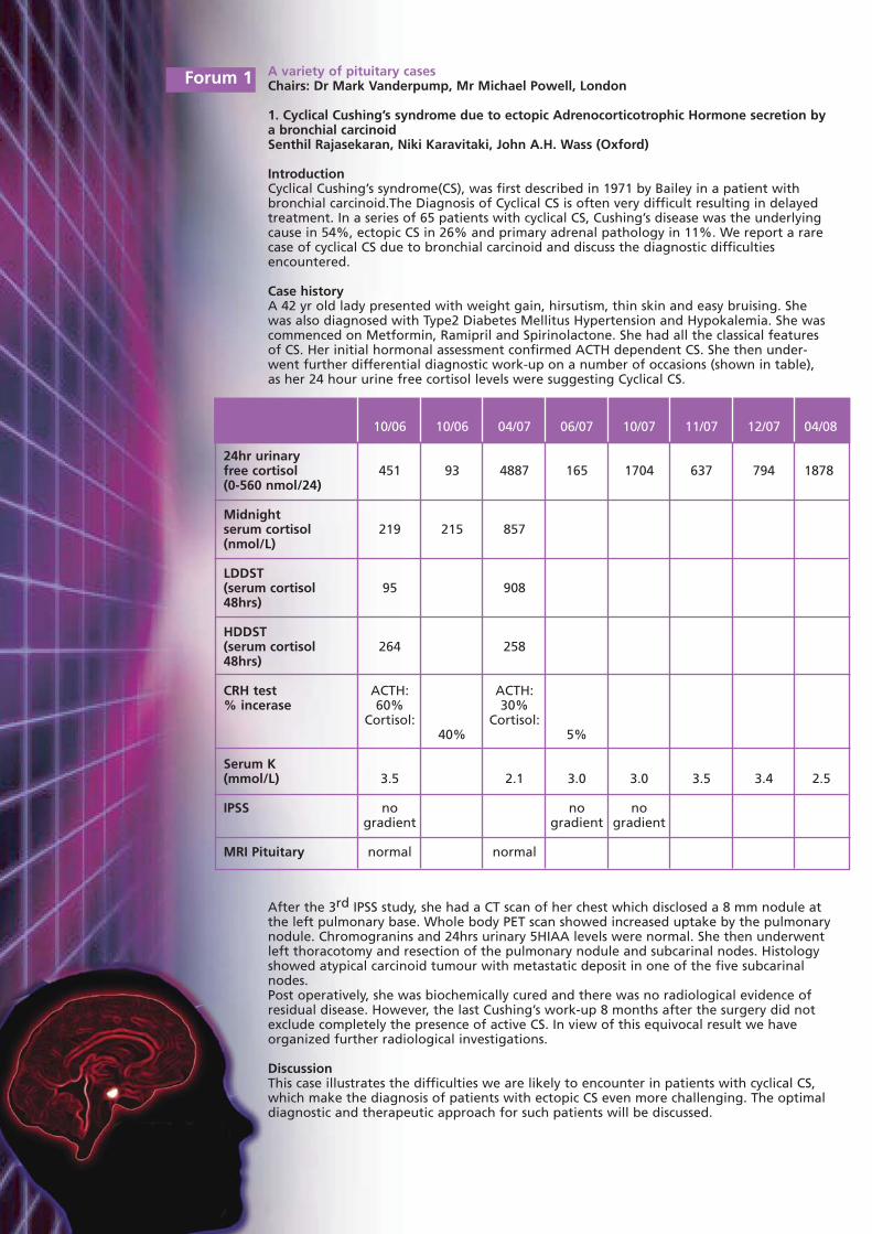
Case historyA 42 yr old lady presented with weight gain, hirsutism, thin skin and easy bruising. Shewas also diagnosed with Type2 Diabetes Mellitus Hypertension and Hypokalemia. She wascommenced on Metformin, Ramipril and Spirinolactone. She had all the classical featuresof CS. Her initial hormonal assessment confirmed ACTH dependent CS. She then under-went further differential diagnostic work-up on a number of occasions (shown in table), as her 24 hour urine free cortisol levels were suggesting Cyclical CS.
After the 3rd IPSS study, she had a CT scan of her chest which disclosed a 8 mm nodule atthe left pulmonary base. Whole body PET scan showed increased uptake by the pulmonarynodule. Chromogranins and 24hrs urinary 5HIAA levels were normal. She then underwentleft thoracotomy and resection of the pulmonary nodule and subcarinal nodes. Histologyshowed atypical carcinoid tumour with metastatic deposit in one of the five subcarinalnodes.Post operatively, she was biochemically cured and there was no radiological evidence ofresidual disease. However, the last Cushing’s work-up 8 months after the surgery did notexclude completely the presence of active CS. In view of this equivocal result we haveorganized further radiological investigations.
DiscussionThis case illustrates the difficulties we are likely to encounter in patients with cyclical CS,which make the diagnosis of patients with ectopic CS even more challenging. The optimaldiagnostic and therapeutic approach for such patients will be discussed.
10/06 10/06 04/07 06/07 10/07 11/07 12/07 04/08
24hr urinary free cortisol 451 93 4887 165 1704 637 794 1878(0-560 nmol/24)
Midnight serum cortisol 219 215 857(nmol/L)
LDDST(serum cortisol 95 90848hrs)
HDDST(serum cortisol 264 25848hrs)
CRH test ACTH: ACTH: % incerase 60% 30%
Cortisol: Cortisol:40% 5%
Serum K (mmol/L) 3.5 2.1 3.0 3.0 3.5 3.4 2.5
IPSS no no nogradient gradient gradient
MRI Pituitary normal normal
Forum 1

2. A family with early-onset growth hormone and prolactin-secreting pituitary tumoursTurner L, Robertson I, Barwell J, Greening J, Levy MJ, Howlett TA (Leicester, Nottingham)
We present a 7 year old girl (Case A) who experienced sudden onset of severe headacheand right-sided third nerve palsy. Prior to acute admission, she had been referred to thepaediatric endocrinologists because of a several month history of accelerated growth (>99th centile for height) and new onset headache. Her initial CT scan in casualty showed amass in the pituitary fossa with invasion of the right cavernous sinus. Preliminaryendocrine investigation showed an elevated IGF-1 level with incomplete suppression ofgrowth hormone (GH) after oral glucose tolerance testing. She was transferred to the neurosurgical Unit with a clinical diagnosis of an apoplectic event within a GH-secretingpituitary tumour. A trans-sphenoidal biopsy showed a somatotroph adenoma and she wasmanaged conservatively. A trial of cabergoline was followed by a reduction in IGF-1 andGH levels to below normal the normal range, with suppression of prolactin. The cabergoline has been discontinued and she is due for dynamic re-assessment of herGH status and re-imaging of the pituitary fossa. Her headache and 3rd nerve palsy haveresolved and her growth continues to be monitored closely. Numerous members of the proband’s family are known to our clinic with early onset GHand prolactin-secreting pituitary tumours. A’s mother (Case B) presented with a micro-prolactinoma at the age of 15 with primary amenorrhoea and galactorrhoea andwas treated with a variety of dopamine agonists and pituitary surgery. B’s cousin was diagnosed with acromegaly at the age of 32 years (Case C), having presented witholigomenorrhoea, hyperprolactinaemia, reduced fertility and headache and had subsequent trans-sphenoidal hypophysectomy, radiotherapy and octreotide. C’s fathergrew rapidly at the age of 17 and subsequently developed of classical acromegalic features (Case D). A glucose tolerance test performed in 1973 (at age 31) and again in1993 showed incomplete suppression of GH, but subsequent GH / IGF-1 levels and pituitaryimaging have been normal over the last decade despite no specific treatment. D’s brotherhas been treated elsewhere with a dopamine agonist for prolactinoma and possiblyacromegaly. None of the family has hypercalcaemia or pancreatic neoplasia, and genetesting for MEN-1 is negative. Case A and her mother have been seen by our clinicalgeneticists who have commented on the presence of pigmented skin lesions but there isno evidence of Cushing syndrome or cardiac myxomas in the family.
This family appear to have a rare pedigree presenting exclusively with GH and prolactin-secreting pituitary tumours. Given the very young age of presentation of the proband, thepossibility of genetic anticipation is raised. We would be interested to hear the views ofthe audience about the unifying diagnosis, and whether further genetic analysis mightlead to new insights into the hypothalamic control of the GH / prolactin axes.
3. Macroprolactinoma and Cushing’s Disease: An Unusual Combination with InitialTreatment Response to Dopamine Agonist Therapy: What next? Nagi D, Azzan R, Jenkins R (Wakefield)
Secretion of multiple pituitary hormones by pituitary adenomas is relatively rare. It is usually due to a single tumour producing more than one hormone. Two distinct adenomas secreting different hormones have been described (1), but this is uncommon. Ithas also been documented that the hormonal production of a particular tumour maychange with time, therefore behaving like a tumour secreting multiple hormones but notsimultaneously (2,3). The commonest combination of hormone secretion is growth hormone (GH) and prolactin(PRL) but the combined secretion of GH/thyroid stimulating hormone (TSH) and PRL/TSH aswell as GH/PRL/TSH, have all been documented (4,5). The combination of adrenocorti-cotrophic hormone (ACTH)/PRL is very rare indeed, with only a few cases reported to date(2, 6-9).
History and examinationAn 18 year old young man was first seen in the ENT department with bilateral swelling ofthe supraclavicular fossae. A clinical diagnosis of Cushing’s syndrome was made and thepatient was referred to the Endocrine team. His past medical history is unremarkable andhe was not on any regular medications. On examination he was grossly Cushingoid withtypical facial features (Fig. 1A), abdominal striae, proximal muscle weakness and elevatedblood pressure at 150/100. His visual field testing in clinic suggested a mild bitemporalhemianopia.
InvestigationsRenal function and electrolytes were normal. Thyroid function tests showed a low fT4 at8.7 pmol/L with a TSH of 0.69 mIU/L. Prolactin was 68280 mIU/ml and 24-hr urinary freecortisol (UFC) was 3860 nmol/24 hr. Random cortisol was 985 nmol/L with an ACTH of 104ng/L. Testosterone was 13.5 nmol/L, SHBG 6 nmol/L, FSH 4.4 IU/L and LH 6.7 IU/L. Theresults of a cortisol day curve are shown in Table 1A. A high dose dexamethasone suppression test showed a basal cortisol of 809 nmol/L suppressing to 247 nmol/L, indicating Cushing’s disease. An MRI showed a large pituitary mass with minimal suprasellar extension and very little normal pituitary tissue and this is shown in Fig. 2A.
ManagementThe patient was commenced on bromocriptine but developed skin rash and arthralgia andwas therefore switched to cabergoline 500 mcg twice weekly. Four weeks later, he wasadmitted for a cortisol day curve as a baseline for commencing metyrapone therapy for hisCushing’s disease. During his admission, it was noted that the patient’s physical appearance had improved dramatically (Fig. 1B). Clinically, it was felt that his Cushing’sdisease has remitted, and his BP normalised to 105/60 with no postural hypotension. Theclinical suspicion of disease remission was confirmed by a cortisol day curve (Table 2B). Hisrepeat endocrine tests showed a prolactin of 1801 mIU/L, testosterone 28.3 nmol/L, SHBG70 nmol/L, FSH 2.9 IU/L, LH 5.2 IU/L, fT4 9.2 pmol/L and TSH 2.55 mIU/L. His UFC was lessthan 36 nmol/24 hr. Repeat MRI performed 6 weeks after starting cabergoline treatmentshowed 30% reduction in the volume of the pituitary tumour (Fig. 2B).
Follllow upThe patient has been under follow up for 28 months now. He remains on cabergoline andhis Cushing's disease has remained in complete remission. Fig. 1C and Fig. 1D show thepatient at 7 months and 28 months post treatment. The response to cabergoline treat-ment was quick and impressive with almost complete shut down in cortisol production.Our patient was indeed hypoadrenal on repeat cortisol day curve and was treated withhydrocortisone replacement therapy for few months. We anticipated that with time hewould resume normal cortisol production, which had been the case (data not shown) andhis hydrocortisone treatment was subsequently discontinued.Repeat MRI showed 30% reduction in the volume of the pituitary tumour.. The case isunusual in that a single pituitary tumour would appear to be responsible for prolactin aswell as ACTH secretion. The patient had a dramatic response to Cabergoline therapy withresolution of his symptoms and shrinkage of the pituitary tumour. This patients now hasbeen on Cabergoline treatment with complete remission of Cushing’s Syndrome and nor-malisation of Prolactin levels, for 6 years. We would like to discuss the future option formanagement of this young man.
4. Pregnancy, prolactin and an expanding problem!Bhake R, Barnfield S, Trinder J and Bradley KJ (Bristol)
A 23 year old lady, who had moved to the UK from Poland 2 years previously, was referredby her GP to the endocrine service at the Bristol Royal Infirmary in March 2008. Her original presentation had been with a six month history of amenorrhoea at the age of19 years. The details of her investigations in Poland were unavailable to us but she wasable to tell us that she had been treated fairly consistently with bromocriptine, obtainingrenewed prescriptions during her visits to Poland. At the point of referral she had experienced four months of amenorrhoea having run out of her dopamine agonist therapy and her GP had appropriately refused to provide an ongoing prescription withoutfurther clarity regarding the diagnosis. Her clinical examination was entirely normal andher anterior pituitary profile was unremarkable apart from a significantly elevated prolactin at 68,306 mIU/L, which was consistent with the diagnosis of a macroprolactinoma. On direct questioning she expressed a wish to start a family within the near future and weadvised her carefully about the need for adequate contraception in order to postponeconception for a number of months until such a time that we had gained biochemical andradiological control of the tumour. She concurred with this plan and consequently cabergoline was commenced immediately which rapidly restored her menstrual cycle andthe drug was titrated up every two weeks to a final dose of 500mcg twice per week.Although she had tolerated bromocriptine reasonably well over the previous four yearsshe had experienced some side-effects and hence we opted for cabergoline therapy.Formal visual field testing was normal but an urgent MRI of the pituitary gland showed asubstantial pituitary mass of 20x16mm (9mm clear of the chiasm), confirming the diagnosisof a macroprolactinoma. When she was reassessed in May 2008 she was asymptomatic and her prolactin had fallento 3,300mIU/L. A further prolactin level and a repeat MRI were planned for June, threemonths post initiation of dopamine agonist therapy, to stage her response to treatment.However, the patient informed us in June that she had stopped her cabergoline twoweeks previously at the end of May as she had missed a period and that her home pregnancy test was positive. As we had achieved a significant biochemical response afterthree months of treatment, we decided to urgently assess the radiological response and tohold off further dopamine agonist therapy pending this result. If there was evidence ofsignificant residual tumour then we planned to introduce and maintain her on bromocriptine throughout the pregnancy whilst if there was evidence of successfultumour shrinkage then we planned on careful clinical and visual field monitoring. She wasadvised to look out for symptoms indicative of an enlarging adenoma and to contact usimmediately if they developed. The planned repeat MRI examination was expedited tourgent and visual field assessments were booked for each trimester. We managed her ongoing care in our maternal medicine endocrine clinic and she had herinitial assessment within a week of informing us that she was pregnant. However, therewas a break in communication in the ensuing weeks as she was successively unable toattend two clinic appointments, two radiology appointments and a visual field assessmentappointment due firstly, to a holiday and then due to work commitments.
We were also unsuccessful in engaging her either by letter or by telephone. She subsequently attended clinic in October 2008, at 26 weeks gestation, having just hadher MRI scan a few days previously. Her motivation for re-attending was significant andunremitting headaches which were present on waking. Her MRI scan revealed dramatictumour expansion such that it now impinged upon the optic chiasm, encased the rightcarotid artery, indented the right temporal lobe and had also invaded inferiorly into thesphenoid sinus and eroded infero-laterally into the skull base. Her prolactin was69,000mIU/L and she was commenced on a rapid titration regimen with bromocriptine, inview of its robust safety data in pregnancy, to reach a dose of 2.5mg bd. Her headachesresolved within a few days and her prolactin fell to 12,600mIU/L after four weeks of treatment. Her visual fields remained normal. A transient increase in her prolactin to25,900mIU/L was then seen at the same time as she was unable to attend a further clinicappointment but we established on the telephone that she had experienced some nauseaand vomiting with the bromocriptine that she felt had been transient and although wedebated the switch to cabergoline this was not instituted.On 21 December 2008 at 33+6 weeks gestation she went into spontaneous labour anddelivered a baby boy prematurely with no complications. One day post delivery the obstetricians reassessed her prolactin and found it to be 31,100mIU/L. The baby spent thefirst few days on NICU but is developing normally given his gestation. We continued herbromocriptine at 2.5mg bd after discussing the risks and benefits and she subsequentlysuccessfully established and maintained breast feeding. She remains well and her latestprolactin four weeks after delivery is 5,050mIU/L. A repeat MRI is pending.The issues and controversies highlighted by this case relate to various aspects of her care:
Timing of agent withdrawal: Should dopamine agonist therapy be withdrawn when pregnancy arises relatively early in the treatment of a macroprolactinoma when the risk ofenlargement, although still low, is relatively greater.Choice of agent: Given recent MHRA reinforcement of the guidance that cabergolineshould be stopped at least one month prior to conception, should we strongly encouragewomen of child bearing age to persist with bromocriptine despite significant side-effectswhich are impacting upon their everyday life and perhaps influencing compliance?Importantly, prolactinomas may also be less responsive to bromocriptine as opposed tocabergoline. Fibrotic cardiac valvulopathy: the MHRA safety update was issued in October 2008 andconsequently this patient has not yet had cardiac echocardiography. She has no clinicalevidence of cardiac compromise and we fear that she may not engage in ongoing therapyif she learns that cardiac valvopathy is a 'very common' side-effect.Monitoring: How is it clinically appropriate and cost effective to monitor patients withmacro (or micro) prolactinomas during pregnancy given the relatively low risk of complications and the current demand on NHS resources? Should formal visual fields becarried out each trimester or will a clinical assessment suffice and is there ever a role forprolactin measurement given the difficulty interpreting the result in the context of pregnancy. Alternatively should patients not be monitored but simply advised to seek helpif they develop symptoms? All of these approaches are currently utilised by differentendocrine centres in the UK and there is no clearly established consensus.Imaging: When is it appropriate or essential to request a pituitary MRI in pregnancy giventhat, although there are no known risks, radiologists ask us to limit use of this technology,especially in the first trimester? Communication barriers: Significant clinical risk may arise when patients fail to engagefully in their care and how much can this lack of engagement be deemed the responsibility of the health care professional.Lactation: Should a patient on bromocriptine be encouraged to breast-feed if it is physio-logically possible?Future fertility: Once a patients has had significant enlargement of a prolactinoma duringpregnancy how should they be managed prior to and during subsequent pregnancies?
5. Lymphocytic hypophysitis secondary to a ruptured Rathke’s cleft cyst: a diagnostic andmanagement challengeP. Mehta, F. Roncaroli, A. Mehta, M. Bhojak, J. Lawrence, L.R. Bridges, A. Belli, K. Meeran,ECI Hatfield, W.S. Dhillo (London, Salisbury, Edinburgh,Southampton)
We describe a 34-year old female who presented with secondary amenorrhoea andfatigue. There were no features of infection, or autoimmune diseases. Investigationsrevealed panhypopituitarism and she was prescribed replacement corticosteroids, thyroxine and HRT. She subsequently developed diabetes insipidus and was commencedon desmopressin. MRI scans showed a peripherally enhancing pituitary mass abutting theoptic chiasm. The lesion was approached transphenoidally with the aim of taking a diagnostic sample. A follow-up imaging demonstrated re-growth of the lesion 14 monthsafter the initial operation with chiasmal compression. The patient underwent secondtransphenoidal surgery. Pathological examination of the samples from the first operationshowed fragments of Rathke’s cleft cyst and adenohypophysis with moderate chronicinflammation. The inflammatory infiltrate was composed mostly of CD8-positive T lymphocytes. No adenoma was present and no micro-organisms were identified.

The sample from the second operation showed granulation tissue associated with a considerable macrophagic infiltrate. These features were consistent with lymphocytichypophysitis secondary to ruptured Rathke’s cleft cyst. An MRI scan performed sevenmonths after the second operation demonstrated further expansion of the cystic lesion.The patient was given a trial of high-dose prednisolone. A repeat MRI to assess responseto steroids revealed no change in size of the cyst but reduced contrast enhancement. Thepatient remains panhypopituitary on replacement therapy. Given the rarity of the association between Rathke’s cleft cyst and hypophysitis, we wouldlike to ask the expert panel how they would optimally manage this patient?Would further surgery be appropriate?Should further steroid treatment be given?Would radiotherapy be advisable?
Management of pituitary apoplexy- Towards a consensus Chairs: Dr SE Baldeweg (London) and Prof JAH Wass (Oxford)
6. Conservative managment of pituitary apoplexyR.Fikri R, Johnston C, .Kong C (Hemel Hempstead)
A 55-year-old man who presented with a severe headache, which was sudden, togetherwith photophobia and nausea just 6 days after induction chemotherapy for acute myeloidleukaemia.On examination he was febrile and had neck stiffness but no focal neurology. He bloodsshowed that he was thrombocytopenic (platelets 19 x109 /L), mild coagulopathy and neutropenia (neutrophils 0.5 x10⁹ /L.CSF examination showed 1 WBC/mm3, 19 RBCs/mm(traumatic tap), no xanthochromia, normal protein and glucose and no organisms wereseen on CSF culture.Unenhanced CT scan of the brain was normal. However, in view of the strong clinical suspicion of intracranial bleeding an MRI was performed which revealed an enlarged pituitary gland with high signal intensities on T1 weighted images, indicating a haemorrhage within the gland likely to be a previously undiagnosed pituitary adenoma.The pituitary was bulging into the suprasellar fossa with no pressure on the optic chiasm.Pituitary function tests showed evidence of hypopituitarism and add on tests from a sample taken 4 days prior to the event showed no evidence of endocrinopathy, which suggests that the adenoma was non-functioning. (which is the commonest associated pituitary tumour) and that the hypopituitarism was acute as a result of pituitary apoplexy.The patient was started on DDAVP, hydrocortisone and testosterone replacement therapy.The headache resolved, fever subsided and the platelet count remained stable at around50 000 /l. He achieved complete remission of the AML after the first course of treatmentand subsequent courses were uneventful. A follow – up MRI showed an almost completeresolution of the pituitary haemorrhage but he remained in hypopituitarism.Pituitary apoplexy is characterised by headache, meningism, altered mental status, opthalmoplegia or visual loss. It occurs most commonly in patients with previously undiagnosed pituitary adenomas. . Those patients who have a pituitary tumour haemorrhage/haemorrhagic infarction have a more severe clinical picture and worse outcome than those who have a pituitary infarction alone The common precipitating factors are surgery (particularly cardiac), coagulopathy, thrombocytopenia and it has beendescribed after dynamic pituitary testing or hormone stimulation states like pregnancyand HRT. Endocrine recovery is uncommonOur case demonstrates the importance of proceeding to an MRI scan if the diagnosis issuspected since it is diagnostic in only 30 % of cases despite a presence of a sellar mass inupto 70 %. It also illustrates the instantaneous effect a pituitary apoplexy can have on thepituitary function, hence a high index of suspicion is necessary and prompt treatment maybe life saving. Care should be taken when performing surgery, dynamic pituitary testing,giving HRT or anticoagulating patients with a known pituitary adenoma. Finally, it sup-ports the conservative management of this condition, especially if there are neuro-opthalmic complications.
7. Pituitary apoplexy: a challenge for diagnosis and management.Esther Nemati, James Ahlquist and Michael Powell (Southend and London)
Following a family night out at a Chinese restaurant, a 70 year old man developed diarrhoea and vomiting. Two days later he developed sudden onset headache, which wasnot severe, mild confusion, double vision, and mild left sided limb weakness. On presentation, he was febrile and mildly confused. Initially there was a partial right ptosis; over the following three days this progressed to complete right ptosis and partialleft ptosis, with complete ophthalmoplegia of the right eye and a left third nerve palsy.He also had mild left sided hemiparesis. A CT scan of his head showed a 20mm pituitarymass which was initially considered to be an incidental finding.
An diagnosis of suspected botulism was made. The Health Protection Agency was contacted, and he was given botulinum antitoxin to which he had an anaphylactic reaction. Endocrine on day 6 assessment demonstrated complete anterior hypopitu-itarism, and a bitemporal hemianopia. MRI pituitary showed a pituitary mass, 17mm x38mm, arising from an enlarged fossa and compressing the optic chiasm. CSF examinationshowed protein 0.96 g/l with a mild lymphocytosis. He was treated with hydrocortisone, and underwent trans-sphenoidal evacuation of ahaemorrhagic pituitary mass seven days after initial presentation. He made a rapid recovery, with full return of higher mental function and gradual resolution of his diplopiaand visual field defect. There was no recovery of pituitary function. This case demonstrates that pituitary apoplexy can be hard to diagnose, and may mimicother acute neurological illnesses. The optimal timing of pituitary surgery is often debated: in this case surgery was not undertaken until seven days after presentation,without obvious harm arising from the delay.
Forum 2
8. Three Perplexing Cases: Controversies in Managing Pituitary ApoplexyMaruthappu T, Mehta SR, Elgayar H, Wynne K, Mendoza N, Mehta A, Morganstein D, Hatfield E, Tan T and Meeran K (Imperial , London)
Pituitary apoplexy is an uncommon and potentially fatal disorder. Its rarity makes establishing evidence based guidelines difficult. We present 3 cases which have presentedin the past year:Case 1: 30 year old male with no concurrent medical illness, presented with sudden onsetsevere headache, photophobia and fever. Clinical examination was normal. Meningitis wasexcluded. MRI revealed a haemorrhagic suprasellar mass with superior displacement of theoptic chiasm. Pituitary function showed hypogonadotrophic hypogonadism. 9 am cortisolwas 121 nmol/L. This patient was managed conservatively and was successfully dischargedon hydrocortisone. Case 2: a 31 year old lady presented with a 5 day history of severe headache with vomit-ing. Neurological examination and visual fields were normal. MRI showed evidence of apituitary macroadenoma abutting but not compressing the optic chiasm, with evidence ofhaemorrhage. Humphrey’s perimetry was normal. Baseline pituitary function tests showeda normal TSH of 0.47 mU/L, free T4 of 11.5 pmol/L (9-26), prolactin of 486 mU/L, IGF-1 of27.4 nmol/L and cortisol of 367nmol/L. She was discharged on hydrocortisone and is currently awaiting re-evaluation.Case 3: A 29 year old male attended A&E with a 24 hour history of headache, vomitingand diplopia. On neurological examination he had a right VIth nerve palsy. A CT and MRIconfirmed a 2.5cm heterogeneous mass in the pituitary fossa extending into the cavernoussinus and the optic chiasm with evidence of a recent haemorrhage. Perimetry was normal.Baseline pituitary function tests revealed TSH 1.12mU/L, T4 9.9pmol/L, LH 1.8 IU/L, FSH 4.1IU/L, Prolactin 96 mU/l, Testosterone 8 nmol/L, Cortisol 284nmol/L, Growth Hormone 0.37ug/l and ACTH 13.4ng/L. He was commenced on Hydrocortisone. Urgent trans-sphenoidalhypophysectomy was performed due to cranial nerve compression and the risk of visualfield loss. Histology revealed a necrotic gonadotroph adenoma expressing the ß subunit ofFSH. His VIth nerve palsy persists.These cases highlight the heterogeneous presentations of pituitary apoplexy. Prompt diagnosis is essential to ensure that patients receive appropriate hormone replacement.Controversies remain as to the optimal management, viz.:What are the benefits of emergency surgery vs treatment with steroids?What are the indications for surgery?What are the long term outcomes: recurrence and need for hormonal replacement?
9. Experience of pituitary apoplexy in 58 patients Pathak A (Sheffield & Chandigarh, India)
All about Cushing’sChairs: Dr Conway and Mr Powell (London)
10. A silent Corticotroph adenoma leading to aggressive pituitary Cushing’s disease Aung, T, Karavitaki N, Wass JAH(Oxford)
Background: Silent corticotroph adenoma was first described in 1978 by Kovacs. Thesetumours account for 1.1-6% of surgically removed pituitary adenomas and 17-22% ofthose with positive ACTH stains. 87-100% is macroadenomas with suprasellar extensionand they may show potential aggressive behaviour, particularly upon recurrence.
History: A 62 year old gentleman was referred to our Department with complex medicalhistory.
At the age of 47 (1996), he presented elsewhere to a Diabetic Clinic following pancreatitiswith uncontrolled diabetes, hypertension, hyperlipidaemia and obesity. The appropriatemedical treatment was initiated. Unfortunately, he was lost to follow up. Eight years later (2003), he presented with headaches, visual disturbances and right 3rdand 6th nerve palsies. The MRI scan showed expanded fossa with tumour extending intothe right cavernous sinus but no evidence of chiasmal compression. The hormonal evaluation revealed prolactin 999mIu/L (0-400), IGF-I 84 ng/ml (71-290), FT4 7.9 pmol/L(9.14-23.4) with low TSH. Short Synthathan test was normal but ACTH at time zero of113ng/L (0-46). One collection for 24 hour urinary free cortisol was normal. The diagnosisof non-functioning pituitary adenoma was made. Thyroid hormone replacement wasstarted. Preoperatively, atrial fibrillation and inferior myocardial infarction developed andfinally TSA was done in 2004. Following surgery, the eye movements went to normal. Thehistology showed a heavily stained ACTH pituitary adenoma. Post TSA assessment showed9am cortisol of 176nmol/L and thyrotroph and gonadotroph deficiencies.
Current presentation and further investigations: 4 years later (2008), he was referred toour Department with a progressive right 3rd nerve palsy and visual field defects. Two collections for 24 hour urinary free cortisol were high [650 and 891 (normal 0-560nmol/L)]. Midnight cortisols were 792nmol/L and 804nmol/L. He failed to suppress onboth low dose and high dose dexamethasone suppression tests. There was no response toCRH test. MRI pituitary showed a large multi-lobulated sellar (1.7X2.0x1.9cm) and suprasellar mass lesion (1.9X2.1X2.2cm) and right cavernous sinus was encased by tumour.
Treatment: Initially Metyrapone was started. Unfortunately he did not tolerate this medication. After considering all the operative risks and benefits, second TSA was doneon November 2008. The post operative 9 am cortisol results were 767 and 1049 nmol/L.His right 3rd nerve palsy remained. Histology confirmed corticotroph pituitary adenoma.Post operative MRI showed a reduction in size of the sellar component but residualtumour persisted and the suprasellar component had not altered. Medical treatment wasre started with Metyrapone and Fluconazole but stopped again due to side effects. Hehas been referred for radiotherapy.
Discussion: This case illustrates the difficulties encountered in the diagnosis and the management of silent corticotroph adenoma which can progress to aggressive clinicalCushing’s disease. The discussion points are:The management and follow up of silent Corticotroph pituitary adenoma including therole of radiotherapy.The management of aggressive Cushing’s disease after two transsphenoidal surgeries withintolerance to current medical therapies.Role of bilateral adrenalectomy in the management of refractory Cushing’s disease.Diagnosis of Cushing’s disease when the responses to high dose dexamethasone suppression test and to CRH test are not typical.The Importance of the continuity of patient care.
11. A case of recurrent Cushing’s DiseaseHS Chahal, I Sabin, J Evanson, N Plowman, M Korbonits, AB Grossman. St Bartholomew’sHospital, (London)
We report a case of a female patient who presented to her local endocrinologist at theage of 20 years with a 4-year history of secondary amenorrhoea, hirsutism and weightgain of 7 stones. She had clinical signs consistent with Cushing’s syndrome. Biochemistryshowed lack of diurnal cortisol variation and failure of cortisol suppression on both thelow-dose (2+0: cortisol 839nmol/L, 2+48 cortisol 741nmol/L) and high-dose dexamethasonesuppression tests (8+48 cortisol 731nmol/L). An MRI demonstrated a poorly-enhancing areaof pituitary tissue on the left side of the gland, extending into the cavernous sinus.She underwent 3 transsphenoidal operations without any clinical or biochemical cure.Histology from the second operation confirmed Crooke’s hyaline change, but no definiteadenoma was found. She was referred to St Bartholomew’s Hospital at the age of 21 yearsfor a second opinion. An MRI revealed a left-sided pituitary lesion with extension into thecavernous sinus and no optic chiasm involvement. A bilateral petrosal sinus catheter confirmed a left-sided pituitary site of origin for the ACTH. As the patient’s Cushing’s disease had proved resistant to surgical therapy, it was felt that external beam
Forum 3
radiotherapy was the most appropriate option and she was administered 45Gy via 3 portals in 25 fractions over 32 days. However, despite being on metyrapone 750mg tdsand ketoconazole 400mg bd, she still had Cushingoid features and an excess cortisol burden with a mean day-curve cortisol of 382nmol/L, and she underwent a bilateraladrenalectomy at the age of 22 years. Following this procedure, she lost 6 stones inweight with resumption of her menstrual cycle, and her clinical signs improved dramatically. Three years post adrenalectomy her plasma ACTH was elevated at 1053 ng/L(a 15-fold increase from her initial diagnosis at the age of 20 years) with a repeat MRI stillshowing residual disease in the left cavernous sinus, so she underwent targeted radiosurgery. Currently, one year after the radiosurgery, her ACTH level has reduced to 630ng/L, with an MRI showing no increase in size of her residual tumour and she is on oestrogen, hydrocortisone and fludrocortisone replacement. She has an AGHDA score of5/25 with a low IGF-1 level of 109ng/ml (117-358), but on an ITT her peak GH rose to11mu/l. In summary we present a young female patient with recurrent Cushing’s disease who hashad 3 transsphenoidal operations, external beam radiotherapy, bilateral adrenalectomyand targeted radiosurgery. We would like to ask the panel: 1) should a bilateral adrenalectomy have been performed or would the panel have continued with medicaltherapy, awaiting the effects of external beam radiotherapy; 2) given the recurrent natureof her disease, should the patient be treated with growth hormone if she is biochemicallygrowth hormone deficient in the future; and 3) what would be their management plan inthe future if the ACTH levels start rising and there is still residual disease on MRI?
12. Persistent pituitary Cushing’s syndromeV Bravis, D Morganstein, W Dhillo, F Roncaroli, E Hatfield, K Meeran Imperial, (London)
A 36-year old lady presented to another institution with hypertension, weight gain,change in body habitus and glucose intolerance. Her 24-hour urinary cortisol was raised at2,000 nmol/24h and MRI of her pituitary revealed a 9mm adenoma. She failed to suppressher cortisol during a low dose dexamethasone suppression test. The adenoma wasremoved with transphenoidal surgery but her morning cortisol the day after the operationwas raised at 400 nmol/L. She then had a second operation, which led to morning cortisolsuppression down to 60. Two months later, whilst on replacement therapy, her symptomsand hypertension recurred. A repeat MRI revealed evidence of residual tumour. She underwent a third operation, which led to a short-lived fall in cortisol and she representedwith persisting pituitary Cushing’s with hypercortisolaemia and symptoms of fatigue,tremor, bloating and shortness of breath on exertion. She was referred for a second opinion. A cortisol day curve revealed elevated cortisol levels throughout the day (786, 635, 499, 499 nmol/l) and she failed to suppress on a lowdose dexamethasone suppression test, with an inappropriate rise after CRH administration. MRI did not show any residual pituitary adenoma. Review of the histologyfrom her initial surgery confirmed a corticotroph adenoma. Ketoconazole treatment wascommenced but resulted in a drug induced hepatitis. She was therefore restarted on highdose Metyrapone.Questions for the panel:What is the appropriate next step? A bilateral adrenalectomy or 4th pituitary operation?If she has an adrenalectomy what is the risk of Nelson’s and should she have pituitaryradiotherapy?
Acromegaly
Chairs: Drs S Baldeweg (London) & J Ahlquist (Southend)
13. A case of acromegaly without radiological evidence of a pituitary tumourSR Mehta, BMC McGowan, O Chaudhri, A Mehta, ECI Hatfield, T Tan, K Meeran, (Imperial, London)
A 62 year old gentleman was referred with a history of frontal headaches, excessivesweating, lethargy, joint pains, reduced libido and erectile dysfunction. In addition, hehad noticed an increase in his shoe size and that his rings no longer fitted him. His pastmedical history comprised of primary hypothyroidism treated with thyroxine, multipleangiomatous glomus tumours, and a previous transient ischaemic attack. On examinationhe had a deep voice, some coarsening of his facial features, and enlargement of his handsand tongue. He had a prominent kyphosis of his spine and multiple pigmented skin lesions(consistent with angiomatous glomus tumours). Tinel’s sign was positive, suggesting thepresence of carpal tunnel syndrome. Baseline blood tests showed an elevated IGF-1 of 56.3nmol/L (6-30) and evidence of secondary hypogonadism [LH <0.5 IU/L (2-12), FSH 1.4 IU/L(1.7-8.0), Testosterone 2.5 nmol/L (10-30)]. TSH was 1.18 mU/L(0.3-4.2), Free T4 16.5pmol/L(9-26) and prolactin 198 mU/L(75-375). His growth hormone failed to suppress during an oral glucose tolerance test (basal 3.90 mg/L, nadir 3.71 mg/L) though glucosetolerance was normal (fasting glucose 4.7 mmol/L; 2h glucose 6.7 mmol/L). Based on theabove clinical and biochemical features, a diagnosis of acromegaly was made. MRI pituitary, though technically challenging in view of his kyphosis, failed to demonstrate aconvincing intra-pituitary lesion, but showed some evidence of skull vault thickening.Glucagon stress test showed an inadequate rise in cortisol from a baseline of 201nmol/L toa peak of 375 nmol/L, so hydrocortisone replacement therapy was commenced. We havenot been successful in locating a laboratory to do a GHRH assay.Points for discussion include:(1) Does this gentleman have pituitary acromegaly?(2) What is the likelihood that he has a GHRH secreting tumour, and what tests should beperformed?
14. Acromegaly and colorectal carcinoma.Miras A, Russell-Jones D, Hordern V
We would like to present the case of a 81 year old gentleman presenting with acromegalyand colorectal carcinoma.Mr NH of good health and on no medication, presented in November 2005 to the colorectal team with anaemia and rectal bleeding. A colonoscopy and biopsy revealed anadenocarcinoma of the ascending colon. He underwent a laparoscopic right hemicolecto-my. Histology unfortunately showed evidence of blood vessel and lymph node invasion. CTimaging of his chest, abdomen and pelvis did not show evidence of distant metastases.Following surgery he underwent adjuvant chemotherapy with 5FU and Leucovorin.Serial CT imaging in 2006-7 showed no evidence of recurrence or metastases and his CEAremain undetectable. In April 2007 it was noticed by the surgical team that he hadacromegalic characteristics. An IGF-1 level came back as 72 and an OGTT did not suppressGH levels (23.4mu/l). MRI of his pituitary (available) showed a pituitary microadenoma8mm in diameter. The rest of his hormonal axes were intact but he did have impaired glucose tolerance. Pictures of the patient over the years (available) show a definite changeof his habitus and facial characteristics from the 1970s onwards.
While he was undergoing endocrinological workup a staging CT showed two metastaticdeposits in the liver and one in his lung. Following this further chemotherapy was commenced in the form of Oxaliplatin/de Gramont and he also underwent radiofrequencyablation of the metastatic deposits. The patient lost a lot of weight, is frail and pituitary surgery is not without risk.Octreotide was started to control his GH levels but he suffered from severe gastrointestinal side effects even at low doses therefore it was discontinued. Cabergoline has been started and we are waiting to see his response.The questions that this case raises are:Would treatment of this patient’s active acromegaly improve his oncological prognosis?Is cabergoline likely to control his disease?Should Pegvisomant be considered even though its gastrointestinal side effects may beintolerable?Should radiotherapy be considered? Would its effects on GH reduction occur soon enoughfor clinical benefit?
15. Radiation induced meningiomas, a potential complication post treatment foracromegalyLecamwasam V, Abbara A, Lecamwasam K, Rafique A, Bell R, Baynes K (Ealing, London)
A 40-year-old lady was diagnosed with acromegaly in 1993 after presentation with clinicalsymptoms. Pituitary imaging showed a 1 cm pituitary lesion and she underwent transphenoidal hypophysectomy in 1993. Post-operatively she was rendered hypopituitary,but still had biochemical evidence of active acromegaly. Aside from steroid and thyroxinereplacement, bromocriptine was commenced. She also underwent external beam radiotherapy.On routine review in 2006, there were no features of acromegaly. But a surveillance pituitary MRI revealed an asymptomatic incidental 1.5 cm right parafalcine tumour.
Forum 4

This was treated conservatively.In 2007, she complained of right facial neuropathic pain and a fifth cranial palsy was present on clinical examination. A repeat MRI brain imaging showed a new lesion arisinglateral to the right cavernous sinus from the dural tail of the petrous bone. She underwent an orbito-zygomatic craniotomy and excision of this lesion. Histology confirmed an atypical meningothelial meningioma with brisk mitotic activity with 80% ofcells expressing progesterone receptor.We believe that the meningiomas reported in this case were induced by radiation and fulfil the diagnostic criteria.There are no conclusive prospective studies to fully assess the excess risk of non-pituitarycerebral malignancies secondary to radiotherapy.This potential late occurrence should be considered for patients who have had radiotherapy for pituitary disease.
16. A future pregnancy in acromegaly?Pererra S, BaldewegS (UCLH, London)
We present the case of a young patient with severe acromegaly due to a macroadenomadiagnosed in her first pregnancy. She has undergone several surgical procedures andradiotherapy and would like to have another child.