Embed Size (px)
Citation preview
Kobe J. Med. Sci. 48, 13/23 February 2002
Phone: 81-6-6853-2001 Fax: 81-6-6850-1750 E-mail: patho@toneyama. hosp.go.jp 13
Spiral Artery of Placenta: Development and Pathology -Immunohistochemical, Microscopical,
and Electron-Microscopic Study
HIROSHI HIRANO, YUKIHIRO IMAI, and HIROSHI ITO Department of Biomedical informatics, Department of Surgical Pathology,
Kobe University Graduate School of Medicine
Received 22 January 2002/ Accepted 30 January 2002 Key word: placenta; CAM 5.2; spiral artery; electron microscopy; preeclampsia The spiral artery (=SA) is an important muscular artery, which controls the blood volume to the placenta. Preeclampsia is thought to be induced by the failure of the placenta by dysfunction of SA. To clarify the function of SA, we examined forty-eight placentae and its morphological and biological characteristics: 36 normal placentae and 12 placentae with preeclampsia. Gestational age of normal placentas was between 19 and 40 weeks and placentae with preeclampsia was between 31-36 weeks. The wall of the placental segment of SA by both light and electron microscope, and the wall width of SA and gestation age were compared each other. The wall of SA, with the invasion of trophoblast, was thin, but SA without trophoblasts was thick in width. At normal placenta, the diameter of SA was dilative constantly, but the width of the wall showed a tendency of getting thinning as advances. Ultrastructually, we found the trophoblast of thin wall of SA with dilated lumen. These ultrastructual alternations were consistence with the light microscopical findings. In preeclampsia, the lumen of SA between normal pregnancy and one with preeclampsia was almost same, but the wall width was thick, compared with normal pregnancy (P<0.05). We concluded that trophoblastic invasion control the functions of SA. Many descriptions about placental circulations have been reported since Freidlander's 7)
report in eighteenth century. In 1967, the relation of gestation age and morphological changes of SA were published already.3) Some investigators indicated also the relations of alternations of SA and gestational age. The change of SA wall is reported to be associated with the trophoblastic invasion 3). The examination of trophoblasts was done by ultrastructual 6,11,17) and immunohistochemical methods.5,15) Wolf 16) disclosed the transmission electron microscopical (TEM) features of the trophoblasts in SA. Sheppard 12) used the scanning electron microscopy (SEM) in analysis of the surface structure of SA. On the other hand, the many pathological studies on pregnancy of the complicated by pre-eclampsia have been performed.7,9) Veall and Broune 1) reported that the pregnancy with pre-eclampsia and hypertension induced the placental failure, and that the blood flowed from maternity decrease . As well known many concepts, morphological studies of pregnancy with preeclampsia have complicated with hypertension and intrauterine growth.2,6) Brosens examined the morphological change of SA during pregnancy process with hypertension.4) He proposed that the stenosis of SA by atherosis may reduce blood volume to the placenta in pregnancy with hypertension. Many investigators analyzed the uteroplacental segments of SA in normal pregnancy and ones with preeclampsia. The morphological studies by TEM are a few,4,11,16,17) and the reports
H. HIRANO et al.
14
by use of SEM are very rare. Its morphological studies are important to clarify the pathological background of maternal preeclampsia and others.
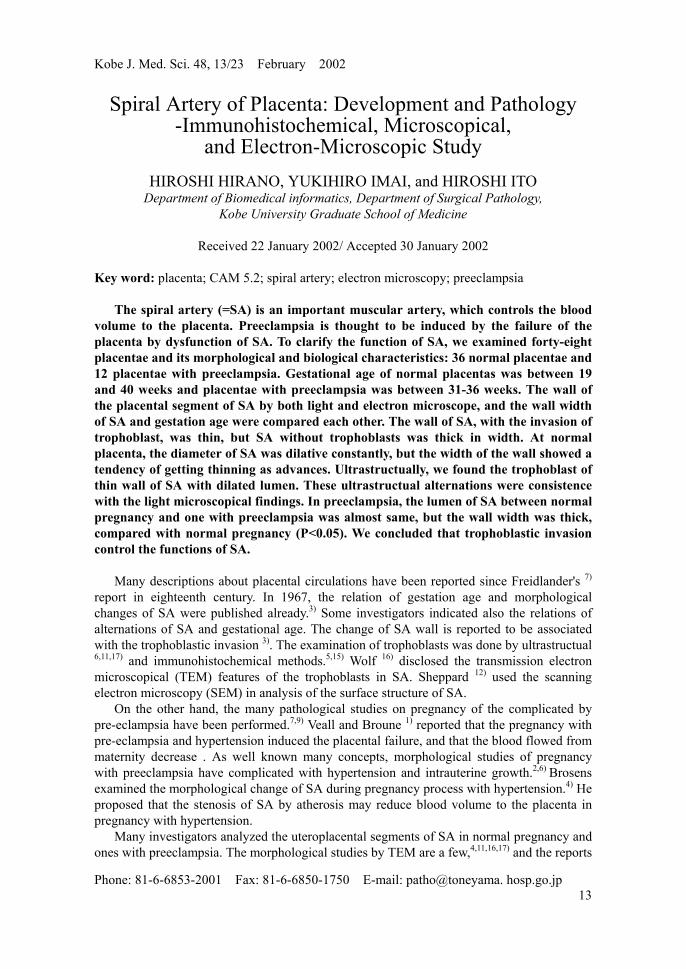
MATERIAL AND METHOD We tried to study the detail features of placental segments of SA and measure these dimension of lumen and wall width of SA and do the observations by the methods of TEM and SEM, to elucidate the mechanism of maternal-fetus circulations and the causes of preeclampsia. Informed consent of all materials of placentae were gotten before pathological examination.
Fig. 1. Cut to the strips; -horizontal with the basal plate.
Patients: We examined 48 placentae: 36 normal placentae and 12 ones with preeclampsia. Normal placentae were between 19 and 40 weeks of gestational age, and placentae with preeclampsia were between 31-36 weeks. In thirty-six cases of normal placentas, there were no any complications such as cardiovascular and endocrine, autoimmune, renal diseases and so furthers. Twelve placentae with preeclampsia showed maternal hypertension, proteinemia, and systemic edema. Clinical history and informations were summarized in Table І-a, І-b. Placentas were fixed in 10% buffered formalin solution, from 6 blocks about 2 by 3 by 0.5 cm in size were made. Many cotyledons are seen on the maternal surface of placenta. The specimens were cut horizontally to the strips on the basal plate of the placentae. Placental specimen had SA (Fig. 1) in the center of block. They were processed routinely in paraffin embedding, and cut 4 µm. Histological sections were stained with HE (Hematoxylin Eosin) and EVG (Elastica van Gieson). We calculated media diameter by internal and external media by EVG and then estimated an idealized diameter. Mean ± SD of the lumen and wall width of SA was calculated finally. A student�s test was used to evaluated clinical and morphological differences between normal pregnancy and ones with preeclampsia by Stat View 4.5 package. Statistical significance was considered at the 0.05 level.
SPIRAL ARTERY OF PLACENTA
15
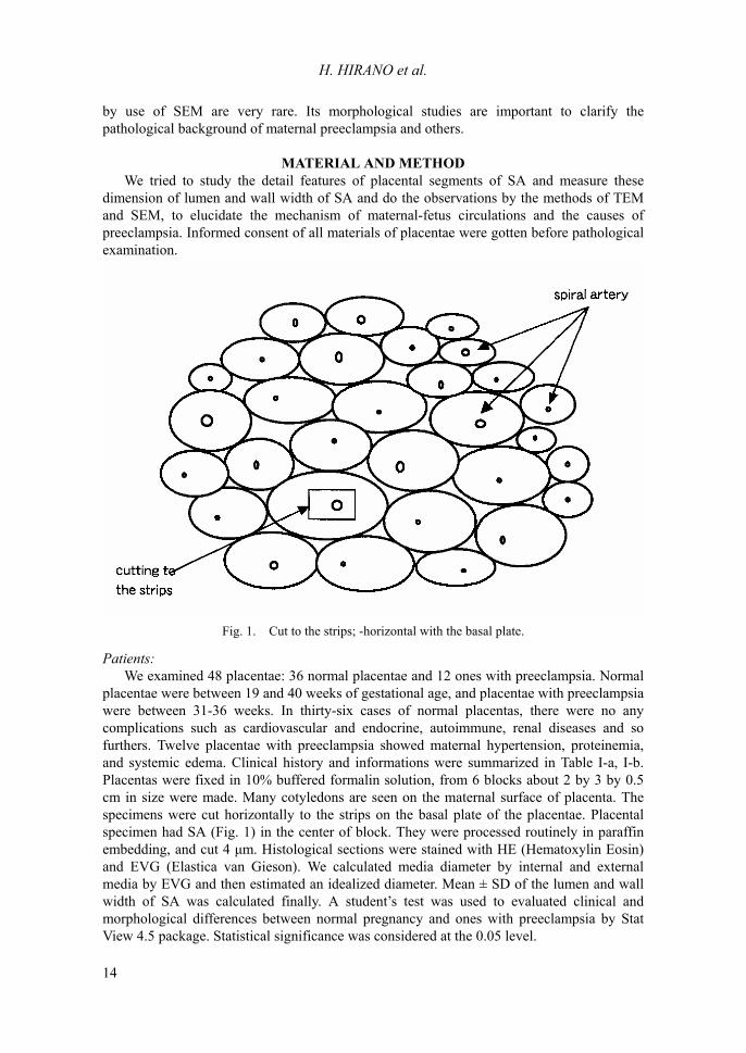
Electron microscopy: We analyzed fifty samples of placental tissue of 31-36 gestation weeks. For TEM analysis, the tissue was minced into 1-mm cubes following surgical removal, fixed overnight in half-strength Karnovsky's fixative, postfixed in osmium tetroxide, dehydrated in alcohols, and embedded in Epon mixture. Thick sections at 1 µm were cut and stained with toluidine blue to trim adequate thinner sectioning. Proper areas containing SA were selected for thin sectioning. Thinner sections stained doubly with uranyl acetate and lead citrate, and observed under transmission electron microscope. At SEM analysis, the specimen was minced in small blocks and fixed by 2.5% glutaraldehyde for 4 hours. They were rinsed several times in 0.1M phosphate buffer and then immersed in 8 N HCl for 45 minutes at 60ûC. They were rinsed again several times in phosphate buffer, treated with a 2% tannic acid solution for one hour, followed by rinsing in buffer. The specimens were then immersed in 0.1% osmium tetroxide for 2 hours at room temperature and transferred into scintillation vials filled with phosphate buffer. The vials were placed in an ultrasonic cleaner bath containing tap water to the level of the buffer in vials where they were subjected to ultrasonic energy at a set frequency of 80 kHz for 2 minutes. The buffer became slightly dark due to release of osmium from the tissue. Tissue blocks were then dried in a series of acetones with graded concentration of acetone and finally in a critical point drier using acetone and CO2. Dried specimens were mounted on specimen stubs with conductive bridges, sputter-coated with gold for 600 seconds, and examined in a SEM at an accelerating voltage of 20kV and a working distance of 20 mm.
Fig. 2. The Relations of the Wall Width of Spiral Artery (SA) between Normal Pregnancy and
Preeclampsia. Immunohistochemistry: We tried immunohistochemical studies to distinguish intermediate trophoblasts from decidual cells. Immunohistochemical staining was performed by the avidin-biotin-peroxidase complex method. Monoclonal antibodies used in this study were CAM5.2 diluted 1:80.
H. HIRANO et al.
16
Table І-a: Clinical data of pregnancy without preeclampsia. Case Age
(year) Gestation week (week)
Placental size (cm)
Placental weight(g)
Number of cotyledon
Clinical diagnosis other than preeclampsia
19-21 gestational weeks 1 26 19 11X 9 172 6 Placenta previa, PROM 2 39 20.5 13X11 230 4 Placental previa, CAM 3 23 22.1 15X12 250 16 NC 4 35 21.6 15X12 260 22 EROM, Abortion 22-24 gestational weeks 5 27 22.5 16X13 330 17 NC 6 29 23 10X 9 170 6 Cervical incompetency 7 28 23.1 14X13 270 9 EROM, Myoma 8 32 23.4 13X10 270 17 NC 9 22 24.3 19X14 450 25 NC 25-27 gestational weeks 10 36 25.1 16X13 350 19 Placenta previa 11 22 25.4 14X12 360 15 Cervical incompetency 12 23 25.6 15X14 230 22 Placental previa 13 35 26.6 17X15 380 13 Chorioamnionitis 14 33 27.3 16X16 445 16 Chorioamnionitis 28-30 gestational weeks 15 23 29.3 13X12 330 15 Cervical incompetency 16 24 30 14X 8 410 17 NC 17 34 30 14X11 380 13 NC 18 30 30 26X16 850 3 Placenta previa 19 29 30.1 18X14 390 12 NC 20 36 30.2 18X17 470 23 EROM 31-33 gestational weeks 21 21 31.5 20X15 455 18 NC 22 30 31.6 15X14 300 18 PROM, CAM 23 31 32 19X17 500 31 EROM 24 33 29 19X17 390 18 NC 25 30 33.1 16X16 425 14 NC 26 34 33.2 21X16 485 23 NC 34-36 gestational weeks 27 30 34 15X15 480 18 EROM 28 31 34 16X15 540 10 Placenta previa 29 28 34.2 18X14 630 16 EROM 30 35 34.4 14X14 360 13 NC 31 26 34.5 28X21 1100 25 NC 32 34 34.5 18X14 520 13 NC 37-40 gestational weeks 33 30 37 16X12 520 23 Placenta previa 34 33 37.3 21X13 545 16 NC 35 28 37.4 21X17 620 40 NC 36 26 38.1 21X16 560 39 NC PROM=premature rapture of the membrane, EROM=early rupture of the membrane, NC=no complication, CAM=chorioamnionitis
SPIRAL ARTERY OF PLACENTA
17
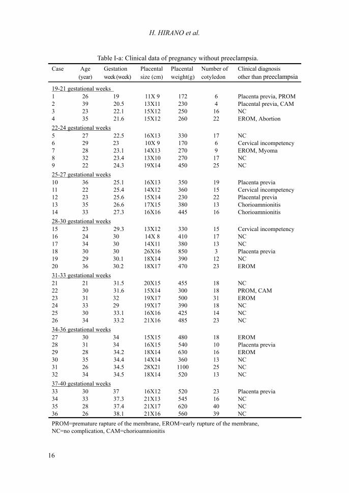
Fig. 3a. The invasion of trophoblasts, showing thinner wall of SA, 38-gestation weeks,
normal pregnancy, HE×200.
Table І-b: Clinical data of pregnancy with preeclampsia.
Case Age (year)
Gestational Week (week)
Placental Size (cm)
Placental Weight(g)
Number of Cotyledon
Clinical Daignosis Other than Toxicosis
31-33 gestational weeks 1 32 31 15X10 390 7 - 2 30 31.6 18X14 400 32 - 3 32 31.2 13X10 380 19 - 4 30 33 23X12 460 20 - 5 35 33.5 17X13 245 16 - 34-36 gestational weeks 6 37 34.2 14X14 370 8 - 7 34 34.2 18X16 480 32 Uterin rapture 8 22 34.3 16X13 440 19 SFD 9 30 34.3 17X13 580 6 - 10 40 34.3 17X12 430 13 - 11 25 36.5 17X14 410 16 - 12 37 36.6 13X10 250 14 IUGR
SFD=small for dates, IUGR=intrauterine growth retardation
H. HIRANO et al.
18
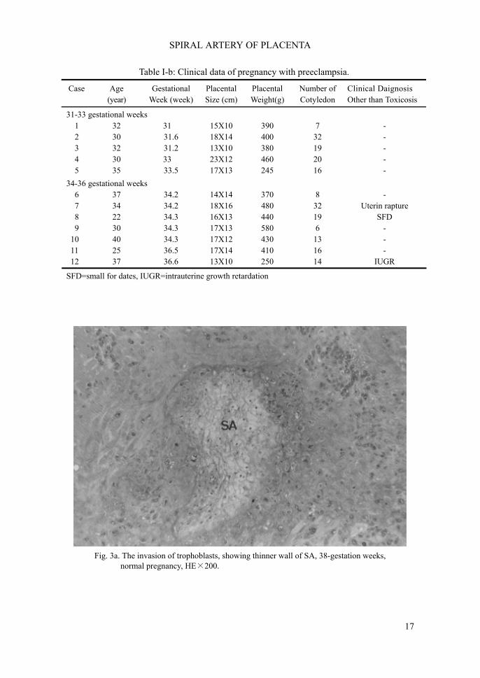
Fig. 3b. No trophoblasts invasion, showing thick wall of SA, 32-gestation weeks, normal
pregnancy, HE×200.
Fig. 4. In the wall of SA, there are remnants of elastic fibers with a few trophoblastic invasion. Preeclampsia, 31-gestation weeks, EF:elastic fiber Elastica van Gieson�s stain,×200.
SPIRAL ARTERY OF PLACENTA
19
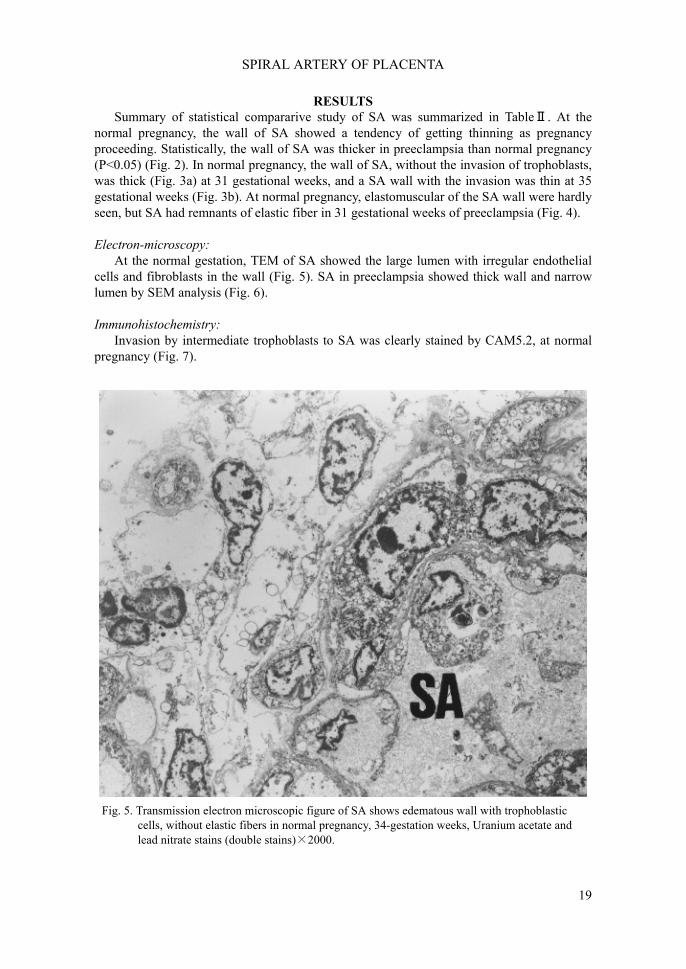
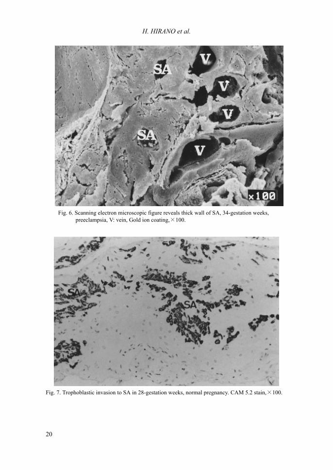
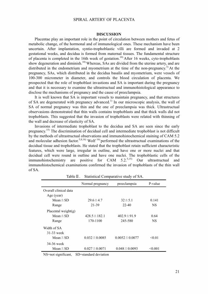
RESULTS Summary of statistical compararive study of SA was summarized in TableⅡ. At the normal pregnancy, the wall of SA showed a tendency of getting thinning as pregnancy proceeding. Statistically, the wall of SA was thicker in preeclampsia than normal pregnancy (P<0.05) (Fig. 2). In normal pregnancy, the wall of SA, without the invasion of trophoblasts, was thick (Fig. 3a) at 31 gestational weeks, and a SA wall with the invasion was thin at 35 gestational weeks (Fig. 3b). At normal pregnancy, elastomuscular of the SA wall were hardly seen, but SA had remnants of elastic fiber in 31 gestational weeks of preeclampsia (Fig. 4). Electron-microscopy: At the normal gestation, TEM of SA showed the large lumen with irregular endothelial cells and fibroblasts in the wall (Fig. 5). SA in preeclampsia showed thick wall and narrow lumen by SEM analysis (Fig. 6). Immunohistochemistry: Invasion by intermediate trophoblasts to SA was clearly stained by CAM5.2, at normal pregnancy (Fig. 7).
Fig. 5. Transmission electron microscopic figure of SA shows edematous wall with trophoblastic cells, without elastic fibers in normal pregnancy, 34-gestation weeks, Uranium acetate and lead nitrate stains (double stains)×2000.
H. HIRANO et al.
20
Fig. 6. Scanning electron microscopic figure reveals thick wall of SA, 34-gestation weeks, preeclampsia, V: vein, Gold ion coating,×100.
Fig. 7. Trophoblastic invasion to SA in 28-gestation weeks, normal pregnancy. CAM 5.2 stain,×100.
SPIRAL ARTERY OF PLACENTA
21
DISCUSSION Placentae play an important role in the point of circulation between mothers and fetus of metabolic change, of the hormonal and of immunological ones. These mechanism have been uncertain. After implantation, syntio-trophoblastic villi are formed and invaded at 2 gestational weeks, and decidua is formed from maternal tissues. The fundamental structure of placenta is completed in the 16th week of gestation.10) After 16 weeks, cyto-trophoblasts show degeneration and diminish.10) Whereas, SAs are divided from the uterine artery, and are distributed in the endometrium and myometrium at the time of the non-pregnancy.3) At the pregnancy, SAs, which distributed in the decidua basalis and myometrium, were vessels of 100-300 micrometer in diameter, and controls the blood circulation of placenta. We prospected that the role of trophoblast invastions and SA is important during the pregnancy and that it is necessary to examine the ultrastructual and immunohistological appearance to disclose the mechanisms of pregnancy and the cause of preeclampsia. It is well known that SA is important vessels to maintain pregnancy, and that structures of SA are degenerated with pregnancy advanced.1) In our microscopic analysis, the wall of SA of normal pregnancy was thin and the one of preeclampsia was thick. Ultrastructual observasions demonstrated that thin walls contains trophoblasts and that thick walls did not trophoblasts. This suggested that the invasion of trophoblasts were related with thinning of the wall and decrease of elasticity of SA. Invasions of intermediate trophoblast to the decidua and SA are seen since the early pregnancy.15) The discrimination of decidual cell and intermediate trophoblast is not difficult by the methods of ultrastructual observations and immunohistochemical staining of CAM 5.2 and molecular adhesion factor.5,8,16) Wolf 16) performed the ultrastructual examinations of the decidual tissue and trophoblasts. He stated that the trophoblast retain sufficient characteristic features, which were large, irregular in outline, and have one or more nuclei and that decidual cell were round in outline and have one nuclei. The trophoblastic cells of the immunohistochemistry are positive for CAM 5.2.5,15) Our ultrastructual and immunohistochemical examinations confirmed the invasion of trophoblasts of the thin wall of SA.
TableⅡ. Statistical Comparative study of SA.
Normal pregnancy preeclampsia P-value
Overall clinical data Age (year) Mean±SD 29.6±4.7 32±5.1 0.141 Range 21-39 22-40 NS
Placental weight(g) Mean±SD 428.5±182.1 402.9±91.9 0.64 Range 170-1100 245-580 NS
Width of SA 31-33 week Mean±SD 0.032±0.0085 0.0052±0.0077 <0.01
34-36 week Mean±SD 0.027±0.0071 0.048±0.0093 <0.001 NS=not significant, SD=standard deviation
H. HIRANO et al.
22
Brozens3) described that remodeling of the SA structures and distruptions of internal elastic lamina and muscular media by fibrinoid material during the normal pregnancy, and he called these alternations �physiological changes�. Whereas, many pathological studies of SA1,2,4,6,7,10) of the complicated pregnancy have been performed, and SA of preeclampsia10) indicated absence of physiological change of SA in utero-placental segment. We examined the SA in the placental segment, which did not show complete physiological changes. Most SA of placental segment were dilated and the little remnant of elastomuscular fiber existed. The remnant of elastic fiber increase resistance of SA. This discrepancy could be explained by the site. At the normal pregnancy, the dilations of SA of placental segment occur at early stage.8,10) Preeclampsia occurs at late stage. We suspected that the physiological changes could not effect on the vessels of the placental segment, which had already been dilated at early stage. However, there were some SA showing thickened wall in the placental segment, compared with one of normal pregnancy. We assumed that the absence of physiological changes may occur in the placental segment. The mechanism of preeclampsia3,7) has been controversy. At preeclampsia, the blood pressure to fetus would decrease, and the narrowing of the lumen by absence of physiological changes induces sufficient blood supply as a compensatory mechanism. We considered trophoblast invasions to the vessels play an important role of controls of blood flow, and the entity of preeclampsia. The controls of the trophoblast invasion to the wall of SA may contribute to the therapy and prophylaxis of preeclampsia. In additions, trophoblast invasions are related to various cytokine and migration factors, and the further investigations of these factors would be necessary.
SPIRAL ARTERY OF PLACENTA
23
REFERENCE 1. Browone, J. and Veall, N. 1953. The maternal placental blood flow in normotensive
and hypertensive women J Obstet Hynaec. Brit. Empire. 60:141-147. 2. Brosens, I. 1964. A study of the spiral arteries of the decidua basalis in normotensive
and hypertensive pregnancy. J. Obstet. Gynecol. 71:222-230. 3. Brosens, I. 1967. The physiological response of the vessels of the placntal bed to
normal pregnancy. J. Path. Bact. 93:569-579. 4. Brosens, I., Dixon, H. G., and Robertson, W.B. 1977. Fetal growth retardation and the
arteries of the placental bed. J. Obstet and Gynecol. 84:656-65. 5. Damsky, C. H., Fitzgerald, M.L., and Fisher, S. J. 1992. Distribution patterns of
extracellular matrix components and adhesion receptors are intricately modulated during first trimeter cytotrophoblast differentiation along the invasive pathway, in vivo. J. Clin. Invest. 89:210-222.
6. Dixon, H.G. and Robertson, W. B. 1953. A study of the vessels of the placental bed in normotensive and hypertensive woman. J. Obstet. Gynecol. 65:803-809.
7. Gerretsen, G., Huisjes, H.J., and Elema J.D. 1981. Morphological changes of the spiral arteries in the placental bed in relation to pre-eclampsia and fetal growth retardation. J. Obstet. Gynecol. 88:876-881.
8. Hamilton W.J. and Boyd J. 1960. Development of the human placenta in the first three months of gestation. J. Anat. 94:297-328.
9. Harijadi, T.K., Nishimura, Y., and Ito, H. 1989. An immunopathological study on the placenta in pre-eclampsia. Kobe J. Med. Sci. 35:217-228.
10. Khong, T.Y. 1991. Acute Atherosis in Pregnancies Complicated by Hypertension, Small-for Gestational-Age Infants, and Diabetes Mellitus. Arch Pathol Lab Med. 115:722-725.
11. Marais W.D. 1968. Human decidual spiral arteries, ultrastructure of the intima in normal vessels. J. Obstet. Gynaecol. Br. Comm. 75:552-567.
12. Sheppard, B.L. and Bonnar, J. 1974. Scanning electron microscopy of the human placenta and decidual spiral arteries in normal pregnancy. 81:20-29.
13. Shin, I.M., Seidman, J.D., and Kurman, J. 1999. Placental site nodule and characterization of distinctive types intermediate trophoblast. Hum. Pathol. 30:687-694.
14. Sternberg, S.S. 1997 Histology for pathologists, second edition Lippincott-Raven, Philadelphia. 961-995. Placenta.
15. Shih, I. M., Nesbit, M., Herlyn M., and Kurman R.J. 1998. A new Mel-CAM (CD146)-specific monoclonal antibody, MN-4, on paraffin-embedded tissue. Mod. Pathol. 11:1098-1106.
16. Wolf, F.D., Wolf-Peeter, C.D., and Brosens, I. 1973. Ultrastructure of the spiral areteries in the human placental bed at the end of normal pregnancy. Am. J. Obstet. Gynecol. 15:833-848.
17. Wynn, R. M. 1967. Cytotrophblastic specializations: An ultrastructural study of the human placenta. Am. J. Obstet. Gynecol. 97:339-355.
18. Zhou, Y., Damsky, C.H., and Fisher, S. J. 1997. Preeclampsia is associated with failure of human cytotrophoblasts mimic a vascular adhesion phenotype. J. Clin. Invest. 99:2152-2164.