Embed Size (px)
Citation preview
This free educational material is provided by
333 N. Commercial Street, Suite 100, Neenah, WI 54956
For additional free educational material regarding symptoms and imaging, please visit www.symptombasedradiology.com
Spine Pain
Donald L. Renfrew, MD
According to Wikipedia1: “Back pain is one of
humanity's most frequent complaints. In the U.S.,
acute low back pain (also called lumbago) is the fifth
most common reason for all physician visits. About
nine out of ten adults experience back pain at some
point in their life, and five out of ten working adults
have back pain every year.” In caring for patients
with spine pain (pain from the neck or low back
and/or radicular symptoms), there are multiple
diagnostic tests and therapeutic options available.
Diagnostic imaging offers plain films, computed
tomography, myelography, combined
myelography/CT, nuclear medicine bone scans,
magnetic resonance imaging, and fluoroscopically
guided injections. Therapy includes doing nothing,
oral medications, physical rehabilitation, spine
injections, surgery, and then sometimes surgery
again. This chapter covers three key concepts to
guide the choice in diagnosis and treatment of spine
pain. These concepts are:
1. “Red flags” in the patient’s presentation call for priority imaging.
2. MRI has supplanted other modalities for the imaging work-up of spine pain.
3. Injections often provide diagnostic or therapeutic benefit for patients with spine pain.
“RED FLAGS” IN THE PATIENT’S PRESENTATION CALL FOR PRIORITY
IMAGING
Gordon Waddell, a Glasgow spine surgeon, uses
the term “red flag” to denote those clinical findings
that indicate the potential of a medically serious
diagnosis, and which should prompt priority
imaging. Waddell’s book The Back Pain Revolution
(Churchill Livingston, 2004) is an excellent book for
anyone who treats those with back pain.
Spine pain is such a common disorder, and so
often runs a benign course, that common advice
(although not necessarily often followed) is to wait
4-6 weeks before pursuing costly diagnostic
measures. However, in the presence of a “red flag”,
it is prudent to expedite imaging. This does not
necessarily mean that the examination has to be
performed in the next five minutes, but it would
probably be better to get imaging done this week
rather than waiting a month.
Red flags include a personal history of
malignancy, unremitting pain, pediatric patients
with back pain, and constitutional symptoms (for
example, weight loss or fever).
Page 74 Spine Pain
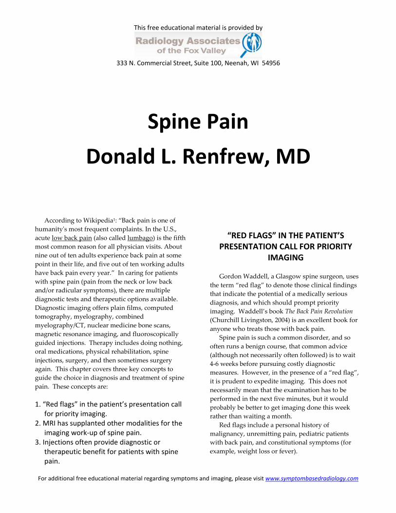
Figure 1. Metastatic lung cancer in a 65 year old man with back pain and known primary malignancy. A. Sagittal thoracic spine T1 weighted MR image shows decreased signal at T6 and T12 (arrows). B. Sagittal thoracic spine fat saturated T2 weighted MR image shows increased signal intensity (arrows), also at T6 and T12.
A personal history of cancer
This particular scenario, with patients presenting
to primary care physicians with spine pain after
successful cancer therapy, will likely increase in
frequency as oncologists become better at curing, or
at least putting into remission, various tumors. In
the case of a patient with the new onset of spine pain
and cancer, it makes most sense to first review any
existing imaging studies, to see if they indicate (even
in retrospect) a malignant cause of the pain. Studies
done for tumor imaging such as CT of the abdomen
and pelvis may show bone destructive changes
which are easy to overlook. Nuclear medicine
studies such as bone scans and PET-CT studies
usually show more conspicuous and easily
appreciated abnormalities which are less likely to be
missed. If these studies do not show an explanatory
abnormality, plain films of the painful region may
be ordered but will likely not be the final study
performed regardless of the outcome: if they are
negative MR will need to be performed (because
Chapter 6 Spine Pain Page 75
plain films are insensitive), and if positive, MR will
likely still need to be performed (to evaluate the
extent of tumor including neural compression)
(Figure 1).
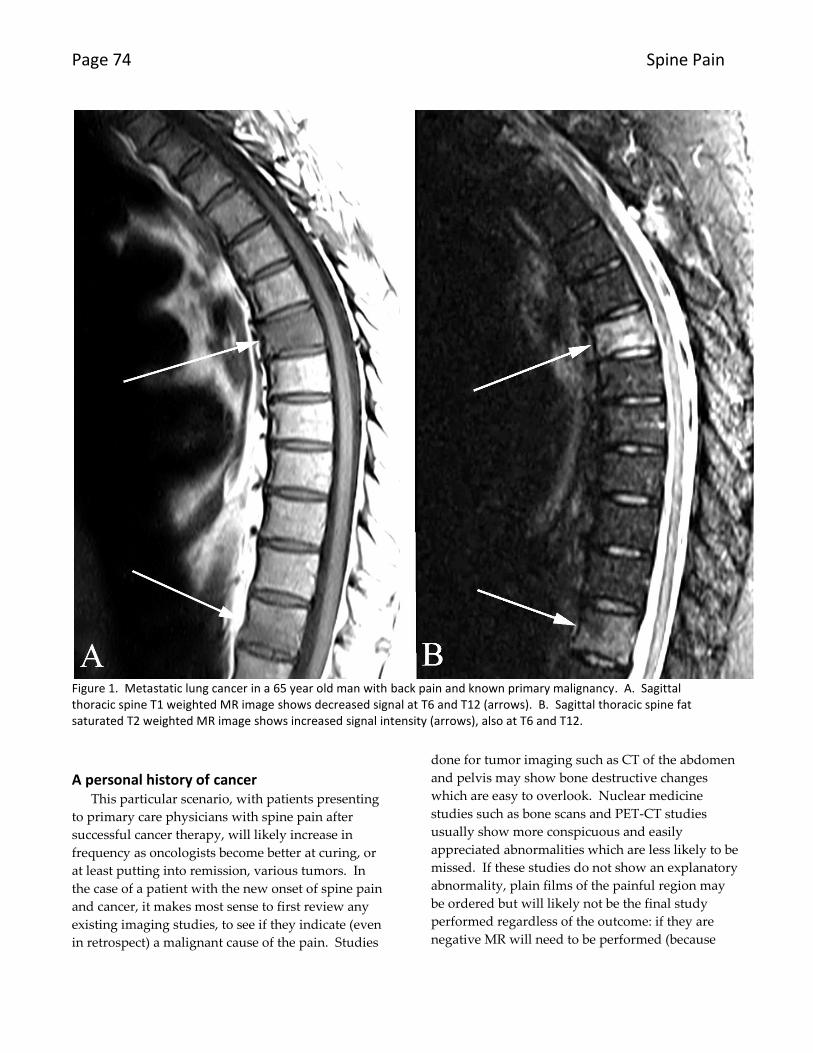
Unremitting pain This red flag emphasizes that, typically, benign
spine pain is “mechanical” in the sense that it is
brought on by mechanical factors (assuming a
certain position, bearing a certain load), whereas
spine pain secondary to such factors as tumor
(Figure 2), osteomyelitis, or fracture is “non-
mechanical”. The patient cannot find comfort
standing, sitting, or lying down, and finds little
relief with medications which would normally offer
benefit. Note that while a young, healthy adult
would not normally sustain a spine fracture without
significant trauma, the amount of trauma necessary
to fracture an elderly, osteoporotic spine can be so
trivial that it escapes notice, and therefore the
patient may present with an osteoporotic fracture
but not recall a specific incident that initiated the
pain.
Figure 2. Lymphoma in a 65 year old man with new onset of unremitting back pain. A. Sagittal lumbar spine T1 weighted spine MR shows decreased signal in the L2 vertebral body and a mass extending into the spinal canal (arrow). B. Sagittal T2 weighted spine MR shows increased signal within the vertebral body, and also demonstrates the soft tissue mass.
Page 76 Spine Pain
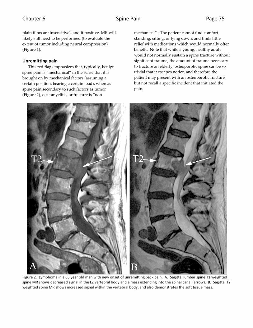
Figure 3. Neuroblastoma in a 2 year old with back pain. fatigue, and fussiness. A. Lateral plain film of the lumbar and thoracic spine shows a mass (arrow) along the posterior upper chest. B. Sagittal T2 MR shows a paraspinal mass (arrow).
The pediatric patient Any child with spine pain should be evaluated
carefully. Children rarely if ever have “degenerative”
causes of backache. Occasionally, teenagers may
present with central low back pain from
spondylolysis. However, any younger child with
spine pain should be suspected of having a possible
serious medical condition such as tumor (Figure 3),
infection, or unreported trauma.
MRI HAS SUPPLANTED OTHER METHODS FOR IMAGING BACK PAIN
In the short space of approximately 25 years,
magnetic resonance imaging has revolutionized
medicine. MRI has changed the way neurologists,
neurosurgeons, and orthopedic surgeons evaluate
and care for patients by providing detailed images
of both pre- and post-operative anatomy that were
the stuff of science fiction a few short decades ago.
Paul C. Lauterber from the University of Illinois and
Peter Mansfield from the University of Nottingham

Chapter 6 Spine Pain Page 77
won the Nobel Prize in Medicine for their key role in
the development of magnetic resonance imaging in
2003, and rightly so, for this technology has allowed
not only academic research on many of the most
devastating diseases, but also found widespread use
in community practice. It would be difficult to find
a family in the United States that has not had a
member to undergo MRI, and we are all familiar
with the sports announcers’ refrain “The team
doctor is waiting for the MR results to make a
decision on the player’s return to action”. MR not
only demonstrates the causes of the “red flags” just
mentioned, but also shows soft tissue and bony
causes of back and leg pain.
Plain films may be obtained prior to performing
an MR, but (as noted in the case of evaluating tumor
patients, above) almost always need to be
supplemented by MR. One possible exception: in
cases of trauma where the plain film documents a
simple compression fracture, MR is typically not
necessary (although often even in this scenario, the
MR will provide significant additional information;
see below).
MR shows the causes of “red flags” As noted above, patients with a history of cancer,
unremitting pain, and pediatric patients may have
serious medical diseases. These patients require
priority imaging and prompt diagnosis and
management. MR is the method of choice for
evaluation of these patients. Please note that while
many patients with tumor, fracture, or infection do
demonstrate these red flags, probably as many do
not, making MR all the more valuable.
MR shows symptom producing, benign soft tissue abnormalities MR’s superiority comes predominantly from its
ability to visualize soft tissues. Prior to MR, imaging
relied on the use of x-rays, either to produce plain
films or myelograms, or CT scans. While a
wonderful invention and tremendously useful, x-ray
based techniques have limitations, the main one of
which is that the x-ray attenuation of different
tissues such as the intervertebral disc, muscle,
synovium, and even tumor is virtually identical, and
the x-ray attenuation of fluid within the
cerebrospinal space is not much different.
Neuroradiologists relied, for decades, on secondary
phenomenon to diagnose spine disease: the lost
intervertebral disc space on plain films as a sign of
disc herniation, or the filling defect on myelography
or myelo-CT. MR easily shows each type of tissue
separately, MR evaluates different physical
properties of protons within the patient to create
pictures showing anatomic detail and unparalleled
demonstration of disease processes.
Disc Herniation
Since the original description by Mixter and Barr
in 19342, the herniated disc has gotten much press.
The North American Spine Society (NASS)
originally proposed, and various other medical
societies have adopted, a specific nomenclature that
distinguishes subtypes of herniation3. If viewed
axially, the normal intervertebral discs are like a tree
trunk with concentrically arranged layers of oblique
fibers constituting the annulus fibrosus. In the
middle of the tree trunk, having a consistency of
toothpaste, is the nucleus pulposus. When the
annular fibers degenerate and/or tear, the nuclear
material may extend or herniate beyond the fibers of
the annulus, and if this happens posteriorly the
effect may be compression and/or inflammation of
adjacent nerves (Figure 4). The NASS terminology
calls small disc herniations “protrusions” and these
are much less likely to be symptomatic. The NASS
terminology calls large disc herniations “extrusions”
and these are much more likely to be symptomatic.
Note that large, acutely symptomatic disc
herniations may show significant regression when
sequentially imaged, even without surgical
intervention.
Page 78 Spine Pain
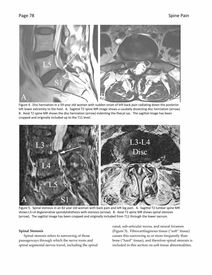
Figure 4. Disc herniation in a 59 year old woman with sudden onset of left back pain radiating down the posterior left lower extremity to the heel. A. Sagittal T2 spine MR image shows a caudally dissecting disc herniation (arrow). B. Axial T2 spine MR shows the disc herniation (arrow) indenting the thecal sac. The sagittal image has been cropped and originally included up to the T11 level.
Figure 5. Spinal stenosis in an 82 year old woman with back pain and left leg pain. A. Sagittal T2 lumbar spine MR shows L3-L4 degenerative spondylolisthesis with stenosis (arrow). B. Axial T2 spine MR shows spinal stenosis (arrow). The sagittal image has been cropped and originally included from T11 through the lower sacrum.
Spinal Stenosis
Spinal stenosis refers to narrowing of those
passageways through which the nerve roots and
spinal segmental nerves travel, including the spinal
canal, sub-articular recess, and neural foramen
(Figure 5). Fibrocartilaginous tissue (“soft” tissue)
causes this narrowing as or more frequently than
bone (“hard” tissue), and therefore spinal stenosis is
included in this section on soft tissue abnormalities.
Chapter 6 Spine Pain Page 79
While plain films usually show degenerative
changes in patients with spinal stenosis, and CT
often better shows the degree of narrowing, MR is
capable of showing not only the narrowing but also
demonstrating the neural structures, and any
associated compression, directly. Compression of
the nerves may result in pain or radiculopathy, but
may also result in less specific generalized leg
weakness and disability, a finding that may be
exacerbated during extension and relieved during
flexion. Indeed, these patients often find relief of
their symptoms at the grocery store, for they use the
grocery cart as an ambulation assistant which allows
them to walk in a forward-flexed position which
opens the spinal canal.
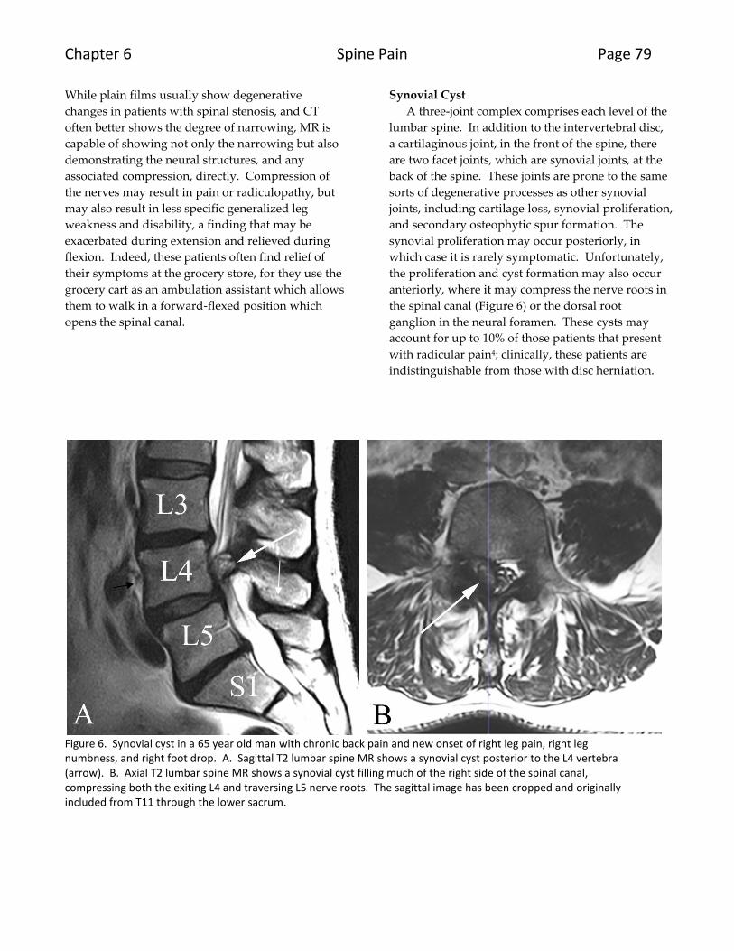
Synovial Cyst
A three-joint complex comprises each level of the
lumbar spine. In addition to the intervertebral disc,
a cartilaginous joint, in the front of the spine, there
are two facet joints, which are synovial joints, at the
back of the spine. These joints are prone to the same
sorts of degenerative processes as other synovial
joints, including cartilage loss, synovial proliferation,
and secondary osteophytic spur formation. The
synovial proliferation may occur posteriorly, in
which case it is rarely symptomatic. Unfortunately,
the proliferation and cyst formation may also occur
anteriorly, where it may compress the nerve roots in
the spinal canal (Figure 6) or the dorsal root
ganglion in the neural foramen. These cysts may
account for up to 10% of those patients that present
with radicular pain4; clinically, these patients are
indistinguishable from those with disc herniation.
Figure 6. Synovial cyst in a 65 year old man with chronic back pain and new onset of right leg pain, right leg numbness, and right foot drop. A. Sagittal T2 lumbar spine MR shows a synovial cyst posterior to the L4 vertebra (arrow). B. Axial T2 lumbar spine MR shows a synovial cyst filling much of the right side of the spinal canal, compressing both the exiting L4 and traversing L5 nerve roots. The sagittal image has been cropped and originally included from T11 through the lower sacrum.
Page 80 Spine Pain
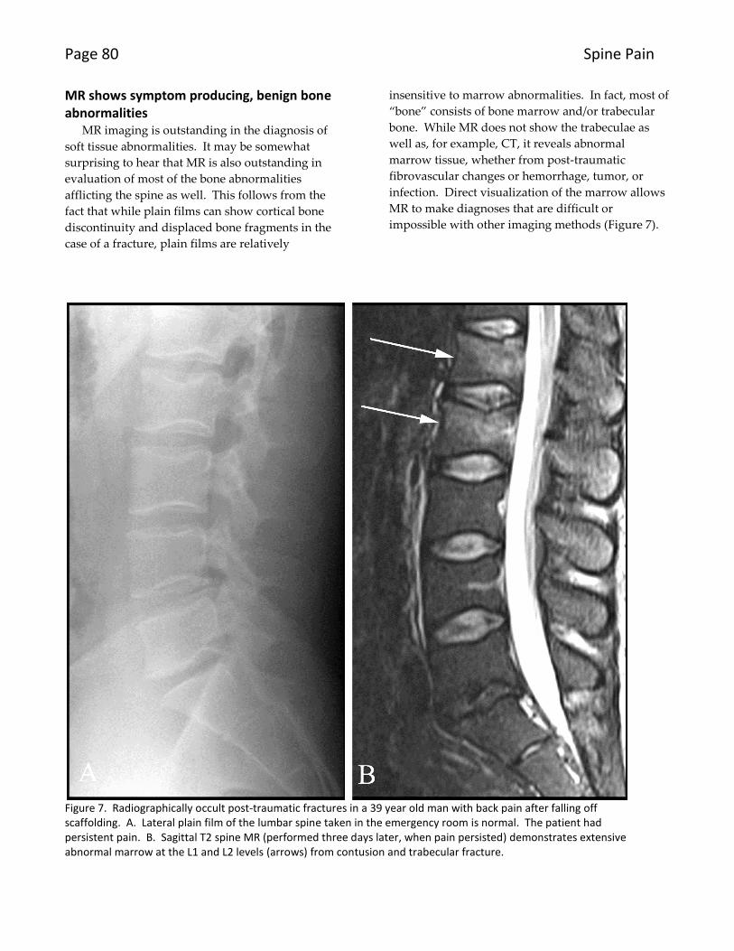
MR shows symptom producing, benign bone abnormalities
MR imaging is outstanding in the diagnosis of
soft tissue abnormalities. It may be somewhat
surprising to hear that MR is also outstanding in
evaluation of most of the bone abnormalities
afflicting the spine as well. This follows from the
fact that while plain films can show cortical bone
discontinuity and displaced bone fragments in the
case of a fracture, plain films are relatively
insensitive to marrow abnormalities. In fact, most of
“bone” consists of bone marrow and/or trabecular
bone. While MR does not show the trabeculae as
well as, for example, CT, it reveals abnormal
marrow tissue, whether from post-traumatic
fibrovascular changes or hemorrhage, tumor, or
infection. Direct visualization of the marrow allows
MR to make diagnoses that are difficult or
impossible with other imaging methods (Figure 7).
Figure 7. Radiographically occult post-traumatic fractures in a 39 year old man with back pain after falling off scaffolding. A. Lateral plain film of the lumbar spine taken in the emergency room is normal. The patient had persistent pain. B. Sagittal T2 spine MR (performed three days later, when pain persisted) demonstrates extensive abnormal marrow at the L1 and L2 levels (arrows) from contusion and trabecular fracture.
Chapter 6 Spine Pain Page 81
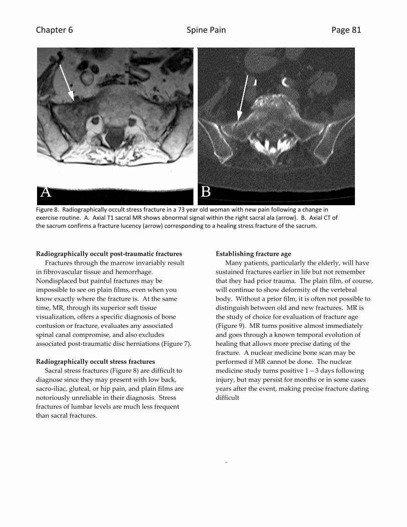
Figure 8. Radiographically occult stress fracture in a 73 year old woman with new pain following a change in exercise routine. A. Axial T1 sacral MR shows abnormal signal within the right sacral ala (arrow). B. Axial CT of the sacrum confirms a fracture lucency (arrow) corresponding to a healing stress fracture of the sacrum.
Radiographically occult post-traumatic fractures
Fractures through the marrow invariably result
in fibrovascular tissue and hemorrhage.
Nondisplaced but painful fractures may be
impossible to see on plain films, even when you
know exactly where the fracture is. At the same
time, MR, through its superior soft tissue
visualization, offers a specific diagnosis of bone
contusion or fracture, evaluates any associated
spinal canal compromise, and also excludes
associated post-traumatic disc herniations (Figure 7).
Radiographically occult stress fractures
Sacral stress fractures (Figure 8) are difficult to
diagnose since they may present with low back,
sacro-iliac, gluteal, or hip pain, and plain films are
notoriously unreliable in their diagnosis. Stress
fractures of lumbar levels are much less frequent
than sacral fractures.
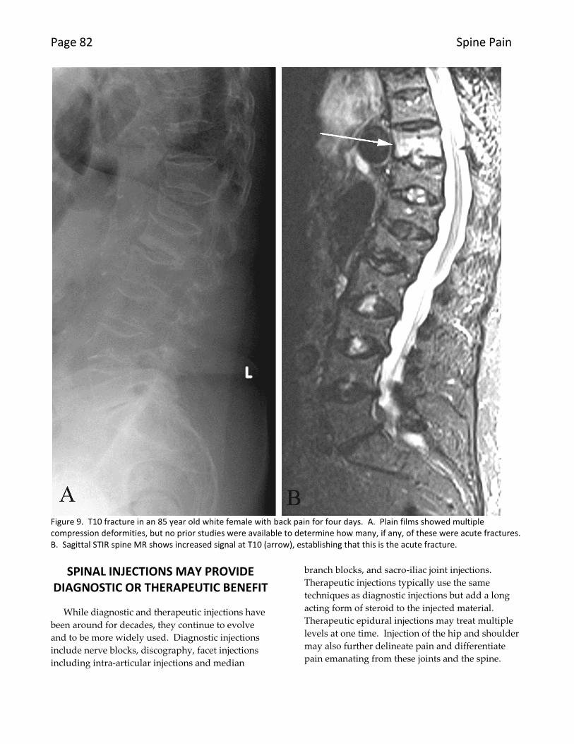
Establishing fracture age
Many patients, particularly the elderly, will have
sustained fractures earlier in life but not remember
that they had prior trauma. The plain film, of course,
will continue to show deformity of the vertebral
body. Without a prior film, it is often not possible to
distinguish between old and new fractures. MR is
the study of choice for evaluation of fracture age
(Figure 9). MR turns positive almost immediately
and goes through a known temporal evolution of
healing that allows more precise dating of the
fracture. A nuclear medicine bone scan may be
performed if MR cannot be done. The nuclear
medicine study turns positive 1 – 3 days following
injury, but may persist for months or in some cases
years after the event, making precise fracture dating
difficult
.
Page 82 Spine Pain
Figure 9. T10 fracture in an 85 year old white female with back pain for four days. A. Plain films showed multiple compression deformities, but no prior studies were available to determine how many, if any, of these were acute fractures. B. Sagittal STIR spine MR shows increased signal at T10 (arrow), establishing that this is the acute fracture.
SPINAL INJECTIONS MAY PROVIDE DIAGNOSTIC OR THERAPEUTIC BENEFIT
While diagnostic and therapeutic injections have
been around for decades, they continue to evolve
and to be more widely used. Diagnostic injections
include nerve blocks, discography, facet injections
including intra-articular injections and median
branch blocks, and sacro-iliac joint injections.
Therapeutic injections typically use the same
techniques as diagnostic injections but add a long
acting form of steroid to the injected material.
Therapeutic epidural injections may treat multiple
levels at one time. Injection of the hip and shoulder
may also further delineate pain and differentiate
pain emanating from these joints and the spine.
Chapter 6 Spine Pain Page 83
Injections may localize or treat a “pain generator”
The three fundamental assumptions of diagnostic
and therapeutic injections include5:
1. Needle placement and injection close to or at
the site of a symptomatic structure will
stimulate nociceptors and thus reproduce
the patient’s typical pain.
2. Anesthetic placed through the needle will
(at least temporarily) decrease activity
within nociceptors and thus relieve the
patient’s typical pain.
3. Pain may be secondary to inflammation
contributing to nociceptor stimulation and
may respond to steroid injection.
Note that because of the placebo effect,
regression to the mean, and the intermittent natural
history of back pain, it is difficult to be certain that
relief of pain upon injection of a structure is genuine
“proof” that the structure is the cause of that pain.
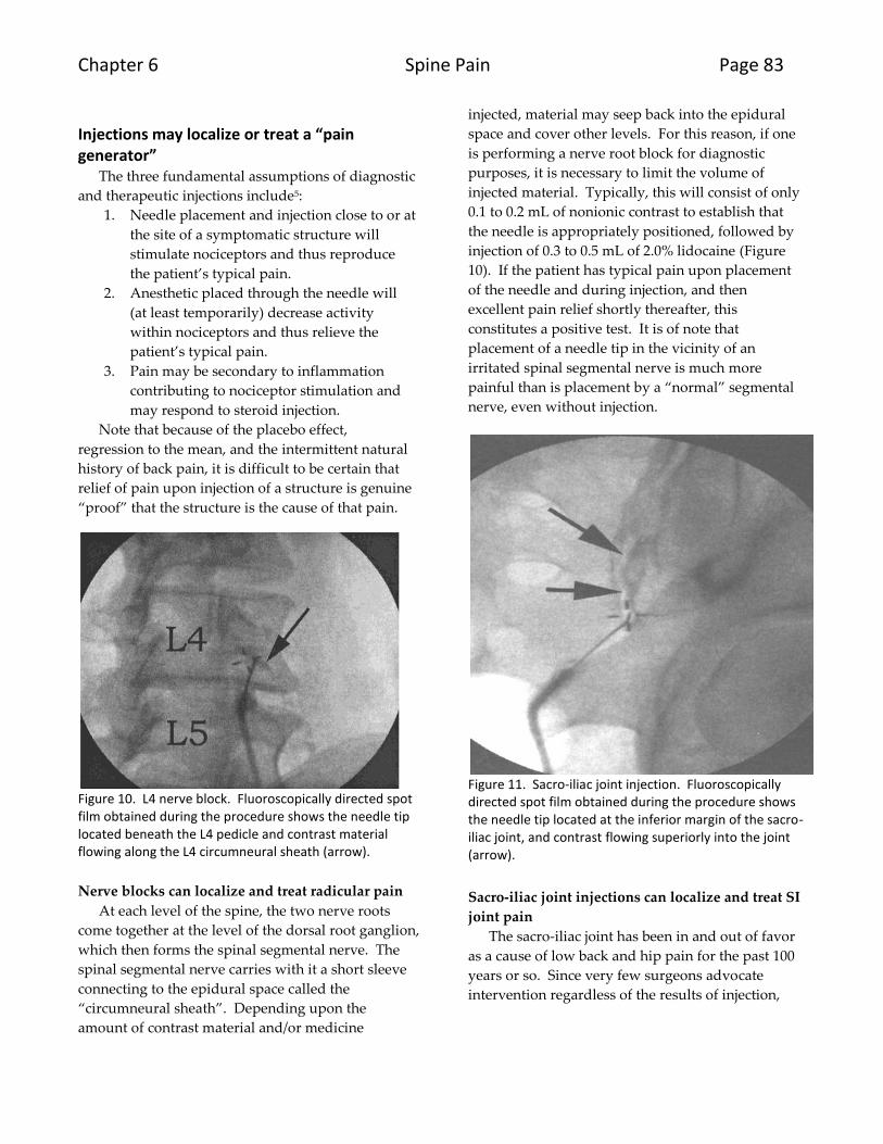
Figure 10. L4 nerve block. Fluoroscopically directed spot film obtained during the procedure shows the needle tip located beneath the L4 pedicle and contrast material flowing along the L4 circumneural sheath (arrow).
Nerve blocks can localize and treat radicular pain
At each level of the spine, the two nerve roots
come together at the level of the dorsal root ganglion,
which then forms the spinal segmental nerve. The
spinal segmental nerve carries with it a short sleeve
connecting to the epidural space called the
“circumneural sheath”. Depending upon the
amount of contrast material and/or medicine
injected, material may seep back into the epidural
space and cover other levels. For this reason, if one
is performing a nerve root block for diagnostic
purposes, it is necessary to limit the volume of
injected material. Typically, this will consist of only
0.1 to 0.2 mL of nonionic contrast to establish that
the needle is appropriately positioned, followed by
injection of 0.3 to 0.5 mL of 2.0% lidocaine (Figure
10). If the patient has typical pain upon placement
of the needle and during injection, and then
excellent pain relief shortly thereafter, this
constitutes a positive test. It is of note that
placement of a needle tip in the vicinity of an
irritated spinal segmental nerve is much more
painful than is placement by a “normal” segmental
nerve, even without injection.
Figure 11. Sacro-iliac joint injection. Fluoroscopically directed spot film obtained during the procedure shows the needle tip located at the inferior margin of the sacro-iliac joint, and contrast flowing superiorly into the joint (arrow).
Sacro-iliac joint injections can localize and treat SI
joint pain
The sacro-iliac joint has been in and out of favor
as a cause of low back and hip pain for the past 100
years or so. Since very few surgeons advocate
intervention regardless of the results of injection,
Page 84 Spine Pain
there seems to be little diagnostic role for injections
(Figure 11). The injections may provide pain relief.
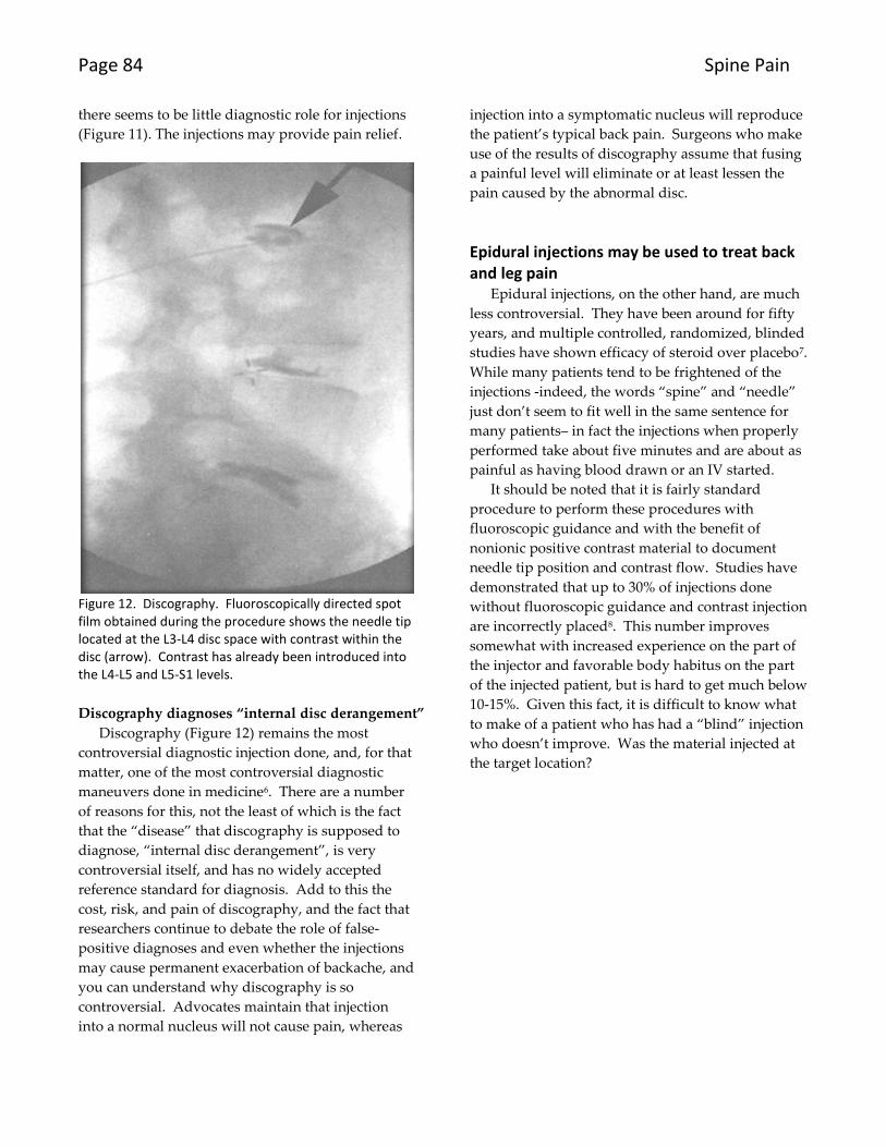
Figure 12. Discography. Fluoroscopically directed spot film obtained during the procedure shows the needle tip located at the L3-L4 disc space with contrast within the disc (arrow). Contrast has already been introduced into the L4-L5 and L5-S1 levels.
Discography diagnoses “internal disc derangement”
Discography (Figure 12) remains the most
controversial diagnostic injection done, and, for that
matter, one of the most controversial diagnostic
maneuvers done in medicine6. There are a number
of reasons for this, not the least of which is the fact
that the “disease” that discography is supposed to
diagnose, “internal disc derangement”, is very
controversial itself, and has no widely accepted
reference standard for diagnosis. Add to this the
cost, risk, and pain of discography, and the fact that
researchers continue to debate the role of false-
positive diagnoses and even whether the injections
may cause permanent exacerbation of backache, and
you can understand why discography is so
controversial. Advocates maintain that injection
into a normal nucleus will not cause pain, whereas
injection into a symptomatic nucleus will reproduce
the patient’s typical back pain. Surgeons who make
use of the results of discography assume that fusing
a painful level will eliminate or at least lessen the
pain caused by the abnormal disc.
Epidural injections may be used to treat back and leg pain
Epidural injections, on the other hand, are much
less controversial. They have been around for fifty
years, and multiple controlled, randomized, blinded
studies have shown efficacy of steroid over placebo7.
While many patients tend to be frightened of the
injections -indeed, the words “spine” and “needle”
just don’t seem to fit well in the same sentence for
many patients– in fact the injections when properly
performed take about five minutes and are about as
painful as having blood drawn or an IV started.
It should be noted that it is fairly standard
procedure to perform these procedures with
fluoroscopic guidance and with the benefit of
nonionic positive contrast material to document
needle tip position and contrast flow. Studies have
demonstrated that up to 30% of injections done
without fluoroscopic guidance and contrast injection
are incorrectly placed8. This number improves
somewhat with increased experience on the part of
the injector and favorable body habitus on the part
of the injected patient, but is hard to get much below
10-15%. Given this fact, it is difficult to know what
to make of a patient who has had a “blind” injection
who doesn’t improve. Was the material injected at
the target location?
Chapter 6 Spine Pain Page 85
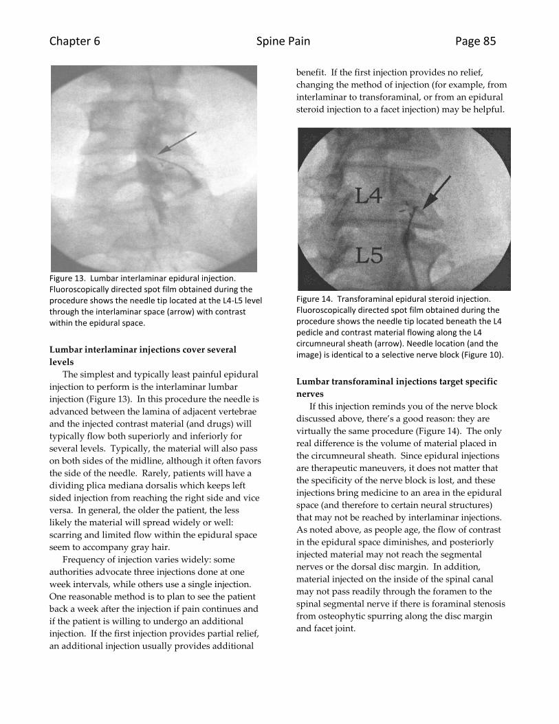
Figure 13. Lumbar interlaminar epidural injection. Fluoroscopically directed spot film obtained during the procedure shows the needle tip located at the L4-L5 level through the interlaminar space (arrow) with contrast within the epidural space.
Lumbar interlaminar injections cover several
levels
The simplest and typically least painful epidural
injection to perform is the interlaminar lumbar
injection (Figure 13). In this procedure the needle is
advanced between the lamina of adjacent vertebrae
and the injected contrast material (and drugs) will
typically flow both superiorly and inferiorly for
several levels. Typically, the material will also pass
on both sides of the midline, although it often favors
the side of the needle. Rarely, patients will have a
dividing plica mediana dorsalis which keeps left
sided injection from reaching the right side and vice
versa. In general, the older the patient, the less
likely the material will spread widely or well:
scarring and limited flow within the epidural space
seem to accompany gray hair.
Frequency of injection varies widely: some
authorities advocate three injections done at one
week intervals, while others use a single injection.
One reasonable method is to plan to see the patient
back a week after the injection if pain continues and
if the patient is willing to undergo an additional
injection. If the first injection provides partial relief,
an additional injection usually provides additional
benefit. If the first injection provides no relief,
changing the method of injection (for example, from
interlaminar to transforaminal, or from an epidural
steroid injection to a facet injection) may be helpful.
Figure 14. Transforaminal epidural steroid injection. Fluoroscopically directed spot film obtained during the procedure shows the needle tip located beneath the L4 pedicle and contrast material flowing along the L4 circumneural sheath (arrow). Needle location (and the image) is identical to a selective nerve block (Figure 10).
Lumbar transforaminal injections target specific
nerves
If this injection reminds you of the nerve block
discussed above, there’s a good reason: they are
virtually the same procedure (Figure 14). The only
real difference is the volume of material placed in
the circumneural sheath. Since epidural injections
are therapeutic maneuvers, it does not matter that
the specificity of the nerve block is lost, and these
injections bring medicine to an area in the epidural
space (and therefore to certain neural structures)
that may not be reached by interlaminar injections.
As noted above, as people age, the flow of contrast
in the epidural space diminishes, and posteriorly
injected material may not reach the segmental
nerves or the dorsal disc margin. In addition,
material injected on the inside of the spinal canal
may not pass readily through the foramen to the
spinal segmental nerve if there is foraminal stenosis
from osteophytic spurring along the disc margin
and facet joint.
Page 86 Spine Pain
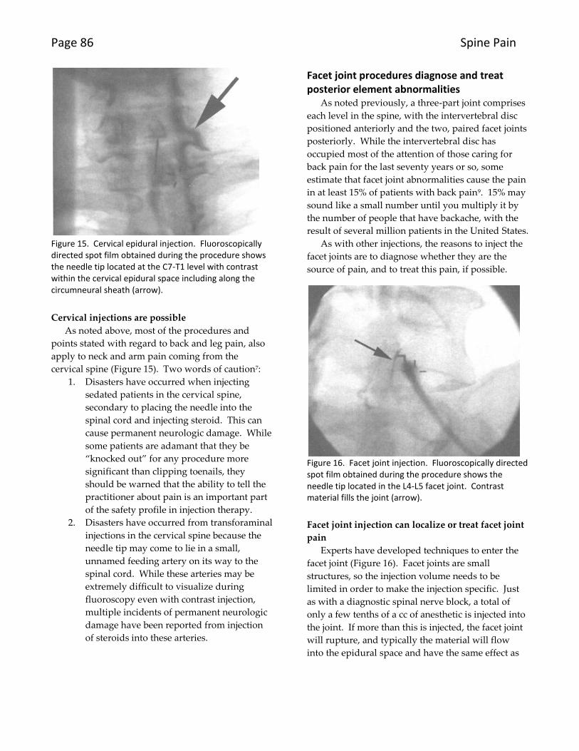
Figure 15. Cervical epidural injection. Fluoroscopically directed spot film obtained during the procedure shows the needle tip located at the C7-T1 level with contrast within the cervical epidural space including along the circumneural sheath (arrow).
Cervical injections are possible
As noted above, most of the procedures and
points stated with regard to back and leg pain, also
apply to neck and arm pain coming from the
cervical spine (Figure 15). Two words of caution7:
1. Disasters have occurred when injecting
sedated patients in the cervical spine,
secondary to placing the needle into the
spinal cord and injecting steroid. This can
cause permanent neurologic damage. While
some patients are adamant that they be
“knocked out” for any procedure more
significant than clipping toenails, they
should be warned that the ability to tell the
practitioner about pain is an important part
of the safety profile in injection therapy.
2. Disasters have occurred from transforaminal
injections in the cervical spine because the
needle tip may come to lie in a small,
unnamed feeding artery on its way to the
spinal cord. While these arteries may be
extremely difficult to visualize during
fluoroscopy even with contrast injection,
multiple incidents of permanent neurologic
damage have been reported from injection
of steroids into these arteries.
Facet joint procedures diagnose and treat posterior element abnormalities
As noted previously, a three-part joint comprises
each level in the spine, with the intervertebral disc
positioned anteriorly and the two, paired facet joints
posteriorly. While the intervertebral disc has
occupied most of the attention of those caring for
back pain for the last seventy years or so, some
estimate that facet joint abnormalities cause the pain
in at least 15% of patients with back pain9. 15% may
sound like a small number until you multiply it by
the number of people that have backache, with the
result of several million patients in the United States.
As with other injections, the reasons to inject the
facet joints are to diagnose whether they are the
source of pain, and to treat this pain, if possible.
Figure 16. Facet joint injection. Fluoroscopically directed spot film obtained during the procedure shows the needle tip located in the L4-L5 facet joint. Contrast material fills the joint (arrow).
Facet joint injection can localize or treat facet joint
pain
Experts have developed techniques to enter the
facet joint (Figure 16). Facet joints are small
structures, so the injection volume needs to be
limited in order to make the injection specific. Just
as with a diagnostic spinal nerve block, a total of
only a few tenths of a cc of anesthetic is injected into
the joint. If more than this is injected, the facet joint
will rupture, and typically the material will flow
into the epidural space and have the same effect as
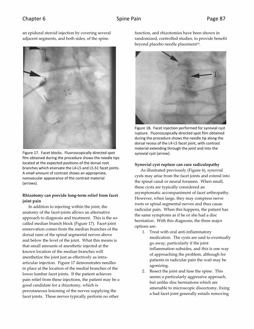
Chapter 6 Spine Pain Page 87
an epidural steroid injection by covering several
adjacent segments, and both sides, of the spine.
Figure 17. Facet blocks. Fluoroscopically directed spot film obtained during the procedure shows the needle tips located at the expected positions of the dorsal root branches which enervate the L4-L5 and L5-S1 facet joints. A small amount of contrast shows an appropriate, nonvascular appearance of the contrast material (arrows).
Rhizotomy can provide long-term relief from facet
joint pain
In addition to injecting within the joint, the
anatomy of the facet joints allows an alternative
approach to diagnosis and treatment. This is the so-
called median branch block (Figure 17). Facet joint
ennervation comes from the median branches of the
dorsal rami of the spinal segmental nerves above
and below the level of the joint. What this means is
that small amounts of anesthetic injected at the
known location of the median branches will
anesthetize the joint just as effectively as intra-
articular injection. Figure 17 demonstrates needles
in place at the location of the medial branches of the
lower lumbar facet joints. If the patient achieves
pain relief from these injections, the patient may be a
good candidate for a rhizotomy, which is
percutaneous lesioning of the nerves supplying the
facet joints. These nerves typically perform no other
function, and rhizotomies have been shown in
randomized, controlled studies, to provide benefit
beyond placebo needle placement10.
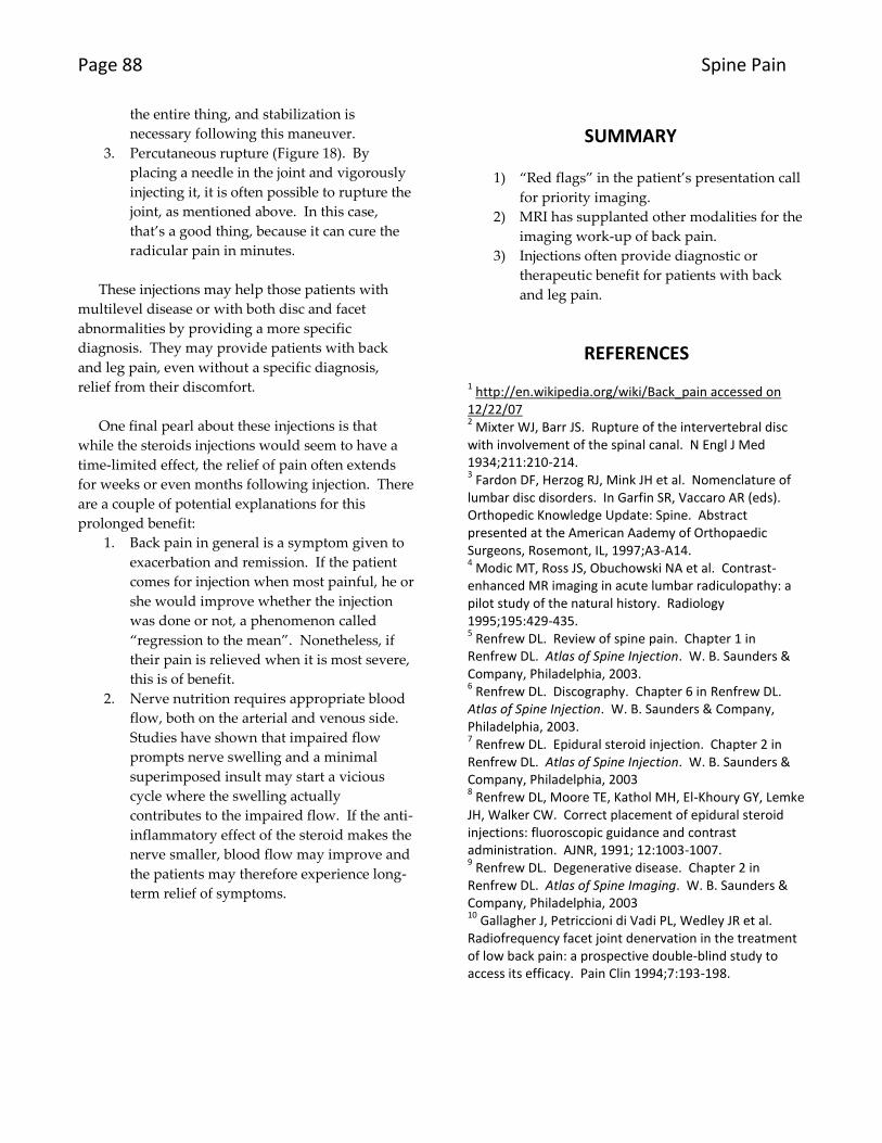
Figure 18. Facet injection performed for synovial cyst rupture. Fluoroscopically directed spot film obtained during the procedure shows the needle tip along the dorsal recess of the L4-L5 facet joint, with contrast material extending through the joint and into the synovial cyst (arrow).
Synovial cyst rupture can cure radiculopathy
As illustrated previously (Figure 6), synovial
cysts may arise from the facet joints and extend into
the spinal canal or neural foramen. When small,
these cysts are typically considered an
asymptomatic accompaniment of facet arthropathy.
However, when large, they may compress nerve
roots or spinal segmental nerves and thus cause
radicular pain. When this happens, the patient has
the same symptoms as if he or she had a disc
herniation. With this diagnosis, the three major
options are:
1. Treat with oral anti-inflammatory
medication. The cysts are said to eventually
go away, particularly if the joint
inflammation subsides, and this is one way
of approaching the problem, although for
patients in radicular pain the wait may be
agonizing.
2. Resect the joint and fuse the spine. This
seems a particularly aggressive approach,
but unlike disc herniations which are
amenable to microscopic discectomy, fixing
a bad facet joint generally entails removing
Page 88 Spine Pain
the entire thing, and stabilization is
necessary following this maneuver.
3. Percutaneous rupture (Figure 18). By
placing a needle in the joint and vigorously
injecting it, it is often possible to rupture the
joint, as mentioned above. In this case,
that’s a good thing, because it can cure the
radicular pain in minutes.
These injections may help those patients with
multilevel disease or with both disc and facet
abnormalities by providing a more specific
diagnosis. They may provide patients with back
and leg pain, even without a specific diagnosis,
relief from their discomfort.
One final pearl about these injections is that
while the steroids injections would seem to have a
time-limited effect, the relief of pain often extends
for weeks or even months following injection. There
are a couple of potential explanations for this
prolonged benefit:
1. Back pain in general is a symptom given to
exacerbation and remission. If the patient
comes for injection when most painful, he or
she would improve whether the injection
was done or not, a phenomenon called
“regression to the mean”. Nonetheless, if
their pain is relieved when it is most severe,
this is of benefit.
2. Nerve nutrition requires appropriate blood
flow, both on the arterial and venous side.
Studies have shown that impaired flow
prompts nerve swelling and a minimal
superimposed insult may start a vicious
cycle where the swelling actually
contributes to the impaired flow. If the anti-
inflammatory effect of the steroid makes the
nerve smaller, blood flow may improve and
the patients may therefore experience long-
term relief of symptoms.
SUMMARY
1) “Red flags” in the patient’s presentation call
for priority imaging.
2) MRI has supplanted other modalities for the
imaging work-up of back pain.
3) Injections often provide diagnostic or
therapeutic benefit for patients with back
and leg pain.
REFERENCES 1 http://en.wikipedia.org/wiki/Back_pain accessed on
12/22/07 2 Mixter WJ, Barr JS. Rupture of the intervertebral disc
with involvement of the spinal canal. N Engl J Med 1934;211:210-214. 3 Fardon DF, Herzog RJ, Mink JH et al. Nomenclature of
lumbar disc disorders. In Garfin SR, Vaccaro AR (eds). Orthopedic Knowledge Update: Spine. Abstract presented at the American Aademy of Orthopaedic Surgeons, Rosemont, IL, 1997;A3-A14. 4 Modic MT, Ross JS, Obuchowski NA et al. Contrast-
enhanced MR imaging in acute lumbar radiculopathy: a pilot study of the natural history. Radiology 1995;195:429-435. 5 Renfrew DL. Review of spine pain. Chapter 1 in
Renfrew DL. Atlas of Spine Injection. W. B. Saunders & Company, Philadelphia, 2003. 6 Renfrew DL. Discography. Chapter 6 in Renfrew DL.
Atlas of Spine Injection. W. B. Saunders & Company, Philadelphia, 2003. 7 Renfrew DL. Epidural steroid injection. Chapter 2 in
Renfrew DL. Atlas of Spine Injection. W. B. Saunders & Company, Philadelphia, 2003 8 Renfrew DL, Moore TE, Kathol MH, El-Khoury GY, Lemke
JH, Walker CW. Correct placement of epidural steroid injections: fluoroscopic guidance and contrast administration. AJNR, 1991; 12:1003-1007. 9 Renfrew DL. Degenerative disease. Chapter 2 in
Renfrew DL. Atlas of Spine Imaging. W. B. Saunders & Company, Philadelphia, 2003 10
Gallagher J, Petriccioni di Vadi PL, Wedley JR et al. Radiofrequency facet joint denervation in the treatment of low back pain: a prospective double-blind study to access its efficacy. Pain Clin 1994;7:193-198.