Embed Size (px)
Citation preview

Spine diseases and MRI / Radiology
Markus MaierOrthopädische Praxis
Starnberg am See
ESWT Summer meetingBergen, Norway, 24. – 26. August 2007

• Diagnostic standard• Helps for better understanding the clinical
sympotoms• How important is the image message?• Before imaging: Anamnesis, clinical
examination• Occasional LBP of mid40 male• Increasing LBP of a child• Everyone who indicates imaging should be
able to evaluate the images
Why need imaging?

• X-ray radiographs
– Vertebral statics– Disturbed growth
– Degenerative changes– Fractures– (Tumor)– (Inflammation)
Which imaging technique?

• X-ray radiographs/ vertebral statics– Not allways 2-plane imaging needed– In children no need for full spine
imaging– Hip (vara/valga – HD – Perthes etc.)– Form of the pelvis– Functional vs. structural scoliosis
Which imaging technique?

• X-ray radiographs/ disturbed growth– Scoliosis– Morbus Scheuermann– To controll progression: 1 plane
Which imaging technique?

• MRI– Nerval root compression– Spinal cord compression (stenosis)– Tumor (with x-ray and CT)– Infection (Leucocytes, CRP) – Combined with contrast application (MR-
myelographia)– Fracture (fresh – old)– Bony stress reactions
Which imaging technique?

• CT–Fracture
Which imaging technique?

• Ultrasound• Bone scintigraphia
–Occult lesions– Inflammation–Metastasis
Which imaging technique?

Degeneration - Chondrosis
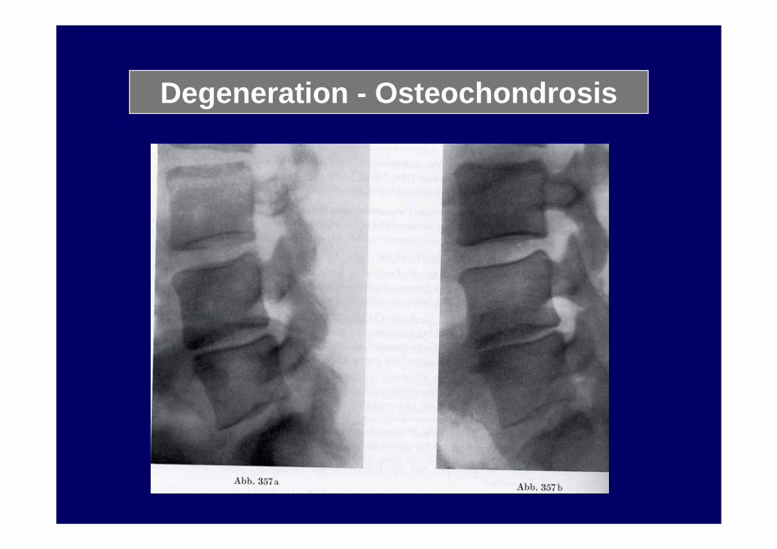
Degeneration - Osteochondrosis

Degeneration - Osteochondrosis

Degeneration - Osteochondrosis
Vacuumsign

Degeneration - Osteochondrosis
Vacuumsign

Degeneration

L5-Ileum nearthros

Degeneration

Degeneration – facett joint

Degeneration

Degeneration - Disc

Degeneration - Osteochondrosis

Degeneration - Disc

Degeneration - Disc

Degeneration - Disc

Degeneration - Disc

Degeneration – Black disc disease
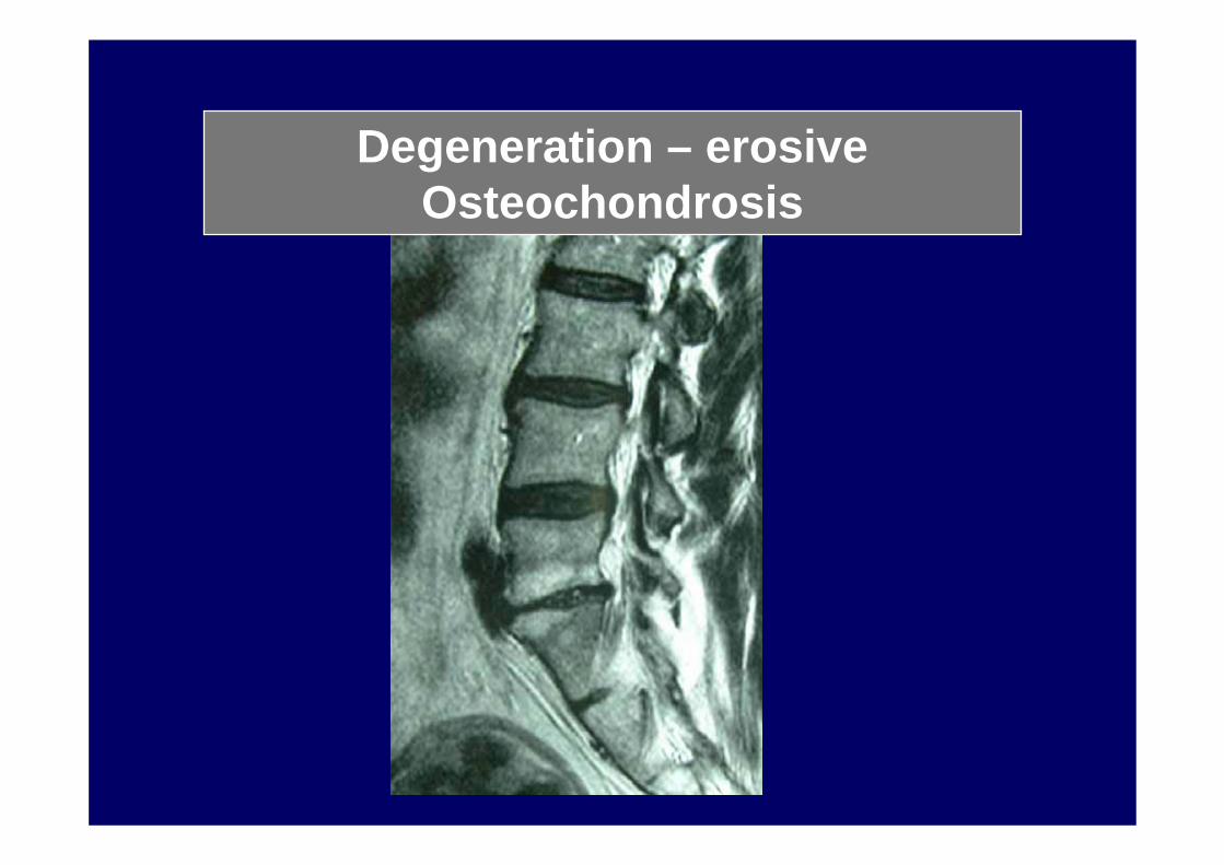
Degeneration – erosiveOsteochondrosis
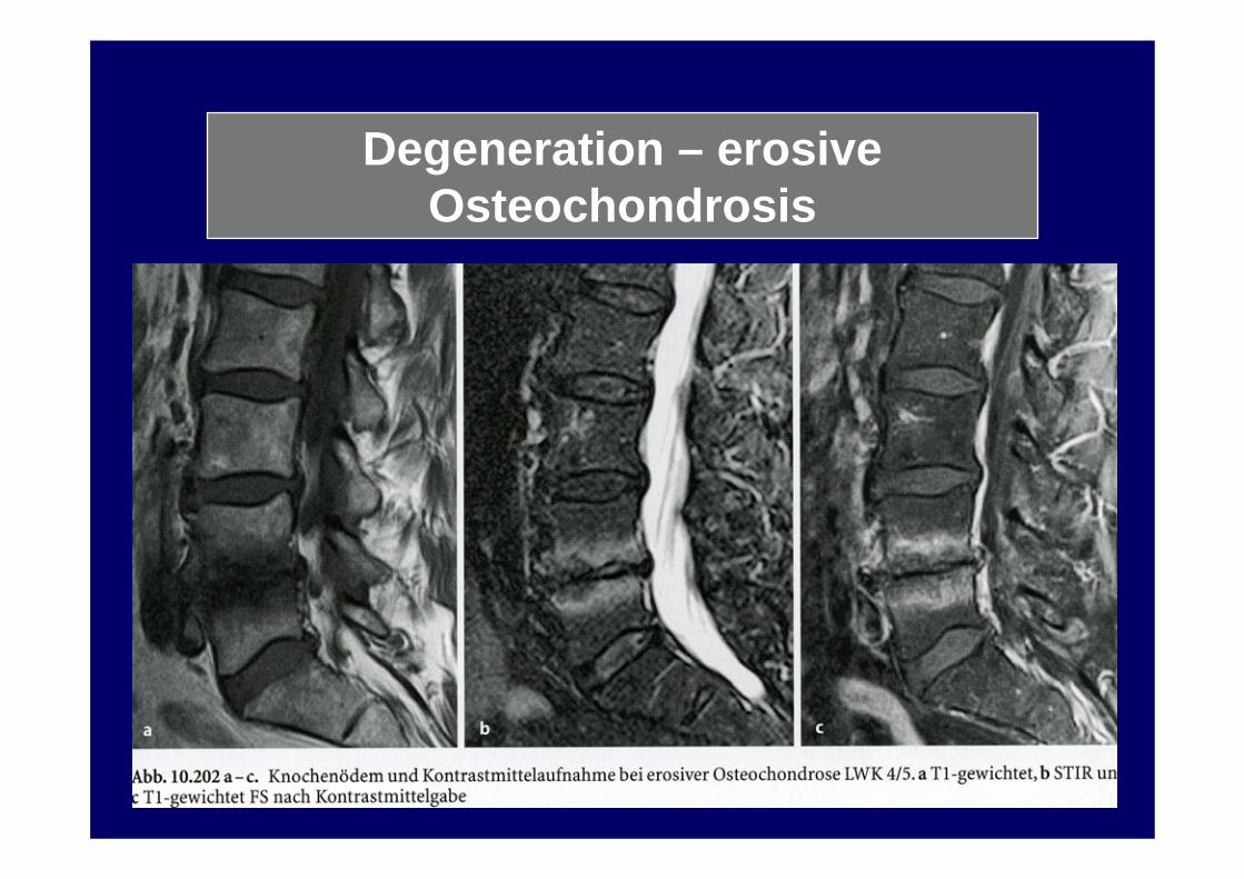
Degeneration – erosiveOsteochondrosis

? Degeneration/ Infection / Tumor ?

Degeneration – Stenosis

Degeneration – Black disc disease

Bastrup phaenomenon