Embed Size (px)
Citation preview
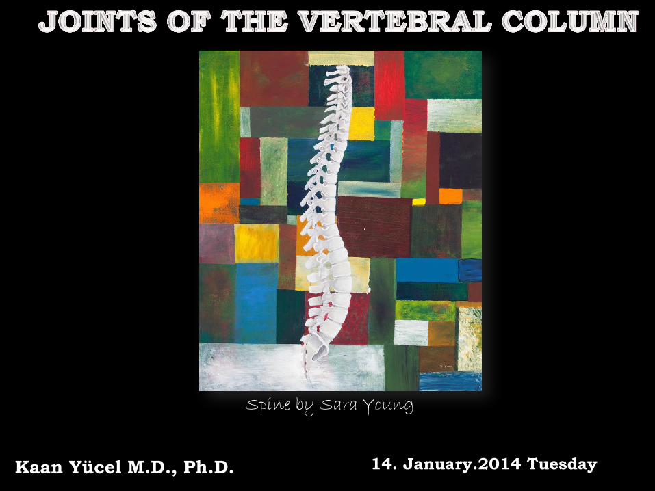
Kaan Yücel M.D., Ph.D. 14. January.2014 Tuesday
Spine by Sara Young

JOINTS OF THE VERTEBRAL COLUMN
1) Craniovertebral (atlanto-axial and atlanto-occipital) joints 2) Costovertebral joints 3) Sacroiliac joints
a total of 6 joints between two vertebrae
2
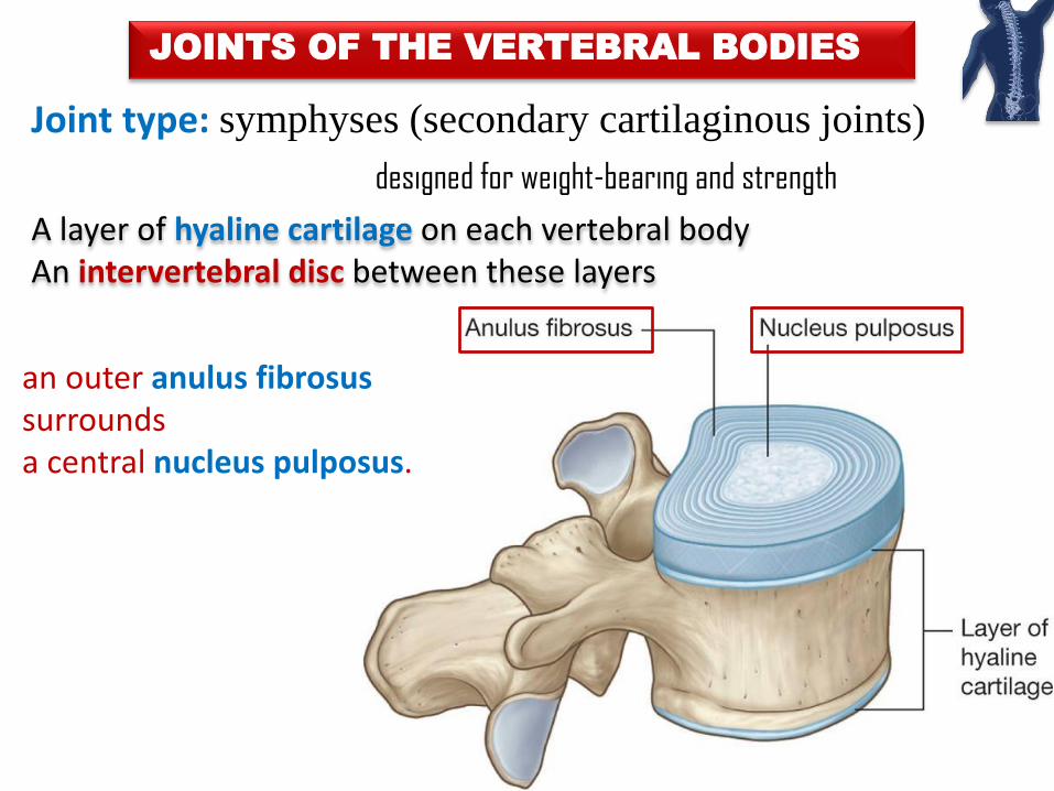
JOINTS OF THE VERTEBRAL BODIES
3
Joint type: symphyses (secondary cartilaginous joints)
designed for weight-bearing and strength
A layer of hyaline cartilage on each vertebral body An intervertebral disc between these layers
an outer anulus fibrosus surrounds a central nucleus pulposus.
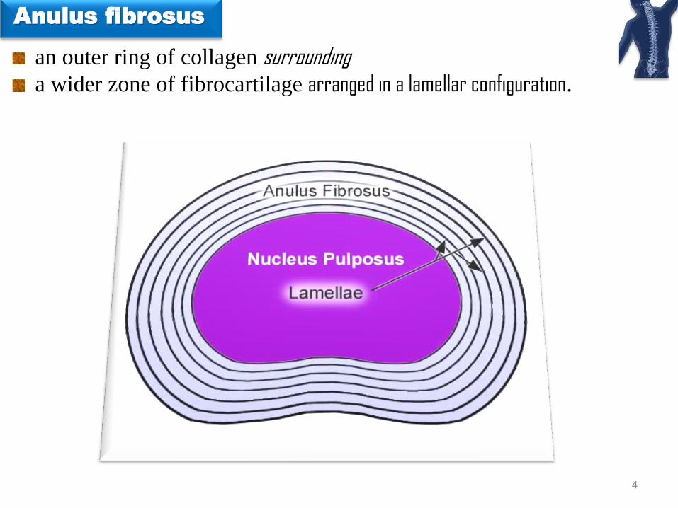
Anulus fibrosus
an outer ring of collagen surrounding a wider zone of fibrocartilage arranged in a lamellar configuration.
4

core of the intervertebral disc fills the center of the intervertebral disc gelatinous absorbs compression forces between vertebrae.
5
Nucleus pulposus L. pulpa, fleshy
semifluid nature responsible for much of the flexibility & resilience of the intervertebral disc and
of the vertebral column as a whole

INTERVERTEBRAL DISCS
Provide strong attachments between the vertebral bodies Unite vertebral bodies into a continuous semirigid column Form the inferior ½ of anterior border of the intervertebral foramen. 20-25% of the length (height) of the vertebral column.
6

INTERVERTEBRAL DISCS
7
Diagram of sagittal section of vertebral body and disc showing
relationship of endplate and longitudinal ligament to the disc and the
vertebrae.
1, vertebral body; 2, annulus fibrosus; 3, nucleus pulposus; 4,
endplate; 5, spinal nerve root

No intervertebral disc between C1 and C2 vertebrae Most inferior functional disc between L5 and S1 vertebrae
INTERVERTEBRAL DISCS
8
1. Anterior arch of the atlas (C1) 2. Dens (odontoid peg around which atlas
rotates) of axis (C2) 3. Posterior arch of the atlas (C1) 4. Soft palate (roof of the mouth) 5. Root of the tongue 6. Transverse process 7. Intervertebral disc 8. Inferior articular process 9. Superior articular process 10. Zygapophyseal (facet) joint 11. Spinous process of C7 2nd-7th: The bodies of 2nd to 7th cervical vertebrae

Thickness of the discs vertebral column descends
The range (amount) of movement relative thickness to body
greatest @ cervical & lumbar regions, movements of vertebral column greatest thickness most uniform in the thoracic region
INTERVERTEBRAL DISCS
9
L4
L5

FUNCTIONS OF INTERVERTEBRAL DISCS
Thanks to the semifluid nature One vertebra rock forward or backward on another during flexion & extension
10

INTERVERTEBRAL DISCS BY AGING
11
Collagen fibers of the anulus degenerate. Thin & less elastic discs
Nucleus & anulus not distinguishable
fibrocartilage.
Water content of the nucleus pulposus

JOINTS OF THE VERTEBRAL ARCHES
ZYGAPOPHYSIAL JOINTS, FACET JOINTS plane synovial joints
between superior & inferior articular processes of adjacent vertebrae. @ cervical region articular capsule especially thin
wide range of movement
12

JOINTS OF THE VERTEBRAL ARCHES
ZYGAPOPHYSIAL JOINTS, FACET JOINTS
permit gliding movements between articular processes
shape & disposition of the articular surfaces determine the types of movement
possible.
Accessory ligaments unite the laminae, transverse processes, and
spinous processes and help stabilize the joints.
13

UNCOVERTEBRAL (LUSCHKA’S) JOINTS
commonly between unci of the bodies of C3 or 4-C6 or 7 vertebrae @ the lateral & posterolateral margins of the intervertebral discs
synovial joints or degenerative spaces (clefts) in the discs occupied by extracellular
fluid
14
Uncinate process elevations@ lateral margins of the upper surface
Articulation with the body of the vertebra above

LIGAMENTS OF THE VERTEBRAL COLUMN
Joints between vertebrae reinforced & supported by numerous ligaments
pass between vertebral bodies
interconnect components of vertebral arches.
15

LIGAMENTS OF THE VERTEBRAL COLUMN
16
Anterior & posterior longitudinal ligaments Ligamenta flava Supraspinous ligament & ligamentum nuchae Interspinous ligaments
between two laminae

17
Anterior longitudinal ligament Posterior longitudinal ligament
From base of the skull to anterior surface of
sacrum
Along its length attached to vertebral bodies and intervertebral discs
Tectorial membrane
Posterior longitudinal ligament connecting
axis to base of the skull
18
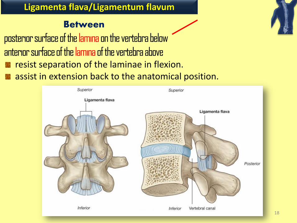
Ligamenta flava/Ligamentum flavum
Between
posterior surface of the lamina on the vertebra below
anterior surface of the lamina of the vertebra above resist separation of the laminae in flexion. assist in extension back to the anatomical position.

19
Supraspinous ligament

triangular, sheet-like structure in the median sagittal plane External occipital protuberance to magnum
tip of spinous process of C7 deep side attached to
posterior tubercle of vertebra C1 & spinous processes of other cervical vertebrae.
20
Ligamentum nuchae

supports the head. resists flexion . facilitates returning the head to the anatomical position. provide attachment for adjacent muscles broad lateral surfaces & posterior edge
21
Ligamentum nuchae

between adjacent vertebral spinous processes
22
Interspinous ligaments
from base to apex of each spinous process
blend with supraspinous ligament posteriorly
ligamenta flava anteriorly
on each side

CRANIOVERTEBRAL JOINTS
a wider range of movement than in the rest of the vertebral column.
23
atlanto-occipital joints between atlas (C1) & occipital (condyle) bone atlanto-axial joints between atlas (C1) & axis (C2)
Synovial joints with no intervertebral discs

ATLANTO-OCCIPITAL JOINTS
nodding of the head, “yes” movement also sideways tilting of the head. Main movement flexion, with a little lateral flexion and rotation.
24
Superior articular surfaces of lateral masses Occipital condyles

LIGAMENTS OF ATLANTO-OCCIPITAL JOINTS
Anterior atlanto-occipital membrane (continuation of anterior long.lig.)
connects anterior arch of the atlas to anterior margin of the foramen magnum
Posterior atlanto-occipital membrane (similar to the ligamentum flavum)
connects the posterior arch of the atlas to the posterior margin of the foramen magnum.
25

ATLANTO-AXIAL JOINTS
26
Right & left lateral atlantoaxial joints
between inf. facets of lateral masses of C1 & superior facets of C2 Median atlantoaxial joint
between dens of axis & anterior arch of atlas
p lane
IVOT

MOVEMENTS OF ATLANTO-AXIAL JOINTS
27
During rotation of the head
Dens/pivot held in a collar
anteriorly anterior arch of atlas
posteriorly transverse ligament of atlas
between tubercles on medial sides of lateral masses of atlas
Cranium & atlas rotate on axis as a unit.
Head turns from side to side, disapproval (“no” movement).

LIGAMENTS OF ATLANTO-AXIAL JOINTS
Superior and inferior longitudinal bands Apical ligament Alar ligaments Cruciate ligament of the atlas Tectorial membrane (Membrana tectoria)
.
28

COSTOVERTEBRAL JOINTS
essential for altering the volume of the thoracic cavity during breathing
29
A typical rib articulates with:
bodies of adjacent vertebrae joint with the head of the rib
transverse process of its related vertebra costotransverse joint
Necks rotate around their longitudinal axis mainly in upper ribs Ribs ascend descdend relative to the spine mainly in lower ribs

Joint with head of rib
divided into two synovial compartments by an intra-articular ligament
30
Head of the rib Two facets face of articulation
1- with superior facet of its own vertebra 2- with inferior facet of the vertebra above

Costotransverse joints
costotransverse ligament medial to the joint lateral costotransverse ligament lateral to the joint attaches the tip of the transverse process to nonarticular part of the tubercle of the rib. superior costotransverse ligament attaches the superior surface of the neck of the rib to the transverse process of the vertebra above.
31
Slight gliding movements
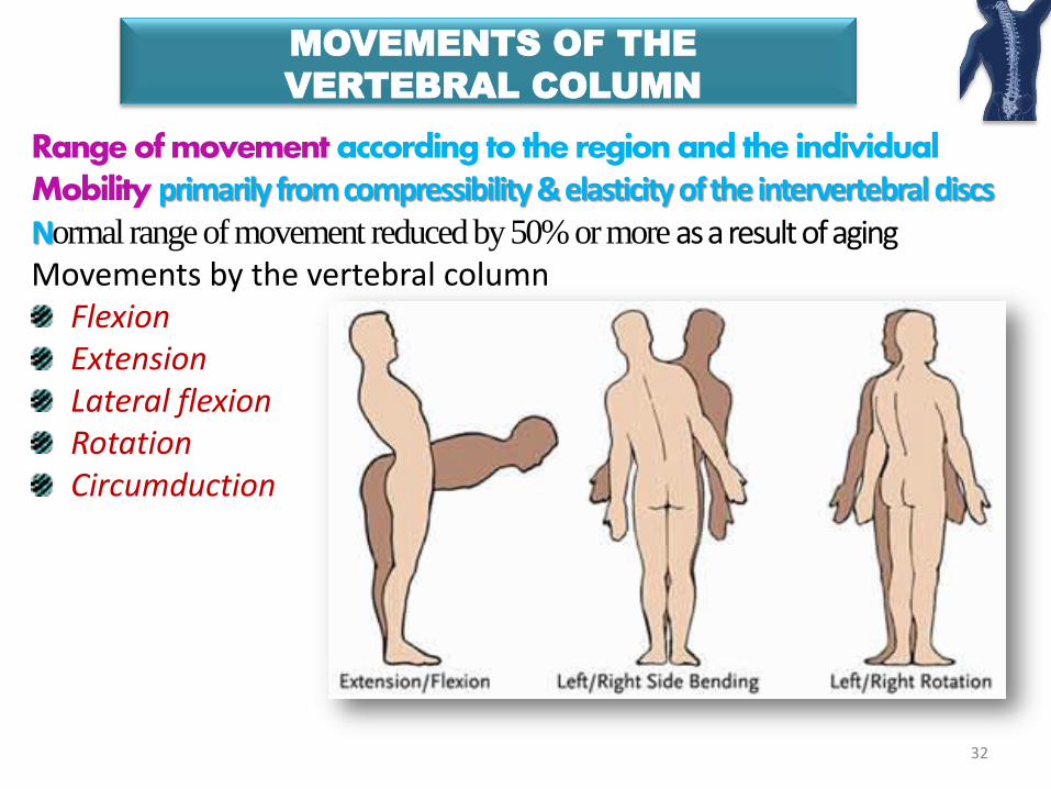
MOVEMENTS OF THE
VERTEBRAL COLUMN
Range of movement according to the region and the individual Mobility primarily from compressibility & elasticity of the intervertebral discs Normal range of movement reduced by 50% or more as a result of aging Movements by the vertebral column
Flexion Extension Lateral flexion Rotation Circumduction
32

MOVEMENTS OF THE
VERTEBRAL COLUMN
Movements in a specific region (cervical, thoracic, and lumbar) determined by
shape & orientation of joint surfaces
on the articular processes & on the vertebral bodies
33

MOVEMENTS OF THE
VERTEBRAL COLUMN
Range of movement limited by
1) Thickness, elasticity, and compressibility of the IV discs
2) Shape & orientation of the zygapophysial joints
3) Tension of the joint capsules of the zygapophysial joints
4) Resistance of the back muscles and ligaments
5) Attachment to the thoracic (rib) cage
6) Bulk of surrounding tissue 34

A tear within the anulus fibrosus
Material of the nucleus pulposus can track
This material tracks into the vertebral canal or into the intervertebral foramen
Pressure on neural structures
common cause of back pain

37
Anatomy and pathophysiology of intervertebral disc disease. Techniques in Regional Anesthesia
and Pain Management. Volume 13, 2009, Pages 67–75.
Quantitative MRI as a diagnostic tool of intervertebral disc matrix composition and integrity.
Eur Spine J. 2008;17 Suppl 4:432-440.
Degeneration and regeneration of the intervertebral disc: lessons from development. Dis Model
Mech. 2011;4:31-41.
Clinical Evaluation and Treatment Options for Herniated Lumbar Disc. Am Fam Physician.
1999;59:575-582.
The use of diagnostic imaging in sports medicine Med J Aust 2005; 183: 482-486.

38
.
A prolapsed intervertebral disc may impinge upon meningeal sac spinal cord most commonly the nerve root
producing symptoms attributable to that level.
PHYSIOTHERAPY OR INTERVENTION BY A NERUOSURGEON

39
Level of the disc protrusion identified before surgery.
MRI scanning and on-table fluoroscopy to prevent operating on the wrong level.
In some instances removal of the lamina will increase the potential space and may
relieve symptoms.