Embed Size (px)
Citation preview
RESEARCH ARTICLE
Spindle Assembly Checkpoint Acquisition atthe Mid-Blastula TransitionMaomao Zhang1,2, Priyanka Kothari1, Michael A. Lampson1,2*
1 Department of Biology, University of Pennsylvania, Philadelphia, Pennsylvania, United States of America,2 Cell and Molecular Biology Graduate Group, University of Pennsylvania, Philadelphia, Pennsylvania,United States of America
AbstractThe spindle assembly checkpoint (SAC) maintains the fidelity of chromosome segregation
during mitosis. Nonpathogenic cells lacking the SAC are typically only found in cleavage
stage metazoan embryos, which do not acquire functional checkpoints until the mid-blastula
transition (MBT). It is unclear how proper SAC function is acquired at the MBT, though sev-
eral models exist. First, SAC acquisition could rely on transcriptional activity, which in-
creases dramatically at the MBT. Embryogenesis prior to the MBT relies primarily on
maternally loaded transcripts, and if SAC signaling components are not maternally sup-
plied, the SAC would depend on zygotic transcription at the MBT. Second, checkpoint ac-
quisition could depend on the Chk1 kinase, which is activated at the MBT to elongate cell
cycles and is required for the SAC in somatic cells. Third, SAC function could depend on a
threshold nuclear to cytoplasmic (N:C) ratio, which increases during pre-MBT cleavage cy-
cles and dictates several MBT events like zygotic transcription and cell cycle remodeling. Fi-
nally, the SAC could by regulated by a timer mechanism that coincides with other MBT
events but is independent of them. Using zebrafish embryos we show that SAC acquisition
at the MBT is independent of zygotic transcription, indicating that the checkpoint program is
maternally supplied. Additionally, by precociously lengthening cleavage cycles with exoge-
nous Chk1 activity, we show that cell cycle lengthening and Chk1 activity are not sufficient
for SAC acquisition. Furthermore, we find that SAC acquisition can be uncoupled from the
N:C ratio. Together, our findings indicate that SAC acquisition is regulated by a maternally
programmed developmental timer.
IntroductionThe spindle assembly checkpoint (SAC) ensures that sister chromatids are correctly attachedto spindle microtubules before anaphase onset. In the presence of unattached kinetochores, theSAC is active and inhibits the anaphase promoting complex/cyclosome [1]. This checkpointmaintains genomic integrity by preventing chromosome segregation errors.
PLOSONE | DOI:10.1371/journal.pone.0119285 March 5, 2015 1 / 12
OPEN ACCESS
Citation: Zhang M, Kothari P, Lampson MA (2015)Spindle Assembly Checkpoint Acquisition at the Mid-Blastula Transition. PLoS ONE 10(3): e0119285.doi:10.1371/journal.pone.0119285
Academic Editor: Daniel Foltz, University of Virginia,UNITED STATES
Received: September 8, 2014
Accepted: January 23, 2015
Published: March 5, 2015
Copyright: © 2015 Zhang et al. This is an openaccess article distributed under the terms of theCreative Commons Attribution License, which permitsunrestricted use, distribution, and reproduction in anymedium, provided the original author and source arecredited.
Data Availability Statement: All relevant data arewithin the paper and its Supporting Information files.
Funding: This work was supported by the NationalInstitutes of Health (grants T32HD007516 to M. Z.and GM083988 to M. A. L.), a Searle Scholar awardto M. A. L., and the Penn Genome Frontiers Instituteand a grant with the Pennsylvania Department ofHealth. The funders had no role in study design, datacollection and analysis, decision to publish, orpreparation of the manuscript.
Competing Interests: The authors have declaredthat no competing interests exist.
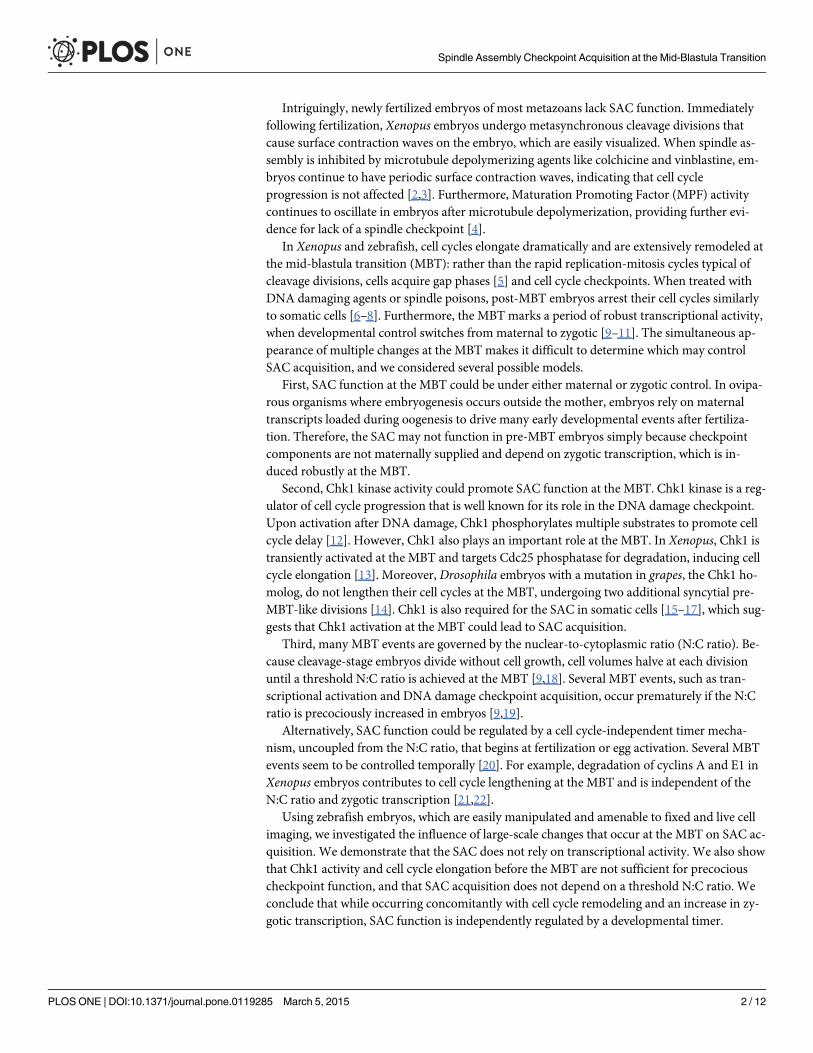
Intriguingly, newly fertilized embryos of most metazoans lack SAC function. Immediatelyfollowing fertilization, Xenopus embryos undergo metasynchronous cleavage divisions thatcause surface contraction waves on the embryo, which are easily visualized. When spindle as-sembly is inhibited by microtubule depolymerizing agents like colchicine and vinblastine, em-bryos continue to have periodic surface contraction waves, indicating that cell cycleprogression is not affected [2,3]. Furthermore, Maturation Promoting Factor (MPF) activitycontinues to oscillate in embryos after microtubule depolymerization, providing further evi-dence for lack of a spindle checkpoint [4].
In Xenopus and zebrafish, cell cycles elongate dramatically and are extensively remodeled atthe mid-blastula transition (MBT): rather than the rapid replication-mitosis cycles typical ofcleavage divisions, cells acquire gap phases [5] and cell cycle checkpoints. When treated withDNA damaging agents or spindle poisons, post-MBT embryos arrest their cell cycles similarlyto somatic cells [6–8]. Furthermore, the MBT marks a period of robust transcriptional activity,when developmental control switches from maternal to zygotic [9–11]. The simultaneous ap-pearance of multiple changes at the MBT makes it difficult to determine which may controlSAC acquisition, and we considered several possible models.
First, SAC function at the MBT could be under either maternal or zygotic control. In ovipa-rous organisms where embryogenesis occurs outside the mother, embryos rely on maternaltranscripts loaded during oogenesis to drive many early developmental events after fertiliza-tion. Therefore, the SAC may not function in pre-MBT embryos simply because checkpointcomponents are not maternally supplied and depend on zygotic transcription, which is in-duced robustly at the MBT.
Second, Chk1 kinase activity could promote SAC function at the MBT. Chk1 kinase is a reg-ulator of cell cycle progression that is well known for its role in the DNA damage checkpoint.Upon activation after DNA damage, Chk1 phosphorylates multiple substrates to promote cellcycle delay [12]. However, Chk1 also plays an important role at the MBT. In Xenopus, Chk1 istransiently activated at the MBT and targets Cdc25 phosphatase for degradation, inducing cellcycle elongation [13]. Moreover, Drosophila embryos with a mutation in grapes, the Chk1 ho-molog, do not lengthen their cell cycles at the MBT, undergoing two additional syncytial pre-MBT-like divisions [14]. Chk1 is also required for the SAC in somatic cells [15–17], which sug-gests that Chk1 activation at the MBT could lead to SAC acquisition.
Third, many MBT events are governed by the nuclear-to-cytoplasmic ratio (N:C ratio). Be-cause cleavage-stage embryos divide without cell growth, cell volumes halve at each divisionuntil a threshold N:C ratio is achieved at the MBT [9,18]. Several MBT events, such as tran-scriptional activation and DNA damage checkpoint acquisition, occur prematurely if the N:Cratio is precociously increased in embryos [9,19].
Alternatively, SAC function could be regulated by a cell cycle-independent timer mecha-nism, uncoupled from the N:C ratio, that begins at fertilization or egg activation. Several MBTevents seem to be controlled temporally [20]. For example, degradation of cyclins A and E1 inXenopus embryos contributes to cell cycle lengthening at the MBT and is independent of theN:C ratio and zygotic transcription [21,22].
Using zebrafish embryos, which are easily manipulated and amenable to fixed and live cellimaging, we investigated the influence of large-scale changes that occur at the MBT on SAC ac-quisition. We demonstrate that the SAC does not rely on transcriptional activity. We also showthat Chk1 activity and cell cycle elongation before the MBT are not sufficient for precociouscheckpoint function, and that SAC acquisition does not depend on a threshold N:C ratio. Weconclude that while occurring concomitantly with cell cycle remodeling and an increase in zy-gotic transcription, SAC function is independently regulated by a developmental timer.
Spindle Assembly Checkpoint Acquisition at the Mid-Blastula Transition
PLOS ONE | DOI:10.1371/journal.pone.0119285 March 5, 2015 2 / 12
Results
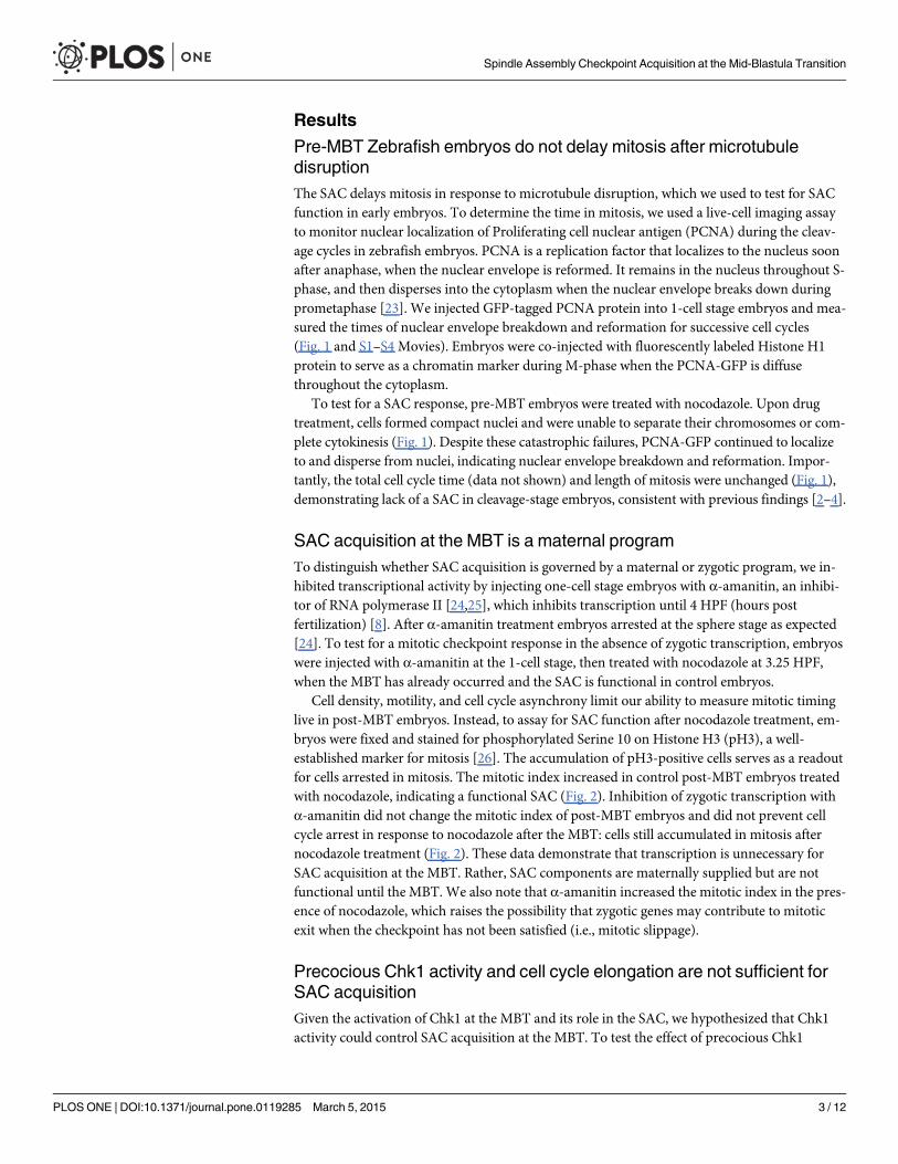
Pre-MBT Zebrafish embryos do not delay mitosis after microtubuledisruptionThe SAC delays mitosis in response to microtubule disruption, which we used to test for SACfunction in early embryos. To determine the time in mitosis, we used a live-cell imaging assayto monitor nuclear localization of Proliferating cell nuclear antigen (PCNA) during the cleav-age cycles in zebrafish embryos. PCNA is a replication factor that localizes to the nucleus soonafter anaphase, when the nuclear envelope is reformed. It remains in the nucleus throughout S-phase, and then disperses into the cytoplasm when the nuclear envelope breaks down duringprometaphase [23]. We injected GFP-tagged PCNA protein into 1-cell stage embryos and mea-sured the times of nuclear envelope breakdown and reformation for successive cell cycles(Fig. 1 and S1–S4 Movies). Embryos were co-injected with fluorescently labeled Histone H1protein to serve as a chromatin marker during M-phase when the PCNA-GFP is diffusethroughout the cytoplasm.
To test for a SAC response, pre-MBT embryos were treated with nocodazole. Upon drugtreatment, cells formed compact nuclei and were unable to separate their chromosomes or com-plete cytokinesis (Fig. 1). Despite these catastrophic failures, PCNA-GFP continued to localizeto and disperse from nuclei, indicating nuclear envelope breakdown and reformation. Impor-tantly, the total cell cycle time (data not shown) and length of mitosis were unchanged (Fig. 1),demonstrating lack of a SAC in cleavage-stage embryos, consistent with previous findings [2–4].
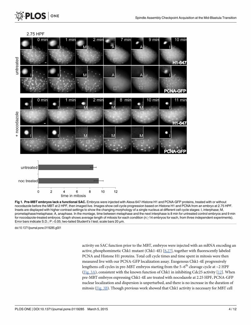
SAC acquisition at the MBT is a maternal programTo distinguish whether SAC acquisition is governed by a maternal or zygotic program, we in-hibited transcriptional activity by injecting one-cell stage embryos with α-amanitin, an inhibi-tor of RNA polymerase II [24,25], which inhibits transcription until 4 HPF (hours postfertilization) [8]. After α-amanitin treatment embryos arrested at the sphere stage as expected[24]. To test for a mitotic checkpoint response in the absence of zygotic transcription, embryoswere injected with α-amanitin at the 1-cell stage, then treated with nocodazole at 3.25 HPF,when the MBT has already occurred and the SAC is functional in control embryos.
Cell density, motility, and cell cycle asynchrony limit our ability to measure mitotic timinglive in post-MBT embryos. Instead, to assay for SAC function after nocodazole treatment, em-bryos were fixed and stained for phosphorylated Serine 10 on Histone H3 (pH3), a well-established marker for mitosis [26]. The accumulation of pH3-positive cells serves as a readoutfor cells arrested in mitosis. The mitotic index increased in control post-MBT embryos treatedwith nocodazole, indicating a functional SAC (Fig. 2). Inhibition of zygotic transcription withα-amanitin did not change the mitotic index of post-MBT embryos and did not prevent cellcycle arrest in response to nocodazole after the MBT: cells still accumulated in mitosis afternocodazole treatment (Fig. 2). These data demonstrate that transcription is unnecessary forSAC acquisition at the MBT. Rather, SAC components are maternally supplied but are notfunctional until the MBT. We also note that α-amanitin increased the mitotic index in the pres-ence of nocodazole, which raises the possibility that zygotic genes may contribute to mitoticexit when the checkpoint has not been satisfied (i.e., mitotic slippage).
Precocious Chk1 activity and cell cycle elongation are not sufficient forSAC acquisitionGiven the activation of Chk1 at the MBT and its role in the SAC, we hypothesized that Chk1activity could control SAC acquisition at the MBT. To test the effect of precocious Chk1
Spindle Assembly Checkpoint Acquisition at the Mid-Blastula Transition
PLOS ONE | DOI:10.1371/journal.pone.0119285 March 5, 2015 3 / 12
activity on SAC function prior to the MBT, embryos were injected with an mRNA encoding anactive, phosphomimetic Chk1 mutant (Chk1-4E) [8,27], together with fluorescently labeledPCNA and Histone H1 proteins. Total cell cycle times and time spent in mitosis were thenmeasured live with our PCNA-GFP localization assay. Exogenous Chk1-4E progressivelylengthens cell cycles in pre-MBT embryos starting from the 5–6th cleavage cycle at ~2 HPF(Fig. 3A), consistent with the known function of Chk1 in inhibiting Cdc25 activity [12]. Whenpre-MBT embryos expressing Chk1-4E are treated with nocodazole at 2.25 HPF, PCNA-GFPnuclear localization and dispersion is unperturbed, and there is no increase in the duration ofmitosis (Fig. 3B). Though previous work showed that Chk1 activity is necessary for MBT cell
Fig 1. Pre-MBT embryos lack a functional SAC. Embryos were injected with Alexa 647-Histone H1 and PCNA-GFP proteins, treated with or withoutnocodazole before the MBT at 2 HPF, then imaged live. Images show cell cycle progression based on Histone H1 and PCNA from an embryo at 2.75 HPF.Insets are displayed with higher contrast settings to show the changing morphology of a single nucleus at different cell cycle stages: I, interphase; M,prometaphase/metaphase; A, anaphase. In the montage, time between metaphase and the next interphase is 8 min for untreated control embryos and 9 minfor nocodazole-treated embryos. Graph shows average length of mitosis for each condition (n�14 embryos for each, from three independent experiments).Error bars indicate S.D.; P>0.05; two-tailed Student’s t test; scale bars 20 μm.
doi:10.1371/journal.pone.0119285.g001
Spindle Assembly Checkpoint Acquisition at the Mid-Blastula Transition
PLOS ONE | DOI:10.1371/journal.pone.0119285 March 5, 2015 4 / 12
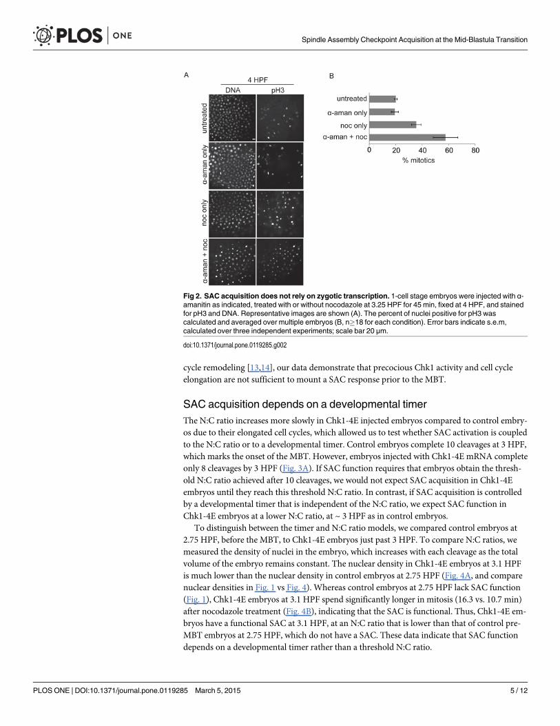
cycle remodeling [13,14], our data demonstrate that precocious Chk1 activity and cell cycleelongation are not sufficient to mount a SAC response prior to the MBT.
SAC acquisition depends on a developmental timerThe N:C ratio increases more slowly in Chk1-4E injected embryos compared to control embry-os due to their elongated cell cycles, which allowed us to test whether SAC activation is coupledto the N:C ratio or to a developmental timer. Control embryos complete 10 cleavages at 3 HPF,which marks the onset of the MBT. However, embryos injected with Chk1-4E mRNA completeonly 8 cleavages by 3 HPF (Fig. 3A). If SAC function requires that embryos obtain the thresh-old N:C ratio achieved after 10 cleavages, we would not expect SAC acquisition in Chk1-4Eembryos until they reach this threshold N:C ratio. In contrast, if SAC acquisition is controlledby a developmental timer that is independent of the N:C ratio, we expect SAC function inChk1-4E embryos at a lower N:C ratio, at ~ 3 HPF as in control embryos.
To distinguish between the timer and N:C ratio models, we compared control embryos at2.75 HPF, before the MBT, to Chk1-4E embryos just past 3 HPF. To compare N:C ratios, wemeasured the density of nuclei in the embryo, which increases with each cleavage as the totalvolume of the embryo remains constant. The nuclear density in Chk1-4E embryos at 3.1 HPFis much lower than the nuclear density in control embryos at 2.75 HPF (Fig. 4A, and comparenuclear densities in Fig. 1 vs Fig. 4). Whereas control embryos at 2.75 HPF lack SAC function(Fig. 1), Chk1-4E embryos at 3.1 HPF spend significantly longer in mitosis (16.3 vs. 10.7 min)after nocodazole treatment (Fig. 4B), indicating that the SAC is functional. Thus, Chk1-4E em-bryos have a functional SAC at 3.1 HPF, at an N:C ratio that is lower than that of control pre-MBT embryos at 2.75 HPF, which do not have a SAC. These data indicate that SAC functiondepends on a developmental timer rather than a threshold N:C ratio.
Fig 2. SAC acquisition does not rely on zygotic transcription. 1-cell stage embryos were injected with α-amanitin as indicated, treated with or without nocodazole at 3.25 HPF for 45 min, fixed at 4 HPF, and stainedfor pH3 and DNA. Representative images are shown (A). The percent of nuclei positive for pH3 wascalculated and averaged over multiple embryos (B, n�18 for each condition). Error bars indicate s.e.m,calculated over three independent experiments; scale bar 20 μm.
doi:10.1371/journal.pone.0119285.g002
Spindle Assembly Checkpoint Acquisition at the Mid-Blastula Transition
PLOS ONE | DOI:10.1371/journal.pone.0119285 March 5, 2015 5 / 12
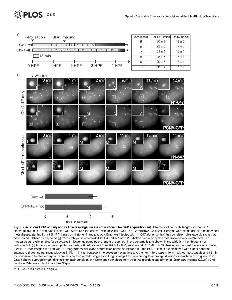
Fig 3. Precocious Chk1 activity and cell cycle elongation are not sufficient for SAC acquisition. (A) Schematic of cell cycle lengths for the first 10cleavage divisions of embryos injected with Alexa 647-Histone H1, with or without Chk1-4E-GFPmRNA. Cell cycles lengths were measured as time betweenmetaphases, starting from 1.5 HPF, based on Histone H1 morphology. Embryos injected with H1-647 alone (control) had consistent cleavage divisions thateach lasted ~16 min as expected [5] while embryos injected with Chk1-4E mRNA and H1-647 had cleavage cycles that progressively lengthened. Themeasured cell cycle lengths for cleavages 5–10 are indicated by the length of each bar in the schematic and shown in the table (n>4 embryos; errorindicates S.D.) (B) Embryos were injected with Alexa 647-Histone H1 and PCNA-GFP proteins and Chk1-4E mRNA, treated with our without nocodazole at2.25 HPF, then imaged live until 3 HPF. Images show cell cycle progression based on Histone H1 and PCNA. Insets are displayed with higher contrastsettings to show nuclear morphology as in Fig. 1. In the montage, time between metaphase and the next interphase is 10 min without nocodazole and 12 minfor nocodazole-treated embryos. There was no measurable progressive lengthening of mitoses during the cleavage divisions, regardless of drug treatment.Graph shows average length of mitosis for each condition (n�12 for each condition, from three independent experiments). Error bars indicate S.D.; P>0.05;two-tailed Student’s t test; scale bars 20 μm.
doi:10.1371/journal.pone.0119285.g003
Spindle Assembly Checkpoint Acquisition at the Mid-Blastula Transition
PLOS ONE | DOI:10.1371/journal.pone.0119285 March 5, 2015 6 / 12
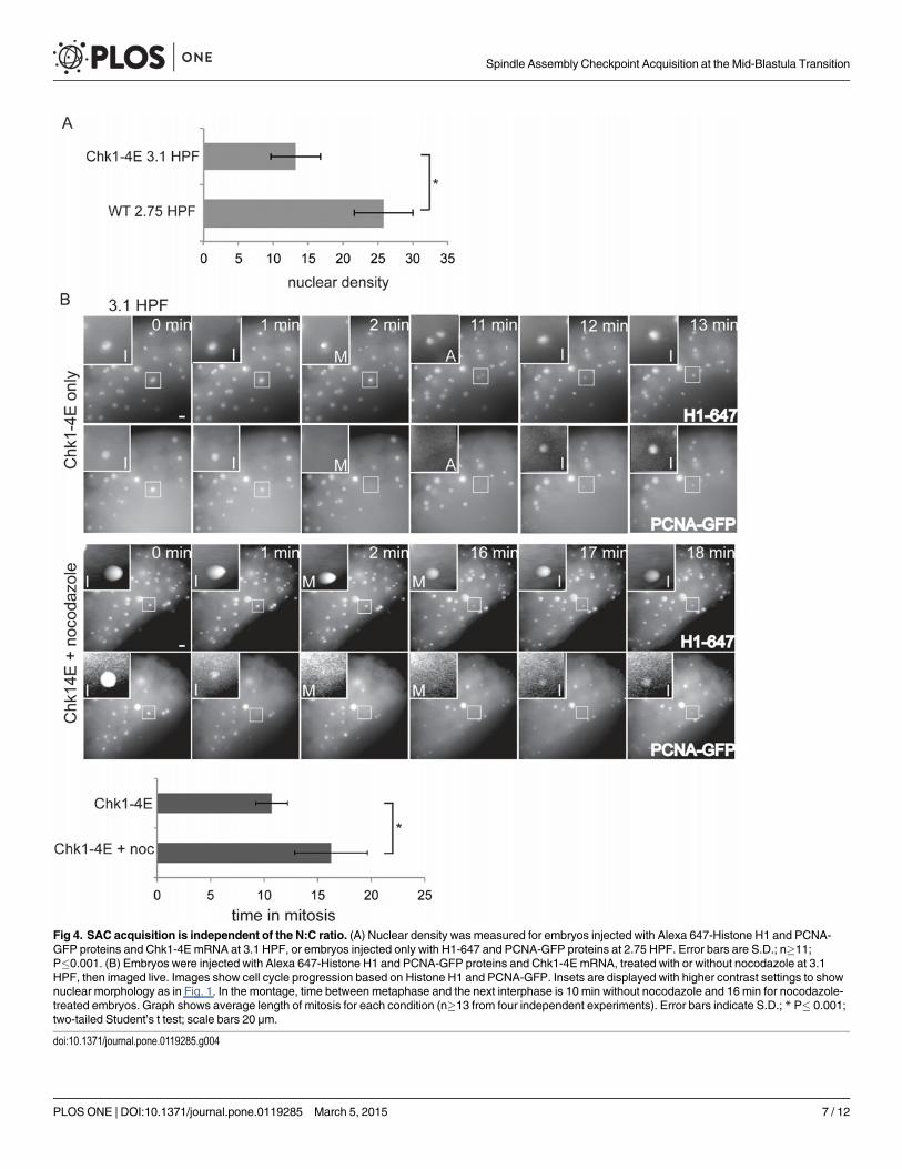
Fig 4. SAC acquisition is independent of the N:C ratio. (A) Nuclear density was measured for embryos injected with Alexa 647-Histone H1 and PCNA-GFP proteins and Chk1-4E mRNA at 3.1 HPF, or embryos injected only with H1-647 and PCNA-GFP proteins at 2.75 HPF. Error bars are S.D.; n�11;P�0.001. (B) Embryos were injected with Alexa 647-Histone H1 and PCNA-GFP proteins and Chk1-4E mRNA, treated with or without nocodazole at 3.1HPF, then imaged live. Images show cell cycle progression based on Histone H1 and PCNA-GFP. Insets are displayed with higher contrast settings to shownuclear morphology as in Fig. 1. In the montage, time between metaphase and the next interphase is 10 min without nocodazole and 16 min for nocodazole-treated embryos. Graph shows average length of mitosis for each condition (n�13 from four independent experiments). Error bars indicate S.D.; * P� 0.001;two-tailed Student’s t test; scale bars 20 μm.
doi:10.1371/journal.pone.0119285.g004
Spindle Assembly Checkpoint Acquisition at the Mid-Blastula Transition
PLOS ONE | DOI:10.1371/journal.pone.0119285 March 5, 2015 7 / 12
DiscussionOur findings provide insight into how the early embryo acquires SAC function. Specifically, weinvestigated whether SAC acquisition is coordinated with other major developmental changesduring the MBT, including cell cycle elongation and Chk1 activity, a threshold N:C ratio, andzygotic transcription. Despite the coincident appearance of these events in the embryo, weshow that SAC function can be uncoupled from other MBT events.
We first investigated whether SAC acquisition is coupled with transcriptional activity, aspast studies have suggested that zygotic transcription and certain aspects of cell cycle remodel-ing are coordinated. For example, addition of the G2 phase of the cell cycle during MBT cellcycle remodeling in zebrafish relies on zygotic transcription [24]. Conversely, cell cycle elonga-tion may be required for zygotic transcription, as transcripts are often aborted in rapid cell cy-cles in Drosophila due to time constraints [28]. Despite the clear co-regulation of cell cycleremodeling and transcriptional activity at the MBT, we show that zygotic transcription is notrequired for SAC acquisition, implying that SAC components are maternally loaded. Similarly,our previous work shows that DNA damage checkpoint acquisition occurs independent oftranscriptional activity[8].
Our data are consistent with similar findings in Xenopus, which showed that blocking tran-scription after the 8-cell stage in dissociated blastomeres does not prevent SAC acquisition[29]. However, gene expression profiling has revealed that many zygotic genes are expressedduring the cleavage stages, some as early as the 4-cell stage [30]. Thus, the previous experi-ments did not fully account for possible early zygotic transcription of SAC components, whichmay provide a sufficient pool of mRNA for SAC protein synthesis and accumulation. In con-trast, we inhibited transcription immediately after fertilization, at the 1-cell stage, ruling outthe possibility of a zygotic contribution of SAC components.
We also investigated the role of the N:C ratio in SAC acquisition. The coordination of manyMBT events seems to stem from the N:C ratio, which increases with every cleavage cycle. Forexample, the replication factors which account for the fast S-phase in pre-MBT embryos are ti-trated as the N:C ratio increases, leading to slowed replication at the MBT and increased inter-phase duration [31]. Additionally, the N:C ratio can affect DNA damage checkpointacquisition: addition of exogenous DNA to pre-MBT Xenopus embryos, to mimic the N:Cratio typical of the MBT, can lead to precocious checkpoint function after DNA damage[32,33].
By precociously increasing cell cycle lengths in pre-MBT embryos with Chk1, we show thatcell cycle lengthening and Chk1 activity are not sufficient for SAC acquisition. Furthermore,lengthening pre-MBT cell cycles slows the increase in N:C ratio, and we find that SAC acquisi-tion does not depend on a threshold N:C ratio. This result is consistent with previous experi-ments which showed that individual blastomeres isolated from dissociated Xenopus embryosacquire a functional SAC with varying N:C ratios [29,34]. Our results indicate that SAC acqui-sition is regulated by a timer mechanism that does not rely on Chk1 activity orzygotic transcription.
Although we find that a threshold N:C ratio is not necessary for SAC acquisition, the SACcan become functional prematurely in Xenopus egg extracts if sperm nuclei are added to in-crease the N:C ratio to a threshold level [35]. Together these findings suggest that both a devel-opmental timer and increases in the N:C ratio can contribute to SAC regulation. Increasing N:C ratio may artificially titrate as-yet unidentified cytosolic SAC inhibitors that are present dur-ing the cleavage stages, or increased numbers of kinetochores may enhance production of aSAC signal. During normal development, when the number of kinetochores is fixed, a set timemay be required for the synthesis and accumulation of SAC proteins that amplify signaling
Spindle Assembly Checkpoint Acquisition at the Mid-Blastula Transition
PLOS ONE | DOI:10.1371/journal.pone.0119285 March 5, 2015 8 / 12
downstream from initial SAC activation at kinetochores. However, the need for accumulationof these proteins could be bypassed if large numbers of kinetochores amplify SAC signaling.
Our findings raise the question of how a maternally-controlled developmental timer regu-lates SAC acquisition at the MBT. Time could be required for either accumulation of SAC pro-teins from maternally supplied transcripts or degradation of a SAC inhibitor. For example,SAC function depends on multiple checkpoint proteins that are recruited to unattached kineto-chores to generate the inhibitory signal that prevents anaphase onset [1]. Though our workshows that mRNAs for these components are provided maternally, the kinetics of checkpointprotein accumulation should be further investigated. Additionally, future work is required todetermine the molecular differences between checkpoint signaling before and after the MBT toelucidate the molecular basis for the developmental timer.
Materials and Methods
Fish husbandryEmbryos were collected from natural mating and incubated in E3 buffer (5 mM NaCl, 0.17mM KCl, 0.33 mM CaCl2, 0.33 mMMgSO4) at 28°C. All experiments were carried out in theTuebingen long fin strain. The animal work performed in this study was approved by the Uni-versity of Pennsylvania Institutional Animal Care and Use Committee. Adult animals wereused only for mating and were not sacrificed. The NIH Policy on Humane Care and Use ofLaboratory Animals applies to zebrafish embryos only after hatching, and all of our experi-ments were performed well before this stage.
Cell cycle length measurementsFor experiments using PCNA-GFP, recombinant protein was purified as described [23] from aGFP-fused human PCNA gene in pENeGFP-PCNA2 provided by Dr Michael Whitaker (Insti-tute of Cell and Molecular Biosciences, University of Newcastle upon Tyne). Histone H1 fromcalf thymus (Sigma) was conjugated to Alexa-Fluor 647 (Invitrogen) following the manufactur-er’s instructions. 1-cell stage embryos were injected with GFP-PCNA and AlexaFluor-taggedhistone H1 and incubated in E3 buffer at 28°C until live imaging. For live imaging, embryoswere dechorionated with 1 mg/mL pronase in E3 buffer for 10 min, washed 2x in E3 buffer,then mounted on a 4-well fluorodish (Grenier Bio-One) in 0.4% agarose dissolved in E3 buffer.For nocodazole-treated embryos, nocodazole was included in the E3 buffer to maintain theworking concentration in the agarose. Images were acquired using a 20x 0.7 NA objective onan inverted fluorescence microscope (DM6000, Leica Microsystems) equipped with an auto-mated XYZ stage and an electron multiplier charge-coupled device camera (QuantEM, 512 SC;Photometrics), controlled by Metamorph Software (MDS Analytical Technologies). Embryoswere imaged every minute by fluorescence. At each time point a z-series of 10 images was col-lected at 10 μm intervals. Cell cycle lengths were measured by manually tracking shuttling ofGFP-PCNA into and out of nuclei defined by AlexaFluor-tagged histone H1, using Metamorphand ImageJ software. Images are shown as maximal intensity projections of the confocal z-series.
For Fig. 1, embryos were imaged from ~2.5–3 HPF to measure the mitotic timing of thecleavage cycles. For Fig. 3A, embryos were imaged live starting from 1.5 HPF, at approximatelythe 4th-5th cleavage cycle. We measured cleavage cycles 5–10 and averaged multiple embryosto estimate the length of each cleavage cycle. For Fig. 3B, embryos were imaged from 2.25 to ~3HPF (the usual time of the MBT), and cell cycle times were averaged over 2–3 cell cycles. ForFig. 4, embryos were imaged starting from 3.1 HPF for one cell cycle until ~3.5 HPF.
Spindle Assembly Checkpoint Acquisition at the Mid-Blastula Transition
PLOS ONE | DOI:10.1371/journal.pone.0119285 March 5, 2015 9 / 12
Embryo drug treatmentsNocodazole was dissolved in DMSO for a stock solution and diluted in E3 buffer for a finalworking concentration of 0.125 ug/mL. For pH3 staining, 3.25 HPF post-MBT embryos weretreated with nocodazole and incubated at 28°C for 45 minutes, then fixed with 4% formalde-hyde in PBS. Control embryos were maintained in E3 buffer containing 0.025% DMSO.
To inhibit transcription, 2 nL of 1 mg/mL α-amanitin dissolved in ddH2O was injected intothe cell of 1-cell stage embryos. Embryos were incubated in E3 buffer at 28°C until drug treat-ment with nocodazole.
ImmunofluorescenceEmbryos were fixed with 4% paraformaldehyde in PBST (PBS with 0.1% Tween-20) overnightat 4°C, then manually dechorionated and dehydrated in 100% methanol overnight at -20°C.Embryos were rehydrated the next day sequentially with 75%, 50% and then 25% methanol inPBST (5 min each), then permeabilized with 100% acetone at -20°C for 7 min, then blockedwith buffer containing 20% Heat-inactivated FBS, 20% Blocking reagent (Roche) and 1%DMSO in PBST for 1 hr at room temperature. Antibodies were diluted in blocking buffer, ap-plied to embryos and incubated overnight at 4°C. Embryos were then washed four times(30 min each) with PBST, incubated with secondary antibody in blocking buffer, washed an-other three times, stained for 5 min with SYTOX Green (Invitrogen), then washed once withPBST. Embryos were mounted on fluorodishes (World Precision) in 4% methylcellulose dis-solved in E3 buffer. Primary antibody was a mouse monoclonal against phospho-Ser10 HistoneH3 (1:1000, Millipore cat #05–806). Secondary antibodies were Alexa-Fluor 647-conjugatedanti-mouse or anti-rabbit (1:200, Invitrogen).
All fixed embryos were imaged with a spinning disk confocal: a microscope (DM4000, Leica)with a 20x 0.7 NA objective, a XY piezo-z stage (Applied Scientific Instrumentation), a spinningdisk (Yokogawa), an electron multiplier charge-coupled device camera (ImageEM, HamamatsuPhotonics), and an LMM5 laser merge module equipped with 488 and 593 nm lasers (SpectralApplied Research) controlled by Metamorph software. Percentages of mitotic cells were quanti-fied by counting pH3 positive nuclei as a fraction of total nuclei (based on DNA staining).
Nuclear density measurementsControl and Chk1-4E embryos were imaged live as described above, and a z-projection of 10images with 10 μm distance between slices was made for each embryo. We manually countedthe number of nuclei within an isolated field measuring 118 x 118 μm for individual embryosfor each condition, and used this number to represent the nuclear density.
mRNA injectionsWildtype zebrafish Chk1 cDNA was purchased from ATCC (Cat no. 5410666) and cloned intoa GFP-pCS2+ expression vector. The constitutively active, phosphomimetic zChk1 was createdby mutating four residues (S256E, S280E, T292E, S301E) in zebrafish Chk1. These residueshave been shown to be critical for Chk1 kinase activity [27]. The Ambion mMessage mMa-chine SP6 in vitro transcription kit was used to make Chk1-4E mRNA.
Supporting InformationS1 Movie. Live imaging of Histone H1 in an untreated embryo. The embryo was injectedwith Alexa 647-Histone H1 and PCNA-GFP proteins, corresponding to Fig. 1.(AVI)
Spindle Assembly Checkpoint Acquisition at the Mid-Blastula Transition
PLOS ONE | DOI:10.1371/journal.pone.0119285 March 5, 2015 10 / 12
S2 Movie. Live imaging of PCNA-GFP in an untreated embryo. The embryo was injectedwith Alexa 647-Histone H1 and PCNA-GFP proteins, corresponding to Fig. 1.(AVI)
S3 Movie. Live imaging of Histone H1 in a nocodazole treated embryo. The embryo was in-jected with Alexa 647-Histone H1 and PCNA-GFP proteins and treated with nocodazole, cor-responding to Fig. 1.(AVI)
S4 Movie. Live imaging of PCNA-GFP in a nocodazole treated embryo. The embryo was in-jected with Alexa 647-Histone H1 and PCNA-GFP proteins and treated with nocodazole, cor-responding to Fig. 1.(AVI)
AcknowledgmentsWe thank Michael Whitaker for supplying the GFP-PCNA protein expression plasmid. Wealso thank Jie He and the Zebrafish Core facility for helping with zebrafish husbandry.
Author ContributionsConceived and designed the experiments: MZ MAL. Performed the experiments: MZ PK. Ana-lyzed the data: MZ PK. Contributed reagents/materials/analysis tools: MZ. Wrote the paper:MZ MAL.
References1. Lara-Gonzalez P, Westhorpe FG, Taylor SS. The spindle assembly checkpoint. Curr Biol. Elsevier Ltd;
2012; 22: R966–80. doi: 10.1016/j.cub.2012.10.006 PMID: 23174302
2. Hara K, Tydeman P, Kirschner M. A cytoplasmic clock with the same period as the division cycle inXenopus eggs. Proc Natl Acad Sci. 1980; 77: 462–6. PMID: 6928638
3. Kimelman D, Kirschner M, Scherson T. The events of the midblastula transition in Xenopus are regulat-ed by changes in the cell cycle. Cell. 1987; 48: 399–407. PMID: 3802197
4. Gerhart J, Wu M, Kirschner M. Cell cycle dynamics of an M-phase-specific cytoplasmic factor in Xeno-pus laevis oocytes and eggs. J Cell Biol. 1984; 98: 1247–1255. doi: 10.1083/jcb.98.4.1247 PMID:6425302
5. Kane D, Kimmel C. The zebrafish midblastula transition. Development. The Company of Biologists Lim-ited; 1993; 119: 447–56.
6. Ikegami R, Rivera-Bennetts A, Brooker DL, Yager TD. Effect of inhibitors of DNA replication on earlyzebrafish embryos: evidence for coordinate activation of multiple intrinsic cell-cycle checkpoints at themid-blastula transition. Zygote. 1997; 5: 329–350. PMID: 9563681
7. Ikegami R, Zhang J, Rivera-Bennetts A, Yager T. Activation of the metaphase checkpoint and an apo-ptosis programme in early zebrafish embryo, by treatment with the spindle-destabilizing agent nocoda-zole. Zygote. 1997; 5: 329–350. PMID: 9563681
8. Zhang M, Kothari P, Mullins M, Lampson MA. Regulation of zygotic genome activation and DNA dam-age checkpoint acquisition at the mid-blastula transition. Cell Cycle. 2014; 13:24: 3828–3838. doi: 10.4161/15384101.2014.967066 PMID: 25558827
9. Newport J, Kirschner M. A major developmental transition in early Xenopus embryos: II. Control of theonset of transcription. Cell. 1982; 30: 687–96. PMID: 7139712
10. Newport J, Kirschner M. A major developmental transition in early Xenopus embryos: I. characteriza-tion and timing of cellular changes at the midblastula stage. Cell. 1982; 30: 675–86. PMID: 6183003
11. Clute P, Masui Y. Development of Microtubule-Dependence of the Chromosome Cycle at the Midblas-tula Transition in Xenopus laevis Embryos. Dev Growth Differ. 1992; 34: 27–36.
12. Bartek J, Lukas J. Chk1 and Chk2 kinases in checkpoint control and cancer. Cancer Cell. 2003; 3:421–9. PMID: 12781359
Spindle Assembly Checkpoint Acquisition at the Mid-Blastula Transition
PLOS ONE | DOI:10.1371/journal.pone.0119285 March 5, 2015 11 / 12
13. Shimuta K, Nakajo N, Uto K, Hayano Y, Okazaki K, Sagata N. Chk1 is activated transiently and targetsCdc25A for degradation at the Xenopus midblastula transition. EMBO J. Oxford University Press; 2002;21: 3694–703. doi: 10.1093/emboj/cdf357
14. Sibon OC, Stevenson V a, Theurkauf WE. DNA-replication checkpoint control at the Drosophila mid-blastula transition. Nature. 1997; 388: 93–7. doi: 10.1038/40439 PMID: 9214509
15. Carrassa L, Sanchez Y, Erba E, Damia G. U2OS cells lacking Chk1 undergo aberrant mitosis and failto activate the spindle checkpoint. J Cell Mol Med. 2009; 13: 1565–1576. doi: 10.1111/j.1582-4934.2009.00362.x PMID: 19778378
16. Petsalaki E, Akoumianaki T, Black EJ, Gillespie D, Zachos G. Phosphorylation at serine 331 is requiredfor Aurora B activation. J Cell Biol. 2011; 195: 449–66. doi: 10.1083/jcb.201104023 PMID: 22024163
17. Zachos G, Black EJ, Walker M, Scott MT, Vagnarelli P, EarnshawWC, et al. Chk1 is required for spin-dle checkpoint function. Dev Cell. 2007; 12: 247–60. doi: 10.1016/j.devcel.2007.01.003 PMID:17276342
18. Edgar B, Schubiger G. Parameters controlling transcriptional activation during early Drosophila devel-opment. Cell. 1986; 44: 871–7. PMID: 2420468
19. Pritchard DK, Schubiger G. Activation of transcription in Drosophila embryos is a gradual process medi-ated by the nucleocytoplasmic ratio. Genes Dev. 1996; 10: 1131–1142. doi: 10.1101/gad.10.9.1131PMID: 8654928
20. TadrosW, Lipshitz HD. The maternal-to-zygotic transition: a play in two acts. Development. 2009; 136:3033–42. doi: 10.1242/dev.033183 PMID: 19700615
21. Howe J, Newport J. A developmental timer regulates degradation of cyclin E1 at the midblastula transi-tion during Xenopus embryogenesis. Proc Natl Acad Sci. 1996; 93: 2060–4. PMID: 8700885
22. Howe J, Newport JW. Identification of a developmental timer regulating the stability of embryonic cyclinA and a new somatic A-type cyclin at gastrulation. Genes Dev. 1995; 9: 1164–1176. doi: 10.1101/gad.9.10.1164 PMID: 7758942
23. Kisielewska J, Lu P, Whitaker M. GFP-PCNA as an S-phase marker in embryos during the first andsubsequent cell cycles. EMBO J. 2005; 97: 221–229.
24. Nogare DED, Pauerstein PT, Lane ME. G2 acquisition by transcription-independent mechanism at thezebrafish midblastula transition. Dev Biol. Elsevier Inc.; 2009; 326: 131–42. doi: 10.1016/j.ydbio.2008.11.002
25. Meinecke B. and Meinecke-Tillmann S. Effects of alpha-amanitin on nuclear maturation of porcine oo-cytes in vitro. J Reprod Fertil. 1993; 195–201. PMID: 8283438
26. Paulson JR, Taylor SS. Phosphorylation of histones 1 and 3 and nonhistone high mobility group 14 byan endogenous kinase in HeLa metaphase chromosomes. J Biol Chem. 1982; 257: 6064–6072. PMID:6281254
27. Katsuragi Y, Sagata N, Phosphorylation A. Regulation of Chk1 Kinase by Autoinhibition and ATR-mediated Phosphorylation. Mol Biol Cell. 2004; 15: 1680–1689. doi: 10.1091/mbc.E03 PMID:14767054
28. Shermoen AW, Farrell PHO. Progression of the cell cycle through mitosis leads to abortion of nascenttranscripts. Cell. 1991; 67: 303–310. PMID: 1680567
29. Clute P, Masui Y. Regulation of the appearance of division asynchrony and microtubule-dependentchromosome cycles in Xenopus laevis embryos. Developmental Biology. 1995. pp. 273–285. PMID:7556912
30. Tan MH, Au KF, Yablonovitch A, Wills A, Chuang J, Baker J, et al. RNA sequencing reveals diverseand dynamic repertoire of the Xenopus tropicalis transcriptome over development. Genome Research.2012. doi: 10.1101/gr.141424.112
31. Collart C, Allen GE, Bradshaw CR, Smith JC, Zegerman P. Titration of four replication factors is essen-tial for the Xenopus laevis midblastula transition. Science. 2013; 341: 893–6. doi: 10.1126/science.1241530 PMID: 23907533
32. Conn CW, Lewellyn AL, Maller JL. The DNA damage checkpoint in embryonic cell cycles is dependenton the DNA-to-cytoplasmic ratio. Dev Cell. 2004; 7: 275–81. doi: 10.1016/j.devcel.2004.07.003 PMID:15296723
33. Peng A, Lewellyn AL, Maller JL. Undamaged DNA transmits and enhances DNA damage checkpoint sig-nals in early embryos. Mol Cell Biol. 2007; 27: 6852–62. doi: 10.1128/MCB.00195-07 PMID: 17664286
34. Clute P, Masui Y. Microtubule Dependence of Chromosome Cycles in Xenopus laevis Blastomeresunder the Influence of a DNA Synthesis Inhibitor, Aphidicolin. Dev Biol. 1997; 13: 1–13.
35. Minshull J, Sun H, Tonks N, Murray A. A MAP kinase-dependent spindle assembly checkpoint in Xeno-pus egg extracts. Cell. 1994; 79: 475–86. PMID: 7954813
Spindle Assembly Checkpoint Acquisition at the Mid-Blastula Transition
PLOS ONE | DOI:10.1371/journal.pone.0119285 March 5, 2015 12 / 12