Embed Size (px)
Citation preview
_________________________________________________________________________
*Corresponding author: Email: [email protected];
International Journal of TROPICAL DISEASE& Health
3(4): 308-317, 2013
SCIENCEDOMAIN internationalwww.sciencedomain.org
Spinal Neurobrucellosis is an Unusual Cause ofNontraumatic Paraplegia in Zaria, NorthernNigeria: A report of 3 Cases and Review of
Current Literature
O. R. Obiako1*, S. A. Abubakar1, J. A. Kehinde1, E. U. Iwuozo1
and A. U. Hamidu1
1Neurology Unit, Department of Medicine, Radiology1, Ahmadu Bello University TeachingHospital (ABUTH) Zaria, Nigeria.
Authors’ contributions
This work was carried out in collaboration between all authors. Author ORO designed thestudy, wrote the protocol and the first draft of the manuscript. Author SAA managed theliterature searches. Authors JAK, EUO and AUH performed the laboratory analysis, andmanaged the analyses of the study. All authors read and approved the final manuscript.
Received 25th April 2013Accepted 3rd August 2013
Published 24th August 2013
ABSTRACT
Introduction: Brucellosis is a zoonotic febrile infection common among farmers orherdsmen who come into contact with animals or animal products. Neurologicalcomplications are uncommon, but when they occur can be confused with otherneurological diseases, particularly those due to tuberculosis (TB).Aim: This report is intended to remind health workers and people living in Brucellaendemic communities that spinal neurobrucellosis can mimic Potts’ disease as the causeof nontraumatic paraparesis or paraplegia.Study Design: longitudinal case series.Methodology: We report the cases of three patients who presented with paraplegiafollowing months of constitutional symptoms of fever, headache, malaise and weight loss.All were exposed to cows, goats and sheep. One patient had received antituberculoustherapy for 18 months with minimal recovery. Serology and neuroimaging were used to
Case Study
International Journal of TROPICAL DISEASE & Health, 3(4): 308-317, 2013
309
confirm the diagnosis.Results: All patients recovered within 6 to 12 weeks of rifampicin, doxycycline,trimethoprim-sulfamethoxazole or streptomycin, but with residual paraparesis.Conclusion: spinal neurobrucellosis can be confused with Pott’s disease (TB of thespine) with consequent poor treatment outcome.
Keywords: Spinal neurobrucellosis; zoonosis; paraplegia; serology; neuroimaging.
1. INTRODUCTION
Brucellosis, also called Malta fever or Mediterranean fever (it was first described in Malta inthe Mediterranean region) is caused by intracellular gram-negative bacteria of the genusBrucella. It is the most common zoonosis in the world, accounting for the annual occurrenceof more than 500,000 cases [1]. All Brucella infections are caused by direct or indirectexposure to animals or animal products (e.g., milk, milk products and raw meat), althoughpossibility of aerosolized person to person transmission exists. Human disease is caused byany of four species: Brucella melitensis (affecting goats, sheep, camel); Brucella abortus(cattle); Brucella suis (pigs, hogs) and Brucella canis (dog) [2]. Symptoms are variable andnon specific because any organ or system in the human body can be affected, but the mainfeatures are remittent fever which can be intermittent, relapsing and undulant in nature.Others are somatic symptoms of headache, body pains, night sweats, anorexia, fatigue,malaise, weight loss, and depression [2].
Neurobrucellosis is a rare complication of Brucellosis [3, 4], in which any part of the neuraxismay be involved. Chronic spinal localization is a rare but well documented event, and spinalcord compression can simulate Pott’s disease [3, 5]. Sometimes the patients receiveantituberculous drugs with some recovery [3].
In endemic area we advise screening for TB, brucellosis, typhoid and paratyphoid infections.This paper presents some difficulties in the diagnosis and management of neurobrucellosis.
2. CASE REPORTS
1. A.R, a 15 year old Fulani milkmaid, who also ingested unpasteurized milk regularly,developed intermittent fever of 5-7days intervals, profuse night sweats, anorexia,fatigue, malaise and hearing loss for 8 weeks. At the 7th week she developed insidiousnon- radiating band-like, low back pain, and progressive lower limb weaknessculminating in paraplegia and double incontinence at the 8th week. She thereforebecame bedbound leading to development of multiple gluteal sores and anterior rightthigh abscess. At 10th week, she presented to our centre when she developed severetetanus of one day duration.
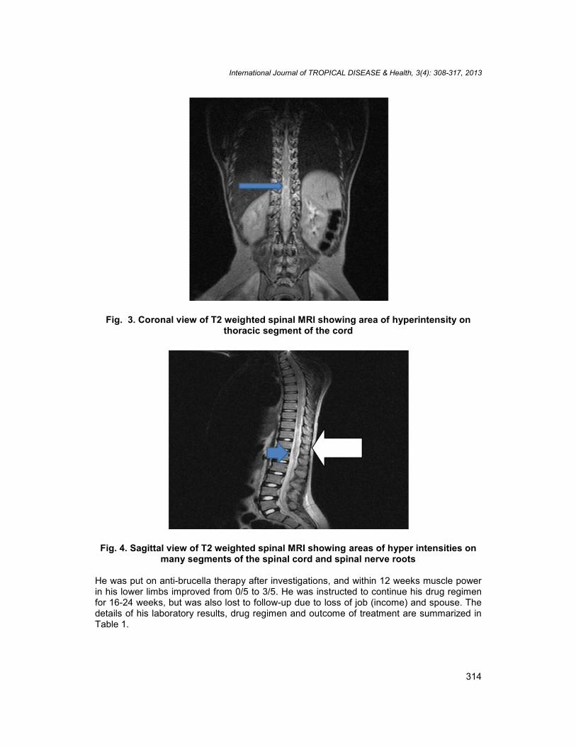
On examination, she was chronically ill-looking, asthenic, conscious, but febrile(temperature 38.7ºC), diaphoretic and pale. She had multiple gluteal and trochantericulcers discharging cheesy/putrid materials; and episodic titanic spasms with locked-jaw,trismus, opisthotonus and rigidity. She also had spastic paraplegia, bilateral extensorplantar reflexes, and sensory loss at level T12, but no obvious vertebral deformity orgibbus. She was incontinent of both faeces and urine, and had a diaper on. X-rays of thespine, pelvis and hips did not reveal abnormalities, but spinal MRI revealed area of hyper-intensity on the thoracic segment of spinal cord (Fig. 3)
International Journal of TROPICAL DISEASE & Health, 3(4): 308-317, 2013
310
She was treated with intravenous human tetanus immunoglobulins 10,000 internationalunits (I.U) stat after a test dose, metronidazole infusions 500 mg 8 hourly, andintermittent diazepam 20-40 mg in 5% dextrose saline 8-12 hours until spasm free. Shereceived 3 pints of packed cells blood transfusion, and recovered from tetanus within 2weeks of admission. She was also investigated for brucellosis, found positive and wastreated with antibiotics. She recovered and was discharged home with residualparaparesis at 12th week of admission. The results of laboratory investigations, drugregimen and outcome of treatment are summarized in Table 1.
2. J.N, a 13 year old student developed intermittent fever of 5-7 days intervals, headache,neck pain and loss of consciousness for 2 weeks. He was treated for possible pyogenicmeningitis with parenteral ceftriaxole at a private hospital where he first presented; andregained consciousness after 10 days. However, the fever continued and he developedparaplegia, urinary retention and incontinence within 4 weeks of illness. There was nohistory of antecedent diarrhea, upper respiratory tract infection, or recent vaccination.Eight weeks later, he developed a sinus above the gluteal cleft which discharged acheesy material. He worked in his father’s animal farm which consisted of pigs, goatsand sheep, but did not ingest unpasteurized milk.
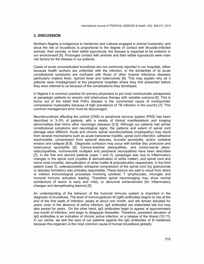
On examination, he was fully conscious, febrile (temperature 37.6ºC), pale anddehydrated. He also had nuchal rigidity with positive Brudzinski and Kernig’s signs,flaccid paraplegia, and loss of sensation below the knee joints (L 4 and below). Therewas a sinus which was discharging cheesy material from a slit-like opening above thegluteal cleft on the midline. He was incontinent of urine, but not of faeces, and there wasno obvious vertebral deformity or no gibbus. However, spinal MRI revealed areas ofhyper-intensities on many segments of spinal cord & spinal nerve roots (Fig. 4)
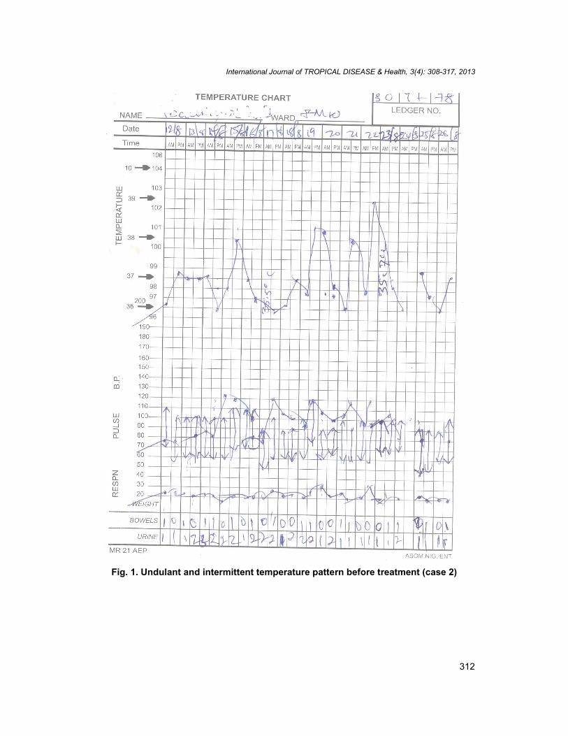
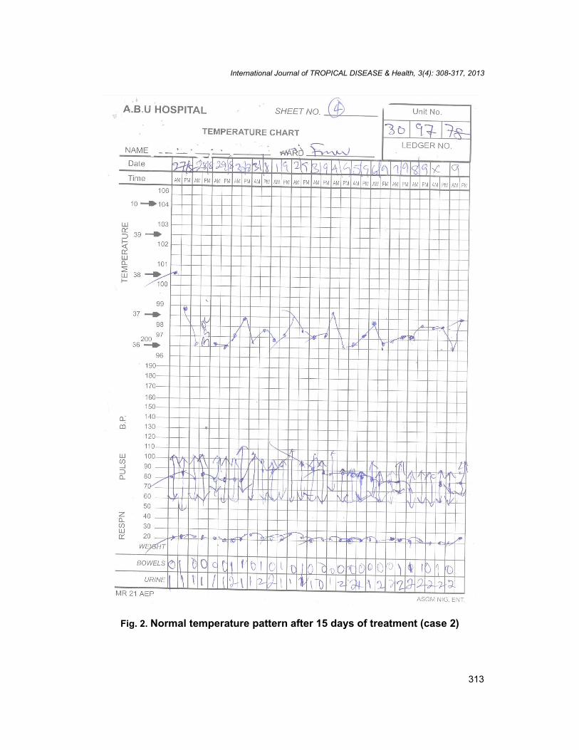
He was investigated for brucellosis, found positive and treated with antibiotics.Constitutional symptoms of brucellosis resolved within 2 weeks of treatment as shownby the pattern of fever at presentation and 15 days after (in Figs. 1 and 2 respectively),but his neurologic deficit persisted. He was instructed to continue his drug regimen for16- 24 weeks but was discharged on request with residual paraparesis at 12th week oftherapy. He was lost to follow-up. The results of his laboratory tests, drug regimen andoutcome of treatment are summarized in Table 1.
3. M.D, a 56 year old man who worked as a veterinary health extension worker in KadunaState Ministry of Agriculture and Animal resources presented with 2 years history ofheadache, anorexia, weight loss, fatigue and progressive lower limb weakness resultingin paraplegia and urinary incontinence. He had completed 18 months course ofantituberculous therapy for suspected Pott’s disease with minimal improvement. Hisdaily routine of more than two decades was vaccination of herds of cows, sheep, goatsand pigs. He never ingested unpasteurized milk. On examination, he was fullyconscious, pale, afebrile and mildly wasted. He had spastic paraplegia, bilateralextensor plantar reflexes, sensory loss at level T10, and urinary incontinence. Therewere multiple vertebral deformities and gibbus.
International Journal of TROPICAL DISEASE & Health, 3(4): 308-317, 2013
311
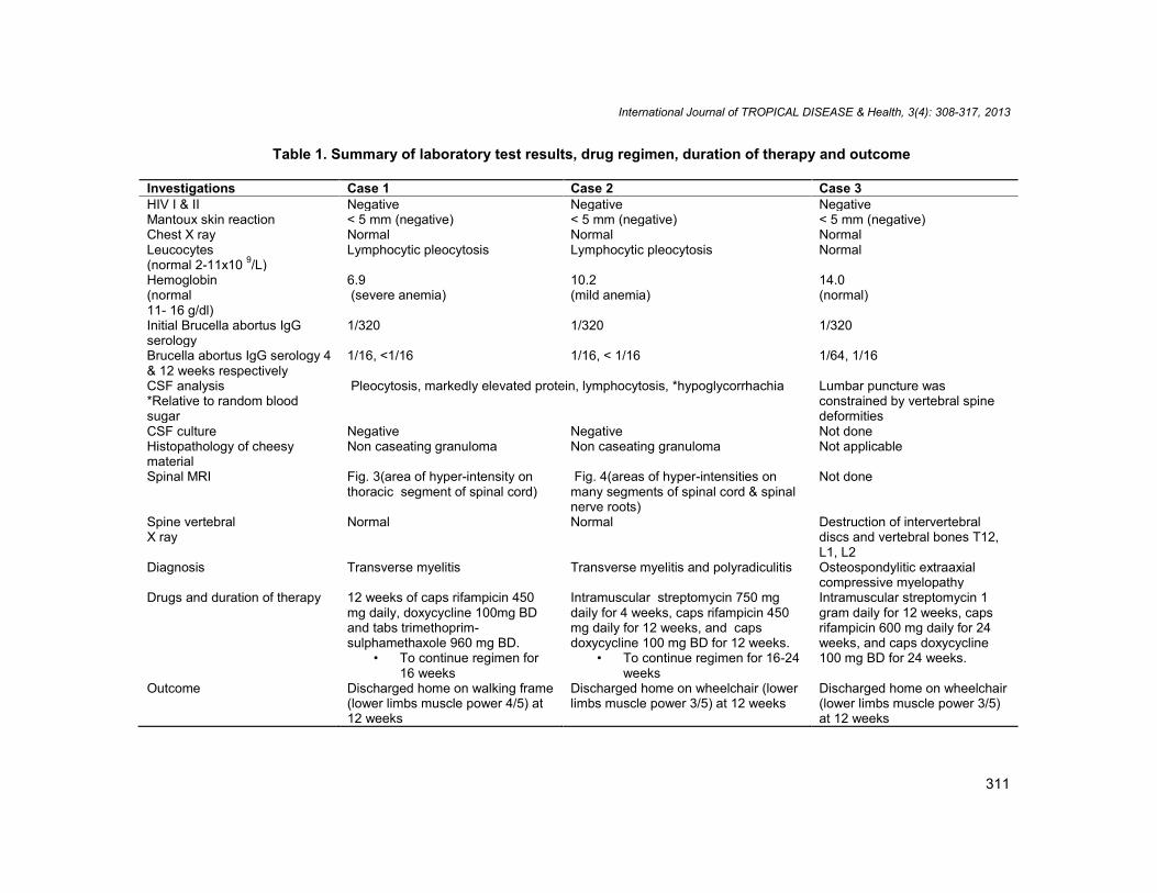
Table 1. Summary of laboratory test results, drug regimen, duration of therapy and outcome
Investigations Case 1 Case 2 Case 3HIV I & II Negative Negative NegativeMantoux skin reaction < 5 mm (negative) < 5 mm (negative) < 5 mm (negative)Chest X ray Normal Normal NormalLeucocytes(normal 2-11x10 9/L)
Lymphocytic pleocytosis Lymphocytic pleocytosis Normal
Hemoglobin(normal11- 16 g/dl)
6.9(severe anemia)
10.2(mild anemia)
14.0(normal)
Initial Brucella abortus IgGserology
1/320 1/320 1/320
Brucella abortus IgG serology 4& 12 weeks respectively
1/16, <1/16 1/16, < 1/16 1/64, 1/16
CSF analysis*Relative to random bloodsugar
Pleocytosis, markedly elevated protein, lymphocytosis, *hypoglycorrhachia Lumbar puncture wasconstrained by vertebral spinedeformities
CSF culture Negative Negative Not doneHistopathology of cheesymaterial
Non caseating granuloma Non caseating granuloma Not applicable
Spinal MRI Fig. 3(area of hyper-intensity onthoracic segment of spinal cord)
Fig. 4(areas of hyper-intensities onmany segments of spinal cord & spinalnerve roots)
Not done
Spine vertebralX ray
Normal Normal Destruction of intervertebraldiscs and vertebral bones T12,L1, L2
Diagnosis Transverse myelitis Transverse myelitis and polyradiculitis Osteospondylitic extraaxialcompressive myelopathy
Drugs and duration of therapy 12 weeks of caps rifampicin 450mg daily, doxycycline 100mg BDand tabs trimethoprim-sulphamethaxole 960 mg BD.
• To continue regimen for16 weeks
Intramuscular streptomycin 750 mgdaily for 4 weeks, caps rifampicin 450mg daily for 12 weeks, and capsdoxycycline 100 mg BD for 12 weeks.
• To continue regimen for 16-24weeks
Intramuscular streptomycin 1gram daily for 12 weeks, capsrifampicin 600 mg daily for 24weeks, and caps doxycycline100 mg BD for 24 weeks.
Outcome Discharged home on walking frame(lower limbs muscle power 4/5) at12 weeks
Discharged home on wheelchair (lowerlimbs muscle power 3/5) at 12 weeks
Discharged home on wheelchair(lower limbs muscle power 3/5)at 12 weeks
International Journal of TROPICAL DISEASE & Health, 3(4): 308-317, 2013
312
Fig. 1. Undulant and intermittent temperature pattern before treatment (case 2)
International Journal of TROPICAL DISEASE & Health, 3(4): 308-317, 2013
313
Fig. 2. Normal temperature pattern after 15 days of treatment (case 2)
International Journal of TROPICAL DISEASE & Health, 3(4): 308-317, 2013
314
Fig. 3. Coronal view of T2 weighted spinal MRI showing area of hyperintensity onthoracic segment of the cord
Fig. 4. Sagittal view of T2 weighted spinal MRI showing areas of hyper intensities onmany segments of the spinal cord and spinal nerve roots
He was put on anti-brucella therapy after investigations, and within 12 weeks muscle powerin his lower limbs improved from 0/5 to 3/5. He was instructed to continue his drug regimenfor 16-24 weeks, but was also lost to follow-up due to loss of job (income) and spouse. Thedetails of his laboratory results, drug regimen and outcome of treatment are summarized inTable 1.
International Journal of TROPICAL DISEASE & Health, 3(4): 308-317, 2013
315
3. DISCUSSION
Northern Nigeria is indigenous to herdsmen and cultures engaged in animal husbandry; andsince the risk of brucellosis is proportional to the degree of contact with Brucella-infectedanimals, their excreta, or their edible byproducts; the disease is expected to be endemic inour environment [5]. Prolonged contact with animals and their edible byproducts were mainrisk factors for the disease in our patients.
Cases of acute uncomplicated brucellosis are not commonly reported in our hospitals, eitherbecause health workers are unfamiliar with the infection, or the similarities of its acuteconstitutional symptoms are confused with those of other tropical infectious diseasesparticularly malaria fever, typhoid fever and tuberculosis [6]. This may explain why all 3patients were misdiagnosed at the peripheral hospitals where they first presented beforethey were referred to us because of the complications they developed.
In Nigeria it is common practice for primary physicians to put most nontraumatic paraparesisor paraplegic patients on empiric anti tuberculous therapy with variable outcome [5]. This isborne out of the belief that Pott’s disease is the commonest cause of nontraumaticcompressive myelopathy because of high prevalence of TB infection in the country [7]. Thiscommon management error must be discouraged.
Neurobrucellosis affecting the central (CNS) or peripheral nervous system (PNS) has beendescribed in 3–5% of patients, with a variety of clinical manifestations and imagingabnormalities that mimic other neurologic diseases [3,5]. Although our patients had similarconstitutional symptoms and neurological signs, the patterns and extent of spinal corddamage were different. Acute and chronic spinal neurobrucellosis (myelopathy) may resultfrom several mechanisms such as acute transverse myelitis, spinal cord infarction, adhesivearachnoiditis, compression from epidural abscess, brucella spondylitis, and/or vertebralerosion and collapse [8,9]. Diagnostic confusion may occur with lumbar disc protrusion andtuberculous spondylitis [8]. Cervico-brachial plexopathies, and lumbo-sacral plexo-radiculopathies, mononeuritis multiplex and peripheral neuropathies have been described[7]. In the first and second patients (case 1 and 2), paraplegia was due to inflammatorychanges in the spinal cord (myelitis & demyelination of white matter), and spinal cord andnerve roots (myelitis, demyelination of white matter & polyradiculitis) respectively; in the thirdpatient (case 3), osteospondylitic extraaxial compression of the spinal cord (by granulomasor abscess formation) was probably responsible. These lesions are said to result from director indirect immunological processes involving cytotoxic T lymphocytes, microglia andhumoral immune activation leading. Therefore spinal neuroimaging may show normalarchitecture (if lesion is early and mild), or abnormal enhancement (for inflammatorychanges and demyelinating lesions) [5].
An understanding of the behavior of the humoral immune system is important in thediagnosis of brucellosis. The level of immunoglobulin M (IgM) antibodies begins to rise at theend of the first week of infection, peaks at about one month, and will remain elevated foryears; even in the absence of active infection. IgA antibodies are elaborated late but mayalso persist for years. On the other hand, IgG antibodies begin to appear at approximatelyone month of infection, and begin to disappear thereafter. Therefore, persistent elevation ofIgG antibodies is an indication of chronic active infection, or a relapse of the illness [10,11].In our centre, we test the sera of our patients against the IgG antibodies of B melitensisbecause this organism is the most common cause of human brucellosis globally.
International Journal of TROPICAL DISEASE & Health, 3(4): 308-317, 2013
316
Recovery of Brucella organisms from cultures of CSF or blood is usually very low inneurobrucellosis because the organisms are chiefly intracellular in locations, although theCSF may exhibit pleocytosis, hypoglycorrhachia, and elevation of protein concentration. TheCSF findings of this disease can thus mimic those of other intracellular organisms such asmycobacteria, fungi and toxoplasma [12].
In endemic areas, isolation of bacteria from serum or cerebrospinal fluid (CSF) is the goldstandard diagnostic method, but appropriate serological tests such as IgG agglutination titersof >1:160 in CSF or >1:320 in serum can be diagnostic, particularly if there are rising titers inserial testing [10]. Rapid diagnosis and treatment often leads to prompt and completerecovery in acute infection, but response to appropriate antibiotic is variable in chronicbrucellosis [13]. Treatment entails use of triple drugs selected from rifampicin, doxycycline,gentamicin, streptomycin, trimethoprim-sulfamethoxazole and ciprofloxacin, usually forperiods of 6 weeks (streptomycin) and 3 months (for others). Standard treatment for adultswith acute spinal brucellosis comprises capsule doxycycline 100 mg BD and tabletstrimethoprim-sulphamethaxole 960 mg BD, or capsule rifampicin 600 mg for at least 12weeks, combined during the first 3-4 weeks with IM streptomycin. Treatment is prolonged for18-24 weeks in chronic neurobrucellosis [13]. Clinical improvement as well as improvementin CSF pleocytosis and fall in CSF and blood Brucella titers should occur after appropriatetreatment. Poor outcomes are associated with complications such as raised intracranialpressure, stroke, endocarditis, intracranial mycotic aneurysm and haemorrhage, andosteospondylitis with compressive myelopathy or radiculopathy [3].
4. CONCLUSION
In conclusion, neurobrucellosis is a treatable disease with a favorable outcome, but becauseacute symptoms are similar to many tropical fevers, the infection may be misdiagnosed withdare consequences. Also, the presence of complications like meningoencephalitis andmyelopathy worsens the prognosis. Therefore the disease will continue to be an importanthealth problem in developing countries unless certain steps are taken to reduce itsincidence. The steps must include avoidance of unpasteurized dairy products, animalcontact or improperly cooked meat; regulation of abattoirs; surveillance, culling andvaccination of herds of sheep, goats, cows and pigs.
CONSENT
Written informed consent was obtained from the patient (or other approved parties) forpublication of this case report and accompanying images.
ETHICAL APPROVAL
Not applicable.
COMPETING INTERESTS
Authors have declared that no competing interests exist.
REFERENCES
1. Edwards C, Jawad AS. History of brucellosis. J R Soc Med. 2006;99(2):54.
International Journal of TROPICAL DISEASE & Health, 3(4): 308-317, 2013
317
2. Young EJ. Human brucellosis. Rev Infect Dis. 1983;5(5):821-842.3. Ceran N, Turkoglu R, Erdem I, Inan A, Engin D, Tireli H, et al. Neurobrucellosis:
clinical, diagnostic, therapeutic features and outcome. Unusual clinical presentationsin an endemic region. Braz J Infect Dis. 2011;15(1):52-59.
4. Obiako OR, Ogoina D, Danbauchi SS, Kwaifa SI, Chom ND, Nwokorie E.Neurobrucellosis- a case report and review of literature. Niger J Clin Pract.2010;13(3):10-15.
5. Al-Sous MW, Bohlega S, Al-Kawi MZ, Alwatban J, McLean DR. Neurobrucellosis:clinical and neuroimaging correlation. Am J Neuroradiol. 2004;25:395-401.
6. Badiaga S, Imbert G, La Scola B. Imported brucellosis associated with plasmodiumfalciparum malaria in a traveler returning from the tropics. J Travel Med.2005;12(5):282-284.
7. Nigeria Tuberculosis Fact Sheet. United States Embassy in Nigeria.http://photos.state.gov/libraries/nigeria/487468/pdfs/JanuaryTuberculosisFactSheet.pdf. Accessed online 30/05/2013.
8. Bahemu Ka M, Shemena AR, Panyiotopoulis CP. Neurologic syndromes ofbrucellosis. J Neurol Neurosurg psychiatry. 1988;51:1017-1021.
9. Shakir RA, Al-Din AS, Araj GF, Lulu AR, Mousa AR, Saadah MA. Clinical categories ofneurobrucellosis; a report on 19 cases. Brain. 1987;110:213-223.
10. Young EJ. Serologic diagnosis of human brucellosis: analysis of 214 cases byagglutination tests and review of the literature. Rev Infect Dis. 1991;13(3):359-372.
11. Araj GF, Kattar MM, Fattouh LG, Bajakian KO, Kobeissi SA. Evaluation of the PANBIOBrucella IgG and IgM enzyme-linked immunosorbent assays for the diagnosis ofhuman brucellosis. Clin Diagn Lab Immunol. 2005;12(11):1334-1335.
12. Bouza E, Garcia de la Torre M, Parras F. Brucellar meningitis. Rev Infect Dis.1987;9(4):810-822.
13. Alp E, Doganay M. Current therapeutic strategy in spinal brucellosis. Int J Infect Dis.2008;12(6):573-577.
© 2013 Obiako et al.; This is an Open Access article distributed under the terms of the Creative CommonsAttribution License (http://creativecommons.org/licenses/by/3.0), which permits unrestricted use, distribution, andreproduction in any medium, provided the original work is properly cited.
Peer-review history:The peer review history for this paper can be accessed here:
http://www.sciencedomain.org/review-history.php?iid=257&id=19&aid=1912