Embed Size (px)
Citation preview
SPINAL MUSCULAR ATROPHIES (SMA) Spin23 (1)
Spinal Muscular Atrophies (SMA) Synonyms: PROGRESSIVE SPINAL MUSCULAR ATROPHY, PROGRESSIVE SPINAL ATROPHY
Last updated: August 8, 2020
ETIOPATHOPHYSIOLOGY ........................................................................................................................ 1
EPIDEMIOLOGY ...................................................................................................................................... 1
CLINICAL FEATURES .............................................................................................................................. 1
DIAGNOSIS ............................................................................................................................................. 2
TREATMENT ........................................................................................................................................... 3 PROGNOSIS ............................................................................................................................................ 3
OTHER FORMS OF LMN DEGENERATION ............................................................................................... 3
SMA - progressive degeneration and loss of LMN (midbrain ÷ spinal cord).
replacement of lost cells by gliosis (e.g. atrophic spinal cord at autopsy).
UMN is not affected! (vs. in ALS)
Type Inheritance Age of Onset Presenting Symptoms Prognosis
SMA type I (infantile /
acute / fatal SMA,
Werdnig-Hoffman
disease)
AR In utero ÷ 6
months
Hypotonia and generalized
weakness, problems with
sucking, swallowing, and
breathing; never able to sit
Average life
expectancy - 8
months;
95% dead before
age of 18 months
SMA type II (intermediate between
type I and type III)
AR 6 ÷ 15 months < 25% learn to sit; never
able to stand, facial
muscles spared Depends on
respiratory
complications SMA type III (chronic
SMA, Kugelberg-
Welander disease)
AR, AD 15 months ÷
teen years
Proximal leg weakness,
delayed motor milestones
Kennedy's disease (bulbospinal muscular
atrophy)
X-linked
recessive
After age 40
yrs
Bulbar → distal limb
weakness; endocrine
dysfunction
Normal lifespan
Fazio-Londe disease
(progressive bulbar
palsy of childhood)
Late
childhood ÷
adolescence
Bulbar weakness
SMA type IV (adult-
onset SMA)
AD, AR, X-
recessive
(very rare)
median ≈ 37
years
Proximal weakness,
variable within families,
more severe in AD
Life expectancy
not markedly
reduced
Distal SMA (Charcot-
Marie-Tooth type-
SMA)
AR, AD AR: birth ÷
infancy;
AD:
adulthood
Distal weakness
Very slow
clinical
progression; does
not alter lifespan
ETIOPATHOPHYSIOLOGY
Autosomal recessive SMA types I, II, III (allelic heterogeneity) have been linked to 5q11.3-13.1 -
gene for survival of motor neurons (SMN):
Defect in neuronal apoptosis!
contains multiple copies of genes and pseudogenes;
characterized by instability: deletions (98%), truncations, point mutations.
protein product has no known homolog, and its function is not yet known.
no correlation between genotype and phenotype! - but most affected siblings exhibit same
phenotype - may be additional modifying factors, e.g. another gene tightly linked to
pathogenic gene:
a) contiguous deletion of nearby neuronal apoptosis inhibitory protein gene (NAIP) is
associated with most severe phenotype (occurs in 45-65% SMA type I and in 20-40%
SMA type II and III cases).
b) homozygous deletions in exons 7 and 8 in SMNt (telomeric copy of SMN) → SMA
type I; mutations that convert SMNt to centromeric copy (SMNc) → SMA type II
and III.
SMN protein is implicated in the trafficking of RNA in and out of the nucleus and in the formation
of complexes that are important in RNA splicing.
SMN locus on chromosome 5 has two almost identical copies of the SMN gene - one produces a
full length SMN protein, whereas the second expresses a small amount of full-length SMN and a
shortened SMN; loss of full-length SMN from mutations at the main locus can be mitigated to
some degree by the shortened SMN protein expressed at the second locus.
EPIDEMIOLOGY
SMA type I (most common SMA) INCIDENCE ≈ 4-10 in 100,000 (2nd most common neuromuscular
disease, following Duchenne muscular dystrophy).
similar numbers are affected with milder forms and forms with later onset.
carrier frequency of SMNt mutation - 1 in 50.
CLINICAL FEATURES
Clinical hallmarks:
1. Insidious onset of symmetrical WEAKNESS.
proximal muscles > distal muscles.
legs > arms.
greatest decline in muscular power occurs at onset* and then slows (i.e. great loss of
motoneurons initially, followed by stabilization in any remaining neurons) - difference
between SMAs and other neurodegenerative disorders.
*results in large number of complications: scoliosis, contractures (e.g.
arthrogryposis multiplex congenita), disuse atrophy, respiratory /
nutritional / sleep problems.
2. HYPOTONIA, ATROPHY, LOSS OF TENDON REFLEXES After immediate neonatal period, spinal muscular atrophy is most common cause of
infantile hypotonia (“floppy infant”)!
3. CRANIAL NERVE PALSIES (CN3, 4, 6 are typically spared!).
No sensory symptoms or loss, no myalgias!
No heart involvement!
Intelligence normal! (children often appear brighter than their normal peers!)
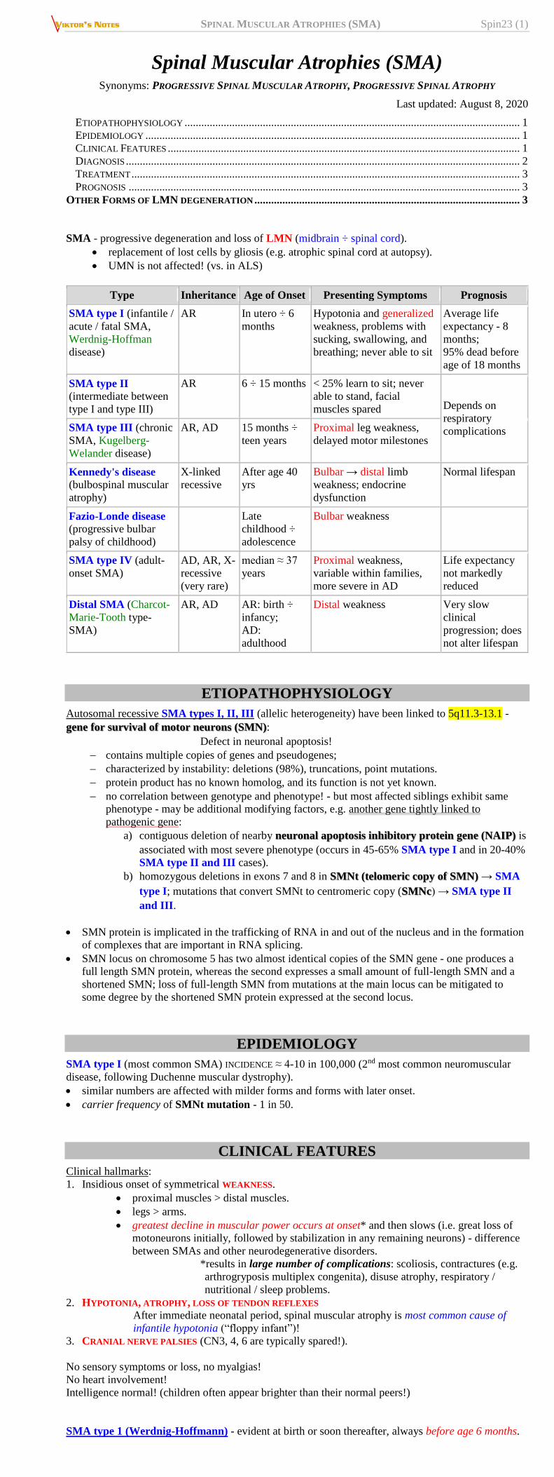
SMA type 1 (Werdnig-Hoffmann) - evident at birth or soon thereafter, always before age 6 months.

SPINAL MUSCULAR ATROPHIES (SMA) Spin23 (2)
mothers notice decreased intrauterine movements.
one of most common forms of floppy infant syndrome (infants lie flaccid with little
movement, unable to overcome gravity).
tongue is often seen to fasciculate (rarely in limb muscles - because of ample subcutaneous
fat).
ultimately, complete flaccid quadriplegia results with compromised respiration.
all dead by age 4 yrs.
SMA type 3 (Kugelberg-Welander) – slowly progressive gait disorder in late childhood or
adolescence.
proximal limb muscle weakness and wasting (simulates muscular dystrophy!); tendon
reflexes are lost.
relative sparing of bulbar muscles.
course relatively benign - many continue to function socially with normal life span (others
may be handicapped); many children are highly intelligent.
DIAGNOSIS
genetic test – homozygous SMN deletion (sensitive test in 95% cases!).
prenatal testing is available only on research basis.
serum CK can be elevated (correlates with illness duration);
in SMA 3, may be 20 times normal (in range of many myopathies!).
ECG – normal.
without DNA diagnosis, it is essential to verify neurogenic process via:
1) EMG – denervation.
2) nerve conduction studies – normal.
3) muscle biopsy (with histochemistry) – denervation & reinnervation: large numbers of
atrophic fibers, often only few micrometers in diameter; atrophic fibers often involve entire
fascicle (panfascicular atrophy!!!); scattered groups of large fibers that are 2-4 times
normal size. also see p. D30 >>
Source of picture: Ramzi S. Cotran “Robbins Pathologic Basis of Disease”, 6th ed. (1999); W. B. Saunders Company; ISBN-13: 978-
0721673356 >>
SPINAL MUSCULAR ATROPHIES (SMA) Spin23 (3)
TREATMENT
multidisciplinary approach aimed at preventing contractures, skeletal deformities, respiratory
complications, and social isolation.
NNUUSSIINNEERRSSEENN (Spinraza®) intrathecal injection - antisense therapy - the first FDA approved drug to treat
children and adults with spinal muscular atrophy.
sham-controlled study in 78 children with infantile SMA showed that treatment with nusinersin
leads to a 50% reduction in deaths or early ventilation. Finkel RS, Mercuri E, Darras BT, et al; ENDEAR Study Group. Nusinersen versus sham control in
infantile-onset spinal muscular atrophy. N Engl J Med. 2017;377:1723-1732.
RRIISSDDIIPPLLAAMM (Evrysdi®) taken by mouth or via a feeding tube – gene-splicing modulator that increases
production of survival of motor neuron protein (SMN), needed for survival of motor neurons - FDA
approved for treatment of SMA in adults and children age 2 months or more.
PROGNOSIS
Earlier onset – more rapid decline.
Other Forms of LMN degeneration
Poliomyelitis – viral disease of LMN – see p. 259 (1) >>
do not map to 5q11.
most are autosomal recessive.
Fazio-Londe disease (progressive bulbar palsy of childhood) - brainstem LMN degeneration of all
brainstem nuclei (vs. most juvenile SMAs).
presents in late childhood or adolescence with stridor → ptosis, dysarthria, facial palsy,
dysphagia.
weakness of arms & legs may occur later, and respiration may be affected.
death in early childhood?
Scapuloperoneal and facioscapulohumeral SMA forms
distinction from muscular dystrophy depends on DNA analysis.
Kennedy's disease - X-linked recessive disorder (expansion of CAG trinucleotide repeats in first
exon of androgen receptor gene Xq11-12) - affects males:
1) progressive bulbospinal muscular atrophy (preferentially bulbar* → distal limb
muscles) *incl. ocular!
2) endocrine dysfunction – androgen insensitivity (testicular atrophy, gynecomastia,
oligospermia), diabetes mellitus.
3) subtle sensory sign in some patients. (e.g. abnormal sensory-evoked potentials, affected
spinal sensory tracts, distal degeneration of sensory axons).
midlife onset, after age 40 yrs. (direct correlation between number of -CAG- repeats and
disease severity).
most common form of adult-onset SMA!
may be readily screened from blood DNA analysis.
slowly progressive, normal lifespan.
SPINAL MUSCULAR ATROPHIES (SMA) Spin23 (4)
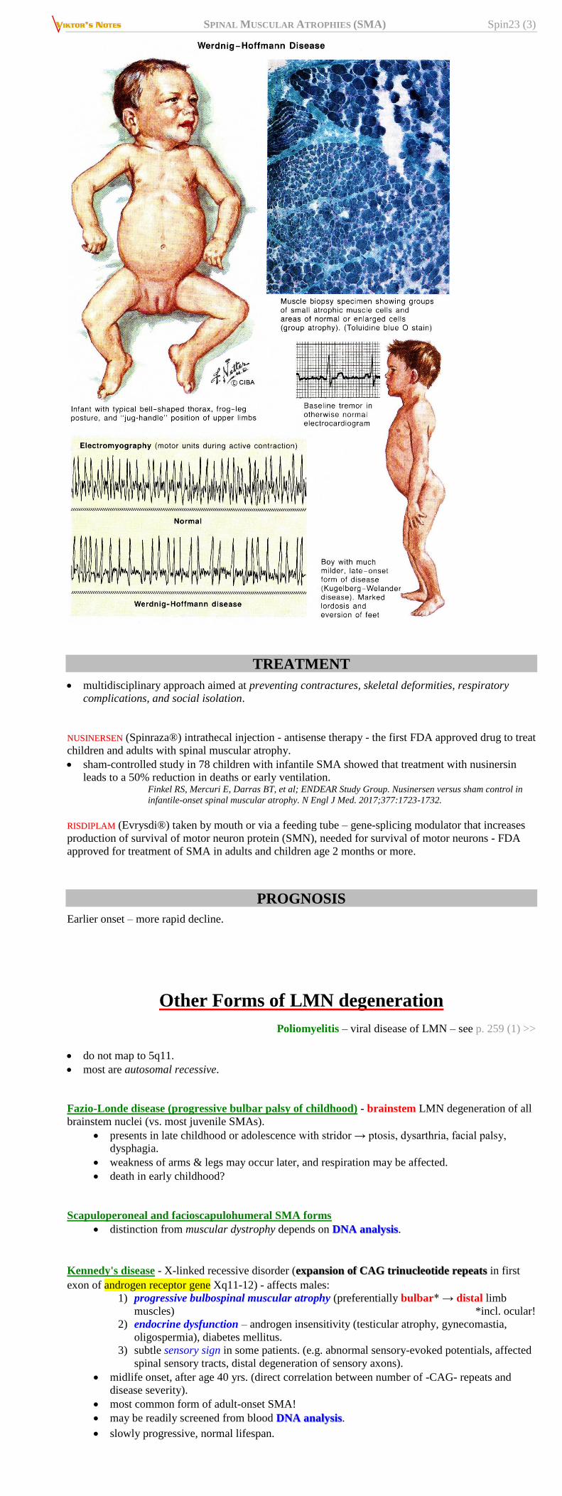
Adult Tay-Sachs disease (hexosaminidase A deficiency)
primarily in Ashkenazi Jewish families.
adult-onset (vs. classical Tay-Sachs disease), very slowly progressive.
dysarthria and cerebellar atrophy.
Baby with Tay-Sachs disease - enlarged, pale neurons:
Source of picture: “WebPath - The Internet Pathology Laboratory for Medical Education” (by Edward C. Klatt, MD) >>
BIBLIOGRAPHY for ch. “Spinal Disorders” → follow this LINK >>
Viktor’s Notes℠ for the Neurosurgery Resident
Please visit website at www.NeurosurgeryResident.net