Embed Size (px)
Citation preview

Spinal Cord Injury: Assessment Criteria to Determine the Need
for Spinal Movement Restriction

Section I – Spinal Anatomy and Function • Introduction……………………………………………………………..4 • The Spine…………………………………………………….…………6 • Cervical Spine………………………………………………..…………6 • Thoracic Spine……………………………………………….…………6 • Lumbar Spine…………………………………………………...………7 • Sacral and Coccygeal Spine…………………………………….………7 • Anatomical Spinal Tracts……………………………………….………7
• Anterior Cord…………………………………………….……..7 • Central Cord……………………………………………….……8 • Posterior Cord……………………………………………….….8
• Nerve Roots…………………………………………………………….8 Section II – Spinal Injury
• Introduction……………………………………………………………..9 • Primary Injury vs. Secondary Injury……………………………………9 • Spinal Shock………………………………………………………….…9 • Spinal Neurological Shock……………………………………………...9 • Complete Spinal Cord Injury…………………………………………..10
• Quadriplegia…………………………………………………....10 • Paraplegia………………………………………………………10
• Incomplete Spinal Cord Injury…………………………………………10 • Anterior Cord Syndrome……………………………………….10 • Central Cord Syndrome…………………………………………10 • Brown – Sequard Syndrome…………………………………...10
• C-Spine Injuries………………………………………………………..11 • T-Spine Injuries………………………………………………………..11 • L/S Spine Injuries……………………………………………………...11 • “Spine Injury” vs. “C-Spine Injury”…………………………………...11
Section III – The Key to Assessment
• Introduction………………………………………………………….…13 • Mechanism of Injury (MOI)…………………………………………….13 • Clinical Assessment Criteria………………………………………..….13
• “Reliability” of Patient Exam……………………………………….14 • Altered Mentation…………………………………………..14 • Intoxication………………………………………………….14 • Age…………………………………………………………15 • Communication Barrier……………………………………15
• Distracting Injury………………………………………………..…16 • Abnormal Sensory/Motor Assessment………………………….…17
• Neurologic Exam – Motor……………………..………17 • Motor Function – Upper Extremities……..……………17 ! Finger abduction/adduction……………..…………17

! Finger/hand extension………………………………17 • Motor Function – Lower Extremities………………………………18
! Foot plantar flexion………………………………….18 ! Foot/great toe dorsiflexion…………………………...18
• Motor Exam Interpretation…………………………………………18 • Neurological Exam – Sensory……………………………………...18
! Abnormal sensation………………………………...19 ! Pain sensation – upper extremities…………………19 ! Pain sensation – lower extremities…………………19 ! Sensory Exam Interpretation……………………….19
• Spinal Pain and Tenderness……………………………………...…20 • Positive Assessment Findings………………………………………20
Section IV – Prehospital Management
• Spinal Injury = Bone Injury………………………………………….…22 • Initial Treatment = Complete Spine Immobilization…………………...22 • Splinting The Spine In Normal Anatomic Position…………………….23 • Helmet Removal………………………………………………………..23 • Cervical Collar Sizing…………………………………………………..24 • Standing Take Down……………………………………………………24 • Spinal Movement Restriction in a Car Seat …………………..………..25 • Kendrick Extrication Device (KED)……………………………………26 • Straddle Slide…………………………………………………………….27 • Scoop Stretcher…………………………………………………………28 • Log Roll………………………………………………………………….28 • Rapid Extrication……………………………………………………….28 • Full Spinal Movement Restriction…………..………………………….29 • Padding Voids…………………………………………………………..29 • Spinal Movement Restriction of Pregnant Patients…….………………30
Appendices
• Sensory Dermatomes……………………………………………………31 • Austin-Travis County EMS System Spinal Assessment Algorithm…….32 • Spinal Assessment Algorithm Pocket Card……………………………..33 • Standards of Care Replacement Page (p.136a)………………………….34

4
Introduction If somebody asked you, “What kind of as-sessment you do the most,” what do you think you would say: respiratory, chest pain, spinal injury? It’s a toss up, but many would say “spinal.” Assessment for spinal cord injury is one of the most common assessments per-formed in the prehospital environment. Eve-ryday ambulances respond to dozens of as-saults, falls, and motor vehicle collisions, each with the potential for spinal injury and all requiring a thorough assessment. So how do you assess whether a patient is at risk for spinal cord injury? Is it the mecha-nism of injury, do you use physical findings, do preexisting conditions play a role? The National Association of EMS Physicians (NAEMSP) says, “Currently spinal immobili-zation is the only EMS procedure performed based only on the mechanism of injury with-out consideration of the patient’s clinical find-ings and the assessment skills of prehospital providers.”i That is, of course, until now. In 1997, the Journal of Emergency Medical Services (JEMS) chronicled the experience of one of the first EMS systems – the State of Maine - to implement a spinal injury assess-ment protocol. Dr. Peter Goth – a former Re-gional Medical Director in Maine – developed the “Maine Protocol.” He recognized that EMS training didn’t include evidence based spinal assessment and that EMS providers were left to use mechanism as the primary criteria for spinal restriction decision-making. In the article, Goth was quoted as saying: “We set up the impossible and expected EMS to perform perfectly. Spine injury manage-ment, in general, has been a black hole in EMS…It’s been fraught by a lack of good direction, and [providers] were always nerv-ous about doing the wrong thing, but were never told the right thing to do. We had been
assessing spinal injury using impractical rules, and I saw the conflict.”ii Even before Dr. Goth, the National Associa-tion of EMS Physicians (NAEMSP) wrote a position paper, “Indications for Prehospital Spinal Immobilization.” Like Goth, NAEMSP advocated the use of clinical as-sessment criteria for the assessment of spinal injury and the determination of the need for spinal movement restriction. In the years since, additional studies have fol-lowed adding to the body of research support-ing the efficacy of the role of spinal assess-ment criteria in accurately identifying those with a low risk of spinal cord injury. In September 1999, the Austin-Travis County EMS System (A/TCEMS) introduced an as-sessment standard based on the position of the NAEMSP and the medical literature available on field spinal assessment and decision-making. Since that introduction, more evi-dence has been published supporting the use of assessment criteria; including a large study known as the National Emergency X-Radiography Utilization Study (NEXUS). The NEXUS study looked at 34,069 blunt trauma patients. 818 patients had a cervical-spine fracture, and of those, 810 were identi-fied by the assessment criteria in the absence of MOI.iii That means the study found that 99.9 percent of the patients assessed, who ac-tually had an injury, were accurately identi-fied and, of the 818 patients with cervical spine injury, 99.0 percent were correctly iden-tified using assessment criteria. 99.0-99.9 percent accurate! Studies like these make it easy to appreciate the significance of using scientifically tested assessment criteria to assess for spinal cord injury. This was further reinforced when the Clinical Practice Division did a retrospective

5
review of 100 percent of the spinal cord inju-ries in Travis County in 2000 and found that the assessment criteria identified every single injury and did so even in the absence of mechanism of injury as a factor. The Spinal Assessment standard has been an important evolution in our practice. How-ever, even with all of the literature supporting the assessment criteria, it hasn’t been without challenge in its application and providers have struggled to do the right thing and apply the standard appropriately. Many providers view the assessment criteria as an exclusionary process - missing that the intent is to identify those that DO need to have spinal movement restriction - and mechanism of injury continues to confuse, as providers revert to using it as a leading con-tributor in their decision. In addition, some providers have struggled with taking patient complaints at face value and attempt to assess the reported injury’s value. Recognition of the importance of a spinal as-sessment standard to our practice, and the challenges providers face in understanding its application, led to this new Spinal Assessment program. The program consists of three (3) components: 1. The Learning Guide: providing detailed
descriptions of spinal anatomy, injury types, and how to successfully assess and manage these patients.
2. The Reference Poster: located in every EMS and fire station and in all of the emergency departments to provide a quick resource outlining the assessment criteria.
3. A Video: a short video presenting clips explaining each criteria of the spinal as-sessment standard.
Together, this program has been designed to provide a well-balanced approach to under-standing the spinal assessment standard. i Domeier, RM; NAEMSP Standards and Practice Committee; “Indications for Prehospital Spinal Immo-bilization, NAEMSP Position Paper. ii Munk, Marc-David, “Maine Taps Medics’ Spinal Skills: Ruling Out C-spine Injuries in the Field,” JEMS, 1997. p.78. iii N Engl J Med 2000 Jul 13;343(2):94-9; http://www.ncbi.nlm.nih.gov/entrez/query.fcgi?cmd=Re-trieve&db=PubMed&list_uids=10891516&dopt=Abstract

6
Section I – Spinal Anatomy & Function Introduction For a prehospital clinician to assess and man-age spinal cord injuries it is important that s/he have a solid understanding of the anat-omy of the spine and its basic functions. This section will provide a basic foundation of un-derstanding of spinal anatomy and function. Selections of the following section are reprinted from “Spine Injury: Clinical Criteria for Assessment and Management” with written permission from Peter Goth, MD. The Spine Although the bony spine actually consists of twenty-six (26) individual bones stacked to-gether in a linear column, the functional anat-omy is more like that of a single “long bone” with a “joint” at either end. The flexible cer-vical spine represents the “joint” at one end of the spine “long bone,” and the flexible hip is the joint at the other end. As with other mus-culoskeletal structures, nerves and blood ves-sels run next to the bones and joints, and these sensitive structures are often damaged during injury to the adjacent bony structures. This “long bone” model of the spine will be help-ful later in understanding the principles of in-jury treatment.i The spine is divided into five (5) regions: • Cervical = seven (7) vertebrae (C1-C7), • Thoracic = twelve
(12) vertebrae (T1-T12),
• Lumbar = five (5) vertebrae (L1-L5)
• Sacral = five (5) fused sacral vertebrae (S1-S5)
• Coccygeal = three – five (3-5) vertebrae fused into one or two bones.ii
Memory Hint: An easy way to remember the number of vertebrae is to think of the numbers like mealtimes. “I eat breakfast at 7am (cer-vical vertebrae = 7), lunch at 12pm (thoracic vertebrae = 12), and dinner at 5pm (lumbar, sacra, Coccygeal vertebrae each = 5).” Cervical Spine The cervical spine consists of the first seven (7) vertebrae in the neck (C1-C7). This re-gion starts at the base of the skull and runs down to the shoulders. The cervical region of the spine has a wide range of motion, which allows the head to move side to side and back and forth.iii The cervical region is associated with func-tion of the:
• Head and neck • Diaphragm • Deltoids • Biceps • Wrist extenders • Triceps • Hands
There is little added support surrounding this region of the spine, which can make it more susceptible to injury. Injury to the cervical spine can be detrimental to a patient and have lifelong debilitating implications. Thoracic Spine The thoracic spinal region consists of twelve (12) vertebrae. The first ten (10) vertebrae are supported on either side by pairs of ribs that make up the rib cage of the thorax.iv

7
The thoracic region is associated with func-tion of the:
• Chest Muscles • Abdominal
Muscles
This region of the spine is less flexible than the cervical spine and has greater protection from potential injury. Lumbar Spine The lumbar spinal region makes up the spinal column of the lower back. It consists of five (5) rather large vertebrae and supports much of the upper torso. The spinal cord ends at the beginning of the lumbar region and continues on as flexible nerve roots (cauda equina).v vi The lumbar region is associated with the func-tion of:
• Hip flexion • Knee flexion and extension • Ankle dorsiflexion • Great toe extension
Sacral and Coccygeal Spine The sacral and coccygeal regions make up the remainder of the spine. The sacrum consists of five (5) fused vertebrae that secure to the back of the pelvis and the coccygeal spine continues on from there with three to five (3-5) vertebrae that are the remnants of a tail.vii The sacral and coccygeal regions are associ-ated with:
• Knee flexion • Foot planter flexion
• Anal sphincter tone The following section is reprinted from “Spine Injury: Clini-cal Criteria for Assessment and Management” with written permission from Peter Goth, MD. Anatomical Spinal Tracts The spinal cord consists of long fibers origi-nating in the brain and running together through the spinal canal in bundles, much like telephone lines run together in cables. These bundles, or spinal tracts, travel in right and left pairs, and these pairs cross the midline together at specific levels of the cord. Spinal tracts, running in pairs and crossing the midline, are always found in spe-cific anatomical regions of the cord; assess-ment of spinal cord injury is easier with an understanding of the structure and function of these tracts:
Anterior Cord The anterior portion of the cord repre-sents the largest portion of the cord; it is supplied by the anterior spinal arter-ies. The anterior cord contains two (2) separate tracts, each running in right and left pairs:
The corticospinal tracts carry fi-bers that control motor function; the right and left tracts cross the midline, but this crossing occurs late and high in the brain. The corticospinal tract component of the anterior cord, therefore, con-trols motor function on the same (ipsilateral) side of the body. The spinothalamic tracts, or lat-eral columns, carry pain (pinprick)

8
and temperature sensation; they also carry sensation to light touch. These paired tracts cross early and low in the cord, so they carry sen-sation from the opposite (contra-lateral) side of the body.
Central Cord The corticospinal tracts of the anterior cord are arranged like concentric cir-cles; motor fibers controlling the hands run through the most central portion of the tracts, with the rest of the upper extremity in the interme-diate zone, the lower extremity in the most outer zone of the tracts. Think of it as a man with hands pointed to-gether over his head in the “diving po-sition,” aiming toward the center of the spinothalamic tract.
Posterior Columns The paired posterior columns cross late and high, carrying the sensations of motion, position, and vibration from the same (ipsilateral) side of the body. The posterior columns, like the spinothalamic tracts in the anterior cord, also carry sensation to light touch. Note that sensation to light touch is carried by multiple independ-ent tracts in the spinal cord.
Nerve Roots
Nerve roots refer to cord fibers as they leave the bony spinal column to travel to a specific region of the body. A dermatome is a specific region of the “body map” that is controlled by a specific nerve root in the spinal cord. While it is not necessary to memorize the function of each nerve root, it is important to appreciate that each level of the spinal cord controls a specific motor and sensory function, and that
these functions can be accurately mapped and accurately tested in the assessment of cord in-jury. The nerve roots listed in the chart in the appendices have the most practical significance in the assessment of spine injury. i Goth, P; Spine Injury: Clinical Criteria for Assess-ment and Management; Augusta, ME; May1995. ii Image: www.jmk.su.se/global99/access/ physi-cal/medphys.html iii Image: www.chiro.org/chimages/ b/cervlat.gif iv Image: www.israeldance.co.il/ mitosimdmimot3.htm v Goth, P; Spine Injury: Clinical Criteria for Assess-ment and Management; Augusta, ME; May1995. vi Image: www.emba.uvm.edu/~iatridis/ re-search/research.html vii Bledsoe BE, Porter RS, Cherry RA: Paramedic Care: Principles & Practice, Vol. IV, Trauma Emergencies; Prentice Hall; 2001; pg 327

9
Section II – Spinal Injury Introduction There are approximately 11,000 new spinal cord injuries (SCI) each year in the United States.i Depending on the degree of injury, the impact can be substantial and can greatly alter the life of the victims. In this section, a brief description of various spinal injuries will be discussed. This is meant to provide a baseline foundation of understand-ing and a general awareness of the signs and symptoms of injuries commonly encountered. Understanding spinal anatomy and appreciat-ing the injury types enhances the ability to manage this patient population.ii The following section is reprinted from “Spine Injury: Clini-cal Criteria for Assessment and Management” with written permission from Peter Goth, MD. Primary Injury vs. Secondary Injury Injury to the cord can be caused by sharp or unstable bony structures pushing directly on the cord tissue; it can also be caused by pres-sure from bony fragments or swelling which interrupts the blood supply to the cord, caus-ing cord damage from ischemia. Primary in-jury to the spinal cord occurs at the time of impact; secondary injury occurs later from swelling, ischemia, or movement of sharp or unstable bony fragments. Spinal Shock The term spinal shock correctly refers to a temporary loss of all types of spinal cord
function distal to the site of injury, along with loss of autonomic function, resulting in hy-potension, vasodilatation, loss of bladder and bowel function, priapism, and loss of thermal control. Again like a brain concussion, spinal shock resolves spontaneously over a period of hours to weeks, usually within 24 hours. Spinal shock is an important concept in the management of spinal injuries; complete flac-cid paralysis immediately following impact does not always involve complete and perma-nent primary injury to the cord. What looks like complete primary injury can be the result of spinal shock; it is still important to manage the spine carefully and hope to avoid further permanent secondary cord injury that can oc-cur during extrication and treatment. Spinal Neurogenic Shock Spinal neurogenic shock, or spinal vascular shock, is that specific component of spinal shock that involves the temporary loss of the autonomic component of cord function which controls cardiovascular function. The usual presentation involves:
• Loss of sympathetic tone, resulting in hypotension
• Systolic pressures typically between 80-100 mmHg systolic
• Skin that is pink, warm, and dry be-cause of cutaneous vasodilatation
• Bradycardia instead of tachycardia Spinal neurogenic shock is rare; true shock following violent injury usually indicates vol-ume loss, which is often hidden in the chest or abdomen. Even when spinal neurogenic shock does occur, it can still be accompanied by volume shock. Spinal neurogenic shock, therefore, is physiologically interesting, but it is generally well tolerated in otherwise healthy patients. The treatment of shock in

10
the context of violent injury should be aimed primarily at volume shock. Complete Spinal Cord Injury Complete spinal cord injuries refer to loss of all cord function distal to the site of injury. The regrowth of nervous tissue through scar generally does not provide return of function; complete loss of cord function following in-jury essentially results in permanent loss of function. Note that cord function can only be determined accurately after spinal shock has resolved – i.e. after at least twenty-four (24) hours following injury.
Quadriplegia Complete cervical cord
injury resulting in loss of function to all four (4) extremities.
Paraplegia
Complete thoracic or lumbar cord injury with a loss of function to the lower extremities, but normal function of the upper extremities.
Incomplete Spinal Cord Injury Incomplete cord injury involves incomplete damage to spinal tracts, with reduced function (determined after spinal shock has resolved) distal to the site of the injury. Incomplete in-jury also refers to selective but complete damage to specific spinal cord tracts, leaving some tracts damaged but other tracts function-ing normally distal to the site of injury. There are three (3) principal patterns of incomplete cord injury involving isolated damage to se-lective tracts; it is important to have some knowledge of these clinical patterns in order to better understand the principles of assess-ment, which will be discussed later:
Anterior Cord Syndrome
The anterior cord syndrome can be caused by direct damage from bony fragments or more commonly from pressure on the paired anterior spinal arteries. The resulting injury involves both right and left spinothalamic and corticospinal tracts, which make the anterior cord; these tracts carry motor function, as well as, sensation to pain, temperature, and light touch. The pos-terior columns are spared, carrying sensation to light touch, motion, posi-tion and vibration. The net result is the complete loss of motor function distal to the site of injury and also loss of pain (pinprick) and temperature sensation. Light-touch, motion, posi-tion, and vibration sensations are pre-served distal to the injury site.
Central Cord Syndrome The central cord syndrome usually oc-curs with a hyperextension mechanism of the C-spine in elderly patients, re-sulting in isolated and incomplete damage to the most central zone of the corticospinal tracts of the anterior cord. Because of the anatomical arrange-ment of tract fibers, the clinical pattern involves isolated weakness of the up-per extremities, but normal strength in the lower extremities.
Brown – Sequard Syndrome
Brown-Sequard syndrome is usually caused by gunshot wounds or other similar penetrating mechanisms and involves isolated but complete damage

11
to all spinal tracts on one side of the cord only. The clinical pattern in-cludes isolated loss of all types of function, including motor, pain and temperature, motion, position, vibra-tion, and light touch distal to the site of injury. Because only the spinotha-lamic tract crosses early and low in the cord, pain and temperature are lost on the side of the body that is opposite the damage to the cord. Since the cor-ticospinal tracts and posterior columns cross high and late, motor function, motion, position vibration and light touch are lost on the same side as the cord damage.
The significance of incomplete cord syn-dromes is related to assessment. Note that:
• Sensation to light touch can be pre-served despite serious cord damage
• Function to the upper extremities can be lost but remain intact in the lower extremities
• One side can appear normal while the other is impaired
C-Spine Injuries Because this component of the spine is very flexible and because the head represents a weighted lever at the top, this portion of the spinal column and spinal cord is the most fre-quently injured. The most common injury is at the C-5/C-6 level; this presents as loss of elbow extension/triceps function (C-7), with sparing of elbow flexion/biceps function (C-5, 6), and sparing of shoulder shrug/trapezius (C-3, 4). The possible mechanisms of injury are many and variable, including flexion, ex-tension, lateral bending, rotation, axial load-ing, and axial distraction. Most of these inju-ries are unstable and can cause serious secon-dary injury with improper extrication and packaging. While any significant movement
away from anatomical position can be harm-ful, the most common dangerous movement is forward flexion. T-Spine Injuries Although the T-spine is less flexible than the C-spine, the spinal canal is narrow here in re-lation to the cord, and cord injury can occur with minimal injury to the adjacent spinal column. Mechanisms of injury usually in-volve severe flexion with wedge compression of a vertebra, or axial loading, which causes vertebral fragmentation. In either case, bony fragments can push directly on the cord caus-ing damage; since the spinal cord fits tightly through the spinal canal in this region of the spine, any cord damage is usually severe and complete. Most T-spine injuries occur at the junction of T-9 and T-10, which is the junc-tion between the relatively fixed (by rib sup-port) and the relatively flexible components of the T-spine. Since injury usually occurs at a flexible point, they remain unstable during extrication, packaging, and transport. L/S – Spine Injuries Because the L-spine has flexible nerve roots traveling through a relatively roomy spinal canal, the bony column of the L-spine might be injured, but the spinal cord itself and the spinal nerve roots often remain intact. This is a flexible part of the spine, however, and inju-ries are often unstable; secondary injury can still occur during extrication and packaging. Neurological injury is rare in isolated sacral injuries. “Spine Injury” vs. “C-Spine Injury” There is a tradition in emergency medicine of referring to all spine injuries as “C-spine” in-juries, particularly during pre-hospital field management. Even though the flexible C-spine is the site most commonly injured, it is

12
not the only site that can be injured. T-spine and L/S-spine injuries can also be devastating and can certainly be made worse by improper movement during treatment. Even if the C-spine is known to be injured, this injury often coexists with injury to other areas of the same spine in the same patient. Until the specific sites of injury can be determined accurately by X-rays or by clinical exam in a reliable patient, it is best to assume that an injury to the spine involves the whole spine, to refer to it simply as a “spine injury,” and to manage all components of the spine as if they might be injured and potentially unstable. i Spinal Cord Injury Information Network; May 2001; http://www.spinalcord.uab.edu/show.asp?durki=21446 ii Image: www.paraquad.asn.au/introduction/ spi-nal/spinal.html

13
Section III – The Key to Assessment Introduction To board, or not to board, a patient involved in a traumatic event has been a challenging question facing the EMS industry. Now there is medical literature supporting the use of clinical criteria to assess specific indicators that have been found to reliably determine which patients would best benefit from spinal movement restriction and which could be safely transported in a position of comfort. What follows is the culmination of a review of the available medical literature, which en-compasses over forty-four (44) studies that address clinical criteria in the assessment of the potential for spinal cord injury. The result of this review is a standard of care that dra-matically simplifies the decision-making al-gorithm currently in place for the Austin-Travis County EMS System. Mechanism of Injury (MOI) MOI has been the hallmark criteria used by providers in determining the need for spinal movement restriction. In evaluating the available medical research, however, none of the spinal injury assessment criteria used MOI as a determining factor for the identification of spinal cord injury. One study in The American Surgeon stated, “Mechanism of injury has not been shown to be an independent predictor of injury or the lack thereof.”i Another study published in Prehospital Emergency Care concluded, “Mechanism of injury does not affect the ability of clinical criteria to predict spinal injury…”ii No study was identified that advocated the use of MOI as an indicator for the need for spinal movement restriction.iii
The National Emergency X-Radiography Utilization Study (NEXUS), which represents the largest sample of participants (34,069 pa-tients) evaluated solely by clinical criteria, found that the criteria could identify potential spinal cord injury with a 99% accuracy rate.iv In addition, an Austin-Travis County EMS Quality Improvement study found that out of twenty-four (24) spinal cord injuries identi-fied at Brackenridge Trauma Center in 2000, all would have had spinal movement restric-tion under at least one of the criteria in the absence of MOI as a deciding factor.v With the subjective nature of evaluating MOI, coupled with the lack of medical literature to support it as a determining factor in the iden-tification of spinal cord injury, it has been questioned whether MOI should be a consid-eration in determining if a patient requires spinal movement restriction. Based on these findings, MOI will no longer play a promi-nent role in the assessment of spinal cord injuries. MOI should, however, heighten a provider’s suspicion of the potential of a spi-nal cord injury and the need for a spinal as-sessment. Clinical Assessment Criteria So if mechanism of injury (MOI) no longer plays a key role in the assessment of the po-tential of spinal cord injury, what does? After review of the literature, and focusing on the key indicators that have the greatest likeli-hood to capture the patients with a high potential for spinal cord injury, four (4) clinical assessment criteria were identified. The clinical assessment criteria are:
• “Reliability” of Patient Exam • Distracting Injury • Spinal Pain/Tenderness • Abnormal Sensory/Motor Assessment

14
In this section, the four (4) clinical assessment criteria will be described. This will include discussion of the medical literature supporting them and will provide insight into their appli-cation. “Reliability” of Patient Exam An emergency physician in Maine, Dr. Peter Goth, first coined the concept of the “reliable patient.”vi It encompasses several criteria that are present in the medical literature including:
• Altered mentation • Intoxication • Age • Communications barriers
According to Dr. Goth, in order for a patient exam to be “reliable,” a provider must be able to effectively communicate with a “calm, co-operative, sober, and alert” patient.vii Definition of Reliable = The patient must be calm, cooperative, sober, and alert to be reli-
able.viii The ability for a patient to communicate and to participate in his assessment is the corner-stone of a “reliable” exam. If a provider questions the reliability of the patient’s ability to participate in the exam then the assumption is that the exam is “unreliable” and are to have their spinal movement restricted.
Altered Mentation: An assessment of the reliability of a pa-tient exam includes assessing the patient’s level of mentation. If a patient is altered in any way they are unable to effectively participate in a “reliable” patient exam and should have their spinal movement restricted. ix
So what qualifies as altered? What if they are “conscious, alert, and oriented to per-son, place, and time” aren’t they alert? Two studies, one of 286 patients and the other of 4142 patients classified altered mentation as a Glasgow Coma Scale (GCS) of less than 13.x xi This definition is too liberal of a classification for the prehospital environment. Under this defi-nition a patient could have no verbal re-sponse or eye opening and still qualify as not being altered.
A GSC of 13 should, however, act as a de-fault score, automatically identifying an “unreliable” patient. For a more definitive determination, it is important that the as-sessment include a determination of the patient’s cognitive ability. In other words, a patient with a GCS of 15, who is experi-encing retrograde amnesia or cannot per-form basic cognitive functions, such as simple math, qualifies as altered or unreli-able regardless of their GCS.
Definition of Altered Mentation = When a patient is potentially unable to cogni-
tively contribute to a reliable patient exam or their GCS is 13 or less.
If there is any doubt, or when any pro-vider questions the assessment, the exam should be assumed unreliable and the pa-tient should be have their spinal move-ment restricted.
Intoxication: Intoxication is another assessment that must occur in order to determine a “reli-able” patient. The first question that immediately presents is “what’s the definition of intoxicated?” Is it three beers or six beers? Do we use the same types of assessment that the po-lice use like slurred speech and a stagger-

15
ing gait? While many studies cite intoxi-cation as a clinical indicator for immobilization, it is not clearly defined.
As with altered mentation, the assessment of intoxication should rely on the patient’s ability to cognitively contribute to the as-sessment. Can they focus, are they able to comprehend and answer questions, is their level of interaction equal to that of a sober person? If there is any doubt in the pa-tient’s ability to be “calm, cooperative, sober, and alert” then they are unreliable and warrant having their spinal movement restricted. xii
Definition of Intoxication = When a pa-tient has ingested a quantity of alcohol or drugs that has the potential to impair their ability to cognitively participate in a reli-
able patient exam.
Age: The reliable patient concept is also useful when applying the assessment criteria to pediatric and elderly age groups. Why does age fall under the assessment of the “reliable” patient? One of the key reasons is that the literature raises a con-cern about these patient populations and their capacity to cognitively contribute to their assessment. This is why the focus has shifted from a preset age criteria to that of an assessment of cognitive ability.
Pediatric and elderly patients tend to have a high incidence of low mechanism inju-ries requiring EMS assessment. How many times have you responded to the kid who fell off his new birthday scooter or the elderly person who wasn’t wearing slippers who slipped on the hardwood floor in her socks? These scenarios will probably lead to a low risk for spinal in-
jury and the algorithm will likely rule them out, but in order to make that as-sessment the provider should assess the patient to determine their ability to cogni-tively contribute to the assessment.
In the case of children, they must be able to focus on the provider’s assessment and answer all questions appropriately in order to be assessed to be reliable. If a child is able to allow the provider to assess them, and answers questions related to the exam appropriately, then they may be able to contribute to a “reliable” patient exam. On the other hand, if the patient is grip-ping their parent and will not allow a pro-vider to physically assess them, are unable to focus, or are distracted by the event, then they are to be assessed as unreliable and have their spinal movement restricted.
The same is true for the elderly. With the average elderly patient, who is able to communicate effectively and is within the grasp of their faculties, the provider may proceed with their assessment because the patient is reliable. But if the patient is confused or experiences any signs of de-mentia or cognitive challenge, she should be deemed unreliable and have their spi-nal movement restricted.
Communication Barrier: Communications barrier issues are not represented in the literature, but tie into the ability of a provider to assess the reliability of a patient. Communication barriers can only be ruled out when a provider and a patient are able to flu-ently exchange information in a shared language. If providers are unable to communicate with a patient effectively due to a communication barrier, then they

16
are unable to assess the reliability of the patient and must restrict the spinal move-ment of the patient.xiii Definition of Communication Barrier =
When a provider and patient are unable to fluently exchange information due to the
absence of a shared language The exception to this may be instances where a reliable translator is present. Then the assessment turns to the ability of the translator to communicate information to the patient and reliably translate it back. If the use of a translator is determined to be reliable then the assessment may con-tinue, but if the translator cannot facilitate a reliable patient exam then spinal move-ment restriction is indicated.
For a provider, this means that if a patient is unable to actively participate in the spi-nal assessment then spinal movement re-striction is indicated. When patients are determined capable of a reliable assess-ment, the provider continues with the spi-nal assessment criteria.
Distracting Injury The term “distracting injury” is present in multiple studies that have evaluated clinical assessment criteria. Unfortunately, few have ventured to define what qualifies as a distract-ing injury. The NEXUS study explained that defining the criteria would limit its application because no definition could adequately encompass all the possible injury types. xiv The NEXUS group did, however, describe a distracting injury as a “clinically apparent, painful injury that might distract them (the
patient) from the pain of a cervical spine in-jury.”xv Definition of Distracting Injury = a clinically apparent painful injury that could distract the
patient from the pain of a spinal injury.xvi Put plainly, any injury that a provider assesses to be hindering the ability to provide a “reli-able exam” qualifies as a “distracting injury.” While a long bone fracture may, or may not, be distracting to one patient, an acute stress reaction (ASR) may be sufficient for another. The key is the determination made, at the time of the exam, and is based on the provider’s assessment of the injury and the reliability of the exam. The concept of a “distracting injury” is very similar to that of the “reliable patient exam” and it could almost be included under that cri-teria. While rolling it into “reliable patient” was considered, it was determined that it should remain an individual criteria because it was found to be an effective assessment crite-ria in fourteen (14) separate studies and it was identified as one of the two (2) most common pediatric assessment findings.xvii In addition, an Austin-Travis County EMS Quality Im-provement study found twenty-nine (29) per-cent of patients with diagnosed spinal cord injuries evaluated in the Austin-Travis County EMS System in 1999 had a distracting injury as one of their clinical indicators for the need for spinal movement restriction.xviii When assessing a potential distracting injury the key is to ask, “Why is this not a distract-ing injury?” If the answer isn’t acutely ap-parent to the provider, or other clinician peers don’t readily concur, then the injury should be assessed to be a distracting injury and the pa-tient should have their spinal movement re-stricted. Any question as to whether an injury potentially hinders the ability to thoroughly assess a patient should lean toward the con-

17
servative to reduce the risk of missing a po-tential spinal cord injury and result in spinal movement restriction. Abnormal Sensory/Motor Assessment Abnormal neurological assessment, often termed neurological deficit, is probably the most common of all the accepted assessment criteria. Fifteen (15) studies included it as one of the assessment criteria, and it was found to be an effective indicator of spinal cord injury in every one. The Austin-Travis County EMS Quality Improvement study found that sev-enty-one (71) percent of patients diagnosed with a spinal cord injury had a documented neurological deficit assessed as one of the in-dicators for spinal movement restriction.xix xx While neurological deficit is the most com-mon term, it is more conservative than may be appropriate for the prehospital setting. The term misleads many providers to think of deficit as solely paralysis secondary to injury, but a neurological finding may include weak-ness, sensory abnormalities, motor function reduction, as well as temporary symptoms reported to the provider as occurring post in-jury but not present at the time of the exam. Definition of Abnormal Sensory/Motor As-sessment = weakness, sensory abnormalities, motor function reduction, as well as tempo-rary symptoms reported to the clinician as occurring post injury but not present at the
time of the exam.xxi It is important to perform both a sensory and motor assessment on every patient with the potential of spinal cord injury. Any evidence, whether reported or assessed, of an abnormal finding needs to be taken seriously and the patient should have their spinal movement restricted.
The following section is reprinted from “Spine Injury: Clinical Criteria for Assess-ment and Management” with written permis-sion from Peter Goth, MD.
Neurologic Exam - Motor Although it is unlikely, it is possible to have a significant injury to the spine with-out spine pain or tenderness on exam, even in a reliable patient. In these cases, some abnormality of motor or sensory function will be found if the patient and the exam are reliable. Assessment of mo-tor function is the most dependable and precise component of the neurologic exam. The motor exam described below is accurate, simple, quick, and easy to do. Motor Function - Upper Extremities finger abduction/adduction This tests the interosseous muscle func-tion, controlled by the T-1 nerve root. In-struct the patient by demonstration to spread the fingers of both hands and to keep them spread while you try to squeeze the 4th and 2nd fingers together. Normal resistance should feel like a spring the right and left sides should have equal strength. finger/hand extension This tests the extensors of the hand and fingers, both of which are controlled by the C-7 nerve root. Instruct the patient by demonstration to hold both hands and fingers straight out and to keep them out while you try to push them down. Sup-port the arm at the wrist to avoid testing

18
arm function. Normal resistance should resist moderate pressure, and both right and left sides should have equal strength. This test can be easily adapted to accom-modate local injury, such as a fractured finger. While supporting the wrist, push directly on the hand only (not the fingers) to check hand extensors; this still exam-ines the C-7 nerve root, but avoids move-ment and pressure on the fingers. If the hand or wrist is injured, the finger exten-sors and C-7 can still be isolated and checked by supporting the hand at the MP joints and pushing directly on the fingers only. Motor Function - Lower Extremities foot plantar flexion This tests the plantar (down) flexors of the foot, controlled by the S-1, 2 nerve roots. Place your hands on the soles of both feet, and instruct the patient to push against your hands, like "pushing down on the gas pedal.” Both right and left sides should feel strong and equal. foot/great toe dorsiflexion This tests the dorsal (up) flexors of the foot and great toe, controlled by the L-5 nerve root. Hold the foot firmly, and instruct the patient to pull back on the foot, like “pulling your foot back toward your nose.” Both right and left sides should feel strong and equal. Note that an accurate exam can still be done even if the foot or ankle is injured. In these cases, the boots or
shoes are removed, and the exam is lim-ited to the great toe only. MOTOR EXAM INTERPRETATION The motor exam described above tests nerve roots at both C-spine and L/S-spine levels and on both right and left sides of the body. This should pick up even un-usual clinical patterns of incomplete cord injury, such as the central cord syndrome or Brown-Sequard. Note that some re-dundancy is built into the exam; there are two tests for nerve roots in the C-spine and two tests for roots in the L/S-spine. While testing for both functions at each level gives the most complete and accu-rate exam, this might not be possible if a localized injury prevents normal use of a body part. For example, a fractured index finger represents an obvious and minor local injury, but one that would prevent testing of finger abduction/adduction; it would still allow testing of hand/finger extension, however, if the injured finger could be protected from painful move-ment during testing as noted above. As a general rule, if local injury prevents movement of a hand or foot, one normal motor test at the C-spine and one normal test at the L/S-spine, are sufficient to de-termine normal motor function. If no test-ing at all is possible because of local in-jury, the exam is considered unreliable, and the patient is assumed to have a spine injury. NEUROLOGIC EXAM – SENSORY In order to complete the neurologic exam, it is necessary to test sensory function at two levels. The sensory exam can be more difficult than the motor exam to in-terpret if it is abnormal, because of the subjective element in patient response. A normal exam in a reliable patient, how-ever, is relatively clear and straightfor-ward and gives a reasonably accurate as-

19
sessment of sensory function. The sen-sory exam described below is simple and easy and can be completed in a few sec-onds if the exam is normal. abnormal sensation The best way to check sensory function is to question the patient regarding abnormal sensation. If the patient reports weakness, numbness, paresthesia (tingling), or radicular (''electric" or ''shooting") pain in one of the extremities, the sensory exam is considered to be abnormal, and no further testing is necessary. The exception to this rule involves obvious local injury; if the symptoms are localized to an obvious lo-cal injury, such as a contusion of the arm or hand, this is not considered to be an abnormal exam, as long as the rest of the sensory exam is normal. pain sensation - upper extremities This tests sensation to pain (pinprick), which is controlled by the spinothalamic tracts of the anterior cord and gives us the most useful information regarding cord in-jury. Remember that sensation to light touch is carried in many tracts of the cord (posterior columns, spinothalamic tracts) and can remain intact even with signifi-cant cord injury, such as the anterior cord syndrome. It is important to isolate and test for pinprick sensation only and to avoid confusing the exam with response to light touch. Unlike motor testing, it is not important to isolate specific nerve roots in the extremities for sensory testing. The test pinprick sensation and to separate it from light touch, you need both a sharp object and a dull object. A
broken Q-tip is the perfect safe and dis-posable testing instrument (avoid very sharp objects that might completely punc-ture the skin and cause bleeding; also avoid re-use of sharp objects and the risk of bloodborne pathogen contamination). Ask the, patient to close their eyes and to hold out their hands. Touch the patient at an uninjured site on the lower arm or hand with both sharp and dull objects, and de-termine the patient’s ability to distinguish between the two. “Can you feel me touching your hand? Does it feel sharp or dull? Does this _____ feel the same as this _____? Compare pinprick sensation at the same site on both sides of the body. “Does the pinprick on this side _____ feel the same as the pin-prick on this side _____?” pain sensation - lower extremities The same exam that was done on the up-per extremities is also done on the lower extremities. Again it is important to dis-tinguish between pinprick and light touch, but it is not important to isolate specific nerve roots, as long as the same site is tested on both right and left sides. Sensory Exam Interpretation The sensory exam described above tests spinothalamic tracts of the anterior cord at both C-spine and L/S-spine levels and on both right and left sides of the body. This should pick up even unusual clinical pat-terns of incomplete cord injury, such as the anterior cord syndrome or Brown-Sequard. Note that symptoms are an im-portant part of the sensory exam; if the pa-tient reports weakness, numbness, pares-thesia, or radicular pain, the sensory exam is considered to be abnormal. As a gen-eral rule, it would be very unusual to find

20
significant sensory abnormalities without some type of corresponding motor abnor-mality if the cord is injured.
Spinal Pain and Tenderness “In a reliable patient, almost all spine injuries are associated with either pain or tenderness localized to the spine.”xxii The NAEMSP po-sition paper, the Goth program, and all of the medical literature reviewed, including the NEXUS Study, use spinal pain and tenderness as the primary assessment of the spine. Interestingly, most of the literature limited spinal pain and tenderness assessment to just the cervical spine and not the entire spinal column.xxiii xxiv Despite the conclusions of these studies, a more conservative approach is appropriate in the prehospital setting. It is important to take the entire spinal column in consideration when assessing for spinal cord injury. When assessing, the provider should slowly feel down the bony proc-esses of the spinal column and along the musculature that supports it and ask the patient if it hurts.xxv If the patient complains of pain or tenderness at any point during the assessment, or the assessment causes referred pain or tenderness away from the point of assessment, then the patient should have their spinal movement re-stricted.xxvi xxvii
Definition of Spinal Pain or Tenderness = Direct or referred pain caused by the palpa-tion of the spine or the musculature support-
ing the spine.
Positive Assessment Findings Any single positive assessment finding is an indicator for the potential of spinal cord injury and the need to restrict the patient’s spinal movement for transport. If the provider com-pletes the full exam, and assesses that none of the criteria apply, then immobilization is not indicated and the patient may safely be trans-ported in a position of comfort. Any question, by any system provider, as to whether a criterion applies should default to the conservative and spinal movement re-striction should be applied. iAm Surg 2000 Apr; 66(4): 326-30; discussion 330-1; http://www.ncbi.nlm.nih.gov/entrez/query.fcgi?cmd=Re-trieve&db=PubMed&list_uids=10776867&dopt=Abstract iiPrehosp Emerg Care 1999 Oct-Dec; 3 (4): 332-7; http://www.ncbi.nlm.nih.gov/entrez/query.fcgi?cmd=Re-trieve&db=PubMed&list_uids=10534035&dopt=Abstract iii Image: www.depaul.com/html/archives/drive.html ivN Engl J Med 2000 Jul 13;343(2):94-9; http://www.ncbi.nlm.nih.gov/entrez/query.fcgi?cmd=Re-trieve&db=PubMed&list_uids=10891516&dopt=Abstract vWilliams, DM: The Spinal Immobilization Algorithm and its Application on Patients Diagnosed with Spinal Cord Injuries. 2000; Austin, Texas: Austin-Travis County EMS. viGoth P; “Spine Injury; Clinical Criteria for Assess-ment and Management;” 1994, Bristol, ME viiGoth P; “Spine Injury; Clinical Criteria for Assess-ment and Management;” 1994, Bristol, ME viiiGoth P; “Spine Injury; Clinical Criteria for Assess-ment and Management;” 1994, Bristol, ME ix Image: www.ideascapes.com/popculture/aboutpop.htm xJ Emerg Med 1990 Mar-Apr;8(2):177-82; http://www.ncbi.nlm.nih.gov/entrez/query.fcgi?cmd=Re-trieve&db=PubMed&list_uids=2362120&dopt=Abstract xiJ Trauma 1995 May;38(5):692-6; http://www.ncbi.nlm.nih.gov/entrez/query.fcgi?cmd=Re-trieve&db=PubMed&list_uids=7760394&dopt=Abstract

21
xii Image: www.angelfire.com/sports/bearshelp/southern/moonshinejar.gi xiii Image: i.campus.mda.ctm.net/images/sign%20language.gi xiv Image: www.carnet.co.uk/carlaw/images/broken-arm.jpg xvN Engl J Med 2000 Jul 13;343(2):94-9; http://www.ncbi.nlm.nih.gov/entrez/query.fcgi?cmd=Re-trieve&db=PubMed&list_uids=10891516&dopt=Abstract xviN Engl J Med 2000 Jul 13;343(2):94-9; http://www.ncbi.nlm.nih.gov/entrez/query.fcgi?cmd=Re-trieve&db=PubMed&list_uids=10891516&dopt=Abstract xviiViccellio P, Simon H, Pressman BD, Shah MN, Mower WR, Hoffman JR; NEXUS Group; “A prospec-tive multicenter study of cervical spine injury in chil-dren;” Pediatrics 2001 Aug; 108(2);E20; http://www.ncbi.nlm.nih.gov/entrez/query.fcgi?cmd=Re-trieve&db=PubMed&list_uids=11483830&dopt=Abstract xviiiWilliams, DM: The Spinal Immobilization Algo-rithm and its Application on Patients Diagnosed with Spinal Cord Injuries. 2000; Austin, Texas: Austin-Travis County EMS. xixWilliams, DM: The Spinal Immobilization Algo-rithm and its Application on Patients Diagnosed with Spinal Cord Injuries. 2000; Austin, Texas: Austin-Travis County EMS. xx Image: www.mathmuseum.org/map.htm xxi Image: www.mathmuseum.org/map.htm xxiiGoth P; “Spine Injury; Clinical Criteria for Assess-ment and Management;” 1994, Bristol, ME xxiiiPrehosp Emerg Care 1997 Jan-Mar;1 (1):11-5; http://www.ncbi.nlm.nih.gov/entrez/query.fcgi?cmd=Re-trieve&db=PubMed&list_uids=9709313&dopt=Abstract xxivPrehosp Emerg Care 1997 Jan-Mar;1 (1):16-8; http://www.ncbi.nlm.nih.gov/entrez/query.fcgi?cmd=Re-trieve&db=PubMed&list_uids=9709314&dopt=Abstract xxv Williams, DM: “To Board or Not to Board;” MERGInet News; Feb. 3, 2002; xxvi Image: news.bbc.co.uk/hi/english/education/ newsid_547000/547336.st xxviiImage: Goth P; “Spine Injury; Clinical Criteria for Assessment and Management;” 1994, Bristol, ME

22
Section IV – Prehospital Management The management of spinal cord injuries in the prehospital setting is nothing new. Since the founding days of the EMS industry spinal movement restriction has been a hallmark of successful management. Every EMT course devotes hours of practice to this one skill and field providers venture out to the field and perform it daily, over and over. When spinal movement restriction is war-ranted, it is important to have a clear and uni-form understanding of how to effectively and appropriately manage those patients. This includes being able to assess the location of the patient, identify the safest way to extricate them, and chose the most appropriate method that will restrict spinal movement for trans-port. In this section the following prehospital man-agement methods will be discussed:
• Helmet Removal • Cervical Collar Sizing • Standing Take Downs • Spinal Movement Restriction in a Car
Seat • Kendrick Extrication Device (KED) • Straddle Slide • Scoop Stretcher • Logroll • Rapid Extrication • Full Spinal Movement Restriction • Padding Voids • Spinal Movement Restriction of Preg-
nant Patients Excerpts from the following section are re-printed from “Spine Injury: Clinical Criteria for Assessment and Management” with writ-ten permission from Peter Goth, MD.
Spine Injury = Bone Injury For the practical purpose of injury manage-ment, it is best to think of the spine as a “long bone” with a “joint” at either end and to ap-proach spine injury in essentially the same way that we would approach injury to any other bone or joint. Like other musculoskele-tal injuries with potentially unstable bone fragments, the principal goal of field treat-ment is to protect adjacent structures from damage during patient movement and trans-port. In the case of injury or suspected injury to the bony spinal column, the principal goal of treatment is to protect the sensitive spinal cord from further secondary injury from un-stable bony structures during extrication and transport. By some estimates, about 15% of cord injuries actually reflect secondary injury that might have been prevented with proper treatment. Treatment has little or no effect on primary injury; the only effective manage-ment involves seat belts, drunk driving laws, and other measures aimed at prevention. A great deal can be done to minimize secondary cord injury, however, and this is the focus of our treatment efforts. Initial Treatment = Complete Spine Immo-bilization It is impossible to localize a spine injury to a specific area of the spine unless a) the patient and the exam are reliable, or b) x-rays or other similar techniques are available. Since neither of these conditions generally apply to initial management in the field setting, it is best to treat all suspected spine injuries as if the entire spine were injured. Remember that although the C-spine is the most common site injured, it is not the only site injured. Re-member also that C-spine injuries are often associated with injuries to other areas of the spine. So it is best to refer to spine injuries in the field simply as “spine injuries,” and not as

23
“C-spine injuries” unless the injury is known to be localized to the C-spine only. It is best to initially immobilize the entire spine until the specific site of injury is known. Like any other injury to bony structures, spine injuries should be treated by including the “joint above” and the “joint below” in the splint. Since the injury is assumed to be any-where in the spine, full immobilization should include the head, neck, pelvis, and hips. Splinting The Spine In Normal Anatomic Position Like any other injury to bony structures, the spine should generally be brought back to anatomical position and splinted in that posi-tion. Anatomical position is the most stable position for all bony structures, including the spine, and movement toward that position from the position found is generally consid-ered to be safe. Although the DOT curricu-lum still states, “splint the neck or back in the original position of deformity,” many current textbooks disagree with this statement and advise using the anatomical or “eyes forward” position for splinting and transport, unless repositioning causes pain or resistance. Since bony structures are most stable in normal anatomic position, and since movement to-ward anatomic position is generally consid-ered to be safe, it is, therefore, best to reposi-tion suspected spine injuries into normal ana-tomical (“eyes forward”) position for packag-ing and transport. If movement causes in-creased pain, or if there is resistance to movement, it is then best to splint the spine in the position found. Helmet Removal Dealing with helmets is a challenge. As more people think about safety, prehospital provid-ers are more likely to have to manage patients with helmets in place. Recreational sports
such as football, ice hockey, and lacrosse all have the potential of spinal injuries and all require the players to wear protective helmets. In addition, while the State of Texas does not require motorcycle helmets by law, many rid-ers do wear them for protection. It is impor-tant to understand how to manage a patient with a helmet and how to remove the helmet if necessary. The first concerns with any potential spinal cord injury patient wearing a helmet are main-taining a patent airway and stabilizing the spinal column in line. What follows is a dis-cussion of the management of these two con-cerns and the procedure to safely remove a helmet. Many sports helmets allow for removal of the face protection to allow access to the airway without requiring removal of the whole hel-met. This may be sufficient to allow airway access. If the helmet does not allow easy ac-cess, such as many motorcycle helmets, it should be removed. If the helmet is left in place it is important to ensure the spinal column is maintained in-line. In many cases, when a patient is lying supine without padding under the torso, the helmet will cause the neck to be flexed. By padding under the torso it may be possible to maintain in-line stabilization with the helmet in place. Helmets that do not allow easy access to the airway, or that compromise the maintenance of in-line stabilization, should be removed. To effectively remove a helmet and reduce the risk of further injury to the patient, two (2) providers must be involved in the procedure. If the helmet has removable face protection it should be removed if possible. This allows for easy access to the airway and also elimi-nates any impedance when attempting to re-

24
move the helmet. If the face protection can-not be removed the helmet should be re-moved. To remove a helmet, one provider should sta-bilize the patient’s neck from below the hel-met. This may be accomplished by placing one hand under the occiput of the skull and the other grasping the mandible. The other provider should slide their fingers into the helmet below the patient’s ears and slightly pull the helmet outward to facilitate clearing the patient’s ears. As the provider slides the helmet off of the patient’s head they should rotate the helmet slightly forward and back to facilitate clearing the occipital, chin, nose, and brow.i Once the helmet is removed, it is important to recognize that padding may need to be placed under the patient’s head to compensate for the void that has been created and must be pad-ded to maintain inline stabilization. The pro-vider removing the helmet should then take over in-line stabilization in the traditional manner. Any helmet that is removed should accom-pany the patient to the hospital. The receiving physician may wish to exam it and any dam-age it may have sustained as part of their as-sessment of the patient’s injury potential. Cervical Collar Sizing Cervical collars come in many sizes and are designed to allow easy sizing and placement by pre-hospital providers. While one provider is holding the head
in a neutral in-line position a second provider should use their hand to measure the distance from the patient’s shoulder to immediately below their chin. Using their hand as a guide, the provider should then measure the collar from the lower part of the plastic where it meets the shoulder to the appropriate sizing landmark, which may be a labeled line or a protruding object depending on the make of the collar. If using an adjustable collar, adjust the set-tings to meet the necessary size of the collar. If using traditional collars, identify the collar size that is most appropriate for the patient’s size. It is important to have a correctly sized collar. If a collar is too large or too small remove it and place the appropriately sized collar in its place.ii In some instances, especially pediatrics and patients that are obese, it may be difficult to find a collar that is of an appropriate size. In these rare cases it may be necessary to improvise in order to reduce the movement of the cervical spine. A common improvisation is to place a rolled towel or blanket around the patient’s neck and secure the loose ends in the front.iii Standing Take Down In many instances, a patient involved in a traumatic incident is found ambulating on the scene upon arrival. How many times have you arrived on the scene of a motor vehicle collision to find all of the drivers outside their vehicles walking around in the roadway? A common misconception is that a patient who can ambulate without assistance after an acci-

25
dent probably does not have a spinal injury and therefore does not require spinal move-ment restriction for transport. A patient’s ability to ambulate after an acci-dent has nothing to do with their potential of having a spinal injury. Ambulating patients should be assessed in the same fashion as any other patient involved in an accident that has the potential for spinal cord injury. If the spi-nal assessment reveals that spinal movement restriction is not warranted, then the patient may be safely transported to the hospital on the stretcher in a position of comfort. If a provider completes the spinal assessment, and one of the criteria is positive, then spinal movement restriction is required for the safest movement and transport of the patient. Am-bulatory patients should not be asked to climb into the ambulance and lie on a board already placed on the bench or to turn and sit on a board already placed on the stretcher. In-stead, they should have their spinal movement restricted by using a technique called the “standing take down.” A standing take down is a relatively easy pro-cedure to perform and requires three to four (3-4) providers to safely accomplish. One provider should approach the patient from the front and ask the patient not to move. Explain that another provider (preferably a taller pro-vider) will approach them from behind and hold their head inline. Once the head is held inline, an appropriately sized cervical collar should be applied. The other providers should then slide the backboard vertically behind the patient, and in between the arms of the provider holding the head, so that the board is against the posterior side of the patient. With the board now in place, a provider should stand on either side of the patient, fac-
ing them. Both providers should then reach the hand closest to the patient under the pa-tient’s armpits and grab the board at the next highest hand hold. iv The providers should support the bottom of the longboard with their feet. On the direction of the provider holding the head, slowly lower the patient backwards on the board until the patient is lying on the board. Inline stabilization of the head and neck should be maintained throughout. Warn the patient before you begin and keep com-municating to them throughout the procedure. Once the patient is lying on the board, it may be necessary to slide the patient up so that they are lined up appropriately. With the pa-tient’s position corrected, the patient may be strapped to the longboard. Spinal Movement Restriction in a Car Seat Pediatrics can be one of the most challenging patients to attempt to spinally restrict after a traumatic event. Pediatric patients are scared and they do not like being separated from their loved ones or having their movements restricted. When a provider arrives on the scene of a child involved in a motor vehicle crash who is still in a car seat, one alternative to restricting spinal movement on a longboard is to do it in the car seat itself. This offers two main advantages; one it re-duces the chances of causing injury by reduc-ing secondary movement and two it restricts the movement of the child in a structure they are acquainted with. v
26
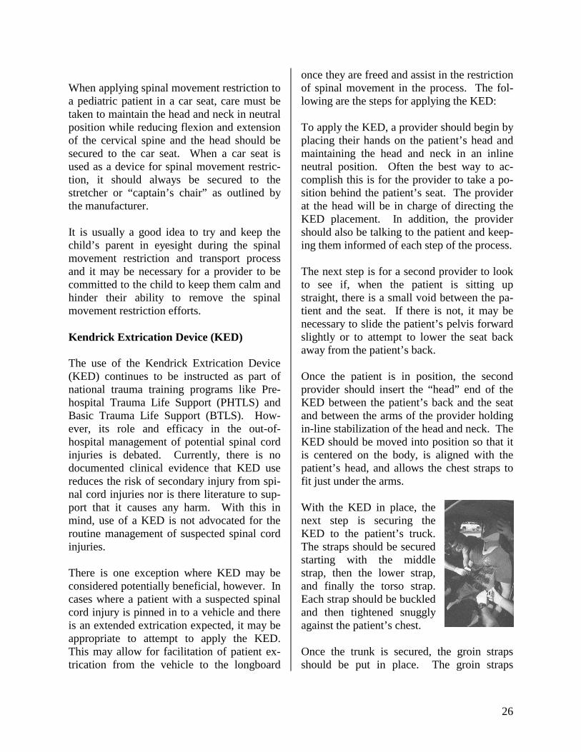
When applying spinal movement restriction to a pediatric patient in a car seat, care must be taken to maintain the head and neck in neutral position while reducing flexion and extension of the cervical spine and the head should be secured to the car seat. When a car seat is used as a device for spinal movement restric-tion, it should always be secured to the stretcher or “captain’s chair” as outlined by the manufacturer. It is usually a good idea to try and keep the child’s parent in eyesight during the spinal movement restriction and transport process and it may be necessary for a provider to be committed to the child to keep them calm and hinder their ability to remove the spinal movement restriction efforts. Kendrick Extrication Device (KED) The use of the Kendrick Extrication Device (KED) continues to be instructed as part of national trauma training programs like Pre-hospital Trauma Life Support (PHTLS) and Basic Trauma Life Support (BTLS). How-ever, its role and efficacy in the out-of-hospital management of potential spinal cord injuries is debated. Currently, there is no documented clinical evidence that KED use reduces the risk of secondary injury from spi-nal cord injuries nor is there literature to sup-port that it causes any harm. With this in mind, use of a KED is not advocated for the routine management of suspected spinal cord injuries. There is one exception where KED may be considered potentially beneficial, however. In cases where a patient with a suspected spinal cord injury is pinned in to a vehicle and there is an extended extrication expected, it may be appropriate to attempt to apply the KED. This may allow for facilitation of patient ex-trication from the vehicle to the longboard
once they are freed and assist in the restriction of spinal movement in the process. The fol-lowing are the steps for applying the KED: To apply the KED, a provider should begin by placing their hands on the patient’s head and maintaining the head and neck in an inline neutral position. Often the best way to ac-complish this is for the provider to take a po-sition behind the patient’s seat. The provider at the head will be in charge of directing the KED placement. In addition, the provider should also be talking to the patient and keep-ing them informed of each step of the process. The next step is for a second provider to look to see if, when the patient is sitting up straight, there is a small void between the pa-tient and the seat. If there is not, it may be necessary to slide the patient’s pelvis forward slightly or to attempt to lower the seat back away from the patient’s back. Once the patient is in position, the second provider should insert the “head” end of the KED between the patient’s back and the seat and between the arms of the provider holding in-line stabilization of the head and neck. The KED should be moved into position so that it is centered on the body, is aligned with the patient’s head, and allows the chest straps to fit just under the arms. With the KED in place, the next step is securing the KED to the patient’s truck. The straps should be secured starting with the middle strap, then the lower strap, and finally the torso strap. Each strap should be buckled and then tightened snuggly against the patient’s chest. Once the trunk is secured, the groin straps should be put in place. The groin straps

27
should be slid under the posterior side of the upper leg into the groin area and then wrapped over the anterior side until the strap can click into the buckle. This should be re-peated for the other leg. The final step in securing the KED is the head. Before the head is secured it is impor-tant to see if there is any void present between the back of the patient’s head and the KED device. If a void is present, it should be pad-ded. Securing the head to the KED is pretty easy. It is done in a similar fashion to that of a regu-lar backboard except it comes with ready-made Velcro straps that go over the chin and brow and fasten to the side head flaps of the KED. It is important to remember that the head is not completely secured until the pa-tient is fully secured to a longboard and that inline stabilization should be maintained until that time.vi Once the patient is secured in the KED, the next step is to extricate the patient from the vehicle onto a long board. This process will require the assistance of four to five (4-5) providers. The first step is to place a longboard on a col-lapsed stretcher and bring the foot end into the open door area. The tip of the foot of the longboard should be slid between the pa-tient’s buttocks and the car seat to allow for the ability to slide the patient out of the seat onto the board. The most common method is for the patient to be extricated headfirst. In order to accom-plish this the provider holding the head needs to coordinate moving the patient as a unit and transferring inline stabilization of the head to a provider outside the vehicle. Moving the patient as a unit, with one provider guiding the legs, another assisting the torso, and two
providers coordinating the head, rotate the patient onto the longboard and slide them down until their head is aligned with the top of the longboard. The groin straps maintain the patient’s legs in the seated position and will require a provider to support them. Now that the patient is cen-tered on the board, the buckles of the groin straps may be released and the patients legs me be guided down to a full supine position. Once the patient is on the longboard it will be necessary to slide the board completely onto the stretcher. Inline stabilization of the head should be maintained until the patient’s body has been fully strapped to the longboard. Straddle Slide The straddle slide is a method for moving a patient onto a longboard. It is not a com-monly used technique, but in some circum-stance it may be very effective. This is espe-cially the case when the patient is located in a tight space or in hallway that does not allow providers to get along the patient’s side. To do a straddle slide you need to have four (4) providers. As with any moving technique used on potential spinal cord injury patients, one provider should position themselves at the patient’s head and maintain inline stabili-zation of the head and neck. This provider should direct any movement of the patient. The second and third providers should stand over the patient, facing the head, and position themselves so that their feet are far enough apart to allow room for the backboard. One provider should be positioned at the shoulders and the other should be positioned at the pa-tient’s pelvis. The fourth provider should place himself or herself at the head or the foot of the patient and have the longboard ready to slide underneath the patient.

28
At the direction of the provider supporting the head, the providers straddling the patient should simultaneously raise the patient off of the ground just enough so that the board can be slid into place underneath the patient. Once the board is in position, the provider at the head will again direct the providers to lower the patient gently to the board. Inline stabilization of the head should be maintained until the patient’s body has been fully strapped to the longboard. Scoop Stretcher In some instances the scoop stretcher may be a useful adjunct to assist in lifting or moving a patient onto a longboard. As a general rule, the scoop stretcher should not be used as the sole spinal movement restriction device. Once the patient has been moved to a long-board the patient should be fully strapped to restrict spinal movement. Log Roll The log roll technique for moving a patient onto a longboard is probably the most com-monly used method and the one that most providers are intimately familiar with. Ideally, to effectively log roll a patient, the process requires four (4) providers. As in the straddle slide method, one provider is at the head maintaining inline stabilization and di-recting the process, while two providers are situated at the torso and pelvis and the fourth is responsible for sliding the long-board in place. The two providers at the torso and pelvis will be primarily responsible for rolling the pa-tient. To best accomplish this, they should kneel closely beside the patient on the side
they wish to roll them on, lean across the pa-tient, and place their hands on the patient’s shoulders, hips, and knees. To ensure that the patient is moved uniformly, the providers should crisscross their inside arms.vii At the direction of the provider stabilizing the head, the patient is rolled as a single unit on their side, toward the providers. With the pa-tient on their side, the fourth provider should insert the longboard at an angle in place be-hind the patient. At the direction of the pro-vider stabilizing the head, the patient is gently lowered down onto the longboard. Often, the patient is not centered on the board on the first try and it may necessitate minor movement of the patient to get them properly aligned. Once the patient is centered on the longboard, the patient should be fully strapped to restrict spinal movement. Inline stabilization of the head should be maintained until the patient’s body has been fully strapped to the longboard. Rapid Extrication A vast majority of motor vehicle collisions encountered by prehospital providers do not involve life-threatening patient conditions and will not require rapid intervention and trans-port. In some cases, however, time is of the essence and providers need to make a deter-mination as to whether the patient needs to be rapidly extracted from the vehicle to facilitate rapid transport to a Trauma Center. In these instances, providers will coordinate moving the patient directly from the vehicle onto the longboard in a rapid fashion. This process will require the assistance of four to five (4-5) providers. The first step is for a provider to place their hands on the pa-tient’s head and maintain the head and neck in an inline neutral position. Often the best way to accomplish this is for the provider to take

29
up position behind the patient’s seat. The provider at the head will be in charge of di-recting the rapid extrication. In addition, if the patient is conscious and alert, the provider should also be talking to the patient and keep-ing them informed of each step of the process. When everyone is ready to move the patient, place a longboard onto a collapsed stretcher and bring the foot end into the open door area. The tip of the foot of the longboard should be slid between the patient’s buttocks and the car seat to allow for the ability to slide the patient off of the seat and onto the board. The most common method is for the patient to be extricated headfirst. The provider holding the head needs to coordinate moving the pa-tient as a unit and transferring inline stabiliza-tion of the head to a provider outside the ve-hicle. Moving the patient as a unit, with one provider guiding the legs, another assisting the torso, and two providers coordinating the head, rotate the patient onto the longboard and slide them down until their head is aligned with the top of the longboard. Once the patient is on the longboard it will be necessary to slide the board completely onto the stretcher. Inline stabilization of the head should be maintained until the patient’s body has been fully strapped to the longboard. Full Spinal Movement Restriction The provider situated at the head should maintain holding inline stabilization until the patient’s body, and their head and neck are secured to the longboard. The patient’s head and neck should always be the last component of restricting spinal movement. There are many acceptable methods for strap-ping a patient to a longboard. The key objec-tive is to stabilize the shoulders and pelvis to the board to restrict movement of the spinal
column. This may be accomplished by either placing straps across the body at the shoulders and pelvis, crisscrossing straps across the torso, or a combination of the two. In addi-tion, a strap should be placed across the knees to secure the legs to the longboard. Once the body has been secured to the long-board, the head and neck should follow. To stabilize the head, place a head block on ei-ther side to effectively “sandwich” the head. Using medical tape, secure the patient’s head and the head blocks to the longboard by plac-ing one strip across the patient’s brow - being cautious of their eyebrows - and another across the patient’s chin. It is important to ensure that a patient’s airway remains unob-structed at all times. Once the body and head are secure, the pro-vider at the head may release manual inline stabilization. The final step is to check and ensure that the straps are not impeding respi-ration and that the patient is maintaining good distal circulation. If not, loosen the straps. Properly placed straps should be snug but it should still be possible for a provider to insert two (2) fingers between the strap and the pa-tient. Padding Voids In most patients whose spinal movement is restricted, the provider will notice that there are voids between the patient and the flat longboard. Common voids include beneath the patient’s head and neck at the lumbar re-gion of the back, between and behind the knees, and along the patient’s body. To effec-tively restrict the potential for spinal move-ment, and to ensure the patient is as comfort-able as possible, these voids should be padded when possible. Towels and blankets have proven effective at padding these voids.

30
Spinal Movement Restriction of Pregnant Patients As a woman progresses in her pregnancy (greater then twenty (20) weeks gestation) the increasing size and weight of her uterus be-comes a concern when backboarding. When a patient lies supine, it is possible for the weight of the gravid uterus to compress the inferior vena cava, lowering venous return to the heart. This can lead to supine-hypotensive syndrome or cause significant problems in a patient who is hemorrhaging. In addition, increased intra-abdominal pres-sure coupled with compression of the inferior vena cava increases venous pressure in the pelvis and lower extremities, engorging ves-sels and increasing the speed of hemorrhage in pelvic fractures and lower limb injuries.viii To reduce the potential of these issues occur-ring, any patient greater than twenty (20) weeks pregnant placed on a longboard for spinal movement restriction should be posi-tioned so that the patient and the board is tilted fifteen (15) degrees to the patient’s left to relieve pressure off of the gravis uterus, the inferior vena cava, and other abdominal struc-tures. Final Thought When managing a patient with any potential of spinal cord injury, remember the goal is to restrict spinal movement. If you encounter a situation where a patient is unable to be man-aged in the traditional manner, it is acceptable to modify those methods in an attempt to meet the objective of restricting spinal movement. If you deviate from standard, please ensure that the patient care record ac-curately depicts the thought process behind the modification and that spinal movement restriction was accomplished.
i Image: Bledsoe BE, et al, Paramedic Care: Principles & Practice Vol.4, Prentice Hall 2001, p. 353 ii Image: Ambu: Directions for use, Ambu Perfit ACE, Adjustable Collar for Extrcication iii Image: AAOS – Emergency Care and Transportation of the Sick and Injured, 8th Edition, pg. 702 iv Image: Bledsoe BE, et al, Paramedic Care: Principles & Practice Vol.4, Prentice Hall 2001, p. 351 v Image: AAOS – Emergency Care and Transportation of the Sick and Injured, 8th Edition, pg. 707 vi Image: PHTLS: Basic and Advanced Prehospital Trauma Life Support, 4th Edition, Pg.220 vii Image: Bledsoe BE, et al, Paramedic Care: Principles & Practice Vol.4, Prentice Hall 2001, p. 354 viii Bledsoe BE, et al, Paramedic Care: Principles & Practice Vol.4, Prentice Hall 2001, p. 432

31
Appendix I: Sensory Dermatomes NERVE ROOT MOTOR SENSORY
C-3, 4 shoulder shrug (trapezius) top of shoulder C-3, 4, 5 Diaphragm top of shoulders C-5, 6 elbow flexion (biceps) thumb C-7 elbow extension (triceps) middle finger
wrist/finger extension C-8/T-1 finger abduction/adduction little finger
T-4 nipple T-10 umbilicus
L-1, 2 hip flexion inguinal crease L-3, 4 quadriceps medial thigh/calf L-5 great toe/foot dorsiflexion lateral calf
S-1 knee flexion lateral foot S-1, 2 foot plantar flexion S-2, 3, 4 anal sphincter tone perianal
Nerve roots exit above their corresponding vertebra; an injury to the bony spinal column at C-6/C-7, therefore, would be expected to affect the C-7 nerve root.

32
Appendix II: Spinal Assessment Algorithm

Appendix III: Spinal Assessment Pocket Cards
Unreliable Patient Exam: The patientmust be calm, cooperative, sober, andalert to be reliable. Are you questioningthe reliability of the patient exam? If theanswer is yes, restrict spinal movement. Abnormal Sensory/Motor: Is thereweakness, sensory abnormality, or motorfunction reduction (new or old)? Is there areport of temporary symptoms followingthe incident that have since resolved? Ifthe answer is yes, restrict spinalmovement. Distracting Injury: Is there a clinicallyapparent painful injury that could distractthe patient from the pain of a spinalinjury? If the answer is yes, restrict spinalmovement. Spinal Pain/Tenderness: Is there director referred pain caused by palpation of thespine or the musculature supporting thespine? If the answer is yes, restrict spinalmovement.
Unreliable Patient Exam: The patientmust be calm, cooperative, sober, andalert to be reliable. Are you questioningthe reliability of the patient exam? If theanswer is yes, restrict spinal movement. Abnormal Sensory/Motor: Is thereweakness, sensory abnormality, or motorfunction reduction (new or old)? Is there areport of temporary symptoms followingthe incident that have since resolved? Ifthe answer is yes, restrict spinalmovement. Distracting Injury: Is there a clinicallyapparent painful injury that could distractthe patient from the pain of a spinalinjury? If the answer is yes, restrict spinalmovement. Spinal Pain/Tenderness: Is there director referred pain caused by palpation of thespine or the musculature supporting thespine? If the answer is yes, restrict spinalmovement.
33

AUSTIN-TRAVIS COUNTY EMS SYSTEM SPINAL MOVEMENT RESTRICTION DECISION ALGORITHM
SPINAL ASSESSMENT
Any traumatic event with the exception of isolated extremity trauma involvement will be assessed for spinal trauma.
Abnormal Sensory / Motor?
? Is there weakness,
sensory abnormality, or motor function reduction (new or old)?
? Is there a report of temporary symptoms following the incident that have since resolved?
If the answer is YES,
restrict spinal movement.
Unreliable Patient Exam?
? The patient must be
calm, cooperative, sober, and alert to be reliable.
? Are you questioning the reliability of the patient exam?
If the answer is YES, restrict spinal movement.
Distracting Injury?
? Is there a clinically
apparent painful injury that could distract the patient from the pain of a spinal injury?
If the answer is YES,
restrict spinal movement.
Spinal Pain / Tenderness?
? Is there direct or
referred pain caused by palpation of the spine or the musculature supporting the spine?
If the answer is YES,
restrict spinal movement.
If the answer is NO
to ALL of the above:
It is the expectation of the Austin-Travis County EMS System to offer transport to all
patients for further evaluation.
Austin-Travis County Emergency Medical Services 1999 Standards of Care Revision
Page 136a
Approved: 06/01/03 Edward M. Racht, MD - EMS Medical Director:
YES
YES YES
YES