Embed Size (px)
Citation preview
Korean J Pain 2013 January; Vol. 26, No. 1: 72-75pISSN 2005-9159 eISSN 2093-0569http://dx.doi.org/10.3344/kjp.2013.26.1.72
| Case Report |
Spinal Arteriovenous Malformation Masquerating Zoster Sine Herpete
Department of Anesthesiology and Pain Medicine, Ajou University Hospital, Suwon, *Korea University Ansan Hospital, Ansan, †Jeju National University Hospital, Jeju, Korea
Ji Young Lee, MD, Se Jin Ok, MD*, Chang Keun Oh, MD, Sun Kyung Park, MD†, Do Wan Kim, MD, and Jong Yeun Yang, MD
Zoster sine herpete (ZSH) is difficult to diagnosis during an acute period due to the absence of the characteristic zosteriform dermatomal rash; therefore, progression to postherpetic neuralgia is more common than typical zoster. In addition, misdiagnosis of other neuropathic pain as ZSH is common in clinical situations. Here, we report a case of spinal arteriovenous malformation that mimics ZSH. This is a rare condition; therefore, high clinical suspicion for a correct diagnosis and proper examination are not easy. However, early diagnosis and definitive treatment are essential to prevent neurologic deficit and mortality. (Korean J Pain 2013; 26: 72-75)
Key Words:
arteriovenous malformation, herpes zoster, postherpetic neuralgia, zoster sine herpete.
Received August 6, 2012. Revised October 19, 2012. Accepted November 8, 2012.Correspondence to: Jong Yeun Yang, MDDepartment of Anesthesiology and Pain Medicine, Ajou University Hospital, San 5, Woncheon-dong, Yeongtong-gu, Suwon 443-721, KoreaTel: +82-31-219-5575, Fax: +82-31-219-5579, E-mail: [email protected]
This is an open-access article distributed under the terms of the Creative Commons Attribution Non-Commercial License (http:// creativecommons.org/licenses/by-nc/3.0/), which permits unrestricted non-commercial use, distribution, and reproduction in any medium, provided the original work is properly cited.Copyright ⓒ The Korean Pain Society, 2013
Herpes zoster (HZ) is a clinical syndrome where topical
skin irritation, pain and paresthesia occur along the dis-
tribution of the dermatome, due to varicella zoster virus
entering through the sensory nerve of the skin and is dor-
mant in the dorsal root ganglia or the cerebral ganglia until
it is reactivated when the immune system of the body be-
comes weak. When pain persists for 1, 3, 4, or 6 months
or more after developing a rash, it is called postherpetic
neuralgia (PHN) [1].
There are rare cases of HZ which do not form vesicular
eruption on the skin. Weber [2] calls this zoster sine her-
pete (ZSH). Rather than early diagnosis, in most cases, it
is diagnosed as ZSH when the patient complains of chronic
continuing dermatomal neuropathic pain. Before diagnosis,
it is important to differentiate from other diseases that can
cause dermatomal neuropathic pain with an accurate
physical examination [3].
This case will examine spinal arteriovenous malforma-
tion (AVM), which was misdiagnosed as ZSH.
CASE REPORT
A 36-year-old man with an itching and tingling sen-
sation in the left L2 dermatome area for 3 months was
referred to our clinic for pain management. The patient did
not have significant past medical history, and before com-
Lee, et al / Spinal Arteriovenous Malformation Masquerating Zoster Sine Herpete 73
www.epain.org
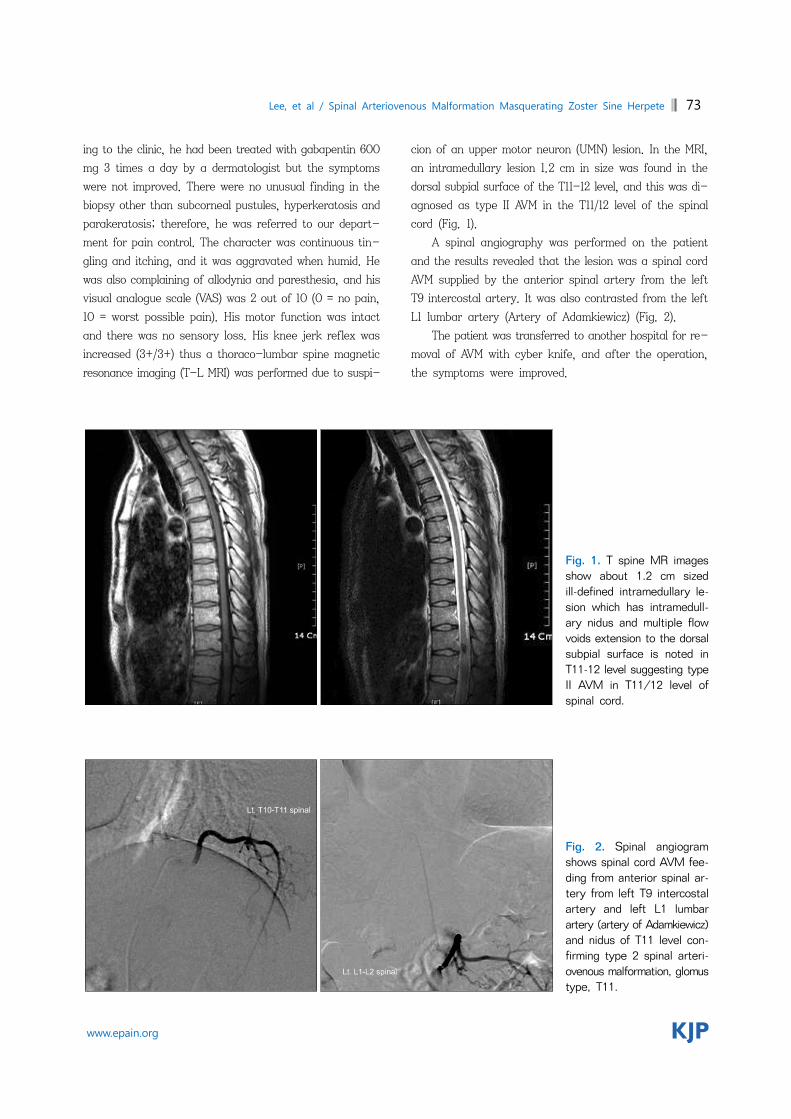
Fig. 1. T spine MR images show about 1.2 cm sized ill-defined intramedullary le-sion which has intramedull-ary nidus and multiple flow voids extension to the dorsal subpial surface is noted in T11-12 level suggesting type II AVM in T11/12 level of spinal cord.
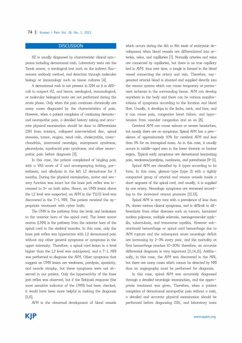
Fig. 2. Spinal angiogram shows spinal cord AVM fee-ding from anterior spinal ar-tery from left T9 intercostal artery and left L1 lumbar artery (artery of Adamkiewicz)and nidus of T11 level con-firming type 2 spinal arteri-ovenous malformation, glomustype, T11.
ing to the clinic, he had been treated with gabapentin 600
mg 3 times a day by a dermatologist but the symptoms
were not improved. There were no unusual finding in the
biopsy other than subcorneal pustules, hyperkeratosis and
parakeratosis; therefore, he was referred to our depart-
ment for pain control. The character was continuous tin-
gling and itching, and it was aggravated when humid. He
was also complaining of allodynia and paresthesia, and his
visual analogue scale (VAS) was 2 out of 10 (0 = no pain,
10 = worst possible pain). His motor function was intact
and there was no sensory loss. His knee jerk reflex was
increased (3+/3+) thus a thoraco-lumbar spine magnetic
resonance imaging (T-L MRI) was performed due to suspi-
cion of an upper motor neuron (UMN) lesion. In the MRI,
an intramedullary lesion 1.2 cm in size was found in the
dorsal subpial surface of the T11-12 level, and this was di-
agnosed as type II AVM in the T11/12 level of the spinal
cord (Fig. 1).
A spinal angiography was performed on the patient
and the results revealed that the lesion was a spinal cord
AVM supplied by the anterior spinal artery from the left
T9 intercostal artery. It was also contrasted from the left
L1 lumbar artery (Artery of Adamkiewicz) (Fig. 2).
The patient was transferred to another hospital for re-
moval of AVM with cyber knife, and after the operation,
the symptoms were improved.
74 Korean J Pain Vol. 26, No. 1, 2013
www.epain.org
DISCUSSION
HZ is usually diagnosed by characteristic clinical sym-
ptoms including dermatomal rash. Laboratory tests are the
Tzank smear, a serological test such as the indirect fluo-
rescent antibody method, and detection through molecular
biology or immunology such as tissue cultures [4].
A dermatomal rash is not present in ZSH so it is diffi-
cult to suspect HZ, and hence, serological, immunological,
or molecular biological tests are not performed during the
acute phase. Only when the pain continues chronically are
many cases diagnosed by the characteristics of pain.
However, when a patient complains of continuing dermato-
mal neuropathic pain, a detailed history taking and accu-
rate physical examination should be done to differentiate
ZSH from sciatica, collapsed intervertebral disc, spinal
stenosis, tumor, angina, renal colic, cholecystitis, costo-
chondritis, intercostal neuralgia, entrapment syndrome,
pleurodynia, myofascial pain syndrome, and other neuro-
pathic pain before diagnosis [3].
In this case, the patient complained of tingling pain
with a VAS score of 2 and accompanying itching, par-
esthesia, and allodynia in the left L2 dermatome for 3
months. During the physical examination, motor and sen-
sory function was intact but the knee jerk reflex was in-
creased to 3+ on both sides. Hence, an UMN lesion above
the L2 level was suspected, an AVM in the T11/12 level was
discovered in the T-L MRI. The patient received the ap-
propriate treatment with cyber knife.
The UMN is the pathway from the brain and brainstem
to the anterior horn of the spinal cord. The lower motor
neuron (LMN) is the pathway from the anterior horn of the
spinal cord to the skeletal muscles. In this case, only the
knee jerk reflex was hyperactive with L2 dermatomal pain
without any other general symptoms or symptoms in the
upper extremity. Therefore, a spinal cord lesion in a level
higher than the L2 level was anticipated, and a T-L MRI
was performed to diagnose the AVM. Other symptoms that
suggest an UMN lesion are weakness, paralysis, spasticity,
and muscle atrophy, but these symptoms were not ob-
served in our patient. Only the hyperactivity of the knee
jerk reflex was observed, but if the Babinski response (the
most sensitive indicator of the UMN) had been checked,
it would have been more helpful in making the diagnosis
[5,6].
AVM is the abnormal development of blood vessels
which occurs during the 4th to 8th week of embryonic de-
velopment when blood vessels are differentiated into ar-
teries, veins, and capillaries [7]. Normally arteries and veins
are connected by capillaries, but there is no true capillary
bed in AVM; thus over time, a tangle is formed in the blood
vessel connecting the artery and vein. Therefore, oxy-
genated arterial blood is shunted and supplied directly into
the venous system which can cause temporary or perma-
nent ischemia in the surrounding tissue. AVM can develop
anywhere in the body and there can be various manifes-
tations of symptoms according to the location and blood
flow. Usually, it develops in the limbs, neck, and face, and
it can cause pain, congestive heart failure, and hypo-
tension from vascular congestion and so on [8].
Cerebral AVM can cause seizure or severe headaches,
but mostly there are no symptoms. Spinal AVM has a prev-
alence of approximately 10% for cerebral AVM and less
than 5% for an intraspinal mass. As in this case, it usually
occurs in middle-aged men in the lower thoracic or lumbar
region. Typical early symptoms are dermatomal lancinating
pain, weakness/paralysis, numbness, and paresthesia [9-11].
Spinal AVM are classified by 4 types according to its
form. In this case, glomus-type (type 2) with a tightly
compacted group of arterial and venous vessels inside a
short segment of the spinal cord, and usually, it is supplied
by one artery. Neurologic symptoms are worsened accord-
ing to the increased venous pressure [12,13].
Spinal AVM is very rare with a prevalence of less than
1%, shows various clinical symptoms, and is difficult to dif-
ferentiate from other diseases such as tumors, herniated
nucleus pulposus, multiple sclerosis, meningovascular syph-
ilis, tuberculosis, and transverse myelitis. However sub-
arachnoid hemorrhage or spinal cord hemorrhage due to
AVM rupture and the subsequent acute neurologic deficit
are increasing by 2-3% every year, and the mortality at
first hemorrhage reaches 10-30%; therefore, an accurate
differential diagnosis is very important [11,14,15]. Additio-
nally, in this case, the AVM was discovered in the MRI,
but there are many cases which cannot be detected by MRI
thus an angiography must be performed for diagnosis.
In this case, spinal AVM was accurately diagnosed
through a detailed neurologic examination, and the appro-
priate treatment was given. Therefore, when a patient
complains of dermatomal neuropathic pain without a rash,
a detailed and accurate physical examination should be
performed before diagnosing ZSH, and laboratory tests
Lee, et al / Spinal Arteriovenous Malformation Masquerating Zoster Sine Herpete 75
www.epain.org
should be performed for the acute phase to differentiate
it from other diseases that cause neuropathic pain.
REFERENCES
1. Straus SE, Oxman MN, Schmader KE. Varicella and herpes zoster. In: Fitzpatrick's dermatology in general medicine. 7th ed. Edited by Wolff K, Goldsmith LA, Katz SI, Gilchrest BA, Paller AS, Leffell DJ. New York, McGraw-Hill. 2008, pp 1885-98.
2. Furuta Y, Ohtani F, Mesuda Y, Fukuda S, Inuyama Y. Early diagnosis of zoster sine herpete and antiviral therapy for the treatment of facial palsy. Neurology 2000; 55: 708-10.
3. Rajbala T, Joel LK, Robert HD. Herpes zoster and posther-petic neuralgia. In: Bonica’s management of pain. 4th ed. Edited by Fishman SM, Ballantyne JC, Rathmell JP. Philadelphia, Lippincott Williams & Wilkins. 2010, pp 338-57.
4. Cohen PR. Tests for detecting herpes simplex virus and varicella-zoster virus infections. Dermatol Clin 1994; 12: 51-68.
5. Murray B, Mitsumoto H. Disorders of upper and lower motor neurons. In: Bradley’s neurology in clinical practice. 6th ed. Edited by Daroff RB, Fenichel GB, Jankovic J, Mazziotta JC. Philadelphia, Elsevier. 2012, pp 1855-86.
6. Lewis SL. An approach to neurologic symptoms. In: Neuro-logy for the non-neurologist. 5th ed. Edited by Weiner WJ, Goetz CG. Philadelphia, Lippincott Williams & Wilkins. 2004, pp 21-33.
7. Fisher WS 3rd. Concomitant intracranial aneurysms and arteriovenous malformations. In: Neurosurgery. 2nd ed. Edited by Wilkins RH, Rengachary SS. New York, McGraw-Hill. 1996, pp 2429-31.
8. Neifeld JP, Doppman JL, Chreitien PB. Congenital pelvic arteriovenous fistulas: report of a case and review of the literature. J Urol 1975; 114: 648-52.
9. Gennuso R, Zappulla RA, Strenger SW. A localized lumbar spinal root arteriovenous malformation presenting with radi-cular signs and symptoms. Spine (Phila Pa 1976) 1989; 14: 543-6.
10. Elhammady MS, Wolfe SQ, Aziz-Sultan MA. Spinal arteri-ovenous malformations. In: Handbook of neuroendovascular surgery. 1st ed. Edited by Deshaies EM, Eddleman CS, Boulos AS. New York, Thieme Medical Publishers. 2012, pp 375-87.
11. Geldmacher DS. Vascular diseases of the nervous system: spinal cord vascular disease. In: Bradley’s neurology in clinical practice. 6th ed. Edited by Daroff RB, Fenichel GB, Jankovic J, Mazziotta JC. Philadelphia, Elsevier. 2012, pp 1095-102.
12. Riina HA, Soni D, Stieg PE. Surgical treatment of spinal arteriovenous malformations. In: Textbook of neurological surgery: principles and practices. Edited by Batjer HH, Loftus CM. Philadelphia, Lippincott Williams & Wilkins. 2003, pp 2569-78.
13. Heros RC, Debrun GM, Ojemann RG, Lasjaunias PL, Naessens PJ. Direct spinal arteriovenous fistula: a new type of spinal AVM. Case report. J Neurosurg 1986; 64: 134-9.
14. Caroscio JT, Brannan T, Budabin M, Huang YP, Yahr MD. Subarachnoid hemorrhage secondary to spinal arterio-venous malformation and aneurysm. Report of a case and review of the literature. Arch Neurol 1980; 37: 101-3.
15. Smith BS, Penka CF, Erickson LS, Matsuo F. Subarachnoid hemorrhage due to anterior spinal artery aneurysm. Neuro-surgery 1986; 18: 217-9.