Embed Size (px)
Citation preview
Spiders of the genus Loxosceles (Araneae, Sicariidae): a review of biological, medical and psychologicalaspects regarding envenomations
Richard S. Vetter: Department of Entomology, University of California, Riverside, California 92521; Biology Division,San Bernardino County Museum, Redlands, California 92373, USA. E-mail: [email protected]
Abstract. Loxosceles spiders are of concern outside of the arachnological world because their bites can cause occasionalnecrotic skin lesions and/or systemic complications; these manifestations are known as loxoscelism. Once these spidersbecame well associated as medical entities, much notoriety was attained through the publication of medical case historiesas well as tales of horrific wounds in the general literature. Although most Loxosceles spider bites are unremarkable,require only general supportive care, and often result in excellent outcome, they are an occasional source of severedermonecrotic injury with long healing times and significant scarring. In rare cases of systemic loxoscelism, seriousintravascular, nephrological and/or multi-organ damage can occur, sometimes resulting in death. However, also of concernis that loxoscelism is diagnosed by medical personnel or presumed by the general public in highly improbable scenariospreventing or delaying proper remedy, which can lead to deleterious outcome. Herein, Loxosceles spider biology andmedical aspects are reviewed. In particular, an extensive discussion of the distribution of the brown recluse spider, L.reclusa Gertsch & Mulaik 1940, is presented along with life history characteristics, which relate to the medical aspects of thegenus. Also presented are manifestations and epidemiology of loxoscelism, misdiagnoses of bites by the medicalcommunity, alternative diagnoses confused with recluse spider bites and a discussion of the psychological basis for theproliferation of the myth of loxoscelism by both the general public and the medical community. North and South Americanspecies are reviewed because this is where the genus predominates and is the region where the most pertinent research hasoriginated.
Keywords: Arachnida, brown recluse spider, dermonecrosis, distribution
There are very few spiders that are well known outside ofthe arachnological community. Almost all are large andconspicuous (tarantulas, orb weavers), medically important(black widows, Australian funnel web spiders) or medicallyimplicated (hobo spiders). The spiders of the genus Loxoscelesare ubiquitously infamous throughout the world because oftheir ability to occasionally cause significant skin necrosis alsoknown as cutaneous loxoscelism.
Loxosceles spiders were not documented in the literature asmedically important until the mid-20th century; previously,they were simply typical brown spiders that evoked littleconcern. In North America, once they were determined to be apublic health threat, there was great interest in defining thedistribution of the brown recluse spider, L. reclusa Gertsch &Mulaik 1940. This was followed by many reports of bites,verified and unverified, in both the medical and popularliterature. Unfortunately, there was a parallel accompanimentof misinformation regarding the spider’s distribution and itsculpability as the etiology of skin lesions. Many advances havebeen made in medical areas in determining the treatment forloxoscelism, epidemiology of envenomations and the physio-logical mechanism of dermonecrosis. However, despite theinfamy of the brown recluse spider, there was a surprisingpaucity of biological life history and distribution informationafter the initial efforts in the 1960s. In recent years, the genushas experienced more attention in biology and toxicologyissues, particularly much excellent work by South Americanresearchers with their native species.
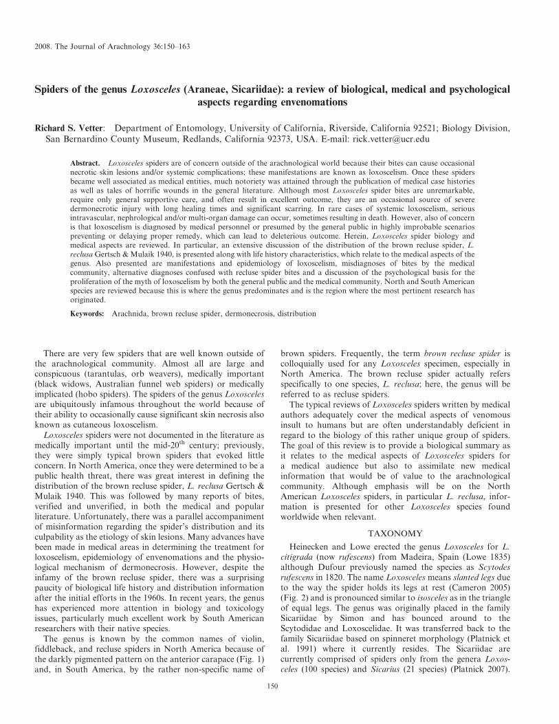
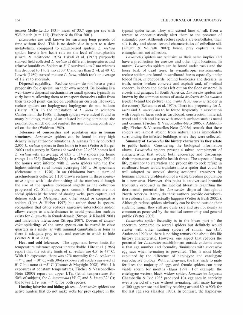
The genus is known by the common names of violin,fiddleback, and recluse spiders in North America because ofthe darkly pigmented pattern on the anterior carapace (Fig. 1)and, in South America, by the rather non-specific name of
brown spiders. Frequently, the term brown recluse spider iscolloquially used for any Loxosceles specimen, especially inNorth America. The brown recluse spider actually refersspecifically to one species, L. reclusa; here, the genus will bereferred to as recluse spiders.
The typical reviews of Loxosceles spiders written by medicalauthors adequately cover the medical aspects of venomousinsult to humans but are often understandably deficient inregard to the biology of this rather unique group of spiders.The goal of this review is to provide a biological summary asit relates to the medical aspects of Loxosceles spiders fora medical audience but also to assimilate new medicalinformation that would be of value to the arachnologicalcommunity. Although emphasis will be on the NorthAmerican Loxosceles spiders, in particular L. reclusa, infor-mation is presented for other Loxosceles species foundworldwide when relevant.
TAXONOMY
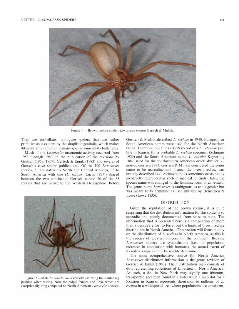
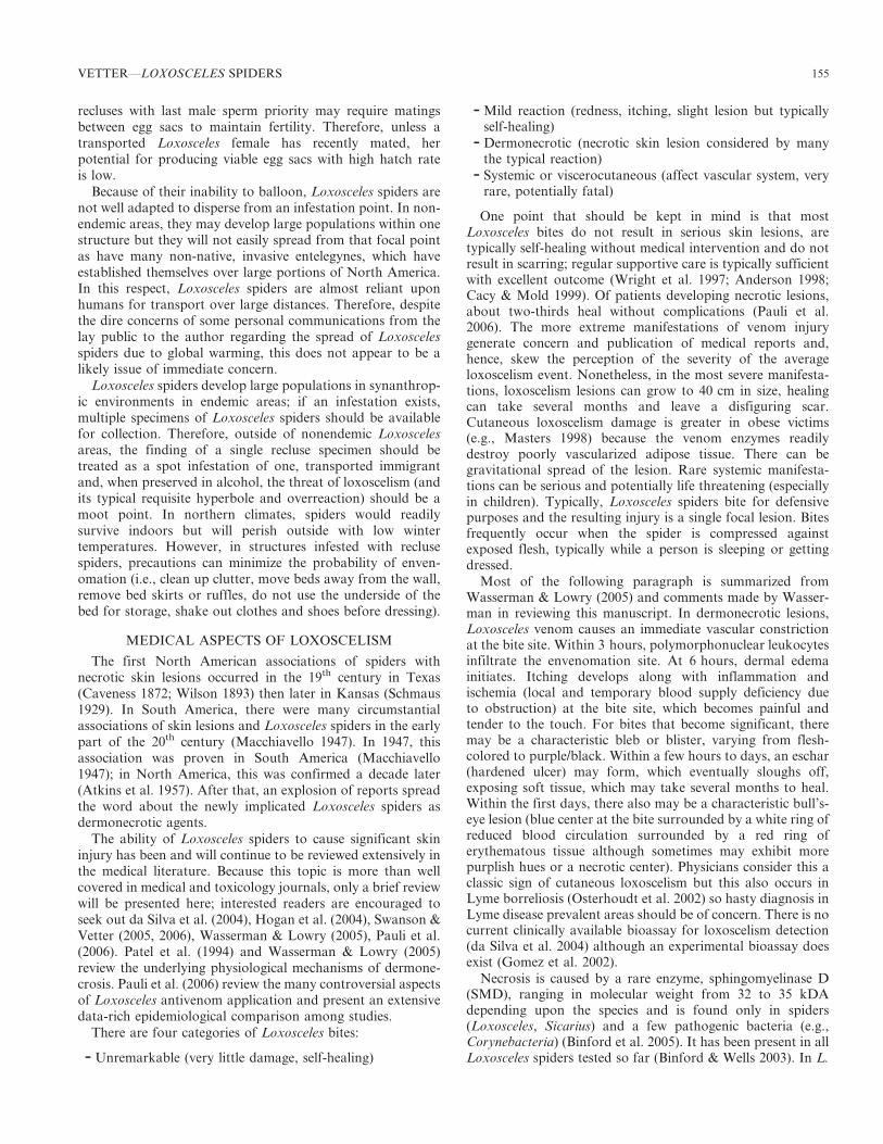
Heinecken and Lowe erected the genus Loxosceles for L.citigrada (now rufescens) from Madeira, Spain (Lowe 1835)although Dufour previously named the species as Scytodesrufescens in 1820. The name Loxosceles means slanted legs dueto the way the spider holds its legs at rest (Cameron 2005)(Fig. 2) and is pronounced similar to isosceles as in the triangleof equal legs. The genus was originally placed in the familySicariidae by Simon and has bounced around to theScytodidae and Loxoscelidae. It was transferred back to thefamily Sicariidae based on spinneret morphology (Platnick etal. 1991) where it currently resides. The Sicariidae arecurrently comprised of spiders only from the genera Loxos-celes (100 species) and Sicarius (21 species) (Platnick 2007).
2008. The Journal of Arachnology 36:150–163
150
They are ecribellate, haplogyne spiders that are ratherprimitive as is evident by the simplistic genitalia, which makesdifferentiation among the many species somewhat challenging.
Much of the Loxosceles taxonomic activity occurred from1958 through 1983, in the publication of the revisions byGertsch (1958, 1967), Gertsch & Ennik (1983) and several ofGertsch’s cave spider publications. Of the 100 Loxoscelesspecies, 51 are native to North and Central America, 33 toSouth America with one (L. rufipes [Lucas 1834]) sharedbetween the two continents. Gertsch named 70 of the 85species that are native to the Western Hemisphere. Before
Gertsch & Mulaik described L. reclusa in 1940, European orSouth American names were used for the North Americanfauna. Therefore, one finds a 1929 record of a L. refescens [sic]bite in Kansas for a probable L. reclusa specimen (Schmaus1929) and the South American name, L. unicolor Keyserling1887, used for the southwestern American desert dweller, L.deserta Gertsch 1973. Gertsch & Mulaik considered the genusname to be masculine and, hence, the brown recluse wasinitially described as L. reclusus (and is sometimes occasionallyincorrectly referenced as such in medical journals); later, thespecies name was changed to the feminine form of L. reclusa.The genus name Loxosceles is ambiguous as to its gender butwas meant to be feminine as used initially by Heinecken &Lowe (Lowe 1835).
DISTRIBUTION
Given the reputation of the brown recluse, it is quitesurprising that the distribution information for this spider is sosporadic and poorly documented from state to state. Theinformation that is presented here is a compilation of morethan a decade’s effort to ferret out the limits of brown reclusedistribution in North America. This section will focus mainlyon the distribution of L. reclusa in North America, as this isthe species of greatest concern on the continent. BecauseLoxosceles spiders are synanthropic (i.e., its populationincreases in association with humans), the actual extent ofits native range cannot be readily determined.
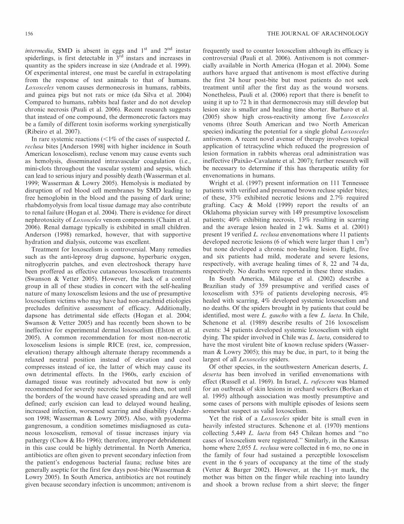
The most comprehensive source for North AmericaLoxosceles distribution information is the genus revision ofGertsch & Ennik (1983). Their distribution map consists ofdots representing collections of L. reclusa in North America.As such, a dot in New York may signify one itinerant,transported specimen found in a hotel while a map dot for alocation in Kansas represents thousands to millions of L.reclusa in a widespread area where populations are consistent,
Figure 1.—Brown recluse spider, Loxosceles reclusa Gertsch & Mulaik.
Figure 2.—Male Loxosceles laeta (Nicolet) showing the slanted legposition when resting. Note the palpal femora and tibia, which areexceptionally long compared to North American Loxosceles species.
VETTER—LOXOSCELES SPIDERS 151
reliably found and spiders plentiful. Unfortunately, this non-specificity has been misinterpreted by non-arachnologists whooverestimate Loxosceles distribution by considering thetransported itinerants to define the boundaries of Loxoscelesdistribution. In addition, there are some areas on the map(e.g., the Texas Panhandle) where few collections are known.This could represent a valid scarcity of the spiders or sparsehuman population with few potential collectors or merelyundersampling due to the spider’s perceived commonness orsome combination of the three factors. Nonetheless, if awareof obvious outliers, the map in Gertsch & Ennik (1983) is anaccurate presentation of L. reclusa presence in North America.An additional study, which offered to identify any arachnid inthe United States thought to be a recluse spider (Vetter 2005),corroborated the distribution as shown in Gertsch & Ennik(1983). However, both of these studies worked on the coarse-grained level of national distribution.
Information is presented below on a state-by-state basis forstates on the periphery of L. reclusa distribution wherepopulations diminish to non-existence. In the central area ofthe range (i.e., Arkansas, Missouri), it appears that entirestates are infested although no actual publications are knownto me that document the brown recluse spider in those statesprobably due to its ubiquity. The information for all the otherstates has been gathered from a wide and disparate number ofsources including species lists by county, unpublished statemaps, minor and arcane publications from state academies ofscience, agricultural experiment station bulletins, local andnon-reviewed museum pamphlets, all corroborated withpersonal communications with arachnologists, entomologists,public and environmental health officials, poison controlcenters, and other authorities who might have decades-longoral history information. This is obviously a very mixed bag ofresources; however, it is the best that could be assembled giventhe paucity of published information on such a well-knownarachnid.
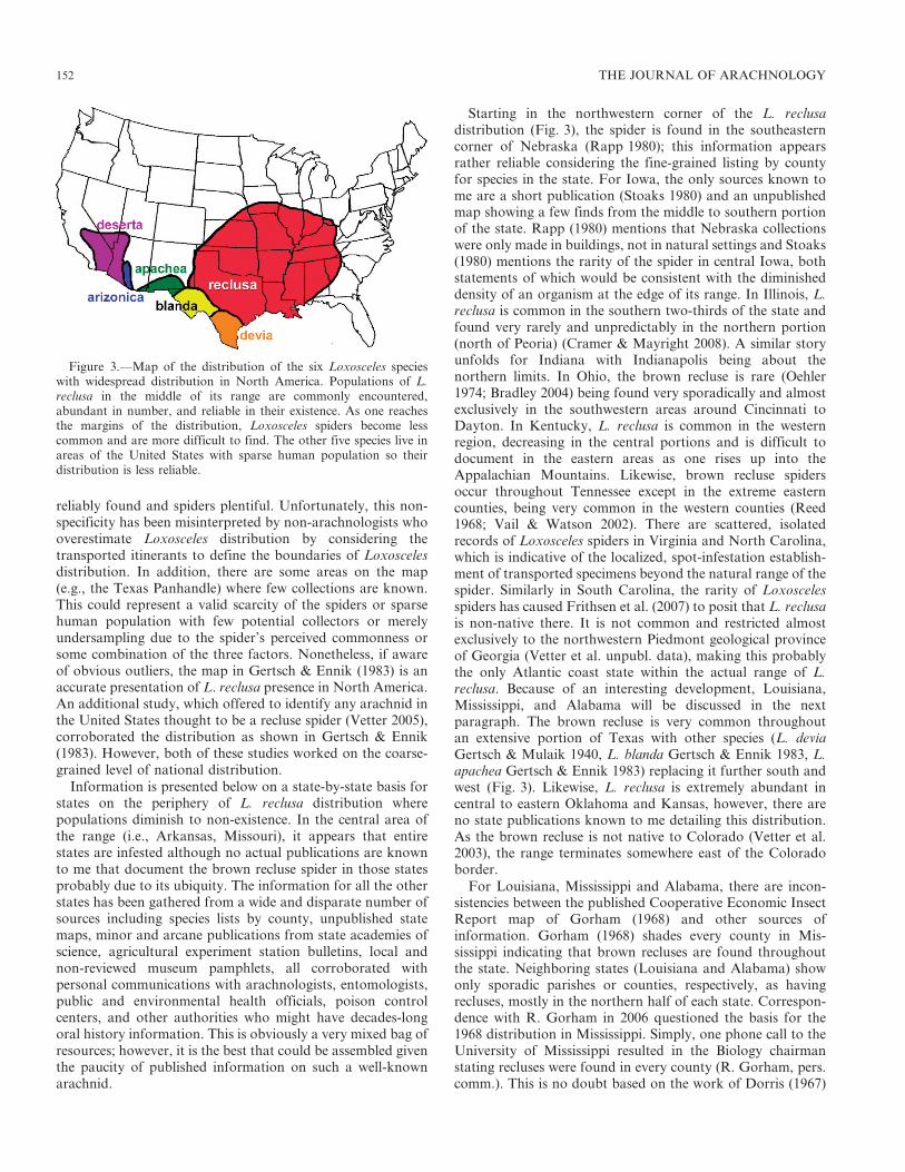
Starting in the northwestern corner of the L. reclusadistribution (Fig. 3), the spider is found in the southeasterncorner of Nebraska (Rapp 1980); this information appearsrather reliable considering the fine-grained listing by countyfor species in the state. For Iowa, the only sources known tome are a short publication (Stoaks 1980) and an unpublishedmap showing a few finds from the middle to southern portionof the state. Rapp (1980) mentions that Nebraska collectionswere only made in buildings, not in natural settings and Stoaks(1980) mentions the rarity of the spider in central Iowa, bothstatements of which would be consistent with the diminisheddensity of an organism at the edge of its range. In Illinois, L.reclusa is common in the southern two-thirds of the state andfound very rarely and unpredictably in the northern portion(north of Peoria) (Cramer & Mayright 2008). A similar storyunfolds for Indiana with Indianapolis being about thenorthern limits. In Ohio, the brown recluse is rare (Oehler1974; Bradley 2004) being found very sporadically and almostexclusively in the southwestern areas around Cincinnati toDayton. In Kentucky, L. reclusa is common in the westernregion, decreasing in the central portions and is difficult todocument in the eastern areas as one rises up into theAppalachian Mountains. Likewise, brown recluse spidersoccur throughout Tennessee except in the extreme easterncounties, being very common in the western counties (Reed1968; Vail & Watson 2002). There are scattered, isolatedrecords of Loxosceles spiders in Virginia and North Carolina,which is indicative of the localized, spot-infestation establish-ment of transported specimens beyond the natural range of thespider. Similarly in South Carolina, the rarity of Loxoscelesspiders has caused Frithsen et al. (2007) to posit that L. reclusais non-native there. It is not common and restricted almostexclusively to the northwestern Piedmont geological provinceof Georgia (Vetter et al. unpubl. data), making this probablythe only Atlantic coast state within the actual range of L.reclusa. Because of an interesting development, Louisiana,Mississippi, and Alabama will be discussed in the nextparagraph. The brown recluse is very common throughoutan extensive portion of Texas with other species (L. deviaGertsch & Mulaik 1940, L. blanda Gertsch & Ennik 1983, L.apachea Gertsch & Ennik 1983) replacing it further south andwest (Fig. 3). Likewise, L. reclusa is extremely abundant incentral to eastern Oklahoma and Kansas, however, there areno state publications known to me detailing this distribution.As the brown recluse is not native to Colorado (Vetter et al.2003), the range terminates somewhere east of the Coloradoborder.
For Louisiana, Mississippi and Alabama, there are incon-sistencies between the published Cooperative Economic InsectReport map of Gorham (1968) and other sources ofinformation. Gorham (1968) shades every county in Mis-sissippi indicating that brown recluses are found throughoutthe state. Neighboring states (Louisiana and Alabama) showonly sporadic parishes or counties, respectively, as havingrecluses, mostly in the northern half of each state. Correspon-dence with R. Gorham in 2006 questioned the basis for the1968 distribution in Mississippi. Simply, one phone call to theUniversity of Mississippi resulted in the Biology chairmanstating recluses were found in every county (R. Gorham, pers.comm.). This is no doubt based on the work of Dorris (1967)
Figure 3.—Map of the distribution of the six Loxosceles specieswith widespread distribution in North America. Populations of L.reclusa in the middle of its range are commonly encountered,abundant in number, and reliable in their existence. As one reachesthe margins of the distribution, Loxosceles spiders become lesscommon and are more difficult to find. The other five species live inareas of the United States with sparse human population so theirdistribution is less reliable.
152 THE JOURNAL OF ARACHNOLOGY
who makes this same statement although examination of herfield notes (copies provided by Pat Miller) and museumspecimens indicates a very incomplete picture. The map ofGorham (1968) then became the basis for the inclusion of theentire Gulf coast area in recent maps in Vetter (2000),Swanson & Vetter (2005) and many publications citing theseworks. Because of discrepancies, studies are currently under-way to systematically examine the distribution of the brownrecluse spider in Mississippi, Alabama and Louisiana.Preliminary data indicate an absence or dearth of L. reclusain the coastal region of the Gulf Coast states, similar toGeorgia. Corroborating this, a Texas entomologist communi-cated that in 25 years, he has had only one brown reclusesubmitted from the Houston area and to collect significantnumber of specimens one must travel about 150 km inland (J.Tucker, pers. comm.).
Of the other American Loxosceles species, only the fiveshown in Fig. 3 have significant widespread distributions.However, because these distributions are in the southwesterndesert where human population is sparse, these species couldhave greater range than currently known. Another aspect thatlimits our knowledge is a behavioral difference: because L.reclusa is a synanthropic spider, it is an urban pest, isabundant in homes and, therefore, is frequently collected bynon-arachnologists. In contrast, the southwestern Loxoscelesspecies appear to be much less adapted to human environ-ments and, in domestic situations, are only found in homesthat are surrounded by native vegetation. For example,although L. deserta is found around Phoenix, AZ and LasVegas, NV, it is not an urban pest in areas where officebuildings, hotels, casinos, and green lawns have arisen in thedesert environment. Because L. reclusa is a synanthrope, liveswhere human population density is comparatively greater andhas a larger distribution, it is involved in more encounters withhumans than other North American species.
Of the medically important Loxosceles species in SouthAmerica, L. laeta (Nicolet 1849) has the greatest distribution,being found in Brazil, Uruguay, Argentina, Chile, Peru, andEcuador (Gertsch 1967). Others include L. intermedia Mello-Leitao 1934 (Brazil, Argentina) and L. gaucho Gertsch 1967(southern Brazil) (Gertsch 1967). From South Africa, L.parrami Newlands 1981 was reported as medically important(Newlands et al. 1982).
The Mediterranean recluse, L. rufescens (Dufour 1820), is aworldwide tramp, originating from the circum-Mediterraneanregion. It has been collected in many localities in the UnitedStates (e.g., Boston, MA; New York City, NY; Philadelphia,PA; Harrisburg, PA; Reading, PA; Washington DC; AnnArbor, MI; Indianapolis, IN; Knoxville, TN; Jacksonville,FL; Baton Rouge, LA; several localities in Ohio and Georgia;Las Animas, CO; Los Angeles and Fresno, CA; Spokane, WA[Gertsch & Ennik 1983; Vetter unpubl. data]). In nonendemicLoxosceles areas in North America, it is more likely to find aspot infestation of the non-native L. rufescens than the nativeL. reclusa. The Mediterranean recluse has also becomeestablished in Australia (Southcott 1976). Gertsch (1967)states that there are no valid specimens of L. rufescens fromSouth America. While others have described this species ascosmopolitan, Gertsch (1967) states that this is a misnomer.Although L. rufescens exists in many localities, in non-endemic
areas it is typically found only indoors and in highlycircumscribed distribution, heavily infesting one building orseveral if interconnected by conduits.
LIFE HISTORY AND BIOLOGY RELEVANT TOMEDICAL ISSUES
After Loxosceles spiders became a medical entity, they werethe subjects of biological and medical articles as researchersrushed to provide information on this new public healththreat. Below is a review of the biological traits as they relateto the features that do or do not show a potential as a publichealth concern.
Longevity, fecundity and resistance to starvation.—Loxosce-les spiders have long life spans compared to many seasonalentelegynes, which pass through a life cycle in , 1 yr. Hite etal. (1966) provide a longevity for L. reclusa of 1.5 yr for malesand 1.7 yr for females with a maximum of 2.5 yr for onefemale when animals were maintained in the lab. Theymention that life spans would probably have been longerhad they been subjected to winter temperatures. Indeed,Horner & Stewart (1967) maintained their animals in winterrefuges to provide a more natural scenario; their spiderssurvived over 5 seasons (spiders were still alive at the time ofpublication). Elzinga (1977) reports average life spans for L.reclusa males (897 da) and females (794 da) with 25% of thefemales living over 1,000 da, including one surviving 4.8 yr.Lowrie (1980) reared L. laeta under sporadic feedingconditions (initially weekly, then once every 3 to 10 mo, thenstarved to death); these spiders took an average of 2.1 yr tomature and lived another 4.8 yr as adults. Similarly, Fischer &Vasconcellos-Neto (2005a) report longevities of 1176 6
478 da for L. intermedia females and 557 6 87 da for males.However, these quantities are for captive animals confined tovials, not exposed to detrimental environmental factors, and,hence, might grossly overestimate the life span in natural orsynanthropic settings.
Compared to many other common spiders, which producehundreds to thousands of eggs per egg sac or over a lifetime,Loxosceles spiders have a more modest fecundity. Female L.reclusa average 50 eggs per egg sac (range 0 to 91, n 5 146),and 2.7 egg sacs per female with a 48% hatch rate (n 5 55)(Hite et al. 1966). For laboratory-reared L. intermediarestricted to one mating, egg sacs contained approximately30 eggs where 70% hatched, however, the egg sacs of field-collected females of unknown mating history averaged around50 eggs with 80% hatch (Fischer & Vasconcellos-Neto 2005b).When kept without access to additional matings, female L.reclusa (Horner & Stewart 1967) and lab-reared, singularly-mated L. intermedia females (Fischer & Vasconcellos-Neto2005b) experience a decrease in fecundity per sac and/or eggviability with successive egg sacs throughout a season. For L.reclusa from figure 5 of Horner & Stewart (1967), from the 1st
to 3rd egg sac, there is a drop in egg number per sac fromabout 27 to 18 and decrease of hatch rate from 66% to 37%.Field-collected L. intermedia females of unknown matinghistory did not show this decline (Fischer & Vasconcellos-Neto 2005b). Similar fecundity numbers are presented forother species: L. laeta – mean of 88.4 eggs per sac (range 22 to138, n 5 81) (Galiano 1967), L. gaucho – mean of 61.3 eggs persac (range 25 to 117, n 5 78) (Rinaldi et al. 1997) and L.
VETTER—LOXOSCELES SPIDERS 153
hirsuta Mello-Leitao 1931– mean of 33.7 eggs per sac with93% hatch (n 5 113) (Fischer & da Silva 2001).
Loxosceles are well known for surviving long periods oftime without food. This is no doubt due in part to a slowmetabolism; compared to similar-sized spiders, L. reclusaspiders have a low heart rate on the level of theraphosids(Carrel & Heathcote 1976). Eskafi et al. (1977) purposelystarved field-collected L. reclusa at different temperatures andrelative humidities. Spiders at 5u C survived 4 to 7 mo whereasthis dropped to 1 to 2 mo at 30u C and less than 2 wk at 40u C.Lowrie (1980) starved mature L. laeta, which took an averageof 1.2 yr to succumb.
Dispersal capability.—Recluse spiders do not have a greatpropensity for dispersal on their own accord. Ballooning is awell-known dispersal mechanism for small spiders, typically asearly instars, allowing them to transport themselves miles fromtheir take-off point, carried on uplifting air currents. However,recluse spiders are haplogynes; haplogynes do not balloon(Beatty 1970). In the infestation of L. laeta in southernCalifornia in the 1960s, although spiders were indeed found inmany buildings, razing of an infested building eliminated thepopulation, which did not reinfest the new building construct-ed on the site (Waldron 1969).
Tolerance of conspecifics and population size in humanstructures.—Loxosceles spiders can be found in very highdensity in synanthropic situations. A Kansas family collected2,055 L. reclusa spiders in their home in 6 mo (Vetter & Barger2002) and a survey in Kansas showed that 22 of 25 homes hadL. reclusa with an average of 83.5 6 114.9 spiders per home(range 1 to 526) (Sandidge 2004). In a Chilean survey, 29% ofthe homes were infested with L. laeta spiders with the fivehighest-infested rural homes averaging 163 6 56 specimens(Schenone et al. 1970). In an Oklahoma barn, a team ofarachnologists collected 1,150 brown recluses in three consec-utive nights with little diminishing of the numbers althoughthe size of the spiders decreased slightly as the collectionprogressed (C. Shillington, pers. comm.). Recluses are notsocial spiders in the sense of sharing webs, prey capture anddefense such as Metepeira and other social or cooperativespiders (Uetz & Hieber 1997) but rather there is species-recognition that either reduces aggressive interactions and/orallows escape to a safe distance to avoid predation such asexists for L. gaucho in female-female (Stropa & Rinaldi 2001)and male-male interactions (Stropa 2007). Dozens of Loxos-celes spiderlings of the same species can be reared in closequarters in a single jar with minimal cannibalism as long asthere is adequate prey to eat and crevices in which to hide(Vetter & Rust 2008).
Heat and cold tolerance.—The upper and lower limits fortemperature tolerance appear unremarkable. Hite et al. (1966)report that the activity limits of L. reclusa are 4.5u to 43u C.With 4-h exposures, there was 47% mortality for L. reclusa at27u C and 210u C; with 30-da exposure all spiders survived at0u C but none at 25u C (Cramer & Mayright 2008). With 1-hexposures at constant temperatures, Fischer & Vasconcellos-Neto (2003) report an upper LT50 (lethal temperatures for50% of subjects) for L. intermedia (35u C) and L. laeta (32u C);the lower LT50 was 27u C for both species.
Hunting behavior and hiding places.—Loxosceles spiders areactive hunters that do not make webs for prey capture in the
typical spider sense. They will extend lines of silk from aretreat to opportunistically alert them to the presence ofentangled prey. Although recluse spiders are ecribellates, theirsilk is dry and shares several characteristics of cribellate silk(Knight & Vollrath 2002); hence, prey capture is viaentanglement not adhesion.
Loxosceles spiders are reclusive as their name implies andhave a predilection for crevices and other tight locations. Innature, Loxosceles spiders can be found under rocks and theloose bark of dead trees. In synanthropic environments,recluse spiders are found in cardboard boxes especially underfolded flaps, in cupboards, behind bookcases and dressers, intrash, under broken concrete and asphalt and, of medicalconcern, in shoes and clothes left out on the floor or stored inclosets and garages. In South America, Loxosceles spiders areknown by the common names of arana de detras de los cuadros(spider behind the picture) and arana de los rincones (spider inthe corner) (Schenone et al. 1970). There is a propensity for L.laeta and L. intermedia to be found frequently in associationwith rough surfaces such as cardboard, construction material,wood and cloth and less so with smooth surfaces such as metaland ceramic (Fischer & Vasconcellos-Neto 2005c). Addition-ally, Fischer & Vasconcellos-Neto (2005c) remark that thesespiders are almost absent from natural areas immediatelysurrounding the infested buildings where they were collected.
Summary of Loxosceles life history characters as they relateto public health.—Considering the biological informationabove, Loxosceles spiders present a mixed complement ofcharacteristics that would both encourage and discouragetheir importance as a public health threat. The aspects of longlife, resistance to starvation and propensity to seek refuge incardboard boxes would translate into a spider that could bewell adapted to survival during accidental transport byhumans allowing proliferation of a viable breeding populationin a new area. However, this point is an overused bromidefrequently espoused in the medical literature regarding thedetrimental potential for Loxosceles dispersal throughoutNorth America. Rarely do these authors provide corrobora-tive evidence that this actually happens (Vetter & Bush 2002a).Although recluse spiders obviously can be found outside theirendemic range, they still are quite rare and are not nearly ascommon as perceived by the medical community and generalpublic (Vetter 2005).
Loxosceles spider fecundity is in the lower part of thespectrum compared to several spiders but definitely wouldcluster with other hunting spiders of similar size (J.F.Anderson 1990) so there is nothing remarkable about this lifehistory characteristic. However, one aspect that reduces thepotential for Loxosceles establishment outside endemic areasis that egg number and fecundity diminishes with successiveegg sacs when re-mating is prevented. This is most likelyexplained by the difference of haplogyne and entelegynereproductive biology. With entelegynes, the first male to matefertilizes the majority of eggs and female spiders can storeviable sperm for months (Elgar 1998). For example, theentelegyne western black widow spider, Latrodectus hesperusChamberlin & Ivie 1935 produced 10+ egg sacs in captivityover a period of a year without re-mating, with many having. 300 eggs per sac and fertility reaching around 80 to 90% forthe last egg sacs (Kaston 1970). In contrast, the haplogyne
154 THE JOURNAL OF ARACHNOLOGY
recluses with last male sperm priority may require matingsbetween egg sacs to maintain fertility. Therefore, unless atransported Loxosceles female has recently mated, herpotential for producing viable egg sacs with high hatch rateis low.
Because of their inability to balloon, Loxosceles spiders arenot well adapted to disperse from an infestation point. In non-endemic areas, they may develop large populations within onestructure but they will not easily spread from that focal pointas have many non-native, invasive entelegynes, which haveestablished themselves over large portions of North America.In this respect, Loxosceles spiders are almost reliant uponhumans for transport over large distances. Therefore, despitethe dire concerns of some personal communications from thelay public to the author regarding the spread of Loxoscelesspiders due to global warming, this does not appear to be alikely issue of immediate concern.
Loxosceles spiders develop large populations in synanthrop-ic environments in endemic areas; if an infestation exists,multiple specimens of Loxosceles spiders should be availablefor collection. Therefore, outside of nonendemic Loxoscelesareas, the finding of a single recluse specimen should betreated as a spot infestation of one, transported immigrantand, when preserved in alcohol, the threat of loxoscelism (andits typical requisite hyperbole and overreaction) should be amoot point. In northern climates, spiders would readilysurvive indoors but will perish outside with low wintertemperatures. However, in structures infested with reclusespiders, precautions can minimize the probability of enven-omation (i.e., clean up clutter, move beds away from the wall,remove bed skirts or ruffles, do not use the underside of thebed for storage, shake out clothes and shoes before dressing).
MEDICAL ASPECTS OF LOXOSCELISM
The first North American associations of spiders withnecrotic skin lesions occurred in the 19th century in Texas(Caveness 1872; Wilson 1893) then later in Kansas (Schmaus1929). In South America, there were many circumstantialassociations of skin lesions and Loxosceles spiders in the earlypart of the 20th century (Macchiavello 1947). In 1947, thisassociation was proven in South America (Macchiavello1947); in North America, this was confirmed a decade later(Atkins et al. 1957). After that, an explosion of reports spreadthe word about the newly implicated Loxosceles spiders asdermonecrotic agents.
The ability of Loxosceles spiders to cause significant skininjury has been and will continue to be reviewed extensively inthe medical literature. Because this topic is more than wellcovered in medical and toxicology journals, only a brief reviewwill be presented here; interested readers are encouraged toseek out da Silva et al. (2004), Hogan et al. (2004), Swanson &Vetter (2005, 2006), Wasserman & Lowry (2005), Pauli et al.(2006). Patel et al. (1994) and Wasserman & Lowry (2005)review the underlying physiological mechanisms of dermone-crosis. Pauli et al. (2006) review the many controversial aspectsof Loxosceles antivenom application and present an extensivedata-rich epidemiological comparison among studies.
There are four categories of Loxosceles bites:
- Unremarkable (very little damage, self-healing)
- Mild reaction (redness, itching, slight lesion but typicallyself-healing)
- Dermonecrotic (necrotic skin lesion considered by manythe typical reaction)
- Systemic or viscerocutaneous (affect vascular system, veryrare, potentially fatal)
One point that should be kept in mind is that mostLoxosceles bites do not result in serious skin lesions, aretypically self-healing without medical intervention and do notresult in scarring; regular supportive care is typically sufficientwith excellent outcome (Wright et al. 1997; Anderson 1998;Cacy & Mold 1999). Of patients developing necrotic lesions,about two-thirds heal without complications (Pauli et al.2006). The more extreme manifestations of venom injurygenerate concern and publication of medical reports and,hence, skew the perception of the severity of the averageloxoscelism event. Nonetheless, in the most severe manifesta-tions, loxoscelism lesions can grow to 40 cm in size, healingcan take several months and leave a disfiguring scar.Cutaneous loxoscelism damage is greater in obese victims(e.g., Masters 1998) because the venom enzymes readilydestroy poorly vascularized adipose tissue. There can begravitational spread of the lesion. Rare systemic manifesta-tions can be serious and potentially life threatening (especiallyin children). Typically, Loxosceles spiders bite for defensivepurposes and the resulting injury is a single focal lesion. Bitesfrequently occur when the spider is compressed againstexposed flesh, typically while a person is sleeping or gettingdressed.
Most of the following paragraph is summarized fromWasserman & Lowry (2005) and comments made by Wasser-man in reviewing this manuscript. In dermonecrotic lesions,Loxosceles venom causes an immediate vascular constrictionat the bite site. Within 3 hours, polymorphonuclear leukocytesinfiltrate the envenomation site. At 6 hours, dermal edemainitiates. Itching develops along with inflammation andischemia (local and temporary blood supply deficiency dueto obstruction) at the bite site, which becomes painful andtender to the touch. For bites that become significant, theremay be a characteristic bleb or blister, varying from flesh-colored to purple/black. Within a few hours to days, an eschar(hardened ulcer) may form, which eventually sloughs off,exposing soft tissue, which may take several months to heal.Within the first days, there also may be a characteristic bull’s-eye lesion (blue center at the bite surrounded by a white ring ofreduced blood circulation surrounded by a red ring oferythematous tissue although sometimes may exhibit morepurplish hues or a necrotic center). Physicians consider this aclassic sign of cutaneous loxoscelism but this also occurs inLyme borreliosis (Osterhoudt et al. 2002) so hasty diagnosis inLyme disease prevalent areas should be of concern. There is nocurrent clinically available bioassay for loxoscelism detection(da Silva et al. 2004) although an experimental bioassay doesexist (Gomez et al. 2002).
Necrosis is caused by a rare enzyme, sphingomyelinase D(SMD), ranging in molecular weight from 32 to 35 kDAdepending upon the species and is found only in spiders(Loxosceles, Sicarius) and a few pathogenic bacteria (e.g.,Corynebacteria) (Binford et al. 2005). It has been present in allLoxosceles spiders tested so far (Binford & Wells 2003). In L.
VETTER—LOXOSCELES SPIDERS 155
intermedia, SMD is absent in eggs and 1st and 2nd instarspiderlings, is first detectable in 3rd instars and increases inquantity as the spiders increase in size (Andrade et al. 1999).Of experimental interest, one must be careful in extrapolatingfrom the response of test animals to that of humans.Loxosceles venom causes dermonecrosis in humans, rabbits,and guinea pigs but not rats or mice (da Silva et al. 2004)Compared to humans, rabbits heal faster and do not developchronic necrosis (Pauli et al. 2006). Recent research suggeststhat instead of one compound, the dermonecrotic factors maybe a family of different toxin isoforms working synergistically(Ribeiro et al. 2007).
In rare systemic reactions (,1% of the cases of suspected L.reclusa bites [Anderson 1998] with higher incidence in SouthAmerican loxoscelism), recluse venom may cause events suchas hemolysis, disseminated intravascular coagulation (i.e.,mini-clots throughout the vascular system) and sepsis, whichcan lead to serious injury and possibly death (Wasserman et al.1999; Wasserman & Lowry 2005). Hemolysis is mediated bydisruption of red blood cell membranes by SMD leading tofree hemoglobin in the blood and the passing of dark urine;rhabdomyolysis from local tissue damage may also contributeto renal failure (Hogan et al. 2004). There is evidence for directnephrotoxicity of Loxosceles venom components (Chaim et al.2006). Renal damage typically is exhibited in small children.Anderson (1998) remarked, however, that with supportivehydration and dialysis, outcome was excellent.
Treatment for loxoscelism is controversial. Many remediessuch as the anti-leprosy drug dapsone, hyperbaric oxygen,nitroglycerin patches, and even electroshock therapy havebeen proffered as effective cutaneous loxoscelism treatments(Swanson & Vetter 2005). However, the lack of a controlgroup in all of these studies in concert with the self-healingnature of many loxoscelism lesions and the use of presumptiveloxoscelism victims who may have had non-arachnid etiologiesprecludes definitive assessment of efficacy. Additionally,dapsone has detrimental side effects (Hogan et al. 2004;Swanson & Vetter 2005) and has recently been shown to beineffective for experimental dermal loxoscelism (Elston et al.2005). A common recommendation for most non-necroticloxoscelism lesions is simple RICE (rest, ice, compression,elevation) therapy although alternate therapy recommends arelaxed neutral position instead of elevation and coolcompresses instead of ice, the latter of which may cause itsown detrimental effects. In the 1960s, early excision ofdamaged tissue was routinely advocated but now is onlyrecommended for severely necrotic lesions and then, not untilthe borders of the wound have ceased spreading and are welldefined; early excision can lead to delayed wound healing,increased infection, worsened scarring and disability (Ander-son 1998; Wasserman & Lowry 2005). Also, with pyodermagangrenosum, a condition sometimes misdiagnosed as cuta-neous loxoscelism, removal of tissue increases injury viapathergy (Chow & Ho 1996); therefore, improper debridementin this case could be highly detrimental. In North America,antibiotics are often given to prevent secondary infection fromthe patient’s endogenous bacterial fauna; recluse bites aregenerally aseptic for the first few days post-bite (Wasserman &Lowry 2005). In South America, antibiotics are not routinelygiven because secondary infection is uncommon; antivenom is
frequently used to counter loxoscelism although its efficacy iscontroversial (Pauli et al. 2006). Antivenom is not commer-cially available in North America (Hogan et al. 2004). Someauthors have argued that antivenom is most effective duringthe first 24 hour post-bite but most patients do not seektreatment until after the first day as the wound worsens.Nonetheless, Pauli et al. (2006) report that there is benefit tousing it up to 72 h in that dermonecrosis may still develop butlesion size is smaller and healing time shorter. Barbaro et al.(2005) show high cross-reactivity among five Loxoscelesvenoms (three South American and two North Americanspecies) indicating the potential for a single global Loxoscelesantivenom. A recent novel avenue of therapy involves topicalapplication of tetracycline which reduced the progression oflesion formation in rabbits whereas oral administration wasineffective (Paixao-Cavalante et al. 2007); further research willbe necessary to determine if this has therapeutic utility forenvenomations in humans.
Wright et al. (1997) present information on 111 Tennesseepatients with verified and presumed brown recluse spider bites;of these, 37% exhibited necrotic lesions and 2.7% requiredgrafting. Cacy & Mold (1999) report the results of anOklahoma physician survey with 149 presumptive loxoscelismpatients; 40% exhibiting necrosis, 13% resulting in scarringand the average lesion healed in 2 wk. Sams et al. (2001)present 19 verified L. reclusa envenomations where 11 patientsdeveloped necrotic lesions (6 of which were larger than 1 cm2)but none developed a chronic non-healing lesion. Eight, fiveand six patients had mild, moderate and severe lesions,respectively, with average healing times of 8, 22 and 74 da,respectively. No deaths were reported in these three studies.
In South America, Malaque et al. (2002) describe aBrazilian study of 359 presumptive and verified cases ofloxoscelism with 53% of patients developing necrosis, 4%
healed with scarring, 4% developed systemic loxoscelism andno deaths. Of the spiders brought in by patients that could beidentified, most were L. gaucho with a few L. laeta. In Chile,Schenone et al. (1989) describe results of 216 loxoscelismevents: 34 patients developed systemic loxoscelism with eightdying. The spider involved in Chile was L. laeta, considered tohave the most virulent bite of known recluse spiders (Wasser-man & Lowry 2005); this may be due, in part, to it being thelargest of all Loxosceles spiders.
Of other species, in the southwestern American deserts, L.deserta has been involved in verified envenomations witheffect (Russell et al. 1969). In Israel, L. rufescens was blamedfor an outbreak of skin lesions in orchard workers (Borkan etal. 1995) although association was mostly presumptive andsome cases of persons with multiple episodes of lesions seemsomewhat suspect as valid loxoscelism.
Yet the risk of a Loxosceles spider bite is small even inheavily infested structures. Schenone et al. (1970) mentionscollecting 5,449 L. laeta from 645 Chilean homes and ‘‘nocases of loxoscelism were registered.’’ Similarly, in the Kansashome where 2,055 L. reclusa were collected in 6 mo, no one inthe family of four had sustained a perceptible loxoscelismevent in the 6 years of occupancy at the time of the study(Vetter & Barger 2002). However, at the 11-yr mark, themother was bitten on the finger while reaching into laundryand shook a brown recluse from a shirt sleeve; the finger
156 THE JOURNAL OF ARACHNOLOGY
turned red and swelled slightly but healed without incident (D.Barger, pers. comm.).
OVERDIAGNOSIS OF SPIDER BITES
In North America, once the brown recluse spider becameknown as a spider of medical importance, the medical aspectswere vigorously researched and reported. In the 1960s, casehistories appeared in medical journals and new county andstate records were documented in the USDA’s weeklyCooperative Economic Insect Report as the brown reclusespider became well known outside of the arachnologicalcommunity. Reports of brown recluse spider bites werecommon in the local media and in national magazines. Asmuch as Loxosceles spiders are a legitimate public healththreat, of equal concern is the overdiagnosis of loxoscelism asa common etiology for skin lesions.
Over the decades, the diagnoses of cutaneous loxoscelismbecame commonplace in the North American medical commu-nity. Although the majority of the reports emanated fromendemic Loxosceles regions such as Tennessee and Oklahoma(Wright et al. 1997; Cacy & Mold 1999), additional reports ofalleged bites (without evidence of a Loxosceles spider) weremade in places such as Montana (Lee et al. 1969), Colorado(Mara & Myers 1977) and Canada (several references inBennett & Vetter 2004). The belief of the existence of Loxoscelesspiders as legitimate and common causes of dermonecroticlesions was widespread and became deeply entrenched in themedical community, which diagnosed bites, the media whichreported this unique and sound-bite friendly health threat, andthe general public who readily believed both entities as trustedsources of knowledge. In contrast, then and now, arachnolo-gists in non-endemic Loxosceles areas familiar with the localspider fauna and who were aware that Loxosceles spiders wereeither completely absent or extremely rare, tried to correct thesemisconceptions, but were often met with vehement resistanceand unequivocal disbelief.
In the 1980s, Dr. Phillip Anderson (University of Missouridermatologist specializing in loxoscelism treatment) and Dr.Findlay Russell (southern California physician, medicaltoxicologist, and one of the world’s foremost authorities onanimal venoms and plant toxins) attempted to alert themedical community to the errors of their ways in regard tojumping so vigorously on the brown recluse spider bitebandwagon (Anderson 1982; Russell & Gertsch 1983; Russell1986). Russell & Gertsch (1983) state that of approximately600 cases seen by them, 80% of the alleged spider bite caseswere caused by other arthropods or other disease states.Russell (1986) further stated that 60% of his loxoscelismconsultations emanated from areas lacking Loxosceles spiders.Other authors also chimed in (e.g., Kunkel 1985); however, byand large, this message was forgotten or trampled under asmedical personnel continued to rely heavily on Loxoscelesspiders as common etiologies to explain idiopathic lesions (i.e.,lesions with unknown causative agents). This message was leftidle until the early 21st century when editorials (e.g., Vetter2000; Vetter & Bush 2002a,b; Bennett & Vetter 2004) and theresearch papers mentioned below were produced to counterthe Loxosceles misinformation.
Because it is impossible to prove a negative (i.e., that noLoxosceles spiders live in the area), a different tack was taken.
The belief in the ubiquity of Loxosceles spiders in an area wasbased almost solely on the number of incidents of skin lesionsattributed to Loxosceles spiders. Therefore, a contradictoryargument was presented: if the great number of skin lesions ina specific geographic area were truly caused by Loxoscelesspiders, then the spiders should be readily collected andverified in the area, both historically and contemporaneously.Using as much taxonomic information as was available(museum and personal arachnological collections, correspon-dence with municipal agencies that receive spiders foridentification [e.g., state diagnostic clinics, departments ofpublic and environmental health, department of food andagriculture]) and comparing it to the number of allegedincidents of Loxosceles envenomation (e.g., published reportor tallies of physician loxoscelism diagnoses, poison controlcenter data bases, physician questionnaire responses), innonendemic Loxosceles regions of North America, the numberof loxoscelism diagnoses always outnumbered the verifiednumber of Loxosceles spiders for such areas as Colorado andthe Pacific coast states (Vetter et al. 2003), Florida (Vetter etal. 2004), Canada (Bennett & Vetter 2004), South Carolina(Frithsen et al. 2007) and Pennsylvania (Vetter et al. unpubl.data). The South Carolina paper was rather spectacular as itwas based on two physician questionnaires in 1990 and 2004where over 1,200 loxoscelism diagnoses were reported byprimary care physicians in just those 2 years for the statewhich had, historically, only 6 disjunct localities producing atotal of 45 Loxosceles spiders. When one considers that inendemic areas one can find great quantities of Loxoscelesspiders in homes (Schenone et al. 1970; Vetter & Barger 2002;Sandidge 2004), mostly without loxoscelism in any occupant,it should be obvious that much misdiagnosis is occurring.These 1,216 diagnoses also represented a fraction of the actualnumber of South Carolina loxoscelism diagnoses because thesurvey response rate was only 42% in 1990 and 19% in 2004and did not include dermatologists or emergency roomphysicians. These papers have been instrumental in helpingto overturn the dogged resistance that the entrenched mythssurrounding loxoscelism create, causing other dermonecroticagents, which are far more likely, to be considered.
MISDIAGNOSES BY PHYSICIANS AND A LIST OFDIFFERENTIAL DIAGNOSES
Unfortunately, in the early years as well as now, physicianspublished unconfirmed bite cases, which confused anderroneously inflated the body of loxoscelism symptomologyby reporting manifestations from a raft of non-arachnidmedical conditions. Loxoscelism dermatologist Philip Ander-son stated, ‘‘Because the well-accepted rules of evidence havebeen ignored, a large part of the total clinical literature onloxoscelism is invalid’’ (P. C. Anderson 1990). It has beensuggested that editors require authors to distinguish betweenproven and presumptive loxoscelism reports in order toprovide a more accurate basis for the information in themedical literature (such as found in de Souza et al. 2008) andthat loxoscelism diagnoses without proof of an envenomingspider are best restricted to endemic Loxosceles regions(Anderson 1982; Vetter & Bush 2002a,b, 2004). Laack et al.(2007) provides a notable exception by documenting a verifiedbite by a Loxosceles spider transported to Minnesota.
VETTER—LOXOSCELES SPIDERS 157
There are many medical maladies that manifest in necroticskin lesions but, unfortunately, the well-known deleteriouseffect of cutaneous loxoscelism causes this condition to bediagnosed far more often than it should. Russell & Gertsch(1983) initiated a list of dermonecrotic etiologies, which wereor could be mistaken for cutaneous loxoscelism; additionalauthors are still adding to this list (Table 1). Some of thereported misdiagnoses include Lyme borreliosis (Osterhoudtet al. 2002), chemical burn (Vetter & Bush 2002c), anthrax(Roche et al. 2001), and Staphylococcus infection (Dominguez2004).
One of the most important developments in medicalarachnology in the last decade is the emergence of a bacterialinfection (methicillin-resistant Staphylococcus aureus [MRSA])as a major etiology of skin and soft tissue injury and therecognition of this infection as a frequent misdiagnosis forspider bite in general (Dominguez 2004; Miller & Spellberg2004; Moran et al. 2006; Vetter et al. 2006; Cohen 2007) andbrown recluse bite in particular (Dominguez 2004). Thisconfusion is caused in part because the general public, wholack sufficient experience to accurately assess their injuries, use‘‘spider bite’’ as the common explanation for idiopathic skinlesions (Miller & Spellberg 2004); of 248 patients who hadMRSA, 29% presented to physicians with complaint of spiderbites (Moran et al. 2006). MRSA awareness is receiving broaddissemination as it is reported routinely in the general media.It is a bacterial infection, which has developed geneticresistance to many broad-spectrum antibiotics. It is consideredoriginally of nosocomial origin (i.e., from hospitals) and, dueto its exposure to many antibiotics, it is quite pernicious.Common risk factors among patients with MRSA includehistories of hospitalization or surgery or long-term careresidence (Klevens et al. 2007). Another strain, community-acquired MRSA (CA-MRSA), manifests in people who do nothave exposure to hospital settings but is common wherepeople are housed in high density for long periods of time suchas in prisons, nursing homes, long-term health care facilities,collegiate and professional sports locker rooms, and militarybarracks (Dominguez 2004; Vetter et al. 2006; Cohen 2007).MRSA is resistant to b-lactam antibiotics such as oralcephalexin; currently, MRSA is treated with antibiotics suchas bactrim (trimethaprim-sulfamethoxazole), rifampin, doxy-cycline, and clindamycin (Benoit & Suchard 2006; Moran etal. 2006). CA-MRSA is susceptible to a larger range ofantibiotics than nosocomial MRSA, possibly because theformer has had less exposure to a wide spectrum of antibiotics.Reports of annual American death rate from invasive MRSAare estimated at 18,000+ per year (Klevens et al. 2007), which,if true, would exceed the annual death rate from AIDS virus(Bancroft 2007).
The continued awareness and education regarding MRSAand CA-MRSA has allowed for better health care asphysicians are now correctly medicating a potentially deadlybacterial infection instead of treating alleged spider bites.Arachnologists who are aware of the communal epidemiolog-ical conditions that breed and spread CA-MRSA havecontradicted medical personnel and correctly assessed allegedspider bite events as MRSA episodes, which allowed forproper remedy (Vetter et al. 2006; G.B. Edwards, pers.comm.). Epidemiological evidence that would suggest MRSAand would contraindicate spider involvement include 1)multiple contemporaneous lesions on one person, 2) sequentiallesions on one person over time, and 3) multiple persons withlesions who live together or are in close contact (Vetter et al.2006). Although Fagan et al. (2003) claim MRSA infectionsecondary to spider bites as a common association (with nocase of definitive spider involvement), this faulty MRSA-spider bite connection has been summarily criticized (Miller &Spellberg 2004; Cohen 2007). Additionally, a study screeningfor MRSA in randomly-collected house spiders in Chicagoshowed no evidence of the bacterium on spider body parts
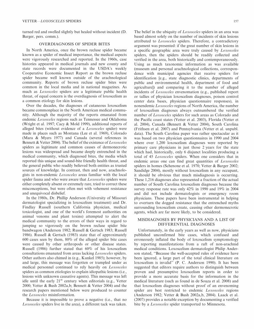
Table 1.—A list of medical conditions that have been or could bemisdiagnosed as cutaneous loxoscelism. Modified from Swanson &Vetter (2005).
InfectionsAtypical mycobacteriaBacterial
- Streptococcus- Staphylococcus (especially MRSA)- Lyme borreliosis- Cutaneous anthrax- Syphilis- Gonococcemia- Ricketsial disease- Tularemia
Deep Fungal- Sporotrichosis- Aspergillosis- Cryptococcosis
Ecthyma gangrenosum (Pseudomonas aeruginosa)Parasitic (Leishmaniasis)Viral (herpes simplex, herpes zoster (shingles))Vascular occlusive or venous diseaseAntiphospholipid-antibody syndromeLivedoid vasculopathySmall-vessel occlusive arterial diseaseVenous statis ulcerNecrotising vasculitisLeukocytoclastic vaculitisPolyarteritis nodosaTakayasu’s arteritisWegeners granulomatosisNeoplastic diseaseLeukemia cutisLymphoma (e.g., mycosis fungoides)Primary skin neoplasms (basal cell carcinoma, malignant melanoma,
squamous cell carcinoma)Lymphomatoid papulosisTopical and Exogenous CausesBurns (chemical, thermal)Toxic plant dermatitis (poison ivy, poison oak)Factitious injury (i.e., self-induced)Pressure ulcers (i.e., bed sores)Other arthropod bitesRadiotherapyOther ConditionsCalcific uremic arteriolopathyCryoglobulinemiaDiabetic ulcerLangerhans’-cell histiocytosisPemphigus vegetansPyoderma gangrenosumSeptic embolism
158 THE JOURNAL OF ARACHNOLOGY

(Baxtrom et al. 2006) further supporting the lack of spiderorigin for a condition well established as a nosocomialinfection.
HUMAN PSYCHOLOGY AND THE PROLIFERATIONOF LOXOSCELISM DIAGNOSES
A large part of the basis for awareness of Loxosceles spidersthroughout North American society is due to the dramatic,psychological nature surrounding the diagnosis of loxoscelism.Although the comments made here are more pertinent fornonendemic Loxosceles areas, there will be some relevancefor endemic areas as well. The diagnosis of loxoscelisminvolves the psychology of both the patient with a lesionand the physician making the diagnosis along with theinteraction of the physician-patient relationship. Much ofthe information here has been developed over the last decadevia conversations and correspondences with medical col-leagues, exposure to hundreds of emails from concernedNorth Americans attempting to discover the cause of theirmysterious skin lesions as well as studies or treatises that delveinto myth proliferation and the psychology of the cognitivemedical diagnostic process. The points presented below are byno means exhaustive.
From the patient standpoint, there are many aspects thatcause loxoscelism to retain a high profile in the generalpublic’s eye.
- Adverse reaction to spiders in western civilization rangesfrom mild dislike to intense arachnophobia (Isbister 2004).Entities perceived in a negative light are readily blamed asculprits for people’s maladies and misfortunes despite thereality of the involvement (Difonzo & Bordia 2006); spidersqualify well as scapegoats. Physicians who likewise sufferfrom arachnophobia or spider disgust will be predisposedto inappropriately blame spiders as idiopathic skin lesionetiologies (Isbister 2004).
- Spiders are commonly encountered, readily recognizableorganisms; therefore, they are embraced as causes ofmedical ills (Isbister 2004). It is difficult for most membersof the non-medical world to visualize or conceptualizeStaphylococcus or pyoderma gangrenosum.
- Patients appear to prefer accepting an exogenous causerather than an endogenous response for a medical affliction(Benoit & Suchard 2006). Blaming a spider over whichthere is no control is more agreeable than admitting thatsome inherent physical weakness or detrimental life stylechoice is causing the illness.
- ‘‘Spider bite’’ is an oddly comforting diagnosis for patientswith skin lesions (Benoit & Suchard 2006). It becomes abadge of courage that they ‘‘survived’’ an encounter with abeast of perceived danger. People who feel they havesuffered loxoscelism recount their stories for years, whichare then retold by others (Vetter, unpubl. data); this is oneof the mechanisms for reinforcing myths in the generalpublic (Difonzo & Bordia 2006). In contrast, one rarelyrecounts to friends and colleagues a personal bout with abacterial infection, especially long after the incident.
- Patients often put blind faith in their physicians (Vetter,unpubl. data). If a physician diagnoses a brown reclusespider bite, this carries far more weight in the patient’s eyesas to the probability of Loxosceles spiders in a local area
than does the lifelong collecting experience of regionalarachnologists (Vetter & Isbister 2008). Physicians know-ingly work in an environment with accepted uncertainty(Montgomery 2006); however, patients feel that physicianswork in a world of absolute knowledge.
For the physician, there are many aspects that maintain thepersistence of loxoscelism as an etiology of idiopathic skinlesions.
- Patients understandably visit a physician because they seekanswers for their illnesses. The physician wants to providean answer because that is his/her job and, hence, this drivesthe desire for a diagnosis. There is an approximate overall15% misdiagnosis rate in medicine (Elstein 1995). Althoughmedicine is described as an art and a science, Montgomery(2006) advocates repeatedly that it should be consideredneither but, rather, ‘‘a rational, science-using practice.’’
- Physicians may be reluctant to request the necessary tests todetermine if a bacterial or viral agent might be the cause ofa skin lesion (Isbister 2004; Benoit & Suchard 2006). This iscaused in part by physicians not sufficiently pursuing thecausative agent (Benoit & Suchard 2006) but also the desireto keep costs low in an era of spiraling medical expenses.
- Medical schools used to instruct their students thatloxoscelism is a common cause of necrotic skin lesions.Colleagues have relayed that these lessons included truismssuch as ‘‘if it is a necrotic wound, it is a brown recluse bite’’and that brown recluse bites were ‘‘deadly’’ despite therarity of such dire outcome. This appears to be changing asthe medical textbooks are incorporating recent research (inparticular, the distribution map of Swanson & Vetter[2005]) along with greater awareness of the differentialdiagnoses for dermonecrosis especially in regard to MRSA.
- The most common cause of cognitive error resulting inmisdiagnosis is premature closure where, once a diagnosisis made, a physician fails to consider other likelydifferential diagnoses (Kuhn 2002; Graber et al. 2005).Senior physicians are just as likely to commit this error asjunior physicians (Kuhn 2002). These mistakes arise as amanifestation of the heuristic diagnostic process, whichwhen done correctly, results in the desired effects ofreducing delay, cost and anxiety (Redelmeier 2005). Othercognitive errors, such as confirmational bias, preventphysicians from considering alternative diagnoses (Groop-man 2007). Again, loxoscelism is a dramatic diagnosis and,once considered, a physician may lock on to this etiology tothe exclusion of more probable causative agents.
- There is conflict in the medical field regarding improbablediagnoses (Montgomery 2006). The conservative-mindedaxiom of ‘‘when you hear hoof beats, think horses, notzebras’’ reinforces the need to first consider commonetiologies with which a patient might present and, moreimportantly, the uncommon manifestation of a commonetiology. The more dramatic zebra diagnoses are recalledmore easily due to their novelty (Kuhn 2002) and, hence,are diagnosed too frequently. Nonetheless, even whenknowingly faced with an improbable diagnosis of once-in-a-career probability, the physician does not want tooverlook this rare condition out of professional duty tothe patient (Montgomery 2006). Hence, the dynamic nature
VETTER—LOXOSCELES SPIDERS 159
of loxoscelism causes medical personnel to diagnose (andpublish articles) where the evidential threads to Loxoscelesspiders are extremely flimsy and sometimes obviouslywrong (Anderson 1982; Vetter & Swanson 2007).
- Spider bites are prematurely embraced as etiologies fordermonecrosis without proper evidence-based medicine.This phenomenon is well demonstrated by an Australianepisode with white-tailed spiders, Lampona cylindrata (L.Koch 1866) and L. murina L. Koch 1873 (Lamponidae) withspeculation that they caused necrotic arachnidism (Suther-land 1983). This lead to a spate of publications documentingalleged effects of white-tailed spider bite based on presump-tive diagnosis without spider involvement (Isbister & Gray2003; White 2003; Isbister 2004). Verified bites with minormanifestation were brushed aside as aberrant; calls forfunding to develop antivenom were made (White 2003).After 20 years of spider incrimination, Isbister & Gray(2003) definitively demonstrated with 130 verified Lamponabites with only minor, non-necrotic manifestation, that thesespiders were not probable causes of necrotic arachnidism.Parallel features exist for loxoscelism in North America andblaming of dermonecrosis on wolf spiders in South America(Isbister 2004; Vetter & Isbister 2008).
CONCLUDING STATEMENT
The medical arachnological world encompassing Loxoscelesspiders is an intriguing mixture of arachnology, toxicology,medicine, psychology, mythology, and even journalism.Without a doubt, Loxosceles spiders present a real envenom-ation threat for many regions of the world from a shy,reclusive spider. However, the exaggeration of this threat hasgiven this genus a reputation that greatly extends past itsactual physical presence. There are many facets to tease out ofthis situation as Loxosceles spiders’ infamy has garneredconcern outside the academic world. The facets are subject tohuman psychology and the checkered ability of non-scientiststo properly interpret scientific data especially for a subject likeloxoscelism, which lends itself so readily to exaggeration andmyth. Although new research is providing the answers to thephysiological mechanisms and treatment of the valid threat ofloxoscelism, there is room for additional research in areas assimple as accurate distribution for states on the border of thecurrently known range of recluse spiders. Loxosceles spiderswill continue to generate significant attention in the worlds ofarachnology and medicine as well as interest and concern fromthe general public.
ACKNOWLEDGMENTS
I thank the dozens of arachnologists and entomologists whohave corresponded with me over the last 15 years in regard toLoxosceles distribution in their state, province, or country aswell as those curators who loaned their museum’s Loxoscelescollection for examination. Thanks also are expressed to themany physicians around the United States who have becomeclose colleagues in building a two-way conduit betweenarachnology and medicine, providing answers to manyquestions about the underpinnings of the physiology ofenvenomation injury and the psychology of medical diagnosesand, conversely, for taking the opportunity to ask manyquestions about spiders and their role in envenomations. Pat
Miller (Northwest Mississippi Community College) graciouslyprovided photocopies of the pertinent sections of Peggy RaeDorris’s dissertation and information regarding Dorris’s fieldnotebook for which I am grateful. The manuscript wasimproved by comments by Paula E. Cushing, G.B. Edwards,Gail Stratton, Gary Wasserman, and two anonymousreviewers. Finally, I would like to dedicate this paper to adear friend and cherished arachnological colleague, H. DonCameron, who took me under his avuncular wing at the 1993AAS meeting in Seattle and has witnessed the development ofmy Loxosceles research program from its inception.
LITERATURE CITED
Anderson, J.F. 1990. The size of spider eggs and estimates of theirenergy content. Journal of Arachnology 18:73–78.
Anderson, P.C. 1982. Letter to the editor. Toxicon 20:533.
Anderson, P.C. 1990. Loxoscelism and the history of the Missouribrown spider: a recollection of Dr. Joseph Flynn. MissouriMedicine 87:747–752.
Anderson, P.C. 1998. Missouri brown recluse spider: a review andupdate. Missouri Medicine 95:318–322.
Andrade, R.M.G., K.C. Oliveira, A.L. Giusti, W.D. Silva & D.Tambourgi. 1999. Ontogenetic development of Loxosceles inter-media spider venom. Toxicon 37:627–632.
Atkins, J.A., C.W. Wingo & W.A. Sodeman. 1957. Probable cause ofnecrotic spider bite in the Midwest. Science 126:73.
Bancroft, E.A. 2007. Antimicrobial resistance: it’s not just forhospitals. Journal of the American Medical Association 298:1803–1804.
Barbaro, K.C., I. Knysak, R. Martins, C. Hogan & K. Winkel. 2005.Enzymatic characterization, antigenic cross-reactivity and neutral-ization of dermonecrotic activity of five Loxosceles spider venomsof medical importance in the Americas. Toxicon 45:489–499.
Baxtrom, C., T. Mongkolpradit, J.N. Kasimos, L.M. Braune, R.D.Wise, P. Sierwald & K.R. Ramsey. 2006. Common house spidersare not likely vectors of community-acquired methicillin-resistantStaphylococcus aureus infections. Journal of Medical Entomology43:962–965.
Beatty, J. 1970. The spider genus Ariadna in the Americas (Araneae:Dysderidae). Bulletin of the Museum of Comparative Zoology139:433–517.
Bennett, R.G. & R.S. Vetter. 2004. An approach to spider bites:erroneous attribution of dermonecrotic lesions to brown recluse orhobo spider bites in Canada. Canadian Family Physician 50:1098–1101.
Benoit, R. & J.R. Suchard. 2006. Necrotic skin lesions: spider bite –or something else? Consultant 46:1386–1394.
Binford, G.J. & M.A. Wells. 2003. The phylogenetic distribution ofsphingomyelinase D activity in venoms of haplogyne spiders.Comparative Biochemistry and Physiology B 135:25–33.
Binford, G.J., M.H.J. Cordes & M.A. Wells. 2005. SphingomyelinaseD from venoms of Loxosceles spiders: evolutionary insights fromcDNA sequences and gene structure. Toxicon 45:547–560.
Borkan, J., E. Gross, Y. Lubin & I. Oryan. 1995. An outbreak ofvenomous spider bites in a citrus grove. American Journal ofTropical Medicine and Hygiene 52:228–230.
Bradley, R.A. 2004. In Ohio’s Backyard: Spiders. Ohio BiologicalSurvey Backyard Series #4. 183 pp.
Cacy, J. & J.W. Mold. 1999. The clinical characteristics of brownrecluse spider bites treated by family physicians: an OKPRN study.Journal of Family Practice 48:536–542.
Cameron, H.D. 2005. An etymological dictionary of North Americanspider genus names. Pp. 274–330. In Spiders of North America: anIdentification Manual. (D. Ubick, P. Paquin, P.E. Cushing & V.Roth, eds.). American Arachnological Society.
160 THE JOURNAL OF ARACHNOLOGY
Carrel, J.E. & R.D. Heathcote. 1976. Heart rate in spiders: influenceof body size and foraging energetics. Science 193:148–150.
Caveness, W.A. 1872. Insect bite, complicated with fever. NashvilleJournal of Medicine and Surgery 10:333–337.
Chaim, O.M., Y.B. Sade, R.B. da Silveira, L. Toma, E. Kalapothakis,C. Chavez-Olortegui, O.C. Mangili, W. Gremski, C.P. vonDietrich, H.B. Nader & S.S. Veiga. 2006. Brown spider dermone-crotic toxin directly induces nephrotoxicity. Toxicology andApplied Pharmacology 211:64–77.
Chow, R.K.P. & V.C. Ho. 1996. Treatment of pyoderma gang-renosum. Journal of the American Academy of Dermatology34:1047–1060.
Cohen, P.R. 2007. Community-acquired methicillin-resistant Staph-
ylococcus aureus skin infections: a review of epidemiology, clinicalfeatures, management and prevention. International Journal ofDermatology 46:1–11.
Cramer, K.L. & A.V. Mayright. 2008. Cold temperature toleranceand distribution of the brown recluse spider Loxosceles reclusa
(Araneae, Sicariidae) in Illinois. Journal of Arachnology 36:136–139.
da Silva, P.H., R.B. da Silveira, M.H. Appel, O.C. Mangili, W.Gremski & S.S. Veiga. 2004. Brown spiders and loxoscelism.Toxicon 44:693–709.
de Souza, A.L., C.M. Malaque, J. Sztanjbok, C.C. Romano, A.J.Duarte & A.C. Seguro. 2008. Loxosceles venom-induced cytokineactivation, hemolysis, and acute kidney injury. Toxicon51:151–156.
Difonzo, N. & P. Bordia. 2006. Rumor Psychology: Social andOrganizational Approaches. American Psychological Association,Washington, DC. 292 pp.
Dominguez, T.J. 2004. It’s not a spider bite, it’s community-acquiredmethicillin-resistant Staphylococcus aureus. Journal of the Amer-ican Board of Family Practice 17:220–26.
Dorris, P.R. 1967. The spiders of Mississippi. Ph.D. dissertation.University of Mississippi, Oxford. 283 pp.
Elgar, M.A. 1998. Sperm competition and sexual selection in spidersand other arachnids. Pp. 307–339. In Sperm Competition andSexual Selection. (T.R. Birkhead & A.P. Møller, eds.). AcademicPress, San Diego, California.
Elstein, A.S. 1995. Clinical reasoning in medicine. Pp. 49–59. InClinical Reasoning in the Health Professions. (J. Higgs & M.Jones, eds.). Butterworth-Heinemann Ltd., Oxford, UK.
Elston, D.M., S.D. Miller, R.J. Young III, J. Eggers, D. McGlasson,W.H. Schmidt & A. Bush. 2005. Comparison of colchicine,dapsone, triamcinolone, and diphenhydramine therapy for thetreatment of brown recluse spider envenomation: a double-blind,controlled study in a rabbit model. Archives of Dermatology141:595–597.
Elzinga, R.J. 1977. Observations on the longevity of the brownrecluse spider, Loxosceles reclusa Gertsch & Mulaik. Journal of theKansas Entomological Society 50:187–188.
Eskafi, F.M., J.L. Frazier, R.R. Hocking & B.R. Norment. 1977.Influence of environmental factors on longevity of the brownrecluse spider. Journal of Medical Entomology 14:221–228.
Fagan, S.P., D.H. Berger, K. Rahwan & S.S. Awad. 2003. Spiderbites presenting with methicillin-resistant Staphylococcus aureusinfection require early aggressive treatment. Surgical Infections4:311–315.
Fischer, M.L. & E.M. da Silva. 2001. Oviposicao e desenvolimento deLoxosceles hirsuta Mello-Leitao, 1931 (Araneae; Sicariidae).Estudos de Biologia Curitiba 47:15–20.
Fischer, M.L. & J. Vasconcellos-Neto. 2003. Determination ofthe maximum and minimum lethal temperatures (LT50) forLoxosceles intermedia Mello-Leitao, 1934 and L. laeta (Nicolet,1849) (Araneae, Sicariidae). Journal of Thermal Biology28:563–570.
Fischer, M.L. & J. Vasconcellos-Neto. 2005a. Development and lifetables of Loxosceles intermedia Mello-Leitao 1934 (Araneae,Sicariidae). Journal of Arachnology 33:758–766.
Fischer, M.L. & J. Vasconcellos-Neto. 2005b. Parameters affectingfecundity of Loxosceles intermedia Mello-Leitao 1934 (Araneae,Sicariidae). Journal of Arachnology 33:670–680.
Fischer, M.L. & J. Vasconcellos-Neto. 2005c. Microhabitats occupiedby Loxosceles intermedia and Loxosceles laeta (Araneae: Sicar-iidae) in Curitiba, Parana, Brazil. Journal of Medical Entomology42:756–765.
Frithsen, I.L., R.S. Vetter & I.C. Stocks. 2007. Reports ofenvenomation by brown recluse spiders exceed verified specimensof Loxosceles spiders in South Carolina. Journal of the AmericanBoard of Family Medicine 20:483–488.
Galiano, M.E. 1967. Ciclo biologico y desarrollo de Loxosceles laeta(Nicolet, 1849) (Araneae, Scytodidae). Acta Zoologica Lilloana23:431–464.
Gertsch, W.J. 1958. The spider genus Loxosceles in North America,Central America, and the West Indies. American MuseumNovitates 1907:1–46.
Gertsch, W.J. 1967. The spider genus Loxosceles in South America(Araneae, Scytodidae). Bulletin of the American Museum ofNatural History 136:117–174.
Gertsch, W.J. & F. Ennik. 1983. The spider genus Loxosceles inNorth America, Central America, and the West Indies (Araneae,Loxoscelidae). Bulletin of the American Museum of NaturalHistory 175:264–360.
Gomez, H.F., D.M. Krywko & W.V. Stoecker. 2002. A new assay forthe detection of Loxosceles species (brown recluse) spider venom.Annals of Emergency Medicine 39:469–474.
Gorham, J.R. 1968. The geographic distribution of the brown reclusespider, Loxosceles reclusa (Araneae, Scytodidae) and relatedspecies in the United States. United States Department ofAgriculture Cooperative Economic Insect Report 18:171–175.
Graber, M.L., N. Franklin & R. Gordon. 2005. Diagnostic error ininternal medicine. Archives of Internal Medicine 165:1493–1499.
Groopman, J. 2007. How Doctors Think. Houghton Mifflin Co.,Boston, Massachusetts. 307 pp.
Hite, J.M., W.J. Gladney, J.L. Lancaster & W.H. Whitcomb. 1966. Thebiology of the brown recluse spider. University of Arkansas,Fayetteville. Agricultural Experiment Station Bulletin No. 711. 26 pp.
Hogan, C.J., K.C. Barbaro & K. Winkel. 2004. Loxoscelism: oldobstacles, new directions. Annals of Emergency Medicine 44:608–624.
Horner, N.V. & K.W. Stewart. 1967. Life history of the brown spider,Loxosceles reclusa Gertsch and Mulaik. Texas Journal of Science19:334–347.
Isbister, G.K. 2004. Necrotic arachnidism: the myth of a modernplague. Lancet 364:549–553.
Isbister, G.K. & M.R. Gray. 2003. White-tail spider bite: aprospective study of 130 definite bites by Lampona species.Medical Journal of Australia 179:199–202.
Kaston, B.J. 1970. Comparative biology of American black widowspiders. Transactions of the San Diego Society of Natural History16:33–82.
Klevens, R.M., M.A. Morrison, J. Nadle, S. Petit, K. Gershman, S.Ray, L.H. Harrison, R. Lynfield, G. Dumyati, J.M. Townes, A.S.Craig, E.R. Zell, G.E. Fosheim, L.K. McDougal, R.B. Carey &S.K. Fridkin. 2007. Invasive methicillin-resistant Staphylococcusaureus infections in the United States. Journal of the AmericanMedical Association 298:1763–1771.
Knight, D.P. & F. Vollrath. 2002. Spinning an elastic ribbon of spidersilk. Philosophical Transactions of the Royal Society of London B357:219–227.
Kuhn, G.J. 2002. Diagnostic errors. Academic Emergency Medicine9:740–750.
VETTER—LOXOSCELES SPIDERS 161
Kunkel, D.B. 1985. The myth of the brown recluse spider. EmergencyMedicine 17(5):124–128.
Laack, T.A., L.G. Stead & M.E. Wolfe. 2007. Images in emergencymedicine. Annals of Emergency Medicine 50:368.
Lee, R.V., R.S. Buker Jr. & K.M. Petersen. 1969. North Americanloxoscelism: two presumptive cases from northern Montana.Rocky Mountain Medical Journal 66:57–59.
Lowe, R.T. 1835. Descriptions of two species of Araneidae, natives ofMadeira. Zoological Journal 5:320–323.
Lowrie, D.C. 1980. Starvation longevity of Loxosceles laeta (Nicolet)(Araneae). Entomological News 91:130–132.
Macchiavello, A. 1947. Cutaneous arachnoidism or cutaneous spot ofChile. Puerto Rico Public Health and Tropical Medicine 22:425–466.
Malaque, C.M.S., J.E. Castro-Valencia, J.L.C. Cardoso, F.O.S. Franca,K.C. Barbaro & H.W. Fan. 2002. Clinical and epidemiologicalfeatures of definitive and presumed loxoscelism in Sao Paulo, Brazil.Revista Instituto de Medicina Tropical de Sao Paulo 44:139–143.
Mara, J.E. & B.S. Myers. 1977. Brown spider bite. Rocky MountainMedical Journal 74:257–258.
Masters, E.J. 1998. Loxoscelism. New England Journal of Medicine339:379.
Miller, L.G. & B. Spellberg. 2004. Spider bites and infections causedby community-associated methicillin-resistant Staphylococcus au-reus. Surgical Infections 5:321–322.
Montgomery, K. 2006. How Doctors Think: Clinical Judgment and thePractice of Medicine. Oxford University Press, Oxford, UK. 246 pp.
Moran, G.J., A. Krishnadasan, R.J. Gorwitz, G.E. Fosheim, L.K.McDougal, R.B. Carey & D.A. Talan. 2006. Methicillin-resistantS. aureus infections among patients in emergency rooms. NewEngland Journal of Medicine 355:666–674.
Newlands, G., C. Isaacson & C. Martindale. 1982. Loxoscelism in theTransvaal, South Africa. Transactions of the Royal Society ofTropical Medicine and Hygiene 76:610–615.
Oehler, C. 1974. The medical significance of spiders at Cincinnati,Ohio. Cincinnati Museum of Natural History 23(3):1–11.
Osterhoudt, K.C., T. Zaoutis & J.J. Zorc. 2002. Lyme diseasemasquerading as brown recluse spider bite. Annals of EmergencyMedicine 39:558–561.
Paixao-Cavalante, D., C.W. van den Berg, R.M. Goncalves-de-Andrade, M.F. Fernandes-Pedrosa, C.K. Okamoto & D.V.Tambourgi. 2007. Tetracycline protects against dermonecrosisinduced by Loxosceles spider venom. Journal of InvestigativeDermatology 127:1410–1418.
Patel, K.D., V. Modur, G.A. Zimmerman, S.M. Prescott & T.M.McIntyre. 1994. The necrotic venom of the brown recluse spiderinduces dysregulated endothelial cell-dependent neutrophil activa-tion. Journal of Clinical Investigation 94:631–642.
Pauli, I., J. Puka, I.C. Gubert & J.C. Minozzo. 2006. The efficacy ofantivenom in loxoscelism treatment. Toxicon 48:123–137.
Platnick, N.I. 2007. The World Spider Catalog, Version 8.0.American Museum of Natural History. Online at http://research.amnh.org/entomology/spiders/catalog/SICARIIDAE.html
Platnick, N.I., J.A. Coddington, R.R. Forster & C.E. Griswold. 1991.Spinneret morphology and the phylogeny of haplogyne spiders(Araneae, Araneomorphae). American Museum Novitates 3016:1–73.
Rapp, W.F. 1980. A catalog of spiders of Nebraska. NovitatesArthropodae 1(2):1–39.
Redelmeier, D.A. 2005. The cognitive psychology of misseddiagnoses. Annals of Internal Medicine 142:115–120.
Reed, H.B. Jr.. 1968. The brown recluse spider and loxoscelism inTennessee. Journal of the Tennessee Academy of Science 43:110–114.
Ribeiro, R.O.S., O.M. Chaim, R.B. da Silveira, L.H. Gremski, Y.B.Sade, K.S. Paludo, A. Senff-Ribeiro, J. de Moura, C. Chavez-Olortegui, W. Gremski, H.B. Nader & S.S. Veiga. 2007. Biological and
structural comparison of recombinant phospholipase D toxins fromLoxosceles intermedia (brown spider venom). Toxicon 50:1162–1174.
Rinaldi, I.M.P., L.C. Forti & A.A. Stropa. 1997. On the developmentof the brown spider Loxosceles gaucho Gertsch (Araneae,Sicariidae): the nympho-imaginal period. Revista Brasileira deZoologia 14:697–706.
Roche, K.J., M.W. Chang & H. Lazarus. 2001. Cutaneous anthraxinfection. New England Journal of Medicine 345:1611.
Russell, F.E. 1986. A confusion of spiders. Emergency Medicine18(11):8–13.
Russell, F.E. & W.J. Gertsch. 1983. Letter to the editor. Toxicon21:337–339.
Russell, F.E., W.G. Waldron & M.B. Madon. 1969. Bites by thebrown spiders Loxosceles unicolor and Loxosceles arizonica inCalifornia and Arizona. Toxicon 7:109–112.
Sams, H.H., S.B. Hearth, L.L. Long, D.C. Wilson, D.H. Sanders &L.E. King Jr. 2001. Nineteen documented cases of Loxoscelesreclusa envenomation. Journal of the American Academy ofDermatology 44:603–608.
Sandidge, J. 2004. Predation by cosmopolitan spiders on themedically significant pest species Loxosceles reclusa (Araneae:Sicariidae): limited possibilities for biological control. Journal ofEconomic Entomology 97:230–234.
Schenone, H., A. Rojas, H. Reyes, F. Villarroel & G. Suarez. 1970.Prevalence of Loxosceles laeta in houses in central Chile. AmericanJournal of Tropical Medicine and Hygiene 19:564–567.
Schenone, H., T. Saavedra, A. Rojas & F. Villarroel. 1989.Loxoscelismo en Chile. Estudios epidemologicos, clınicos yexperimentales. Revista Instituto de Medicina Tropical de SaoPaulo 31:403–415.
Schmaus, L.F. 1929. Case of arachnoidism (spider bite). Journal ofthe American Medical Association 92:1265–1266.
Southcott, R.V. 1976. Spiders of the genus Loxosceles in Australia.Medical Journal of Australia 1:406–408.
Stoaks, R.D. 1980. Occurrence of the brown recluse spider (Araneae:Loxoscelidae) in Iowa. Proceedings of the Iowa Academy ofScience 87:159.
Stropa, A.A. 2007. Social encounters between male brown spiders,Loxosceles gaucho (Araneae, Sicariidae). Journal of Arachnology35:493–498.
Stropa, A.A. & I.M.P. Rinaldi. 2001. Relative tolerance andcommunication in agonistic behaviour between females of Loxos-celes gaucho (Araneae, Sicariidae). Bulletin of the British Arach-nological Society 12:41–45.
Sutherland, S. 1983. Necrotic arachnidism: a possible new Australiansyndrome. Toxicon 21(Supplement 3):435.
Swanson, D.L. & R.S. Vetter. 2005. Bites of brown recluse spidersand suspected necrotic arachnidism. New England Journal ofMedicine 352:700–707.
Swanson, D.L. & R.S. Vetter. 2006. Loxoscelism. Clinics inDermatology 24:213–221.
Uetz, G.W. & C.S. Hieber. 1997. Colonial web-building spiders:balancing the costs and benefits of group-living. Pp. 458–475. InSocial Behavior in Insects and Arachnids. (J.C. Choe & B.J.Crespi, eds.). Cambridge University Press, Cambridge, UK.
Vail, K.M. & J.A. Watson. 2002. The brown recluse spider.University of Tennessee Agricultural Extension Service BulletinPB1191. 16 pp.
Vetter, R.S. 2000. Myth: idiopathic wounds are often due to brownrecluse or other spider bites throughout the United States. WesternJournal of Medicine 173:357–358.
Vetter, R.S. 2005. Arachnids submitted as suspected brown reclusespiders (Araneae: Sicariidae): Loxosceles species are virtuallyrestricted to their known distributions but are perceived to existthroughout the United States. Journal of Medical Entomology42:512–521.
162 THE JOURNAL OF ARACHNOLOGY
Vetter, R.S. & D.K. Barger. 2002. An infestation of 2,055 brownrecluse spiders (Araneae: Sicariidae) and no envenomations in aKansas home: implications for bite diagnoses in nonendemic areas.Journal of Medical Entomology 39:948–951.
Vetter, R.S. & S.P. Bush. 2002a. Reports of presumptive brownrecluse spider bites reinforce improbable diagnosis in regions ofNorth America where the spider is not endemic. Clinical InfectiousDiseases 35:442–445.
Vetter, R.S. & S.P. Bush. 2002b. The diagnosis of brown reclusespider bite is overused for dermonecrotic wounds of uncertainetiology. Annals of Emergency Medicine 39:544–546.
Vetter, R.S. & S.P. Bush. 2002c. Chemical burn misdiagnosed as brownrecluse spider bite. American Journal of Emergency Medicine20:68–69.
Vetter, R.S. & S.P. Bush. 2004. Additional considerations inpresumptive brown recluse spider bites and dapsone therapy.American Journal of Emergency Medicine 22:494–495.
Vetter, R.S., P.E. Cushing, R.L. Crawford & L.A. Royce. 2003.Diagnoses of brown recluse spider bites (loxoscelism) greatlyoutnumber actual verifications of the spider in four westernAmerican states. Toxicon 42:413–418.
Vetter, R.S., G.B. Edwards & L.F. James. 2004. Reports ofenvenomation by brown recluse spiders (Araneae: Sicariidae)outnumber verifications of Loxosceles spiders in Florida. Journalof Medical Entomology 41:593–597.
Vetter, R.S. & G.K. Isbister. 2008. Medical aspects of spider bites.Annual Review of Entomology 53:409–429.
Vetter, R.S., B.B. Pagac, R.W. Reiland, D.T. Bolesh & D.L.Swanson. 2006. Skin lesions in barracks: consider community-
acquired methicillin-resistant Staphylococcus aureus infectioninstead of spider bites. Military Medicine 171:830–832.
Vetter, R.S. & M.K. Rust. 2008. Refugia preferences by the spidersLoxosceles reclusa and Loxosceles laeta (Araneae: Sicariidae).Journal of Medical Entomology 45:36–41.
Vetter, R.S. & D.L. Swanson. 2007. Of spiders and zebras:publication of inadequately documented loxoscelism case reports.Journal of the American Academy of Dermatology 56:1063–1064.
Waldron, W.G. 1969. Loxosceles laeta (Nicolet), an introducedspecies in Los Angeles County. Bulletin of the EntomologicalSociety of America 15:377–379.
Wasserman, G.S., R. Garola, J. Marshall & S. Gustafson. 1999.Death of a 7 year old by presumptive brown recluse spider bite.Journal of Toxicology – Clinical Toxicology 37:614–615.
Wasserman, G.S. & J.A. Lowry. 2005. Loxosceles spiders.Pp. 1195–1203. In Critical Care Toxicology: Diagnosis andManagement of the Critically Poisoned Patient. (J. Brent, K.L.Wallace, K.K. Burkhart, S.D. Phillips & J.W. Donovan, eds.).Elsevier Mosby, Philadelphia, Pennsylvania.
White, J. 2003. Debunking spider bite myths. Medical Journal ofAustralia 179:180–181.
Wilson, J.T. 1893. Poisoning by the bite of the southern spider.Transactions of the Southern Surgical and Gynecological Associ-ation 5:406–411.
Wright, S.W., K.D. Wrenn, L. Murray & D. Seger. 1997. Clinicalpresentation and outcome of brown recluse spider bite. Annals ofEmergency Medicine 30:28–32.
Manuscript received 15 January 2008, revised 27 February 2008.
VETTER—LOXOSCELES SPIDERS 163