Embed Size (px)
Citation preview
Pakistan J. Zool., vol. 40(4), pp. 263-273, 2008.
Spermatogenesis, Maturation, Seasonal Variations and Spawning Season of Silver Pomfret (Pampus argenteus,
Euphrasen) Collected From the Natural Spawning Grounds Off the Shore of Kuwait
KHALID P. LONE*, SALAM AL-ABLANI AND SULAIMAN ALMATAR
Mariculture, Fisheries and Marine Environment Department, Food Resources and Marine Sciences Division, Kuwait Institute for Scientific Research, Kuwait
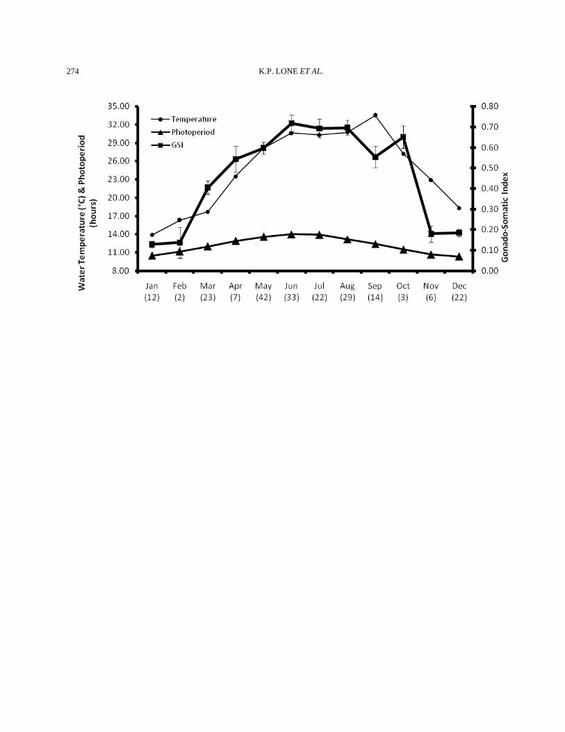
Abstract.- A study on the reproductive biology of male silver pomfret, Pampus argenteus, was undertaken on fish collected from the natural coastal spawning grounds of Kuwait. A total of 215 fish were analyzed for the present studies from a total of 3454 males collected over the three successive spawning seasons. Grossly, testes were classified as mature or immature. The GSI was the lowest (0.13±0.02) in January while the highest values were recorded in June (0.72±0.04). Histologically, the testes were lobular and lobules contained many cysts. Each cyst was comprised of only one type of germ cells and contained either spermatogonia, spermatocytes, or spermatids. The cysts when matured released their contents (spermatids) in to the lobule lumen which were connected with the main sperm duct through anastomosing tubules. In the mature testis the spermatozoa arising from the spermatids filled the lobular lumen and the connected ducts. Sperms were also present in lobular lumen even in non-spawning individuals and winter months. These were residual sperms and were not actively removed from the testis as is the case in many other teleosts. This observation means that testicular regression was not characterized by phagocytosis or apoptosis of the residual sperms. In addition to the germ cells, interstitial cells of Leydig and intra-lobular Sertoli cells were also seen. Based on the collected samples, it appears that the spawning continues from June to October with a decrease in September, due to high temperatures. However, the peak of spawning was in June while a rather small peak was seen in October also. Key words: Butterfish, testis, gross structure; histology, annual cycle.
INTRODUCTION
Interest in the basic biology and reproduction of many species has gained tremendous importance because of the decrease in catches from the traditional natural resources and supplementation of these decreases through aquacultural practices. Several studies have been undertaken on the reproductive biology of fish collected from the wild and/or kept in captivity and have addressed various angles of control of reproduction. Majority of these studies dealt with role of temperature and photoperiod as environmental factors controlling the maturation and spawning of fish (Lam, 1983; Malhotra et al., 1989; Munro, 1990; Acharia et al., 2000; Wang et ________________________________ * Present address: University of Wah, The Mall, Wah Cantt.
Pakistan. 0030-9923/2008/0004-0263 $ 8.00/0 Copyright 2008 Zoological Society of Pakistan. al., 2001; Rutaisire et al., 2003; Bhattacharyya and Maitra, 2006). These studies vividly showed that the reproductive tactics of teleost fish are as varied as their life styles. Teleost testis exhibit tremendous variations in structure, spermatogenic pattern and maturation (Grier, 1981; Billard et al., 1982; Grier and Taylor, 1998; Schulz and Miura, 2002; Maldonado-Garcia et al., 2005). However, more studies are available on female fish than on males. A study on the various aspects of gonad development, maturation and related aspects of reproductive biology of male would be needed for fish that have commercial appeal both for the natural resource protection and sustenance and for developing its aquaculture technology. Pomfrets (butterfishes: Stromateidae) have a wide range of distribution ranging from Japan to
K.P. LONE ET AL.
264
Arabian Gulf. Of the many species, Silver Pomfret (Pampus argenteus Euphrasen), locally known in Kuwait as “zobaidy” (butterfish in Arabic), is the most important commercial species in Japan, India, Pakistan and Arabian Gulf. They are caught in considerable quantities and have tremendous commercial appeal. Very few studies are available on the reproductive biology of this fish (Gopalan, 1969; Pati, 1981; Dadzie et al., 2000; Almatar et al., 2004). Majority of these studies are on female pomfret. The present study, first of its kind on male pomfret, describes in detail the reproductive biology of Pampus argenteus collected over three spawning seasons from the natural spawning grounds in the territorial waters of Kuwait. This stock of Pampus argenteus is shared with Iraq and Iran.
MATERIALS AND METHODS Fish collection and measurements The fish, silver pomfret (Pampus argenteus, Euphrasen) were collected with the help of drift gillnets on its spawning ground, situated in Kuwait waters. The spawning grounds covered an area of 40 km2 and located between latitude 29°22′N and 29°24N′; and longitude 48°00′E and 48°06′E. The nets (mesh size 14 cm) were deployed 2-5 km offshore for around 30 min, where the water depth was 5-12m. Two fiberglass speedboats were used to haul the net. Sampling was conducted during the years 1998 through 2000. Fishing time was between 0600 to 2000 hours. Water temperature and salinity were measured with the help of a portable Yellow Spring (Ohio, USA) temperature and salinity meter. For more details see Almatar et al. (2004). Captured fish were measured to the nearest cm and weighed to the nearest 0.1 g. Fish were brought to the laboratory where they were dissected to remove their gonads, which were weighed to the nearest mg. The gonads were then fixed in 10% buffered formalin solution made with the sea water. Grossly, the testes were classified as running (mature) if the milt was excreted by applying slight pressure on the abdomen or non running (immature). The gonadosomatic index (GSI) was calculated as gonad weight as a percentage of body weight (GSI= WG/WB X 100).
Tissue processing To allow the paraffin wax to infiltrate the preserved tissues prior to sectioning, the tissues were dehydrated gradually in an automatic tissue processor (Citadel 1000, Shandon) by passing them through a sequence of increasing concentrations of isopropyl alcohol (50, 75, 90 and 100%). When the dehydrated tissues reached the final concentration of isopropyl alcohol, they were passed twice through xylene for half an hour each time. Finally, the tissues were passed through two exchanges of paraffin wax (Paraplast, Fisher USA) for duration of one hour for each pass. The paraffin-infiltrated tissues were made into paraffin blocks by means of an Automatic Histo-Embedder (Leica, Germany) to facilitate sectioning.
Tissue sectioning The paraffin blocks were cut into thin transverse sections (3 to 5 µm) by means of an automatic rotary microtome (Jung Supercut, Germany). The sections obtained were placed on microscopic slides and stained with Harris’ haematoxylin and eosin. Sections were mounted with Canada balsam or DPX. The slides were studied in detail for different stages of spermatogenesis and photographed with Olympus Vanox (Japan) microscope. Other details have been reported elsewhere (Almatar et al., 2004).
RESULTS Annual male reproductive (testicular) cycle The data regarding morphometry and related parameters have been given elsewhere (Almatar et al., 2004). Gross and general observations The males were generally smaller than the females in size and ranged from 19.01-21.91 cm in standard length during the three spawning seasons sampled. There was no other difference between the sexes; thus, the sexes could not be distinguished based on any other characteristic or criterion. The testes were small, paired organs. They were present
REPRODUCTIVE BIOLOGY OF MALE SILVER POMFRET
265
on the posterior side of the viscera to which they were attached with the help of a very thin membrane. In most teleost fish the testes are paired, elongated organs attached to the body wall. However, in zobaidy they are closely attached to the viscera and kidneys. This is because of the fact that the peritoneal cavity of the fish is very restricted because of its body shape, which is laterally compressed. A main testicular duct, the vas deferens, runs along the medial part of the both lobes of the testis and merge with each other just prior to the opening to the exterior through the urogenital papilla. There was not much difference in the gross testes structure throughout the year in the fish that were reproductively active, once they had matured. Generally, the immature fish have a very thin, streak like organ which become steadily thicker and bigger as they mature. In mature fish, the organ becomes creamy in color and the blood supply seems to increase a lot. This is clearly visible in the surface arteries that form a network. There is a tradition of distinguishing and classifying (sometimes up to 8 stages) the gonads of wild fish based on their gross structure (Grier, 1981; Billard, 1986; Berois et al., 2004; Utoh et al., 2004; Guerriero et al., 2005; Maldonado-Garcia, 2005); however, no clear-cut classification for testes in zobaidy could be determined. Therefore, only two stages were defined for testes, that is, immature and mature. The immature testes were present in very small males irrespective of the season, while the mature testes were clearly discerned by their shape and structure, once again, irrespective of season. The only seasonal difference was the size of the testis. The testes were bigger in the spawning season, but even during this time, there was a variation in this characteristic depending upon the individual history of the fish. The quantity of milt exuded was quite small (few drops) even in those males which were running or when small pressure was applied to the belly of the fully mature male zobaidy. This could be due to the extended spawning season of the fish in Kuwait’s waters that extends from May to October. This decision of ours of not classifying the testes into the traditional stages of immature, developing, maturing, mature, running, spent and resting, ended up being correct
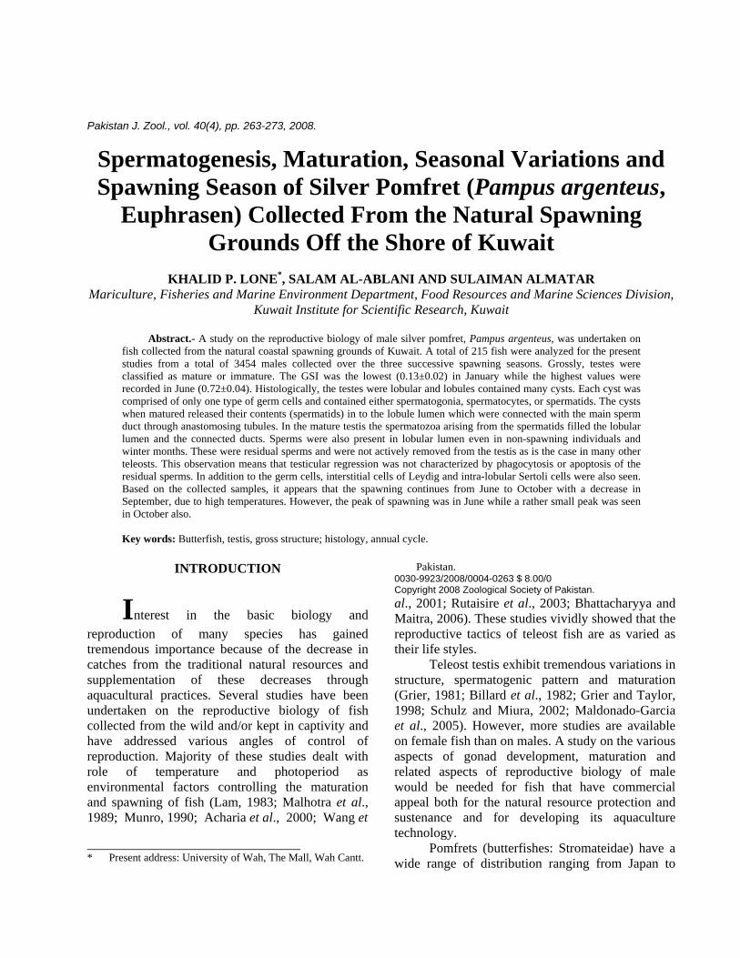
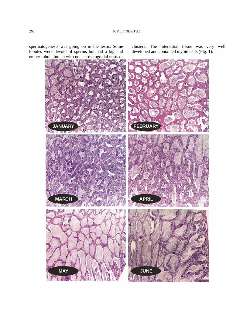
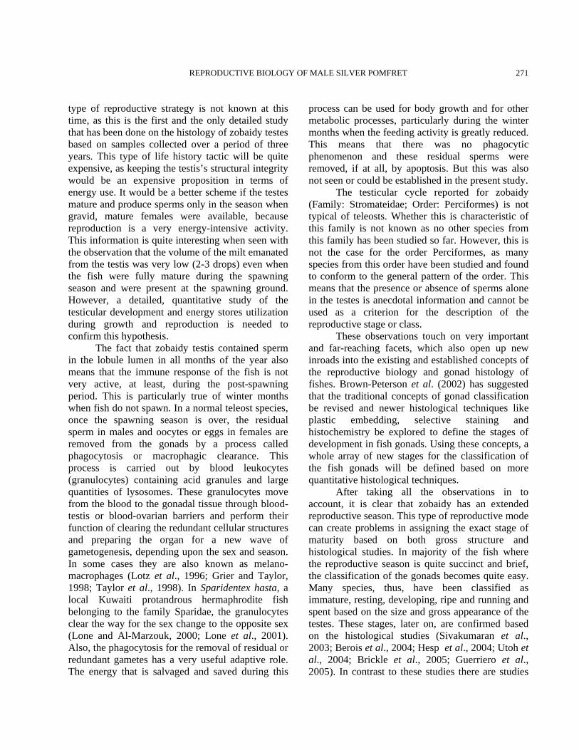
when the detailed histology of the fish collected over three seasons was studied. Therefore, the histological characterization of the zobaidy testes is described based on the season of collection from the spawning grounds present in the coastal waters of Kuwait. Histological observations January The testes in this month were thin in circumference and small in length. The blood vessels on the surface were less pronounced and the color was whitish to dirty white. The GSI (Fig. 3) for this month (n=12) was 0.13±0.02 (mean±SE). The histological picture of the testes showed that the tunica was thicker and the myoid tissue was wavy. Some spermatogonia could be seen scattered in the inner end of the tunica; however, no spermatogonial nests or cords could be seen. The lobular structure of the testis was clearly present. The lobules contained spematocysts that were at different stages of development. All of the stages of the spermatogenesis could be seen in the form of spermatogonia, spermatocytes, spermatids and sperms. The sperms were present in the lumen of the lobules. These could be residual sperms from the previous year. The spermatogenesis was more pronounced in the peripheral or outer tissue of the testis than in the inner tissue towards the main duct of the testis. In fact, the lobules that were adjacent to the ducts were filled with sperm. These lobules only had sperm in them, and no other spermatogenic stages were present. This means that these lobules were just storing the sperm and did not have any role in active spermatogenesis. The germinal epithelium was more or less continuous in the peripheral and distal portion of the testis, while it was clearly discontinuous in the central and deeper parts of the testis. Interstitial tissue was quite clear and contained cells of different sizes (Fig. 1). February The GSI was 0.14±0.08 showing that there was a big difference in the weights of the testes during this month (Fig. 3). Histologically, the tunica was thick, and the lobular structure was present and well established. The lobules contained residual sperm in them because there was no active
K.P. LONE ET AL.
266
spermatogenesis was going on in the testis. Some lobules were devoid of sperms but had a big and empty lobule lumen with no spermatogonial nests or
clusters. The interstitial tissue was very well developed and contained myoid cells (Fig. 1).
JANUARY FEBRUARY
MARCH APRIL
MAY JUNE
REPRODUCTIVE BIOLOGY OF MALE SILVER POMFRET
267
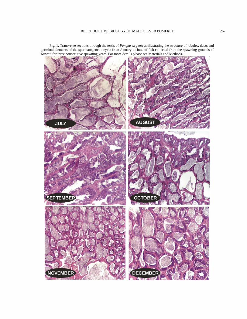
Fig. 1. Transverse sections through the testis of Pampus argenteus illustrating the structure of lobules, ducts and germinal elements of the spermatogenetic cycle from January to June of fish collected from the spawning grounds of Kuwait for three consecutive spawning years. For more details please see Materials and Methods.
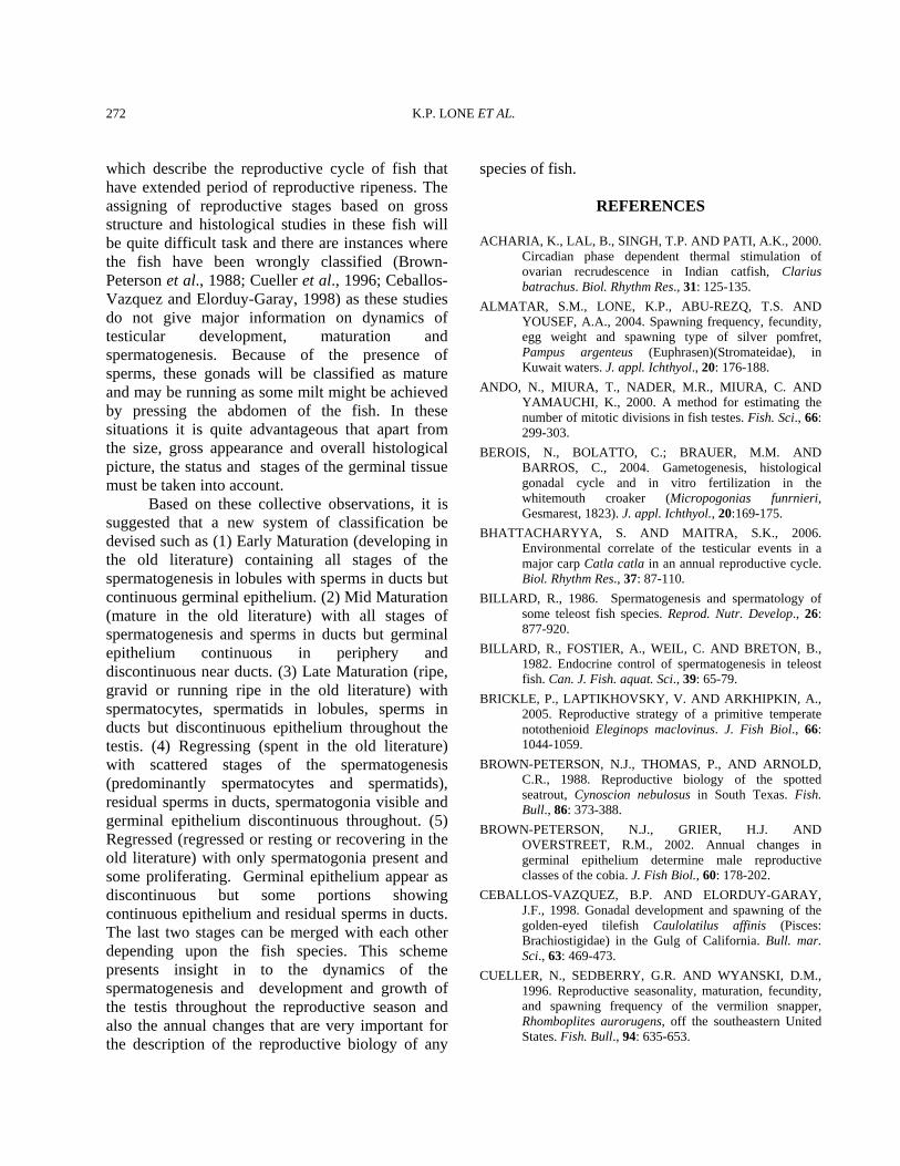
JULY AUGUST
SEPTEMBER OCTOBER
NOVEMBER DECEMBER
K.P. LONE ET AL.
268
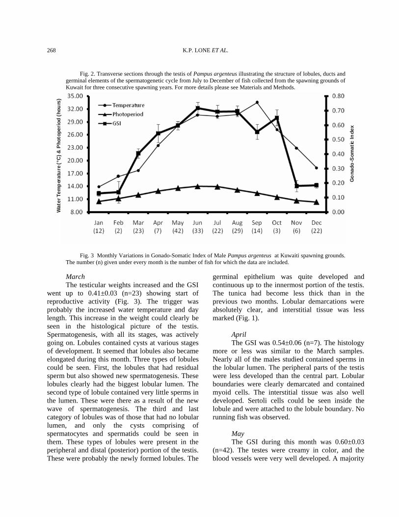
Fig. 2. Transverse sections through the testis of Pampus argenteus illustrating the structure of lobules, ducts and germinal elements of the spermatogenetic cycle from July to December of fish collected from the spawning grounds of Kuwait for three consecutive spawning years. For more details please see Materials and Methods.
Wat
er T
emp
erat
ure
(°C
) & P
hoto
peri
od (h
ours
)
Go
nado
-Som
atic
Inde
x
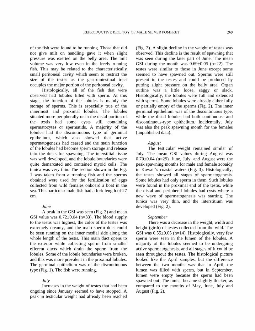
Fig. 3 Monthly Variations in Gonado-Somatic Index of Male Pampus argenteus at Kuwaiti spawning grounds. The number (n) given under every month is the number of fish for which the data are included.
March The testicular weights increased and the GSI went up to 0.41±0.03 (n=23) showing start of reproductive activity (Fig. 3). The trigger was probably the increased water temperature and day length. This increase in the weight could clearly be seen in the histological picture of the testis. Spermatogenesis, with all its stages, was actively going on. Lobules contained cysts at various stages of development. It seemed that lobules also became elongated during this month. Three types of lobules could be seen. First, the lobules that had residual sperm but also showed new spermatogenesis. These lobules clearly had the biggest lobular lumen. The second type of lobule contained very little sperms in the lumen. These were there as a result of the new wave of spermatogenesis. The third and last category of lobules was of those that had no lobular lumen, and only the cysts comprising of spermatocytes and spermatids could be seen in them. These types of lobules were present in the peripheral and distal (posterior) portion of the testis. These were probably the newly formed lobules. The
germinal epithelium was quite developed and continuous up to the innermost portion of the testis. The tunica had become less thick than in the previous two months. Lobular demarcations were absolutely clear, and interstitial tissue was less marked (Fig. 1). April The GSI was 0.54±0.06 (n=7). The histology more or less was similar to the March samples. Nearly all of the males studied contained sperms in the lobular lumen. The peripheral parts of the testis were less developed than the central part. Lobular boundaries were clearly demarcated and contained myoid cells. The interstitial tissue was also well developed. Sertoli cells could be seen inside the lobule and were attached to the lobule boundary. No running fish was observed. May The GSI during this month was 0.60±0.03 (n=42). The testes were creamy in color, and the blood vessels were very well developed. A majority
REPRODUCTIVE BIOLOGY OF MALE SILVER POMFRET
269
of the fish were found to be running. Those that did not give milt on handling gave it when slight pressure was exerted on the belly area. The milt volume was very low even in the freely running fish. This may be related to the characteristically small peritoneal cavity which seem to restrict the size of the testes as the gastrointestinal tract occupies the major portion of the peritoneal cavity. Histologically, all of the fish that were observed had lobules filled with sperm. At this stage, the function of the lobules is mainly the storage of sperms. This is especially true of the innermost and proximal lobules. The lobules situated more peripherally or in the distal portion of the testis had some cysts still containing spermatocytes or spermatids. A majority of the lobules had the discontinuous type of germinal epithelium, which also showed that active spermatogenesis had ceased and the main function of the lobules had become sperm storage and release into the ducts for spawning. The interstitial tissue was well developed, and the lobule boundaries were quite demarcated and contained myoid cells. The tunica was very thin. The section shown in the Fig. 1 was taken from a running fish and the sperms obtained were used for the fertilization of eggs collected from wild females onboard a boat in the sea. This particular male fish had a fork length of 27 cm. June A peak in the GSI was seen (Fig. 3) and mean GSI value was 0.72±0.04 (n=33). The blood supply to the testis was highest, the color of the testes was extremely creamy, and the main sperm duct could be seen running on the inner medial side along the whole length of the testis. This main duct opens to the exterior while collecting sperm from smaller efferent ducts which drain the sperm from the lobules. Some of the lobule boundaries were broken, and this was more prevalent in the proximal lobules. The germinal epithelium was of the discontinuous type (Fig. 1). The fish were running. July Increases in the weight of testes that had been ongoing since January seemed to have stopped. A peak in testicular weight had already been reached
(Fig. 3). A slight decline in the weight of testes was observed. This decline is the result of spawning that was seen during the later part of June. The mean GSI during the month was 0.69±0.05 (n=22). The testes were similar to those in June except some seemed to have spawned out. Sperms were still present in the testes and could be produced by putting slight pressure on the belly area. Organ outline was a little loose, saggy or slack. Histologically, the lobules were full and extended with sperms. Some lobules were already either fully or partially empty of the sperms (Fig. 2). The inner germinal epithelium was of the discontinuous type, while the distal lobules had both continuous- and discontinuous-type epithelium. Incidentally, July was also the peak spawning month for the females (unpublished data). August The testicular weight remained similar of July. The mean GSI values during August was 0.70±0.04 (n=29). June, July, and August were the peak spawning months for male and female zobaidy in Kuwait’s coastal waters (Fig. 3). Histologically, the testes showed all stages of spermatogenesis. Some lobules had only sperm in them. Such lobules were found in the proximal end of the testis, while the distal and peripheral lobules had cysts where a new wave of spermatogenesis was starting. The tunica was very thin, and the interstitium was developed (Fig. 2). September There was a decrease in the weight, width and height (girth) of testes collected from the wild. The GSI was 0.55±0.05 (n=14). Histologically, very few sperm were seen in the lumen of the lobules. A majority of the lobules seemed to be undergoing active spermatogenesis, and all stages of it could be seen throughout the testes. The histological picture looked like the April samples, but the difference between the two months was that in April, the lumen was filled with sperm, but in September, lumen were empty because the sperm had been spawned out. The tunica became slightly thicker, as compared to the months of May, June, July and August (Fig. 2).
K.P. LONE ET AL.
270
October The GSI was 0.65±0.05 (n=3). This increase in testicular weight was most probably due to the decrease in water temperatures. The highest water temperatures (33.5°C) in Kuwait’s coastal waters are encountered in the month of September. These high water temperatures inhibit spawning both in female and male zobaidy (Almatar et al., 2004). However, when the water temperature (27.2°C) become lower in October, the gonads become active again. This is probably the reason that more or less two peaks of zobaidy spawning were observed in the coastal waters of Kuwait. The second October peak, however, was shorter than the first peak in July. Histologically, spermatogenesis continued, and there were signs of recent spermiation as lobule lumens were either devoid of sperms or have very little residual sperm in the them. The germinal epithelium is of the discontinuous type, and the interstitium is well developed (Fig. 2). November A precipitous drop in the GSI was observed in this month samples and values reached 0.18±0.04 (n=6). This decline was the result of a drop in water temperatures in the coastal areas of Kuwait and shorter photoperiods. Histologically, the lobules were well developed and a majority of them had sperm in them. In addition, a lot of lobules also had cysts of spermatocytes and spermatids. The tunica had become thicker. December The weight of the testes remained comparable to the November sample. The GSI was 0.18±0.02 (n=22). However, the water temperature declined from 22.9°C to 18.3°C. The blood vessels had minimal surface area, and the color of the testes had become dirty white. There wasn’t any change in histological picture to the one seen in the previous month.
DISCUSSION The gross testicular structure of the teleosts varies from fish to fish but is mainly of two types: the tubular type and the lobular type. The tubular type of testis is found in very limited types of fishes
that have been studied so far and is typical of Atheriniformes (Grier, 1981; Nagahama, 1983). The lobular type is the more universal of the two types of spermatogenesis. In this type, the testis is composed of numerous lobules which are separated from each other by a thin layer of fibrous connective tissue containing myoid cells. The arrangement and size of the lobules differ extensively between fishes. Within the lobule, there are many spermatocysts. Each cyst is at the same stage of development and develops from a single spermatogonia lying along the germinal epithelium. However, in a lobule, different spermatocysts can be at different stages of maturity. The spermatogonia divide by mitosis for a certain number of cycles (range from 6-14 times). This number seemed to be constant for a species, but very few species have been studied to confirm this claim (Ewing, 1972, Danio rerio; Ando et al., 2000, Hucho perryi, Oncorhynchus masou, Oryzias latipes; Miura et al., 1991, Anguilla japonica; Billard, 1986, Poecilia reticulata; Utoh et al., 2004, Conger myriaster). After a certain number of mitotic divisions, the spermatogonia begin the first meiotic division and become Primary spermatocytes. These cells begin the second meiotic division and become secondary spermatocytes, generally referred to as spermatocytes. The spermatocytes divide and end up as spermatids. After this stage, the cyst ruptures, spermatids are released into the lobule lumen, and the process of spermiogenesis starts, in which fully mature sperm with their characteristic tails appear and are ready to fertilize the egg at the time of spermiation. The testicular structure and spermatogenesis in zobaidy is of the lobular type. The testis of zobaidy had sperm in the lobules irrespective of the season, environmental water temperature and photoperiod. This means, theoretically, that the male zobaidy is ready to provide sperms at all times of the year. It is not known whether these sperms will be active in fertilizing an egg or not. However, this seems to be of no real use because the mature and spawning females are found only during a specific part of the year (late May to October) at the spawning ground. Also, the milt was exuded from the testis only in the spawning season and no milt could be obtained outside the spawning season. The advantage of this
REPRODUCTIVE BIOLOGY OF MALE SILVER POMFRET
271
type of reproductive strategy is not known at this time, as this is the first and the only detailed study that has been done on the histology of zobaidy testes based on samples collected over a period of three years. This type of life history tactic will be quite expensive, as keeping the testis’s structural integrity would be an expensive proposition in terms of energy use. It would be a better scheme if the testes mature and produce sperms only in the season when gravid, mature females were available, because reproduction is a very energy-intensive activity. This information is quite interesting when seen with the observation that the volume of the milt emanated from the testis was very low (2-3 drops) even when the fish were fully mature during the spawning season and were present at the spawning ground. However, a detailed, quantitative study of the testicular development and energy stores utilization during growth and reproduction is needed to confirm this hypothesis. The fact that zobaidy testis contained sperm in the lobule lumen in all months of the year also means that the immune response of the fish is not very active, at least, during the post-spawning period. This is particularly true of winter months when fish do not spawn. In a normal teleost species, once the spawning season is over, the residual sperm in males and oocytes or eggs in females are removed from the gonads by a process called phagocytosis or macrophagic clearance. This process is carried out by blood leukocytes (granulocytes) containing acid granules and large quantities of lysosomes. These granulocytes move from the blood to the gonadal tissue through blood-testis or blood-ovarian barriers and perform their function of clearing the redundant cellular structures and preparing the organ for a new wave of gametogenesis, depending upon the sex and season. In some cases they are also known as melano-macrophages (Lotz et al., 1996; Grier and Taylor, 1998; Taylor et al., 1998). In Sparidentex hasta, a local Kuwaiti protandrous hermaphrodite fish belonging to the family Sparidae, the granulocytes clear the way for the sex change to the opposite sex (Lone and Al-Marzouk, 2000; Lone et al., 2001). Also, the phagocytosis for the removal of residual or redundant gametes has a very useful adaptive role. The energy that is salvaged and saved during this
process can be used for body growth and for other metabolic processes, particularly during the winter months when the feeding activity is greatly reduced. This means that there was no phagocytic phenomenon and these residual sperms were removed, if at all, by apoptosis. But this was also not seen or could be established in the present study. The testicular cycle reported for zobaidy (Family: Stromateidae; Order: Perciformes) is not typical of teleosts. Whether this is characteristic of this family is not known as no other species from this family has been studied so far. However, this is not the case for the order Perciformes, as many species from this order have been studied and found to conform to the general pattern of the order. This means that the presence or absence of sperms alone in the testes is anecdotal information and cannot be used as a criterion for the description of the reproductive stage or class. These observations touch on very important and far-reaching facets, which also open up new inroads into the existing and established concepts of the reproductive biology and gonad histology of fishes. Brown-Peterson et al. (2002) has suggested that the traditional concepts of gonad classification be revised and newer histological techniques like plastic embedding, selective staining and histochemistry be explored to define the stages of development in fish gonads. Using these concepts, a whole array of new stages for the classification of the fish gonads will be defined based on more quantitative histological techniques. After taking all the observations in to account, it is clear that zobaidy has an extended reproductive season. This type of reproductive mode can create problems in assigning the exact stage of maturity based on both gross structure and histological studies. In majority of the fish where the reproductive season is quite succinct and brief, the classification of the gonads becomes quite easy. Many species, thus, have been classified as immature, resting, developing, ripe and running and spent based on the size and gross appearance of the testes. These stages, later on, are confirmed based on the histological studies (Sivakumaran et al., 2003; Berois et al., 2004; Hesp et al., 2004; Utoh et al., 2004; Brickle et al., 2005; Guerriero et al., 2005). In contrast to these studies there are studies
K.P. LONE ET AL.
272
which describe the reproductive cycle of fish that have extended period of reproductive ripeness. The assigning of reproductive stages based on gross structure and histological studies in these fish will be quite difficult task and there are instances where the fish have been wrongly classified (Brown-Peterson et al., 1988; Cueller et al., 1996; Ceballos-Vazquez and Elorduy-Garay, 1998) as these studies do not give major information on dynamics of testicular development, maturation and spermatogenesis. Because of the presence of sperms, these gonads will be classified as mature and may be running as some milt might be achieved by pressing the abdomen of the fish. In these situations it is quite advantageous that apart from the size, gross appearance and overall histological picture, the status and stages of the germinal tissue must be taken into account. Based on these collective observations, it is suggested that a new system of classification be devised such as (1) Early Maturation (developing in the old literature) containing all stages of the spermatogenesis in lobules with sperms in ducts but continuous germinal epithelium. (2) Mid Maturation (mature in the old literature) with all stages of spermatogenesis and sperms in ducts but germinal epithelium continuous in periphery and discontinuous near ducts. (3) Late Maturation (ripe, gravid or running ripe in the old literature) with spermatocytes, spermatids in lobules, sperms in ducts but discontinuous epithelium throughout the testis. (4) Regressing (spent in the old literature) with scattered stages of the spermatogenesis (predominantly spermatocytes and spermatids), residual sperms in ducts, spermatogonia visible and germinal epithelium discontinuous throughout. (5) Regressed (regressed or resting or recovering in the old literature) with only spermatogonia present and some proliferating. Germinal epithelium appear as discontinuous but some portions showing continuous epithelium and residual sperms in ducts. The last two stages can be merged with each other depending upon the fish species. This scheme presents insight in to the dynamics of the spermatogenesis and development and growth of the testis throughout the reproductive season and also the annual changes that are very important for the description of the reproductive biology of any
species of fish.
REFERENCES ACHARIA, K., LAL, B., SINGH, T.P. AND PATI, A.K., 2000.
Circadian phase dependent thermal stimulation of ovarian recrudescence in Indian catfish, Clarius batrachus. Biol. Rhythm Res., 31: 125-135.
ALMATAR, S.M., LONE, K.P., ABU-REZQ, T.S. AND YOUSEF, A.A., 2004. Spawning frequency, fecundity, egg weight and spawning type of silver pomfret, Pampus argenteus (Euphrasen)(Stromateidae), in Kuwait waters. J. appl. Ichthyol., 20: 176-188.
ANDO, N., MIURA, T., NADER, M.R., MIURA, C. AND YAMAUCHI, K., 2000. A method for estimating the number of mitotic divisions in fish testes. Fish. Sci., 66: 299-303.
BEROIS, N., BOLATTO, C.; BRAUER, M.M. AND BARROS, C., 2004. Gametogenesis, histological gonadal cycle and in vitro fertilization in the whitemouth croaker (Micropogonias funrnieri, Gesmarest, 1823). J. appl. Ichthyol., 20:169-175.
BHATTACHARYYA, S. AND MAITRA, S.K., 2006. Environmental correlate of the testicular events in a major carp Catla catla in an annual reproductive cycle. Biol. Rhythm Res., 37: 87-110.
BILLARD, R., 1986. Spermatogenesis and spermatology of some teleost fish species. Reprod. Nutr. Develop., 26: 877-920.
BILLARD, R., FOSTIER, A., WEIL, C. AND BRETON, B., 1982. Endocrine control of spermatogenesis in teleost fish. Can. J. Fish. aquat. Sci., 39: 65-79.
BRICKLE, P., LAPTIKHOVSKY, V. AND ARKHIPKIN, A., 2005. Reproductive strategy of a primitive temperate notothenioid Eleginops maclovinus. J. Fish Biol., 66: 1044-1059.
BROWN-PETERSON, N.J., THOMAS, P., AND ARNOLD, C.R., 1988. Reproductive biology of the spotted seatrout, Cynoscion nebulosus in South Texas. Fish. Bull., 86: 373-388.
BROWN-PETERSON, N.J., GRIER, H.J. AND OVERSTREET, R.M., 2002. Annual changes in germinal epithelium determine male reproductive classes of the cobia. J. Fish Biol., 60: 178-202.
CEBALLOS-VAZQUEZ, B.P. AND ELORDUY-GARAY, J.F., 1998. Gonadal development and spawning of the golden-eyed tilefish Caulolatilus affinis (Pisces: Brachiostigidae) in the Gulg of California. Bull. mar. Sci., 63: 469-473.
CUELLER, N., SEDBERRY, G.R. AND WYANSKI, D.M., 1996. Reproductive seasonality, maturation, fecundity, and spawning frequency of the vermilion snapper, Rhomboplites aurorugens, off the southeastern United States. Fish. Bull., 94: 635-653.
REPRODUCTIVE BIOLOGY OF MALE SILVER POMFRET
273
DADZIE, S., ABOU-SEEDO, F. AND AL-SHALLAL, T., 2000. Reproductive biology of the silver pomfret, Pampus argenteus (Euphrasen), in Kuwait waters. J. appl. Ichthyol., 16: 247-253.
EWING, H.H., 1972. Spermatogenesis in the zebrafish, Brachidanio rerio (Hamilton-Buchanan). Anat. Rec., 172: 308.
GOPALAN, U.K., 1969. Studies on the maturity and spawning of silver pomfret, Pampus argenteus (Euphrasen), in the Arabian Sea. Bull. natl. Inst. Sci. (India), 38: 785-796.
GRIER, H.J., 1981. Cellular organization of the testis and spermatogenesis in fishes. Am. Zool., 21: 345-357.
GRIER, H.J. AND TAYLOR, R.G., 1998. Testicular maturation and regression in the common snook. J. Fish Biol., 53: 521-542.
GUERRIERO, G., FERRO, R. AND CIARCIA, G., 2005. Correlations between plasma levels of sex steroids and spermatogenesis during the sexual cycle of the chub, Leuciscus cephalus L. (Pisces: Cyprinidae). Zool. Sci., 44: 228-233.
HESP, S.A., POTTER, I.C. AND HALL, N.G., 2004. Reproductive biology and protandrous Hermaphroditism in Acanthopagrus latus. Environ. Biol. Fish., 70: 257-272.
LAM, T.J., 1983. Environmental influences on gonadal activity in fish. In: Fish physiology (eds. W.S. Hoar, D.J. Randall and E.M. Donaldson), Vol. 9B, pp. 65-116. Academic Press, New York.
LONE, K.P. AND AL-MARZOUK, A., 2000. First observations on natural sex reversal in a protandrous bream (Sparidentex hasta: Sparidae) from Kuwait. Pakistan J. Zool., 32: 229-243.
LONE, K.P., AL-ABLANI, S. AND AL-YAQOUT, A., 2001. Steroid hormone profiles and correlative gonadal histological changes during natural sex reversal of sobaity kept in tanks and sea cages. J. Fish Biol., 58:305-324.
LOTZ, J.M., OVERSTREET, R.M. AND FRANKS, J.S., 1996. Gonadal maturation in the cobia, Rachycentron canadum, from the north central Gulf of Mexico. Gulf Res. Rep., 9:147-159.
MALDONADO-GARCIA, M., GARCIA-LOPEZ, V., CARRILLO, M., HERNANDEZ-HERRERA, A. AND RODRIGUEZ-JARAMILLO, C. 2005. Stages of gonad development during the reproductive cycle of the blackfin snook, Centropomus medius Gunther.
Aquacult. Res., 36: 554-563. MALHOTRA, Y.R., JYOTI, M.K. AND GUPTA, K., 1989.
Reproductive cycles of freshwater fishes. In: Reproductive cycles of Indian vertebrates (ed. S.K. Saidapur), pp. 59-105, Allied Publishers, New Delhi.
MIURA, T., YAMAUCHI, K., NAGAHAMA, Y. AND TAKAHASHI, H., 1991. Induction of spermatogenesis in male Japanese eel, Anguilla japonica, by a single injection of human chorionic gonadotropin. Zool. Sci., 8: 63–73.
MUNRO AD., 1990. Tropical freshwater fishes. In: Reproductive seasonality in teleosts: environmental influences (eds. A.D. Munro, A.P. Scott, and T.J. Lam), pp. 145-239, CRC Press, Boca Raton.
NAGAHAMA, Y., 1983. The functional morphology of teleost gonads. In: Fish physiology (eds. W.S. Hoar, D.J. Randall and E.M. Donaldson), Vol. 9A, pp. 223-276. Academic Press, New York.
PATI, S., 1981. Fecundity of silver pomfret, Pampus argenteus (Euphrasen) from Bay of Bengal. Indian J. mar. Sci., 10: 103-104.
RUTAISIRI, J., MUWAZI, R.T. AND BOOTH, A.J., 2003. Structure and cytology of the testes of Labeo victorianus (Pisces: Cyprinidae). African Zool., 38: 119-126.
SCHULZ, R.W. AND MIURA, T. 2002. Spermatogenesis and its endocrine regulation. Fish Physiol. Biochem., 26: 43-56.
SIVAKUMARAN, K., BROWN, P., STOESSEL, D. AND GILES, A., 2003. Maturation and reproductive biology of female wild carp, Cyprinus carpio, in Victoria, Australia. Environ. Biol. Fish., 68: 321-332.
TAYLOR, R.G., GRIER, H.J. AND WHITTINGTON, J.A., 1998. Spawning rhythms of common snook in Florida. J. Fish Biol., 53: 502-520.
UTOH, T., OKAMURA, A., YAMADA, Y., TANAKA, S., MIKAWA, N., AKAZAWA, A., HORIE, N. AND OKA, H.P., 2004. Reproductive cycle in reared male common Japanese conger, Conger myriaster. Aquaculture, 240: 589-605.
WANG, H.Y., WENG, C.F., TU, M.C. AND LEE, S.C., 2001. Synchronization of plasma sexual steroid concentrations and gonadal cycles in the sleeper, Eleotris acanthopoma. Zool. Stud., 40: 14-20.
(Received 3 April 2008, revised 15 June 2008)
K.P. LONE ET AL.
274