Embed Size (px)
Citation preview

RESEARCH ARTICLE
Specificity of Signal-Binding via Non-AHLLuxR-Type ReceptorsSophie Brameyer, Ralf Heermann*
Biozentrum, Bereich Mikrobiologie, Ludwig-Maximilians-Universität München, Martinsried/München,Germany
AbstractQuorum sensing is a typical communication system among Gram-negative bacteria used to
control group-coordinated behavior via small diffusible molecules dependent on cell num-
ber. The key components of a quorum sensing system are a LuxI-type synthase, producing
acyl-homoserine lactones (AHLs) as signaling molecules, and a LuxR-type receptor that de-
tects AHLs to control expression of specific target genes. Six conserved amino acids are
present in the signal-binding domain of AHL-sensing LuxR-type proteins, which are impor-
tant for ligand-binding and -specificity as well as shaping the ligand-binding pocket. Howev-
er, many proteobacteria possess LuxR-type regulators without a cognate LuxI synthase,
referred to as LuxR solos. The two LuxR solos PluR and PauR from Photorhabdus lumines-cens and Photorhabdus asymbiotica, respectively, do not sense AHLs. Instead PluR and
PauR sense alpha-pyrones and dialkylresorcinols, respectively, and are part of cell-cell
communication systems contributing to the overall virulence of these Photorhabdus spe-cies. However, PluR and PauR both harbor substitutions in the conserved amino acid motif
compared to that in AHL sensors, which appeared to be important for binding the corre-
sponding signaling molecules. Here we analyze the role of the conserved amino acids in
the signal-binding domain of these two non-AHL LuxR-type receptors for their role in signal
perception. Our studies reveal that the conserved amino acid motif alone is essential but
not solely responsible for ligand-binding.
IntroductionBacteria constantly need to monitor changing environments and hosts to adapt accordinglythe bacterial group-behavior. Typically, this process of cell-cell communication is mediated viaquorum sensing (QS) systems among proteobacteria. Thereby, the bacterial behavior is con-trolled dependent on the population size by communication via small diffusible molecules. Thebasic molecular QS system of Gram-negative bacteria consists of a LuxI-like autoinducersynthase and a LuxR-type receptor that detects the signaling molecule to control expression ofspecific target genes [1]. Typically, Gram-negative bacteria use acyl-homoserine lactones(AHLs) for communication, which are constantly synthesized by LuxI at a basal level, and
PLOSONE | DOI:10.1371/journal.pone.0124093 April 29, 2015 1 / 15
OPEN ACCESS
Citation: Brameyer S, Heermann R (2015)Specificity of Signal-Binding via Non-AHL LuxR-TypeReceptors. PLoS ONE 10(4): e0124093. doi:10.1371/journal.pone.0124093
Academic Editor: Jens Kreth, University ofOklahoma Health Sciences Center, UNITED STATES
Received: January 16, 2015
Accepted: February 25, 2015
Published: April 29, 2015
Copyright: © 2015 Brameyer, Heermann. This is anopen access article distributed under the terms of theCreative Commons Attribution License, which permitsunrestricted use, distribution, and reproduction in anymedium, provided the original author and source arecredited.
Data Availability Statement: All relevant data arewithin the paper and its Supporting Information files.
Funding: This study was funded by the DeutscheForschungsgemeinschaft (www.dfg.de) under grantnumber HE5247/4-2. The funders had no role instudy design, data collection and analysis, decision topublish, or preparation of the manuscript.
Competing Interests: The authors have declaredthat no competing interests exist.
sensed by the cognate LuxR-like receptor when exceeding a threshold concentration. However,several LuxR-type proteins show the modular domain structure of QS LuxR family members,but do not possess a cognate LuxI synthase. These LuxR proteins are referred to as LuxR or-phans [2] or solos [3]. Strikingly, the three enteric and insect pathogenic Photorhabdus species,P. luminescens, P. temperata and P. asymbiotica, harbor an exceptional high number of LuxRsolos [4], however the signals sensed by these LuxR solos were yet not known. Furthermore, allthree Photorhabdus species do not contain any luxI homologous genes. Therefore, all Photo-rhabdus species found so far do not produce AHLs [5].
Recently, we identified the two homologous LuxR-type proteins PluR and PauR of P. lumi-nescens and P. asymbiotica, respectively, detecting each an endogenous signaling molecule usedfor cell-cell communication. Formerly, both PluR and PauR were classified as LuxR solos,however, since the cognate synthase systems were identified, these receptors are designated asLuxR-type receptors in the following. The LuxR-type receptor PluR of P. luminescens senses α-pyrones, named photopyrones (PPYs), as signaling molecules at nanomolar concentrations.Moreover, PPYs are produced by the photopyrone synthase PpyS, which is a ketosynthase-likeenzyme. P. temperata possess a PluR-homolog and a PpyS-homolog revealing a similar cell-cellcommunication via pyrones [6]. Contrarily, P. asymbiotica comprises neither a LuxI nor aPpyS homolog, thus PauR detects dialkylresorcinols (DARs) and cyclohexanediones (CHDs)as signaling molecules instead of AHLs or PPYs [5]. These signaling compounds are used aswell for cell-cell communication and are synthesized by the DarABC operon. Moreover, CHDsare intermediates of the DAR pathway [7]. Upon signal recognition, both LuxR-type receptorsactivate expression of the cognate pcfABCDEF operon leading to cell clumping that contributesto the overall virulence of Photorhabdus species. These are highly pathogenic toward insects,whereas P. asymbiotica is additionally able to colonize and to infect humans. Furthermore, ex-pression of pcfABCDEF of either P. luminescens or P. asymbiotica in normally harmless E. colicells resulted in a pathogenic strain against Galleria mellonella larvae. Therefore, the pcf-depen-dent cell clumping and the PpyS/PluR or the DarABC/PauR QS system contribute to the over-all toxicity of Photorhabdus species towards insect larvae [5, 6].
Certainly, both the LuxR-type receptors PluR and PauR both share the typical domain mod-ularity of QS LuxR proteins, with a N-terminal signal-binding domain (SBD) and a C-terminalhelix-turn-helix DNA-binding domain (DBD) [8] (Fig 1A). Upon binding the cognate signal-ing molecule to the SBD of a LuxR-type regulator, a conformational change is induced, com-monly followed by the recognition of target promoter regions by the DBD and transcriptionalactivation [9]. Furthermore, AHL-binding to AHL-LuxR family proteins is necessary for stabil-ity, correct folding [10] or dimerization [11]. LuxR-type proteins share a low protein sequenceidentity (18%-25%), however, nine highly conserved amino acids are identical in at least 95%of LuxR-type proteins. The SBD harbors six conserved amino acids (W57, Y61, D70, P71,W85 and G113, with respect to TraR, Fig 1C), reflecting a conserved motif for AHL-sensors,which is important for signal-specificity and shaping of the ligand-binding pocket [2]. Threeconserved amino acids are located in the C-terminal DBD important for DNA-binding [2](Fig 1C). The highest conservation of primary structure of several QS LuxR family members islocated in the DBD, as its function and mechanism is similar in all LuxR receptors. Whereasthe SBD is quite diverse, potentially evolved to an adaptation to specific signaling molecules[12]. The non-AHL sensors PluR and PauR both share a high protein sequence identity amongeach other compared to typical AHL-LuxR QS family members like TraR from Agrobacteriumtumefaciens (Fig 1B). Furthermore, PluR and PauR both harbor four substitutions at the simi-lar positions in the conserved WYDPWG-motif of AHL-sensors, displaying a TYDQCS-motifand a TYDQYI-motif, respectively [5]. However, single substitution of the conserved positions
Specificity of Signal-Binding via Non-AHL LuxR-Type Receptors
PLOS ONE | DOI:10.1371/journal.pone.0124093 April 29, 2015 2 / 15

Y66 and D75 with alanine in PluR as well as PauR prevented activation by the cognate signal-ing molecules [5, 6].
In this study we focused on the function of the amino acids of the TYDQCS-motif of PluRand the TYDQYI-motif of PauR in the SBD for binding and/or specificity of the cognate signal-ing molecule and the functionality of the ligand-binding pocket. Therefore, specific aminoacids were replaced one the one hand against A and on the other hand against the respectiveconserved amino acid at the similar position of QS LuxR family proteins. Furthermore, the
Fig 1. Protein sequence comparison of QS LuxR family members and the non-AHL sensors PluR and PauR. (A) Modular domain structure of LuxR-type regulators, with a N-terminal signal-binding domain (SBD) and a C-terminal DNA-binding domain (DBD) [8], containing the helix-turn-helix "HTH LUXR"motif (SMART00421) [25]. (B) Comparison of the protein sequence identity of PluR from P. luminescens, PauR from P. asymbiotica and TraR from A.tumefaciens. The identity was compared either of the full-length protein sequence, only the signal-binding domain (SBD) and only the DNA-binding domain(DBD). To calculate identity of the protein sequences the LALIGN software from SIB (Swiss Institute of Bioinformatics) was used [26]. (C) Sequencealignment of the protein sequences of PauR from P. asymbiotica (P.a.), PluR from P. luminescens (P.l.), QscR from Pseudomonas aeruginosa (P.a.), SdiAfrom Escherichia coli (E.c.), TraR from Agrobacterium tumefaciens (A.t.) and LuxR from Vibrio fischeri (V.f.). The SBD is depicted with a blue bar and theDBD with a green bar. Within the SBD the six conserved amino acids, displaying theWYDPWG-motif of AHL-sensors, are marked with red asterisks and thethree conserved amino acids in the DBD are marked with blue asterisks. Amino acids with a consensus of 60–100% are shown, positions with a lowercoverage are marked with a dot. The RasMol colouring of the amino acids and the alignment was generated with CLCMainworkbench 7 (CLC Bio Qiagen,Hilden, Germany).
doi:10.1371/journal.pone.0124093.g001
Specificity of Signal-Binding via Non-AHL LuxR-Type Receptors
PLOS ONE | DOI:10.1371/journal.pone.0124093 April 29, 2015 3 / 15

conserved motifs of the non-AHL LuxR solos PluR and PauR were restored to possibly senseunrelated signaling molecules to locate the adequate amino acids for AHL- or PPY-sensing.
Materials and Methods
Bacteria and growth conditionsThe bacterial strains used in this study were E. coliDH10α [13] and E. coli LMG194 [14]. Theplasmids used in this study are listed in S1 Table and oligonucleotides in S2 Table. E. colistrains were grown aerobically at 37°C in LB medium [10% (w/v) peptone, 5% (w/v) yeast ex-tract, 10% (w/v) NaCl] or in M63 minimal medium [15] with appropriate antibiotics. Carbeni-cillin and ampicillin were used at 100 μg/ml and gentamicin was used at 20 μg/ml finalconcentration. Synthetic C8-HSL (N-3-oxooctanoyl-L-homoserinelactone) was purchasedfrom Sigma-Aldrich and dissolved in methanol. Photopyrone D (PPYD) were isolated and pu-rified from P. luminescens TT01 supernatant and dissolved in isopropanol [6]. 2,5-dialkylresor-cinol (DAR), 2,5-dialkylcyclohexane-1,3-diones (CHDA and CHDB) and isopropylstilbene(IPS) were isolated and purified from P. asymbiotica PB68.1 supernatant and dissolved in iso-propanol [5].
Generation of plasmidsTo monitor the effect of amino acid substitutions in PluR and PauR site-directed mutagenesiswas performed to generate pluR and pauR derivatives. This was achieved with two-step PCRusing the appropriate primer pairs and P. luminescens or P. asymbiotica genomic DNA, respec-tively, as template (e.g. PluR_T62W_fwd and PluR_T62W_rev for pBAD24-His-pluR-T62W).The overlap PCR was performed using the primers Plu4562-6HisNcoIs and 4562_SalI_rev forpluR derivatives and the primers PAU4062-His-NheI_fwd and 4062_SalI_rev for pauR deriva-tives, and the respective DNA-fragment was cloned into plasmid pBAD24 [14] using restric-tion sites NcoI and SalI or NheI and SalI for pluR or pauR, respectively. Correct insertion wasverified by sequence analyzes using primer pBAD24_Seq_fwd.
Reporter plasmid assaysTo test the specificity of PluR or PauR towards different signaling molecules, E. coliLMG194 was transformed with the plasmids pBAD24-His-pluR and the reporter plasmidpBBR1-pcfAP.l.-luxCDABE or pBAD24-His-pauR and the reporter plasmid pBBR1-pcfAP.a.-luxCDABE, respectively. As controls, E. coli LMG194 was transformed with the plasmidspBAD24-His-pluR or pBAD24-His-pauR and pBBR1-MCS5-TT-RBS-lux (no promoter) orpBAD24 (empty plasmid) and pBBR1-PpcfAP.l.-lux or pBBR1-PpcfAP.a.-lux, respectively. Over-night cultures were grown in M63 minimal medium, adjusted to OD620 = 0.05 and then aerobi-cally cultivated in 96-well plates at 37°C. At OD620 = 0.1, different signaling molecules wereseparately added, and the OD620 and the luminescence were monitored every hour in a Sunriseplate reader (Tecan, Crailsheim) and a Centro luminometer (Berthold Technologies, BadWild-bad), respectively. The signaling molecules photopyrone D (PPYD), 2,5-dialkylresorcinol(DAR), 2,5-dialkylcyclohexane-1,3-diones (CHDA and CHDB), isopropylstilbene (IPS) wereadded in a final concentration of 3.5 nM. The AHL N-3-oxooctanoyl-L-homoserinelactone(C8-HSL) was added in a final concentration of 100 nM. Nomenclature of PPYD is used ac-cording to [6] and nomenclature of IPS, CHDA, CHDB and DAR is used according to [5]. Ascontrol the same amount of isopropanol or methanol was added. For all strains the relativelight unit (RLU) was calculated and subtracted from the respective control strain where only
Specificity of Signal-Binding via Non-AHL LuxR-Type Receptors
PLOS ONE | DOI:10.1371/journal.pone.0124093 April 29, 2015 4 / 15
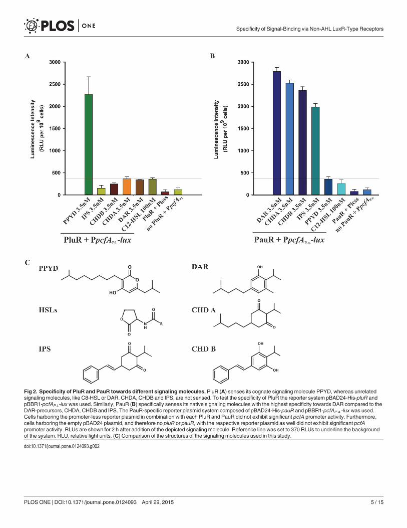
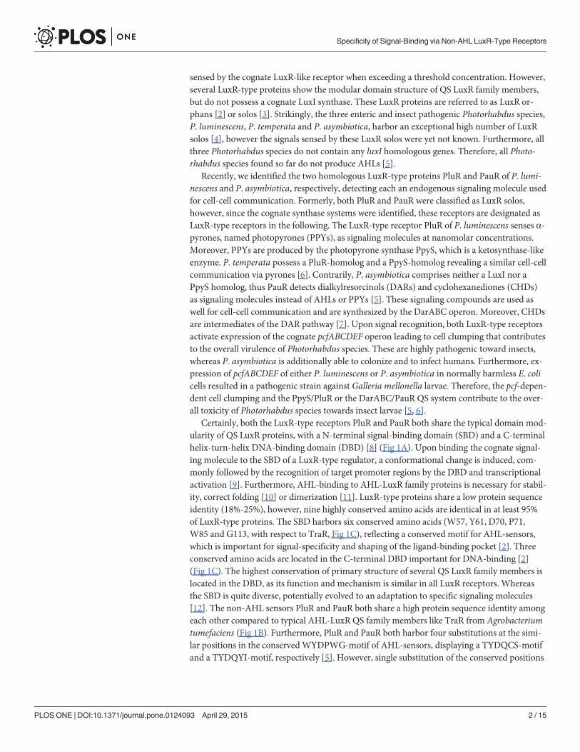
Fig 2. Specificity of PluR and PauR towards different signalingmolecules. PluR (A) senses its cognate signaling molecule PPYD, whereas unrelatedsignaling molecules, like C8-HSL or DAR, CHDA, CHDB and IPS, are not sensed. To test the specificity of PluR the reporter system pBAD24-His-pluR andpBBR1-pcfAP.l.-lux was used. Similarly, PauR (B) specifically senses its native signaling molecules with the highest specificity towards DAR compared to theDAR-precursors, CHDA, CHDB and IPS. The PauR-specific reporter plasmid system composed of pBAD24-His-pauR and pBBR1-pcfAP.a.-lux was used.Cells harboring the promoter-less reporter plasmid in combination with each PluR and PauR did not exhibit significant pcfA promoter activity. Furthermore,cells harboring the empty pBAD24 plasmid, and therefore no pluR or pauR, with the respective reporter plasmid as well did not exhibit significant pcfApromoter activity. RLUs are shown for 2 h after addition of the depicted signaling molecule. Reference line was set to 370 RLUs to underline the backgroundof the system. RLU, relative light units. (C) Comparison of the structures of the signaling molecules used in this study.
doi:10.1371/journal.pone.0124093.g002
Specificity of Signal-Binding via Non-AHL LuxR-Type Receptors
PLOS ONE | DOI:10.1371/journal.pone.0124093 April 29, 2015 5 / 15
isopropanol or methanol was added. Moreover, highest induction was determined at timepoint 2 h after addition of substances.
Amino acid substitutions in PluR or PauR might affect the spatial structure of the proteinsand influence their functionality to bind the cognate pcfA promoter. To quantify the structuralinfluence of amino acid replacements in the signal-binding domain (SBD) of PluR or PauR, theability of PluR wild type or PauR wild type and different derivatives to activate pcfA promoteractivity was determined. Therefore, the similar method was used as described above, however0.1% (w/v) arabinose was added and also derivatives of PluR or PauR were used. For bettercomparison, the values of PluR wild type or PauR wild type was set as 100% in the Figs 3 and 4.
To quantify the influence of amino acid replacements in the SBD of PluR or PauR on sens-ing of signaling molecules, the similar plasmid combinations as described above were used,however the distinct signaling molecules PPYD, CHDA, CHDB and DAR were added. Thesame final concentration of substances was used as described above. For better comparison, thevalues of PluR wild type or PauR wild type was set to 100% in the Figs 3 and 4.
Fig 3. Amino acid replacements within the SBD of PluR caused either functionality or impaired PPYD-sensing. The PluR derivatives D75E andS115G dramatically decreased functionality and hence decreased its ability to bind and activate pcfAP.l. promoter (lower left quadrant). Replacements within theTYDQCS-motif of PluR decreased the ability of PluR to sense PPYD. The most drastic influence on PPYD-sensing is detectable with the replacement of Y66A,D75A, D75N, Q76A, Q76P and S115A in PluR (lower right quadrant). Only the replacement T62W showed no effect and same induction levels as PluR wild type(upper right quadrant). The activity of the pcfAP.l. promoter was measured via luminescence as read-out and the depicted values were taken 2 h after addition of0.1% (w/v) arabinose (lower axis) or 3.5 nMPPYD (left axis) and compared to PluR wild type, which values were set to 100%. To evaluate the different PluRderivatives, a cut-off of 70%was set for each value. RLU (relative light units) values for all PluR derivatives and PluR wild type are depicted in S3 Table.
doi:10.1371/journal.pone.0124093.g003
Specificity of Signal-Binding via Non-AHL LuxR-Type Receptors
PLOS ONE | DOI:10.1371/journal.pone.0124093 April 29, 2015 6 / 15

Generation of αPluR and αPauR antibodiesTo generate specific antibodies against full-length PluR, E. coli BL21 was transformed with theplasmid pBAD24-His-pluR, cultivated at 30°C and expression was induced at OD600 = 0.5 with0.1% (w/v) arabinose. Whole cells were subjected to SDS-PAGE [16], and the amount of 6His-PluR was detected by staining with coomassie solution [40% (v/v) ethanol; 10% (v/v) aceticacid; 0.2% (w/v) coomassie brilliant blue R250] and destained with destaining solution [40%(v/v) ethanol; 10% (v/v) acetate] [17]. The according band with a size of 27.03 kDa was cut andused as an antigen to produce polyclonal antisera and antibodies in two rabbits (Biogenes, Ber-lin). Furthermore, total IgG of αPluR antibody was purified using Protein-A column (Biogenes,Berlin). Highest specificity of αPluR antibody was given in 3% BSA and a dilution of 1:10.000.
To generate specific antibodies against PauR, polyclonal antibodies were generated in tworabbits against a peptide of PauR (amino acids 62–75: CTMGNYDKNDNHDSD) (Biogenes,Berlin). Total IgG of αPauR antibody was purified using Protein-A column (Biogenes, Berlin).Highest specificity of αPauR antibody was given in 5% milk powder and a dilution of 1:10.000.
Fig 4. The TYDQYI-motif in the SBD of PauR is essential for the overall functionality of the receptor and DAR-sensing. The most drastic effects onDAR-sensing were gained with the replacement of S38A, T62A, Y66A, D75A and D75N in the SBD of PauR and a decreased effect on DAR-sensing weregained with the replacement of Y90C and I113S in PauR (lower right quadrant). The PauR derivatives Y40A, Y40F, D75E and Q76A dramatically influencedthe structure of PauR and decrease its ability to bind and activate pcfAP.a. promoter (lower left quadrant). The activity of pcfAP.a. promoter was measured vialuminescence as read-out and pictured values were taken 2 h after addition of 0.1% arabinose (lower axis) or 3.5 nM DAR (left axis) and compared to PauRwild type, which values were set to 100% (upper right quadrant). To evaluate the different derivatives, a cut-off of 70% was set for each value. RLU (relativelight units) values for all PauR derivatives and PauR wild type are depicted in S4 Table.
doi:10.1371/journal.pone.0124093.g004
Specificity of Signal-Binding via Non-AHL LuxR-Type Receptors
PLOS ONE | DOI:10.1371/journal.pone.0124093 April 29, 2015 7 / 15
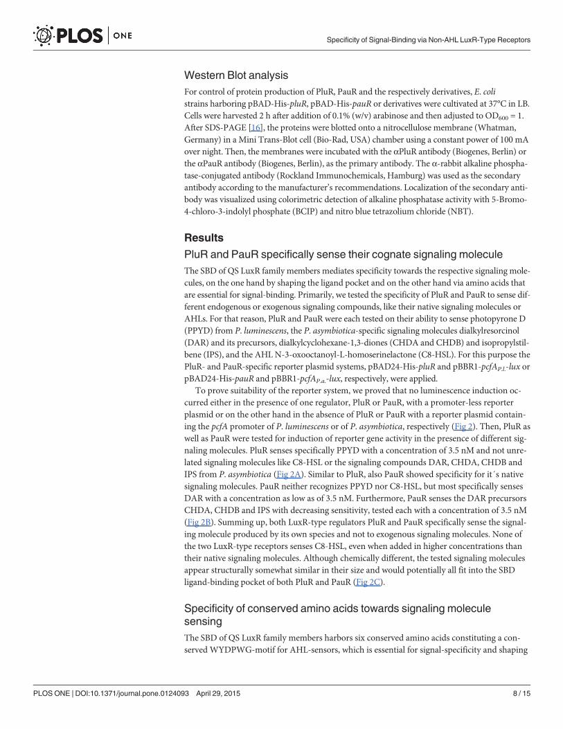
Western Blot analysisFor control of protein production of PluR, PauR and the respectively derivatives, E. colistrains harboring pBAD-His-pluR, pBAD-His-pauR or derivatives were cultivated at 37°C in LB.Cells were harvested 2 h after addition of 0.1% (w/v) arabinose and then adjusted to OD600 = 1.After SDS-PAGE [16], the proteins were blotted onto a nitrocellulose membrane (Whatman,Germany) in a Mini Trans-Blot cell (Bio-Rad, USA) chamber using a constant power of 100 mAover night. Then, the membranes were incubated with the αPluR antibody (Biogenes, Berlin) orthe αPauR antibody (Biogenes, Berlin), as the primary antibody. The α-rabbit alkaline phospha-tase-conjugated antibody (Rockland Immunochemicals, Hamburg) was used as the secondaryantibody according to the manufacturer’s recommendations. Localization of the secondary anti-body was visualized using colorimetric detection of alkaline phosphatase activity with 5-Bromo-4-chloro-3-indolyl phosphate (BCIP) and nitro blue tetrazolium chloride (NBT).
Results
PluR and PauR specifically sense their cognate signaling moleculeThe SBD of QS LuxR family members mediates specificity towards the respective signaling mole-cules, on the one hand by shaping the ligand pocket and on the other hand via amino acids thatare essential for signal-binding. Primarily, we tested the specificity of PluR and PauR to sense dif-ferent endogenous or exogenous signaling compounds, like their native signaling molecules orAHLs. For that reason, PluR and PauR were each tested on their ability to sense photopyrone D(PPYD) from P. luminescens, the P. asymbiotica-specific signaling molecules dialkylresorcinol(DAR) and its precursors, dialkylcyclohexane-1,3-diones (CHDA and CHDB) and isopropylstil-bene (IPS), and the AHL N-3-oxooctanoyl-L-homoserinelactone (C8-HSL). For this purpose thePluR- and PauR-specific reporter plasmid systems, pBAD24-His-pluR and pBBR1-pcfAP.l.-lux orpBAD24-His-pauR and pBBR1-pcfAP.a.-lux, respectively, were applied.
To prove suitability of the reporter system, we proved that no luminescence induction oc-curred either in the presence of one regulator, PluR or PauR, with a promoter-less reporterplasmid or on the other hand in the absence of PluR or PauR with a reporter plasmid contain-ing the pcfA promoter of P. luminescens or of P. asymbiotica, respectively (Fig 2). Then, PluR aswell as PauR were tested for induction of reporter gene activity in the presence of different sig-naling molecules. PluR senses specifically PPYD with a concentration of 3.5 nM and not unre-lated signaling molecules like C8-HSL or the signaling compounds DAR, CHDA, CHDB andIPS from P. asymbiotica (Fig 2A). Similar to PluR, also PauR showed specificity for it´s nativesignaling molecules. PauR neither recognizes PPYD nor C8-HSL, but most specifically sensesDAR with a concentration as low as of 3.5 nM. Furthermore, PauR senses the DAR precursorsCHDA, CHDB and IPS with decreasing sensitivity, tested each with a concentration of 3.5 nM(Fig 2B). Summing up, both LuxR-type regulators PluR and PauR specifically sense the signal-ing molecule produced by its own species and not to exogenous signaling molecules. None ofthe two LuxR-type receptors senses C8-HSL, even when added in higher concentrations thantheir native signaling molecules. Although chemically different, the tested signaling moleculesappear structurally somewhat similar in their size and would potentially all fit into the SBDligand-binding pocket of both PluR and PauR (Fig 2C).
Specificity of conserved amino acids towards signaling moleculesensingThe SBD of QS LuxR family members harbors six conserved amino acids constituting a con-served WYDPWG-motif for AHL-sensors, which is essential for signal-specificity and shaping
Specificity of Signal-Binding via Non-AHL LuxR-Type Receptors
PLOS ONE | DOI:10.1371/journal.pone.0124093 April 29, 2015 8 / 15

of the ligand-binding pocket. PluR and PauR both share only two of the conserved amino acidswithin AHL-sensors, Y66 and D75, and display a TYDQCS-motif and a TYDQYI-motif at thesimilar amino acid positions, respectively. Although, the sequence identity between PluR andPauR represents 83% (Fig 1), both sense different signaling molecules. To identify amino acidswithin each conserved motif essential for signal binding for PluR and PauR, the amino acidswithin the TYDQCS- and TYDQYI-motif were individually replaced either with alanine or therespective amino acids conserved in AHL-sensors or in PluR. As above, the ability of PluR orPauR and their derivatives were tested to bind and activate the corresponding pcfA promoterin presence of the native signaling molecule. To exclude structural influences on the receptorby the amino acid replacements, PluR and PauR wild type and their derivatives were first testedon their general functionality to bind and activate the corresponding pcfA promoter. This wastested by activation of pcfA promoter activity due to simple overproduction independent of thecognate signaling molecule (Fig 3, lower axis). The amino acid substitutions D75E and S115Gin PluR dramatically decreased its ability to activate expression of pcfA promoter region to 50%compared to PluR wild type (Fig 3, left lower quadrant). This decreased ability to bind and acti-vate the corresponding pcfA promoter, which is possibly caused by conformational defectswithin one monomer or defects in dimerization. However, all other PluR derivatives that havebeen tested showed no general functional defects. Furthermore, all tested amino acid replace-ments in PluR did not influence protein production since protein amounts comparable to thewild type could be detected after overproduction (S1 Fig). Then, the ability of all PluR deriva-tives was tested for PPYD-sensing. It emerged that the substitutions Y66A, D75A, D75N,Q76A and Q76P in PluR most dramatically impaired signaling molecule sensing (Fig 3, rightlower quadrant). The PluR derivative C90W showed a 50% reduced reporter gene activity com-pared to the wild type. Only the replacement T62W did not affect PPYD-sensing of PluR(Fig 3, upper right quadrant).
Likewise, PauR and its derivatives carrying amino acid substitutions in the TYDQYI-motifwere analyzed for their functionality and ability of DAR-sensing. Additionally, the amino acidsS38 and Y40 were also analyzed for their impact on DAR-sensing as these have been predictedto be as well involved in DAR-sensing [5]. The amino acid replacements Y40A, Y40F, D75E,D75N and Q76A in PauR dramatically influenced the general functionality of PauR. Thus,over-production of these proteins dramatically reduced pcfAP.a. promoter activity compared towild type (Fig 4, lower left quadrant). The most drastic effect appeared when position D75 wasreplaced with E and when position Y40 was substituted against A (7–25% compared to PauRwild type) (Fig 4, lower left quadrant). Therefore, these amino acids might be important forshaping the SBD and substitutions of these amino acids affect either the monomer structure ordimerization prior to pcfA promoter binding. Protein over-production of PauR wild type andall derivatives were comparable revealing that the amino acid substitutions did not effect pro-tein production (S1 Fig). All tested amino acids replacements within the TYDQYI-motif aswell as S38A decreased the ability of PauR to sense DAR (Fig 4, lower right quadrant). Replace-ment of T62A in PauR completely prevented DAR-sensing (5%) without affecting its overallfunctionality. Furthermore, drastic effects were gained on DAR-sensing with the replacementof S38A, Y66A, D75A and D75N in the SBD of PauR (33–65%) and a decreased effect onDAR-sensing were gained with the replacement of Y90C and I113S in PauR (53–64%) (Fig 4,lower right quadrant).
In both LuxR-type receptors the size of amino acid at position 75 seems to be crucial. WhenD75 is substituted with a bigger amino acid (D75E) the general functionality of both proteinsPluR and PauR is affected (Figs 3 and 4). Whereas charge reversion at position D75N impairedsignal-binding and not the general functionality of both PluR and PauR. Furthermore, aminoacid Y66 in both proteins is essential for recognizing of the corresponding signaling molecule.
Specificity of Signal-Binding via Non-AHL LuxR-Type Receptors
PLOS ONE | DOI:10.1371/journal.pone.0124093 April 29, 2015 9 / 15

Interestingly, also position S38 of PauR is crucial for DAR-sensing (Fig 4). However, thisamino acid is not comprised in the conserved TYDQYI-motif but predicted to be involved inDAR-sensing in a DAR-docking model of the SBD of PauR [5]. Overall, all amino acids withinthe conserved amino acid motif in PluR as well as PauR are important for signal-sensing orfunctionality, supporting the idea that all these amino acids are located in the respective signal-binding pocket.
In another approach, we changed the TYDQYI-motif of PluR to the WYDPWG-motif ofAHL-sensors to possibly achieve AHL-sensing instead of PPYs. For that reason, we generatedquadruple substitutions within the SBD of PluR, resulting in the derivative PluR-T62W/Q76P/C90W/S115G, which was stepwise performed via introducing the next amino acid exchange inPluR-T62W. With successive introduction of more amino acid substitutions within the SBD ofPluR, the general functionality of these derivatives is stepwise decreased. However, these deriv-atives showed comparable protein amounts like wild type (S1 Fig). The quadruple derivativePluR-T62W/Q76P/C90W/S115G strongly impaired the ability to activate the pcfAP.l. promotertested by simple over-production of the protein (Fig 5A). Likewise, PPYD-sensing is stepwiseimpaired with progressive amino acid substitutions in PluR, but AHL-sensing could not begained (Fig 5B). Furthermore, the reporter gene activities after addition of C8-HSL were lowerthan the background values of unspecific signaling molecules (compare with Fig 2A). PluRshows the highest protein sequence identity to QscR of Pseudomonas aeruginosa of LuxR-typeregulators with known crystal structures. The LuxR solo QscR responds to multiple AHLs,like C8-HSL [18], which was therefore used in our study. In conclusion, more amino acidsmust make up the specificity for AHL-sensing besides the conserved WYDPWG-motif.
Since PluR is very homologous to PauR with a protein sequence identity of 83% (Fig 1), wetried to convert PauR to a PPY-sensor. For that reason, the conserved TYDQYI-motif of PauRwas converted to the TYDQCS-motif of PluR to potentially gain PPYD-sensing, though alsothe general functionality of PauR-Y90C/I113S was affected and reduced to approximately 50%compared to PauR wild type (Fig 5C). However, PauR-Y90C/I113S still had the ability to senseDAR, although this was dramatically reduced about 70% compared to PauR wild type (Fig5D). Protein amounts of PauR-Y90C/I113S were comparable to PauR wild type (S1 Fig).
Thus, these six conserved amino acids in the SBD, displaying either the WYDPWG-motif ofAHL-sensors, the TYDQCS-motif of PluR or the TYDQYI-motif of PauR, are all essential forshaping the specific ligand pocket and ligand-binding, however, these are not sufficient for sig-nal-sensing and-specificity. The solely insertion of the specific amino acid motif for AHL-,PPY- or DAR-sensors into a LuxR-type receptor is therefore not sufficient to convert the signalspecificity of the sensor. This reveals that other amino acids in the SBD must also be essentialfor forming the ligand-binding pocket and for signal perception, although these are not highlyconserved.
DiscussionThe two LuxR-type regulators PluR and PauR are both part of a quorum sensing system in P.luminescens and P. asymbiotica depending on non-AHL compounds as signals. Both receptorssense different endogenous signaling molecules, PPYs or DARs, respectively, but both activatethe expression of the corresponding pcfABCDEF operon. Expression of the pcfABCDEF operonin turn leads to cell clumping and contributes to the virulence of Photorhabdus species [5, 6].In this study we focused on signal specificity of the two non-AHL sensing LuxR-type receptorsPluR and PauR.
PluR and PauR harbor two of the conserved amino acids of the conserved WYDPWG-motifof AHL-sensors, comprising a TYDQCS-motif and a TYDQYI-motif, respectively. However,
Specificity of Signal-Binding via Non-AHL LuxR-Type Receptors
PLOS ONE | DOI:10.1371/journal.pone.0124093 April 29, 2015 10 / 15
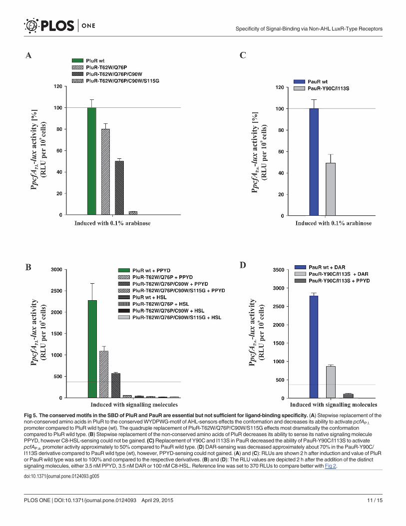
Fig 5. The conservedmotifs in the SBD of PluR and PauR are essential but not sufficient for ligand-binding specificity. (A) Stepwise replacement of thenon-conserved amino acids in PluR to the conservedWYDPWG-motif of AHL-sensors effects the conformation and decreases its ability to activate pcfAP.l.
promoter compared to PluR wild type (wt). The quadruple replacement of PluR-T62W/Q76P/C90W/S115G effects most dramatically the conformationcompared to PluR wild type. (B) Stepwise replacement of the non-conserved amino acids of PluR decreases its ability to sense its native signaling moleculePPYD, however C8-HSL-sensing could not be gained. (C)Replacement of Y90C and I113S in PauR decreased the ability of PauR-Y90C/I113S to activatepcfAP.a. promoter activity approximately to 50% compared to PauR wild type. (D) DAR-sensing was decreased approximately about 70% in the PauR-Y90C/I113S derivative compared to PauRwild type (wt), however, PPYD-sensing could not gained. (A) and (C): RLUs are shown 2 h after induction and value of PluRor PauRwild type was set to 100% and compared to the respective derivatives. (B) and (D): The RLU values are depicted 2 h after the addition of the distinctsignaling molecules, either 3.5 nMPPYD, 3.5 nMDAR or 100 nMC8-HSL. Reference line was set to 370 RLUs to compare better with Fig 2.
doi:10.1371/journal.pone.0124093.g005
Specificity of Signal-Binding via Non-AHL LuxR-Type Receptors
PLOS ONE | DOI:10.1371/journal.pone.0124093 April 29, 2015 11 / 15

our studies reveal that these motifs are as important for signal-specificity and conformation asfor AHL-sensing of QS LuxR family proteins. Substitution of the conserved amino acid D75 ofeach PluR and PauR highly decreased the recognition of the cognate signaling molecule. SinceD75 of PluR and PauR is deduced to form a hydrogen bond to the hydroxy group attached tothe pyrone and the DAR-hydroxy group, respectively [5, 6]. Likewise, in the AHL-sensor TraRthis position (D70) is known to be important for binding the amide group of the N-3-oxoocta-noyl-L-homoserine lactone [19]. Certainly in PluR and PauR the size and charge of the aminoacid at position D75 mediates correct signaling molecule binding as substitution against glu-tamic acid impaired conformation and substitution against asparagine either affect binding ofPPYD or DAR. Docking experiments with PauR and DAR as ligand revealed an arene-areneinteraction between T62 and Y66 and the DAR aromatic ring [5]. This was also confirmed bysingle replacements of both amino acids with alanine, which decreased the ability to sense theligand (Fig 4). However, in PluR only Y66 was deduced to form an arene-arene interactionwith the pyrone ring [6]. Accordingly, substitution of Y66A in PluR showed a dramatically re-duced ability to sense PPYD, whereas substitution of T62 against A showed no effect (Fig 3).The appropriate position to Y66 of PluR and PauR is Y61 in AHL-sensors and this amino acidis known to be involved in binding of the acyl chain of the signaling molecule via hydrophobicinteractions, e.g. in TraR [19] or LuxR [11]. In general, the six conserved amino acids in theSBD are essential for shaping the ligand pocket and ligand-binding, either in QS LuxR familymembers binding AHLs or non-AHLs like PluR and PauR. Also the subfamily of LuxR solos ofplant-associated bacteria have conserved substitutions in the WYDPWG-motif of AHL-sen-sors, which are W57M and Y61W (with respect to TraR), however the specific signaling mole-cules are yet unknown. These substitutions are assumed to allow the binding of plant signalmolecules rather than AHLs [2, 20]. Furthermore, several amino acids beside the WYDPWG-motif are known to be involved either in ligand-binding, dimerization or DNA-binding inSdiA from E. coli [21], TraR from A. tumefaciens [22] and LuxR from V. fischeri [23, 24]. Thisis also true for PauR, in which the position S38 outside of the conserved motif is important forDAR-sensing.
In conclusion, the conserved TYDQCS- and TYDQYI-motifs of PluR and PauR, respective-ly, are essential but not only sufficient for ligand-binding. Hence, other amino acids of the SBDmust also contribute to the signal sensing specificity, although these are not highly conserved.Similarly, QS LuxR family members sensing AHLs contain several important amino acids inthe SBD that are important for AHL-binding beside the conserved WYDPWG-motif. There-fore, each QS LuxR-type protein potentially evolved special amino acids to bind its specific sig-naling molecule to regulate diverse cellular processes. However, PluR and PauR regulate theexpression of the cognate pcfABCDEF operon leading to cell clumping. Therefore, the questionremains why both organisms, P. luminescens and P. asymbiotica, use different molecules forthis QS-regulated process, which results in a similar phenotype. Possibly, both adapted to theirdifferent host, and therefore PPY-signaling might be more appropriate for infection of cold-blooded hosts like insect larvae, and DAR-signaling might be a better choice for infecting endo-therm organisms like humans. This idea is underline by the fact that genomic analysis revealedthat many human pathogens are putative DAR producers and that these pathogens might alsoconstitute a DAR-dependent QS system, possibly besides an intact AHL QS system [5].
In summary, our studies reveal that specific amino acid motifs in the SBD of LuxR-type re-ceptors are important for signal-sensing, but not alone sufficient for signal-specificity. The re-placement of diverse amino acids within the SBD allow LuxR-type receptors to sense diversefamilies of signaling molecules beside AHLs. The specific amino acid motifs for AHL as well asfor PPY and DAR sensors are incomplete to date. Future work has to be performed to identify
Specificity of Signal-Binding via Non-AHL LuxR-Type Receptors
PLOS ONE | DOI:10.1371/journal.pone.0124093 April 29, 2015 12 / 15

the complete amino acid motifs in the SBD of LuxR-type receptors, which can then be used forsignal prediction of yet un-investigated LuxR solos.
Supporting InformationS1 Fig. Protein production of PluR and PauR and their respective derivatives. For analysisof protein production of PluR and its respective derivatives (A) and of PauR and its respectivederivatives (B), E. coli strains harboring pBAD-His-pluR, pBAD-His-pauR or variants werecultivated at 37°C in LB medium. Cells were harvested 2 h after addition of 0.1% (w/v) arabi-nose, as a control no arabinose was added. The figure shows the immunoblots of 12.5% SDSgels. Antibodies directed against the respective protein were used to detect PluR or PauR. PluRhas an estimated size of 27.03 kDa and PauR has an estimated size of 27.14 kDA. The PageRu-ler prestained protein ladder (Thermo Fischer, Schwerte) was used to estimate protein sizes.(TIF)
S1 Table. Plasmids used in this study.(PDF)
S2 Table. Oligonucleotides used in this study. Underlined nucleotides indicate the positionof the site-directed mutagenesis.(PDF)
S3 Table. Influence of amino acid substitutions within the SBD of PluR on general func-tionality and PPYD-sensing. PluR wild type and PluR derivatives were tested for their abilityto activate pcfAP.l. promoter activity controlling the luxCDABE operon in the presence of 0.1%(w/v) arabinose or 3.5 nM PPYD. Reporter gene activity was quantified 2 h after addition of0.1% (w/v) arabinose (functionality [%]) or 3.5 nM PPYD (PPYD-sensing [%]) and comparedto PluR wild type, which values were set to 100%. RLU, relative light units. Std, standard devia-tion of three biological experiments.(PDF)
S4 Table. Influence of amino acid substitutions within the SBD of PauR on general func-tionality and DAR-sensing. PauR wild type and PauR derivatives were tested for their abilityto activate pcfAP.a. promoter activity controlling the luxCDABE operon in the presence of 0.1%(w/v) arabinose or 3.5 nM DAR. Reporter gene activity was quantified 2 h after addition of0.1% (w/v) arabinose (functionality [%]) or 3.5 nM DAR (DAR-sensing [%]) and compared toPauR wild type, which values were set to 100%. RLU, relative light units.(PDF)
AcknowledgmentsWe are grateful to Dr. Helge Bode for providing purified DARs and PPYs. We thank NikolaiPeschek and Patrick Reith for generating amino acid substitutions in PauR.
Author ContributionsConceived and designed the experiments: SB RH. Performed the experiments: SB. Analyzedthe data: SB RH. Wrote the paper: SB RH.
References1. Waters CM, Bassler BL. Quorum sensing: cell-to-cell communication in bacteria. Annu Rev Cell Dev
Biol. 2005; 21: 319–346. doi: 10.1146/annurev.cellbio.21.012704.131001 PMID: 16212498
Specificity of Signal-Binding via Non-AHL LuxR-Type Receptors
PLOS ONE | DOI:10.1371/journal.pone.0124093 April 29, 2015 13 / 15

2. Patankar AV, González JE. Orphan LuxR regulators of quorum sensing. FEMSMicrobiol Rev. 2009;33: 739–756. doi: 10.1111/j.1574-6976.2009.00163.x PMID: 19222586
3. Subramoni S, Venturi V. LuxR-family “solos”: bachelor sensors/regulators of signalling molecules. Mi-crobiology. 2009; 155: 1377–1385. doi: 10.1099/mic.0.026849–0 PMID: 19383698
4. Brameyer S, Kresovic D, Bode HB, Heermann R. LuxR solos in Photorhabdus species. Front Cell InfectMicrobiol. 2014; 4: 1–23. doi: 10.3389/fcimb.2014.00166 PMID: 24478989
5. Brameyer S, Kresovic D, Bode HB, Heermann R. Dialkylresorcinols as bacterial signaling molecules.PNAS USA. 2015; 112: 572–577. 201417685. doi: 10.1073/pnas.1417685112 PMID: 25550519
6. Brachmann AO, Brameyer S, Kresovic D, Hitkova I, Kopp Y, et al. Pyrones as bacterial signaling mole-cules. Nat Chem Biol. 2013; 9: 573–578. doi: 10.1038/nchembio.1295 PMID: 23851573
7. Fuchs SW, Bozhüyük KAJ, Kresovic D, Grundmann F, Dill V, et al. Formation of 1,3-Cyclohexane-diones and Resorcinols Catalyzed by aWidely Occuring Ketosynthase. Angew Chem Int Ed. 2013; 52:4108–4112. doi: 10.1002/anie.201210116 PMID: 23423827
8. Shadel GS, Young R, Baldwin TO. Use of regulated cell lysis in a lethal genetic selection in Escherichiacoli: Identification of the autoinducer-binding region of the LuxR Protein from Vibrio fischeri ATCC7744. J Bacteriol. 1990; 172: 3980–3987. PMID: 2141835
9. Welch M, Todd DE, Whitehead NA, McGowan SJ, Bycroft BW, et al. N-acyl homoserine lactone bindingto the CarR receptor determines quorum-sensing specificity in Erwinia. EMBO J. 2000; 19: 631–641.PMID: 10675332
10. Zhu J, Winans SC. The quorum-sensing transcriptional regulator TraR requires its cognate signaling li-gand for protein folding, protease resistance, and dimerization. PNAS USA. 2001; 98: 1507–1512.PMID: 11171981
11. Nasser W, Reverchon S. New insights into the regulatory mechanisms of the LuxR family of quorumsensing regulators. Anal Bioanal Chem. 2006; 387: 381–390. doi: 10.1007/s00216-006-0702-0 PMID:16953322
12. Vannini A, Volpari C, Gargioli C, Muraglia E, Cortese R, et al. The crystal structure of the quorum sens-ing protein TraR bound to its autoinducer and target DNA. EMBO J. 2002; 21: 4393–4401. PMID:12198141
13. Woodcock DM, Crowther PJ, Doherty J, Jefferson S, DeCruz E, et al. Quantitative evaluation of Escher-ichia coli host strains for tolerance to cytosine methylation in plasmid and phage recombinants. NucleicAcids Res. 1989; 17: 3469–3478. PMID: 2657660
14. Guzman LM, Belin D, Carson MJ, Beckwith J. Tight regulation, modulation, and high-level expressionby vectors containing the arabinose PBAD promoter. J Bacteriol. 1995; 177: 4121–4130. PMID:7608087
15. Sambrook J, Russel DW. Molecular Cloning—A Laboratory Manual. Cold Spring Harbour LaboratoryPress. 2001; Cold Spring Harbour, New York.
16. Laemmli UK. Cleavage of structural proteins during the assembly of the head of bacteriophage T4. Na-ture. 1970; 227: 680–685. doi: 10.1016/j.fgb.2010.11.011 PMID: 5432063
17. Weber K, Osborn M. The reliability of molecular weight determinations by dodecyl sulfate-polyacryl-amide gel electrophoresis. J Biol Chem. 1969; 244: 4406–4412. PMID: 5806584
18. Chugani S, Greenberg EP. An evolving perspective on the Pseudomonas aeruginosa orphan quorumsensing regulator QscR. Front Cell Infect Microbiol. 2014; 4: 1–7. doi: 10.3389/fcimb.2014.00152/abstract PMID: 24478989
19. Churchill MEA, Chen L. Structural basis of acyl-homoserine lactone-dependent signaling. Chem Rev.2011; 111: 68–85. doi: 10.1021/cr1000817 PMID: 21125993
20. González JF, Patel HK. A novel widespread interkingdom signaling circuit. Trends Plant Sci. 2013; 18:167–174. doi: 10.1016/j.tplants.2012.09.007 PMID: 23089307
21. Kim T, Duong T, Wu C, Choi J, Lan N, et al. Structural insights into the molecular mechanism of Escher-ichia coli SdiA, a quorum-sensing receptor. Acta Cryst. 2013; 70: 694–707. doi: 10.1107/S1399004713032355
22. Luo Z-Q, Smyth AJ, Gao P, Qin Y, Farrand SK. Mutational analysis of TraR: Correlating function withmolecular structure of a quorum-sensing transcriptional activator. J Biol Chem. 2003; 278: 13173–13182. doi: 10.1074/jbc.M210035200 PMID: 12569101
23. Trott AE, Stevens AM. Amino acid residues in LuxR critical for its mechanism of transcriptional activa-tion during quorum sensing in Vibrio fischeri. J Bacteriol. 2001; 183: 387–392. doi: 10.1128/JB.183.1.387–392.2001 PMID: 11114940
24. Koch B. The LuxR receptor: the sites of interaction with quorum-sensing signals and inhibitors. Microbi-ology. 2005; 151: 3589–3602. doi: 10.1099/mic.0.27954–0 PMID: 16272381
Specificity of Signal-Binding via Non-AHL LuxR-Type Receptors
PLOS ONE | DOI:10.1371/journal.pone.0124093 April 29, 2015 14 / 15

25. Letunic I, Doerks T, Bork P. SMART 7: recent updates to the protein domain annotation resource. Nu-cleic Acids Res. 2012; 40: D302–D305. doi: 10.1093/nar/gkr931 PMID: 22053084
26. Artimo P, Jonnalagedda M, Arnold K, Baratin D, Csardi G, et al. ExPASy: SIB bioinformatics resourceportal. Nucleic Acids Res. 2012; 40: W597–W603. doi: 10.1093/nar/gks400 PMID: 22661580
Specificity of Signal-Binding via Non-AHL LuxR-Type Receptors
PLOS ONE | DOI:10.1371/journal.pone.0124093 April 29, 2015 15 / 15


![LUXr 1-day workshop, August 15, 2012 [San Francisco]](https://img.pdfslide.us/doc/110x75/54c7b5694a7959c66e8b45d0/luxr-1-day-workshop-august-15-2012-san-francisco.jpg)


![LUXr 1-day workshop, April 27, 2012 [San Francisco]](https://img.pdfslide.us/doc/110x75/54c73ffa4a7959b4048b4588/luxr-1-day-workshop-april-27-2012-san-francisco.jpg)


![LUXr 1-day workshop, June 13, 2012 [San Francisco]](https://img.pdfslide.us/doc/110x75/54c73ff44a7959c27e8b45a1/luxr-1-day-workshop-june-13-2012-san-francisco.jpg)