Embed Size (px)
Citation preview
Special SensesSpecial Senses
The EyeThe Eye
Overview of the EyeOverview of the Eye
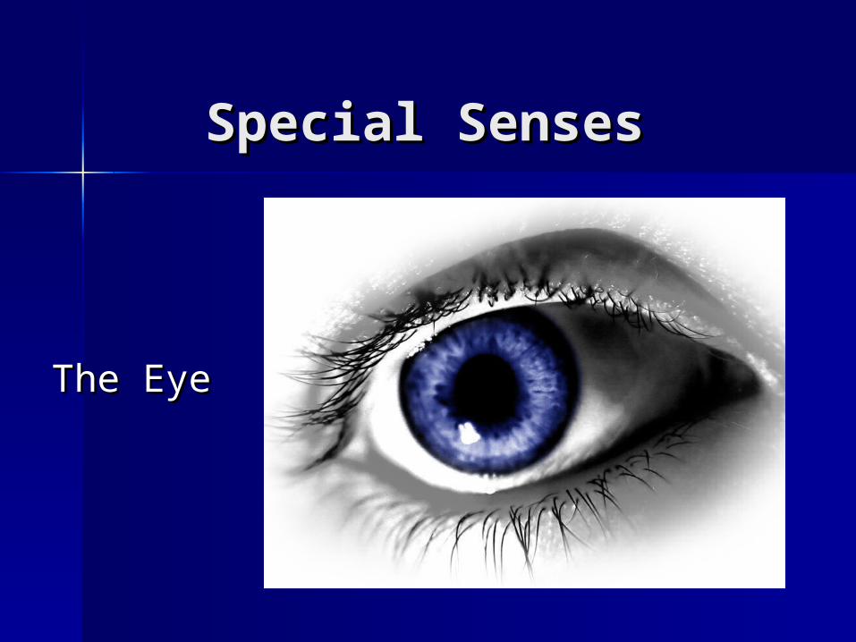
Eye acts like a camera– Lens adjusts to bring object into focus– Pupil constricts to allow less light to
enter in bright setting or dilates to allow more light to enter in darker setting
– Through bending of light rays, images reaches retina
Sensitive nerve cell layer of eye Image is transmitted to brain for
interpretation
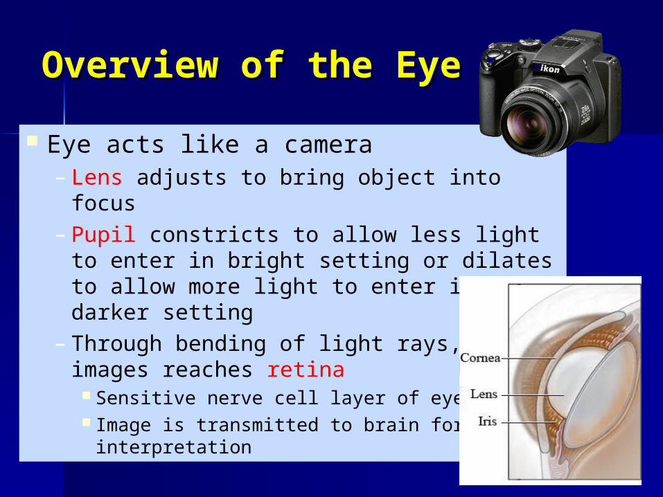
Front view of the eyeFront view of the eye Sclera = white
portion of eye– tough, maintains
shape of eyeball– serves as protective
covering for the eye Iris = colored
portion of the eye Pupil= opening in
center of eye that controls amount of light entering eye
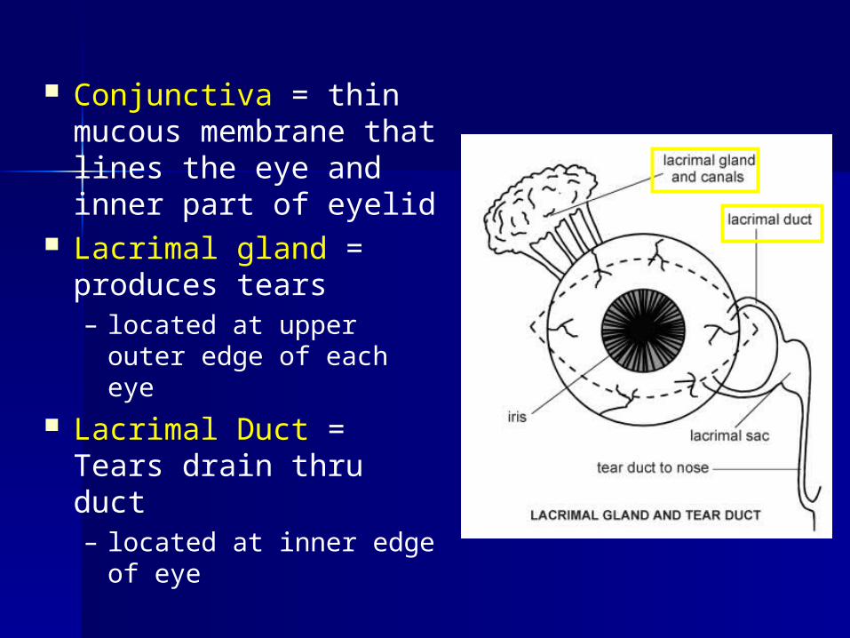
Conjunctiva = thin mucous membrane that lines the eye and inner part of eyelid
Lacrimal gland = produces tears– located at upper outer
edge of each eye Lacrimal Duct =
Tears drain thru duct– located at inner edge
of eye
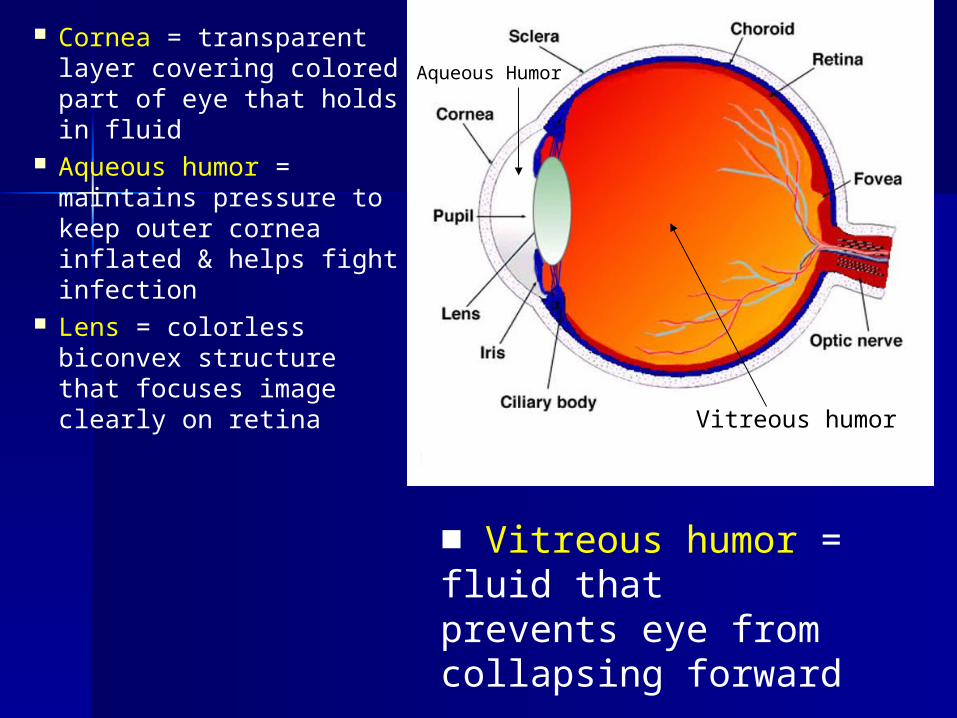
Cornea = transparent layer covering colored part of eye that holds in fluid
Aqueous humor = maintains pressure to keep outer cornea inflated & helps fight infection
Lens = colorless biconvex structure that focuses image clearly on retina
Aqueous Humor
■ Vitreous humor = fluid that prevents eye from collapsing forward
Vitreous humor
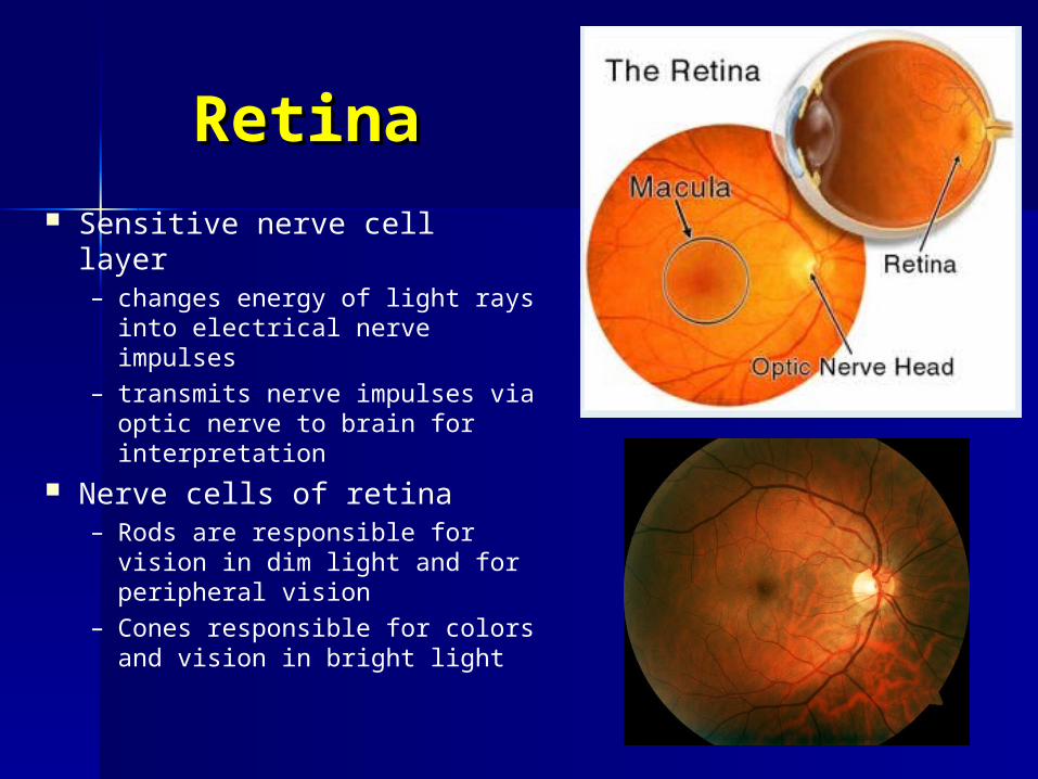
RetinaRetina Sensitive nerve cell layer
– changes energy of light rays into electrical nerve impulses
– transmits nerve impulses via optic nerve to brain for interpretation
Nerve cells of retina– Rods are responsible for
vision in dim light and for peripheral vision
– Cones responsible for colors and vision in bright light
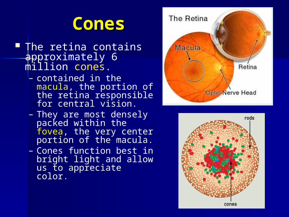
ConesCones The retina contains
approximately 6 million cones. – contained in the
macula, the portion of the retina responsible for central vision.
– They are most densely packed within the fovea, the very center portion of the macula.
– Cones function best in bright light and allow us to appreciate color.
Fovea
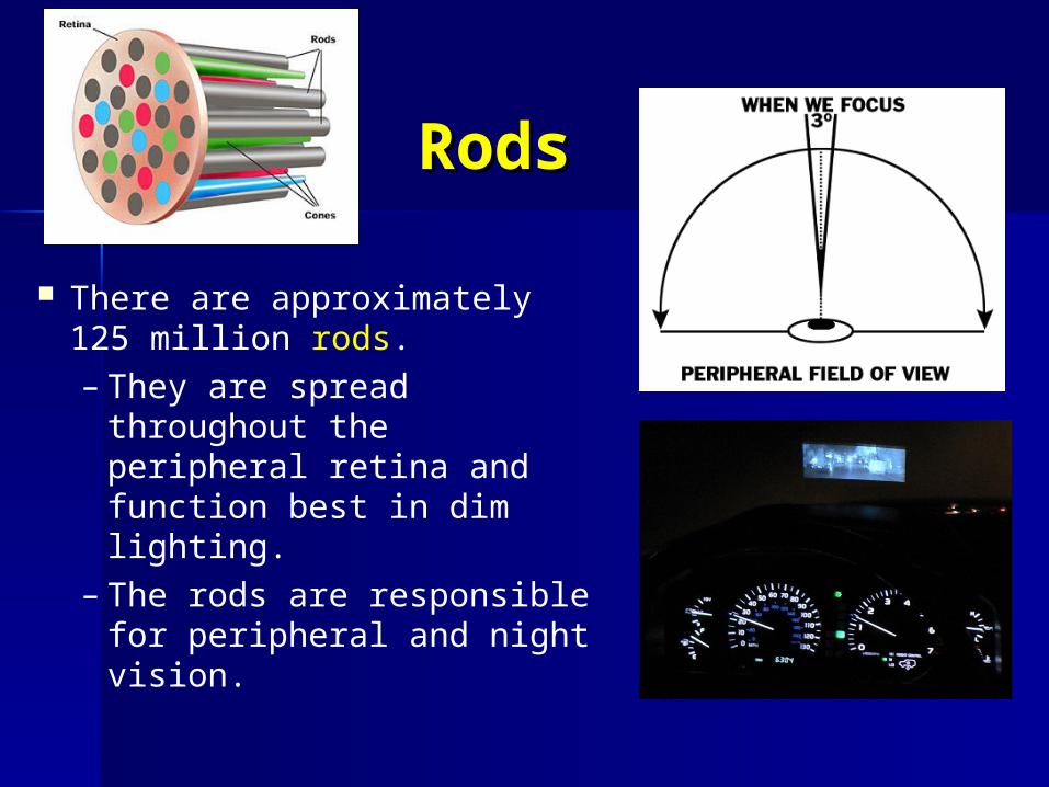
RodsRods
There are approximately 125 million rods. – They are spread
throughout the peripheral retina and function best in dim lighting.
– The rods are responsible for peripheral and night vision.
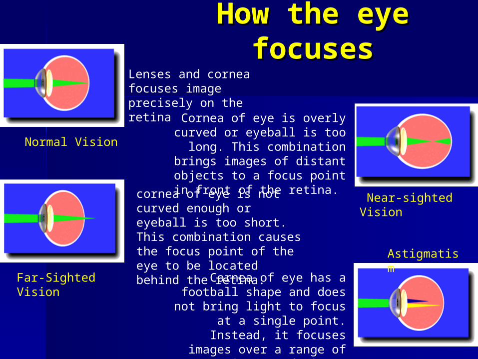
How the eye How the eye focusesfocuses
Lenses and cornea focuses image precisely on the retina
Cornea of eye is overly curved or eyeball is too long. This combination
brings images of distant objects to a focus point in
front of the retina. cornea of eye is not curved enough or eyeball is too short. This combination causes the focus point of the eye to be located behind the retina.
Cornea of eye has a football shape and does not bring
light to focus at a single point. Instead, it focuses
images over a range of points producing a blurred image
Normal Vision
Near-sighted Vision
Far-Sighted Vision
Astigmatism
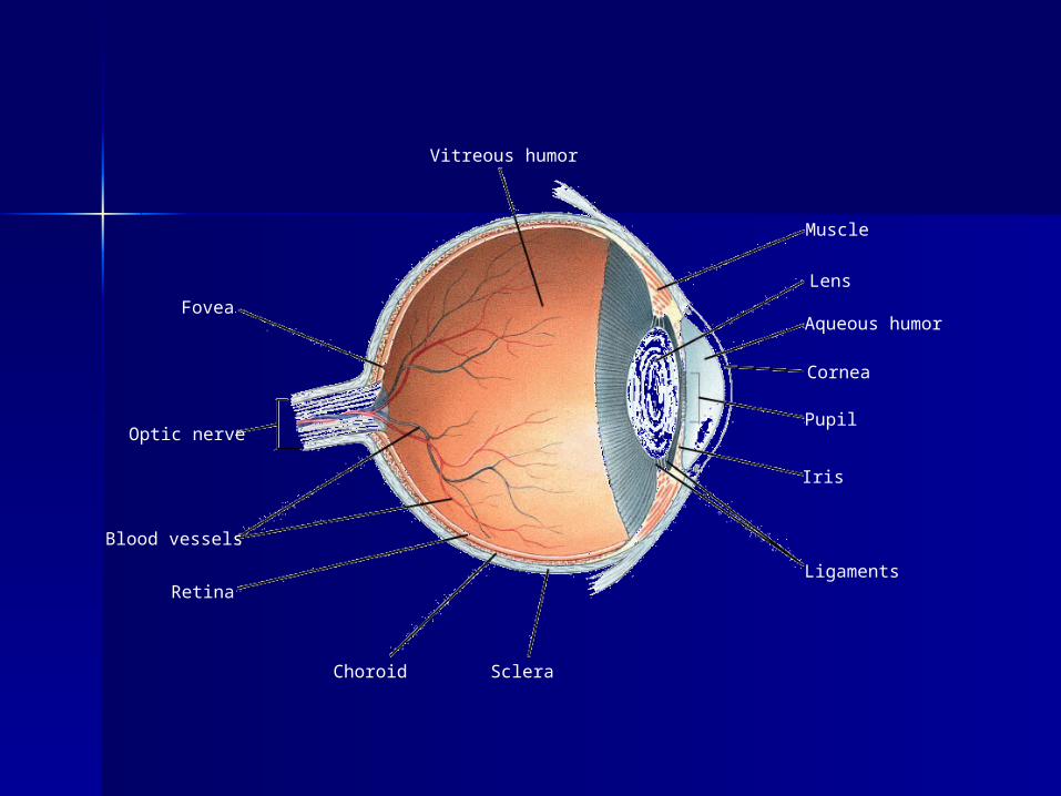
Choroid
Retina
Blood vessels
Optic nerve
Fovea
Vitreous humor
Sclera
Ligaments
Iris
Pupil
Cornea
Aqueous humor
Lens
Muscle
Section 35-4
Figure 35-14 The Eye
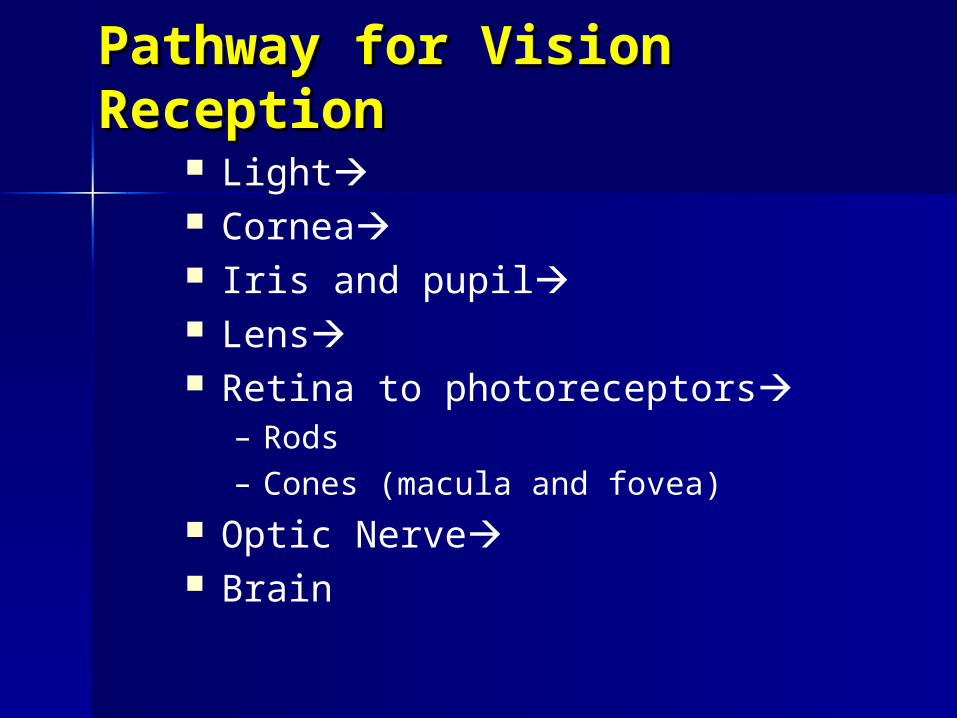
Pathway for Vision Pathway for Vision ReceptionReception
Light Cornea Iris and pupil Lens Retina to photoreceptors
– Rods– Cones (macula and fovea)
Optic Nerve Brain