Embed Size (px)
Citation preview

Spatial Localization of Chaperone Distribution in
the Endoplasmic Reticulum of Yeast
Marc Griesemer1, Carissa Young2, Anne Robinson2, and LindaPetzold1
1Department of Computer Science, University of California, SantaBarbara, CA 93106
2Department of Chemical Engineering, University of Delaware,Newark, DE 19716
Abstract
In eukaryotes, the endoplasmic reticulum (ER) serves as the first membrane-
enclosed organelle in the secretory pathway, with functions including protein
folding, maturation, and transport. Molecular chaperones, of the Hsp70 family
of proteins, participate in assisting these processes and are essential to cellu-
lar function and survival. BiP is a resident Hsp70 chaperone in the ER of
Saccharomyces cerevisiae. In this study we have created a partial differential
equation (PDE) model to examine how BiP interacts with the membrane-bound
co-chaperone Sec63 in translocation, a process in which BiP assists in guiding
a nascent protein into the ER lumen. We found that when Sec63 participates
in translocation through localization at the membrane, the spatial distribution
of BiP is inhomogeneous, with more BiP at the surface. When translocation
is inhibited through a disabling of Sec63’s membrane tether, the concentration
of BiP throughout the ER becomes homogeneous. Our computational simula-
tions suggest that Sec63’s localization and the resulting binding to BiP near the
membrane surface of the ER enable a heterogeneous distribution of BiP within
the ER, and may facilitate BiP’s role in translocation.
1

1 Introduction
Molecular chaperones participate in a wide range of processes essential to cellu-
lar function and survival. Found in all organisms, and ubiquitously distributed
in the major compartments of eukaryotic cells, most are intricate players in the
response to cellular stress [1]. Hsp70s assist in protein folding and maturation,
assembly or disassembly of complexes, ribosomal RNA processing, translocation
of newly synthesized proteins, suppression of aggregation, and protein degrada-
tion (as reviewed in [2, 3]). The versatility among molecular chaperones is
intriguing, for they have a single purpose: to bind protein substrates. The very
high degree of conservation among Hsp70 proteins may favor a unique molecular
mechanism common to all, whereas functional differences may depend on modu-
lating co-chaperones such as Hsp40s and nucleotide exchange factors (NEFs) [4].
This phenomenon of a protein having multiple, sometimes competing functions,
indicates a high level of systems control.
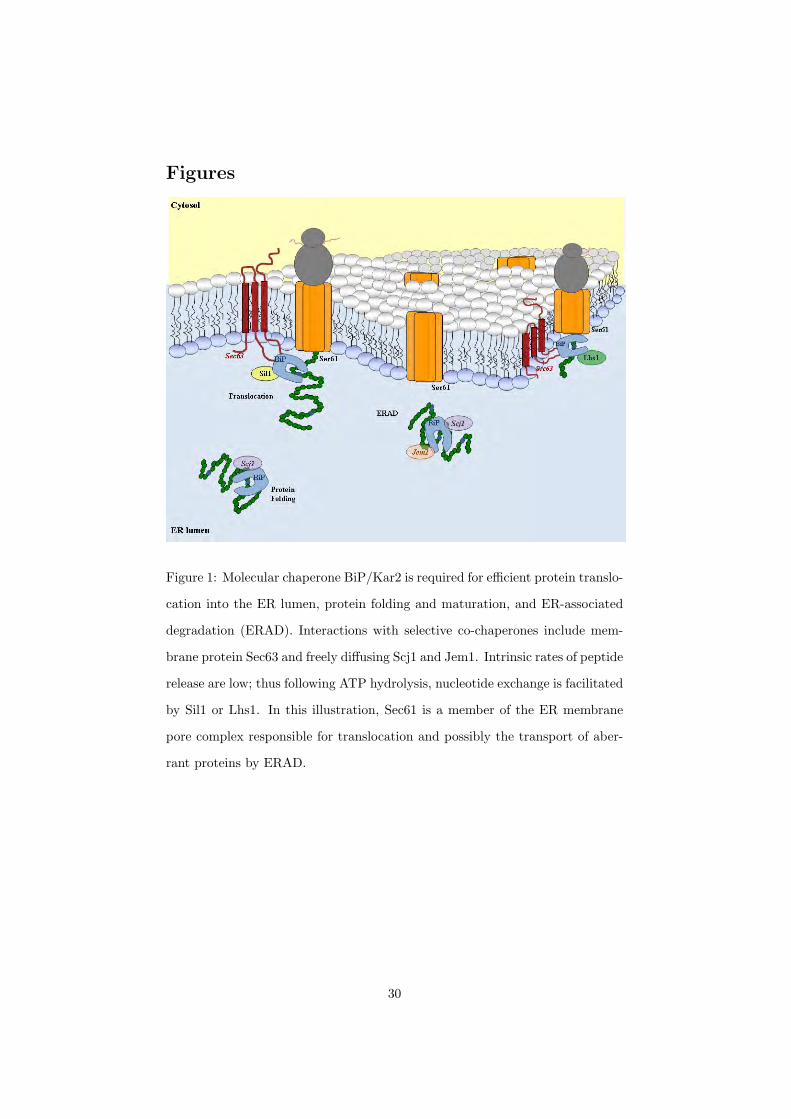
In the lumen of the endoplasmic reticulum (ER), a network of chaperones
and cofactors ensures the proper folding of secretory proteins. One of the most
abundant proteins of the ER is the Hsp70 molecular chaperone, BiP. Through
biochemical and genetic experiments, BiP has been identified in critical cel-
lular processes including protein translocation of newly synthesized precursors
across the ER membrane, folding and maturation, karyogamy, and ERAD (ER-
associated degradation) where unfolded or abnormally folded proteins are sent
back to the cytosol for degradation [5–9]. Like other Hsp70 chaperones, BiP
assists the folding of a protein by repeated ATP-controlled cycles of binding
and release. Co-chaperones, such as Hsp40s, stimulate the binding of molecular
chaperones to the substrate and regulate chaperone activities [10]. In the ER of
S. cerevisiae, co-chaperone Sec63 directly interacts with BiP, increasing its affin-
ity for the nascent proteins proceeding through the translocation pore [11–13]
(Figure 1). Simultaneously within the ER lumenal environment, co-chaperones
Scj1 and Jem1 associate with BiP during the processes of protein maturation
2

and karyogamy, respectively [8, 9, 14]. BiP, Jem1, and Scj1 are all involved in
the degradation of aberrant soluble proteins through ERAD [15]. As illustrated
in Figure 1, Sil1 and Lhs1 are the nucleotide exchange factors (NEF) that play
key roles in these processes by triggering substrate release [16, 17].
The regulation of Hsp70-Hsp40-NEF interactions is best understood for Es-
cherichia coli homologues DnaK, DnaJ, and GrpE. Mechanistic details have
been experimentally explored and mathematically modeled [18–24]. Yet, E.
coli is an organism that does not consist of membrane-bound compartments
to perform distinct cellular functions. Additionally, many chaperone-mediated
processes involve spatial aspects, such as the subcellular localization of messen-
ger RNA leading to translation of their encoded proteins [25]; subcompartments
of the nucleus implicated in the processes of transcription and splicing [26, 27];
and the spatial localization of BiP at the ER membrane maintaining the per-
meability barrier during protein translocation [28].
Yeast, S. cerevisiae, is a simple eukaryotic organism that compartmental-
izes selective processes and protein-protein interactions to specified organelles.
Proteomic studies have verified the location of ER-resident proteins of S. cere-
visiae and identified absolute levels of protein expression [29, 30]. These data
suggest that the concentration of BiP exceeds the level of co-chaperones by at
least an order of magnitude at normal growth conditions, and is significantly
up-regulated during quality control mechanisms of the cell including the heat
shock response (HSR) and unfolded protein response (UPR) [31, 32]. It is also
known that multiple BiPs can bind to substrates with varying affinities [33].
Experimentally, the co-chaperone Sec63 must be spatially localized at a sub-
organelle level – the ER membrane – in order for translocation to occur [34].
Given this evidence, we hypothesize that the spatial localization of BiP and
interactions with co-chaperones regulate its diversity and functionality in the
ER of S. cerevisiae.
Thus, we describe our model development in Section 2, discuss the signifi-
cance of our simulations in Section 3, and examine the sensitivity of our model
3

to parameter perturbations in Section 4. In order to investigate the chaperone
and co-chaperone interaction of BiP and Sec63, respectively, in ER transloca-
tion, we have introduced spatial components to our model. Subsequently, we
have shown that Sec63’s localization and functional interaction with BiP in
translocation provides an explanation for BiP’s heterogeneity in the ER.
2 Models
Modelers have attempted to discern the role of Hsp70 chaperones in assist-
ing and accelerating translocation of proteins across membranes of organelles,
specifically the ER and mitochondria of S. cerevisiae. Previous work has fo-
cused on transport mechanisms, including either the Brownian ratchet model
[35], comparison to the power stroke model [36, 37], or a unifying mechanism of
both termed entropic pulling [4]. We have examined the significance of spatial
effects between BiP and the ER membrane co-chaperone Sec63 and implemented
the reaction rates associated with previous models that evaluate a nascent pro-
tein as it transits through the translocation pore. Specifically, we are interested
in how the BiP-Sec63 interaction enhances translocation of the nascent protein.
Experiments exploring the BiP-Sec63 interaction in vitro suggest that Sec63
acts as an anchor to localize BiP within the proximity of the translocation
channel, and accelerates the transit of a peptide through the membrane pore by
regulating ATP hydrolysis of the chaperone [5, 34].
This work implements the experimental evidence [12, 16, 17, 38–42] with
the added spatial component including the ER membrane and lumenal regions.
Developing spatially-relevant models is important as in vivo experiments such
as single particle tracking (SPT) and technology advancements in fluorescence
imaging begin to capture spatial effects of protein localization (reviewed in [43]).
Estimates of species concentrations have been determined for S. cerevisiae [30]
as well as binding affinities between BiP, Sec63, and synthetic peptides [38, 39].
When experimental data was not available, as in the case of the interactions
4

between NEFs, Sil1 and Lhs1 and BiP, established estimates from the mam-
malian literature were used (Supplementary Material). However, a degree of
uncertainty is inherent when evaluating kinetic rates as a result of in vitro ex-
periments. We have incorporated these estimates into models used to elucidate
in vivo mechanisms and varied the parameters by two orders of magnitude to
test the sensitivity of the particular values. Due to the high degree of homology
between chaperones and co-chaperones amongst eukaryotes, we believe that this
estimation procedure is an appropriate method.
Our goal is to use modeling to better understand the role of Sec63 in parti-
tioning of BiP within the ER. To this end, we first constructed an ODE model to
examine the reaction pathways and then extended this system to a PDE model
in order to capture the spatiotemporal dynamics.
2.1 Model Descriptions
2.1.1 Core ODE model
Our core model is described by a system of ordinary differential equations. It
is a 7 state, 13 parameter model representing the interactions of BiP with the
co-chaperone Sec63 and unfolded proteins. The schematic below, Figure 2,
depicts all plausible states of BiP binding to nascent proteins during protein
translocation into the ER lumen. In general, molecular chaperones alternate
between an ATP-bound state representing fast substrate binding/release rates
and therefore a low affinity for proteins (upper right triangle, Fig.2), and an
ADP-bound state characteristic of slow association/dissociation kinetics and a
high affinity for substrates (lower left triangle, Fig. 2) [44]. The states of the
model are given in Table 1.
Under physiological conditions, BiP preferentially binds to ATP (X1) and
associates with either unfolded proteins in the lumen or nascent proteins of the
translocon (Fig. 1). BiP’s interaction with unfolded proteins can occur in the
presence or absence of co-chaperone mediation (X1 → X6) [38, 40] where BiP
5
preferentially binds to hydrophobic residues [33, 45]. ATP hydrolysis ensues
(X6 → X7), trapping the peptide to form X7. Due to low intrinsic rates of
bound peptide released from the chaperone complex, nucleotide exchange fac-
tors further accelerate the cycle of protein folding (X7 → X5 → X1). The
conventional mechanism of translocation proceeds with the binding of the co-
chaperone Sec63 to BiP (X2) which synergistically stimulates peptide binding
(X3) and ATP hydrolysis (X4). This coupling effect has been shown to mediate
the molecular trapping of proteins that BiP would not bind on its own [38, 39].
However, it is also plausible that BiP could initially bind to the unfolded pro-
tein and then sequentially associate with its co-chaperone during translocation.
This scenario is represented in states X1 → X6 → X3.
We have assumed that a single BiP is activated per Sec63 molecule located
at the ER membrane. In vitro studies have indicated that the BiP-Sec63 inter-
action occurs transiently, and suggest that one Sec63 molecule could potentially
activate at least ten BiP molecules [38]. On average, E. coli experiments have
determined that incoming proteins present a new Hsp70 binding site every 25
to 35 amino acids [46] which is consistent with S. cerevisiae literature that es-
timates a minimum of six to seven molecules of BiP bound to the endogeneous
protein, ppαF [47]. For simplicity, our model assumes a 1:1 stoichiometry be-
tween BiP and unfolded proteins as well as between BiP and Sec63.
Simulations were realized until the values reached a steady-state level and
then the final species concentrations were obtained for a range of initial condi-
tions. We found that when the total amount of BiP is low, the Sec63-dependent
pathway (Fig. 2, outer loop) accounted for most of the bound BiP. As the to-
tal amount of BiP in the system was increased, the Sec63-dependent pathway
(Fig. 2, diagonal) dominated, and states such as BiP-U-ATP (X6) accounted
for most of the BiP. The latter result confirmed an important point: that BiP
strongly interacts with unfolded protein, although it should be mentioned that
the association rate to form X6 was chosen on the higher end of the range of
experimental data [24]. This core ODE model served as a description of reaction
6

kinetics between BiP, Sec63, and unfolded protein, and was a building block for
constructing a spatially-dependent model describing chaperone interactions in
the ER. Details of the model are summarized in the Supplementary Material.
2.1.2 PDE Model
We constructed a PDE model, making use of the reaction kinetics from the
ODE model, to determine the spatial distribution of BiP in the ER. The model
incorporates: (1) chemical reactions representing transitions between states in
the ODE system that take place on the inner membrane, and (2) diffusion
into the lumen of the ER. This spatially dependent system of equations was
approximated by the method of finite differences (Figure 3). The irregular
geometry of the ER was simplified to a sphere (surrounding a spherical nucleus)
and assumed to be symmetric. With these assumptions, the (time-dependent)
system can be modeled in one spatial dimension. We believe a PDE model is
justified because BiP and (wild-type) Sec63 have a localized interaction at the
membrane that precludes a well-mixed system.
We define the membrane-associated zone as the membrane portion of the
ER where protein-protein interactions take place between BiP and the other
proteins in the system. Images attained by electron microscopy (EM) show
that the membrane has a defined thickness [48], shown as 35 nm in Figure
3. Sec63 spans the membrane [49] and associates with the Sec61-constituted
pore channel [50]. We do, however, consider the entire region as well-mixed,
represented by the boundary condition of the PDE.
The rate of change of the concentration of species k is the sum total of the
concentrations of the free and bound species plus the diffusion in the interior.
∂Cz,k
∂t= Rk + D
∂2Cl,k
∂x2
∣∣∣x=0
(1)
Reactions and (one-dimensional) diffusion occurs in the lumen
7

∂Cl,k
∂t= Rk + D
∂2Cl,k
∂x2(2)
with the nuclear-ER boundary condition given by
∂Cl,k
∂x
∣∣∣x=Ll
= 0. (3)
Here, Cz,k is the membrane-associated zone concentration and Cl,k repre-
sents the concentration in the lumen for diffusing species k. If this species exists
only in the membrane-associated zone, then this term is absent. D is the dif-
fusion coefficient (assumed to be the same for all ER lumenal species), and Rk
is the reaction term for species k. Lz is the length of the membrane-associated
zone (in one dimension), and Ll is the annular radius of the lumen. The variable
x represents the distance from the membrane-associated zone for a particular
point on the grid.
Discretizing in space, we obtain
∂(Cz,k)∂t
= Rk + DC1
l,k − Cz,k
∆x2(4)
∂C1l,k
∂t= Rk + D
Cz,k − 2C1l,k + C2
l,k
∆x2(5)
...
∂CNl,k
∂t= D
CN−1l,k − CN
l,k
∆x2, (6)
where Cil,k represents the concentration of species k at grid point i in the lumen,
and ∆x is the spatial separation between two consecutive grid points. The
boundary at x = 0 is the ER membrane, and the boundary at x = Ll is the
nucleus. Given this formulation, the concentration of each species is tracked
spatially and temporally.
Several assumptions were made to simplify the model and approximate the
8

dynamics of the system. Reactions can occur in both the membrane-associated
zone and the lumen. We define unfolded proteins as one species without dis-
tinguishing between proteins diffusing in the lumen from those transiting the
translocation pore. Loss of proteins at the translocon (due to completed translo-
cation) is offset by a production reaction (∅ → U) that allows for replenishment
of the U pool. The translocated U is designated as a non-reacting product Ut
(Figure 2). Thus, the total amount of unfolded protein in the model remains
constant.
3 Simulation Results
3.1 Model Scenarios
Three scenarios were constructed to simulate different conditions in the ER.
In each scenario, different species were allowed to diffuse in the lumen. In our
model, we assume that if all the constituent species in a bound state can diffuse
in a particular scenario, then the bound state can diffuse as well. The scenarios
are:
(1) The wild-type case that assumes that ER-resident chaperone BiP, nu-
cleotide exchange factors Sil1 and Lhs1, soluble U, and selective com-
plexes, diffuse freely throughout the lumen [16, 41, 51, 52]. Sec63 is an
integral membrane protein and therefore remains embedded in the ER
lipid bilayer [53].
(2) In addition to the species outlined in Scenario 1, Sec63 and membrane-
associated complexes are now allowed to diffuse into the lumen. Exper-
imental data has shown that the presence of a soluble variant of Sec63
(63Jp) not localized to the ER membrane inhibits efficient protein translo-
cation. These results suggest that 63Jp competes with the endogenous
membrane-localized Sec63 by sequestering BiP from the vicinity of the
translocon pore (Sec61 complex of Fig. 1) [34]. In this case, we can test
9
whether the variant’s ability to diffuse into the lumen leads to a loss of
BiP’s heterogeneous distribution in the ER.
(3) When the BiP-Sec63 protein interactions are severely impaired, or in the
absence of Sec63 within the system of scenario 1 (i.e. zero concentration),
we have been able to mimic a situation in which ER protein translocation
fails. Mutations in either Sec63 or BiP have been shown to inhibit translo-
cation due to defects in Sec63 interacting with BiP (sec63-1, [5, 54]) and,
to varying degrees, BiP mutants display different translocation efficiencies
into the lumen (kar2-113, kar2-159, and kar2-203; [55]).
It should be noted that only scenario one represents physiological conditions.
Scenarios 2 and 3 are special conditions that have been experimentally obtained
using either protein variants or thermosensitive yeast strains. The species and
states that diffuse in each scenario are given in Table 2.
3.2 Ratio Metric
From the ER PDE model, we determined the ratio of total BiP concentration
localized to the membrane-associated zone compared to the total BiP concentra-
tion of the interior. This metric gives an indication of BiP’s spatial localization
within the ER sub-compartments. The default scenario is to tether Sec63 to the
membrane, while BiP, NEF and U are allowed to diffuse. From these scenarios,
one can make predictions of the importance of these species on translocation.
This is described by the equation:
RatioBiP =
[BiPtotal
]z
[BiPtotal]l, (7)
where the subscript z means the membrane-associated zone and l means the
lumen. At steady-state, the concentrations of diffusing species in the membrane-
associated zone and the lumen are the same, with the concentration of the non-
diffusing species on the membrane-associated zone determining the divergence
of the BiP ratio from unity.
10

3.3 Model Results
Using the DASPK ODE/DAE solver [56], we ran simulations for each of the
scenarios until steady-state (t = 105 s). The system starts from conditions
where all proteins are free and present in the membrane-associated zone. Dif-
fusion is fast, equalizing gradients across the ER. Reactions then occur in the
membrane-associated zone and in the lumen on a slower time scale. We an-
alyzed the concentration of each state in the membrane-associated zone and
at interior grid points and then calculated the ratio of total BiP concentration
in the membrane-associated zone compared to the total BiP concentration in
the lumen. Simulations accounted for total Sec63 ranging from 0.55 to 1.37
mM (10, 000-25, 000 molecules) while BiP and NEF concentrations were varied
from 0.55 to 55 mM (104-106 molecules). These initial conditions were based
on a range around populations given in the Yeast Genome Database [30]. The
amount of total unfolded protein (U) was 5.5 mM or 105 molecules (in Section
4, Sensitivity Analysis).
When Sec63 is appropriately integrated in the membrane, BiP preferen-
tially remains in the membrane-associated zone (Figure 4), resulting in an in-
homogeneous distribution throughout the ER. This is expected from the model
since BiP is reacting in the membrane-associated zone and forming non-diffusing
species. When Sec63 is allowed to diffuse or is removed from the system entirely,
BiP is uniformly distributed in the ER, resulting in a BiP ratio (Equation 7) of
one for all total values of Sec63 and BiP. Thus, when translocation is inhibited,
the concentration is homogeneous. When total BiP levels are low, most of it is
involved in translocation, so the BiP ratio is high (left side of Fig. 4 graph).
In the limit of high BiP levels, relatively less BiP is used in translocation, so
its distribution tends towards homogeneity (right side of Fig. 4 graph). This
calculation was initially performed with 11 spatial grid points and repeated for
several grid sizes in order to increase the resolution of our results (e.g. up to
1000), yielding identical results for the ratios.
We examined the results of an ODE model to describe the impact of the
11

interaction between BiP and Sec63 on the chaperone distribution in the ER. In
this model, we assumed that fast diffusion produced a well-mixed system for
all species. We then scaled the concentrations of Sec63-derived states (i.e. X2,
X3, and X4) with respect to the volume of the membrane-associated zone and
the concentrations of the remaining states using the combined volume of the
membrane zone and lumen. The calculation of the BiP ratio then proceeded as
defined in the last section.
In comparison with the PDE model, we found that this modified ODE model
resulted in a higher BiP ratio at low total BiP populations as shown in Figure
4. This discrepancy occurs because in the ODE model, Sec63 has access to the
entire BiP population for translocation. The PDE model, on the other hand,
allows for the presence of BiP in both the membrane-associated zone and the
lumen. In this case, a percentage of BiP does not participate with Sec63 in
translocation. Especially when BiP is scarce, this difference produces a higher
BiP ratio in the ODE model versus the PDE model. Given BiPs known presence
in both membrane-associated zone and in the lumen, we believe that a spatial
model best describes the chaperone dynamics in the ER. Furthermore, the PDE
model takes into account that the lumenal BiP is involved in other processes
(e.g. protein folding and degradation).
The determination of an actual physiological value of the BiP ratio in Fig-
ure 4 is complicated; many cellular factors affect it. For example, if BiP binds
at higher stoichiometric ratios to Sec63 and U than 1:1, then there would be
much more BiP found in the membrane-associated zone, and the BiP ratio
would increase for the same initial conditions. BiP is involved in multiple non-
translocation cellular functions [57], which may result in a smaller percentage
available to translocation, and would describe the right side of Figure 4. If either
BiP or Sec63 are partially defective in their interaction with each other [39, 58],
this scenario would lead to a flattening of the curves toward homogeneity. Ad-
ditionally, perturbations to the cellular environment, such as the induction of
stress conditions in the ER lead to membrane expansion [59] and a resulting
12

increase in ER volume [60], yet simultaneously, the unfolded protein response
(UPR) increases the amount of BiP in the lumen to handle an unfolded protein
load [32]. Thus, the BiP ratio would change dynamically in time. This project
does not attempt a thorough investigation of protein folding in the ER nor
perturbations to the system that, in turn, induce various quality control mech-
anisms; rather our model introduces the effect of dimensionality — specifically
ER compartmentalization — and shows that BiP’s interaction with Sec63 at
the membrane brings about spatial localization in the chaperone’s distribution
within the ER, which will ultimately have implications in protein folding and
other ER-related functions.
4 Sensitivity Analysis
Although we found BiP’s concentration in the ER under wild-type conditions
to be inhomogeneous (scenario 1), we also wanted to examine how the degree
of heterogeneity (as quantified in the BiP ratio) would change under variation
in model parameters and initial conditions, to examine the influence of various
model inputs on the system.
We first simulated the variation in total number of each protein species in
the ER. The Yeast Genome Database reports the total physiological population
of BiP and Sec63 proteins at 337, 000 and 17, 700 molecules, respectively, but
with those estimates there is a great deal of uncertainty. It is known that BiP
levels in the yeast ER can fluctuate and are regulated by the unfolded protein
response. Cellular conditions also dictate a large possible range in concentration
of unfolded proteins. Thus we simulated the model over a large range of values
for all protein species, as summarized in Table 3. The resulting BiP ratio for
variation in Sec63 is given in Figure 4. We also examined a range of unfolded
protein concentrations in our simulations from 0.55 to 8.25 mM (10, 000-150, 000
molecules). The ratio results for U are shown in Figure 5. An estimate for U
came from Surface Plasmon Resonance (SPR) experiments [39] using the peptide
13

p5 at 2 mM or roughly 40, 000 molecules in the ER. We found that this value
of protein concentration resulted in less than a one percent deviation in the BiP
ratio from our standard conditions (at 5.5 mM or 100, 000 molecules). Most of
the U binds with free BiP, but the vast excess remains free and is not a factor
of the BiP ratio calculation. Overall, our simulations with the aforementioned
Sec63 and U concentrations have confirmed our previous result that when BiP
levels are low, the BiP ratio is high and is affected by the amount of Sec63 in
the system. As BiP levels are increased, the BiP ratio becomes insensitive to
changes in all initial conditions.
We next considered the binding rate between BiP and U as variable parame-
ters (kinetic parameters k2 and k7 , Figure 6). BiP interacts with varying affini-
ties to numerous types of unfolded proteins in the ER and these interactions are
dependent on the exposed hydrophobic residues of the unfolded proteins [45].
Multiple BiPs can bind to a single translocating protein resulting in stoichio-
metric effects on the reaction, impacting the overall rate as well [35]. We varied
the association rates over five orders of magnitude and recorded the BiP ratio
for each simulation. As the association rate between BiP and U increases, there
is less of these species in the free (as opposed to bound) state, but this does not
affect the total amount of BiP bound to Sec63 at the membrane. Therefore, we
found that the BiP ratio was essentially the same for all kinetic values.
We examined the effect on the BiP ratio given a change in the Sec63 disso-
ciation rate from the trimeric complex of BiP, Sec63, and U (Figure 7). This
is described by the kinetic rate constants k5 and k−3. The values from the lit-
erature are 0.0086 and 0.038 s−1 [38], respectively. Rate k5 is slower than the
reactions upstream and downstream in the pathway (e.g., k4 = 0.016 s−1 and
k6 = 0.267 s−1). Thus, a bottleneck occurs, resulting in a high concentration
of the BiP-Sec63-U-ADP (X4) state. In vitro experiments [39, 61, 62] suggest
that Sec63 may have a more transient interaction with BiP, which would allow
for higher translocation efficiency. We increased the Sec63 dissociation rate ten,
one hundred, and a thousand fold, and found that the BiP ratio decreased at
14
low levels of BiP and Sec63. We surmise this is due to translocating protein
being shuttled out of the Sec63-dependent pathway at a faster rate, which in
the wild-type scenario accounts for the excess BiP on the membrane.
These results were also verified by local sensitivity analysis. Using SUNDI-
ALS [63], we computed the sensitivities of the BiP ratio to perturbations of the
reaction-rate parameters. If the BiP ratio is defined as a function G, by the
chain rule we get the derived function,
dG
dp=
∂G
∂C∂C∂p
+∂G
∂p(8)
where C and p represent the states and parameters, respectively. The derivative
∂C/∂p is the standard sensitivity matrix while the second term ∂G/∂p equals
zero because the BiP ratio has no explicit dependence on parameters. The BiP
ratio sensitivities are then scaled by the BiP ratios and parameter values to
allow for comparison. The results are summarized in Table 4.
We took the minimum and maximum of 13 sets of initial conditions as de-
fined in the last section as the range of our sensitivities. We then ranked the
parameters by their maximum and calculated the mean of the ensemble. As
Table 4 illustrates, the BiP ratio is robust to perturbations in parameters.
Finally, we examined the sensitivity of the BiP ratio by varying all the
kinetic parameters simultaneously. We used uniform random numbers to select
each parameter value in the range [0.1pi, 10pi], where pi is the nominal value
of the parameter. From this method, we generated 105 sets of parameters to
serve as the input for the simulations. We then simulated the model, increasing
the total BiP concentration from 0.55 to 55 mM , and recording the resulting
distributions of the BiP ratio in a histogram as presented in Figure 8. Other
metrics were considered [64], but we wanted to examine the global sensitivity
of the BiP ratio and to vary more than one parameter at a time.
We found that at the low end of the BiP concentration range (0.55 mM), the
BiP ratio had a wide spread of values for the simulations. When BiP is scarce,
15

changes in kinetic parameters have a significant effect on whether the BiP is
bound to Sec63 at the membrane or diffuses freely in the lumen. Despite this
sensitivity, most simulations yielded a highly heterogeneous BiP distribution
(BiP Ratio = 3.5-163.7, 95 percent confidence interval for total BiP = 0.55
mM), confirming that most of the BiP participates in translocation. The tails
of the BiP ratio distributions are due to the combination of parameters all being
near their maximum values. As the total BiP in the ER increases, more of the
BiP is not interacting with Sec63 (but rather freely diffusing in the lumen); thus
the reaction parameters have much less effect. Further studies concluded that
no one parameter dominates the variation of the BiP ratio. The qualitative
behavior of the model was reproduced in all 105 simulations.
In summary, the BiP distribution is heterogeneous with a high concentration
of BiP near the membrane for the wild-type condition where Sec63 is tethered to
the membrane. In contrast, for situations where Sec63 is allowed to freely diffuse
into the lumen, the BiP concentration is homogeneous. Is Sec63 recruiting BiP
to the membrane? In the model there is no driving force for BiP diffusion
toward the membrane. However, BiP has a relatively fast diffusion rate, which
means that a BiP molecule readily traverses both the lumen and the membrane-
associated zone. When it arrives at the membrane-associated zone, it has a
high probability of binding to Sec63 and this binding results in a preferential
localization at the membrane. This supports the model that the J-domain co-
chaperones act to modulate BiP localization in the ER.
5 Conclusion
We have developed a spatial PDE model describing the chaperone activity in the
ER of S. cerevisiae through reaction-diffusion equations. From the simulations,
we found that the concentration of BiP in the system was inhomogeneous, with
the concentration being greater on the membrane, particularly as BiP levels
in the ER decrease. Our simulations showed, however, that when Sec63 was
16

untethered and allowed to diffuse freely into the lumen, the BiP distribution
was homogeneous. This is consistent with the absence of BiP (translocation
failure) at the membrane observed experimentally in this situation. Sec63’s
localization and functional interaction with BiP in translocation provides an
explanation for BiP’s heterogeneity in the ER.
6 Acknowledgements
The authors gratefully acknowledge funding from NIH Grant GM07529 and
NSF IGERT Grant DGE02-21715. We also would like thank David Raden (U.
Delaware) and Theresa Yuraszeck (UC Santa Barbara) for helpful discussions.
References
[1] Georgopoulos, C. and Welch, W.: ‘Role of the Major Heat Shock Proteins
as Molecular Chaperones’, Annu. Rev. Cell Biol., 9, 1993, pp. 601–634
[2] Mayer, M. P. and Bukau, B.: ‘Hsp70 Chaperones: Cellular Functions and
Molecular Mechanisms’, Cell Mol. Life Sci., 62, 2005, pp. 670–684
[3] Buck, T., Wright, C., and Brodsky, J.: ‘The Activities and Function of
Molecular Chaperones in the Endoplasmic Reticulum’, Seminars in Cell
and Developmental Biology, 18, 2007, pp. 751–761
[4] de los Rios, P., Ben-Zvi, A., Slutsky, O., Azem, A., and Goloubinoff, P.:
‘Hsp70 Chaperones Accelerate Protein Translocation and the Unfolding of
Stale Protein Aggregates by Entropic Pulling’, Proc. Natl. Acad. Sci., 103,
2006, pp. 6166–6171
[5] Brodsky, J. L. and Schekman, R.: ‘A Sec63p-BiP complex from Yeast is
Required for Protein Translocation in a Reconstituted Proteoliposome’, J.
Cell Biol., 123, (6 Pt 1), 1993, pp. 1355–1363
17
[6] Latterich, M. and Schekman, R.: ‘The Karyogamy Gene KAR2 and Novel
Proteins are Required for ER-Membrane Fusion’, Cell, 78, (1), 1994, pp.
87–98
[7] McCracken, A. A. and Brodsky, J. L.: ‘Evolving Questions and Paradigm
Shifts in Endoplasmic Reticulum-Associated Degradation (ERAD)’, BioEs-
says, 25, (9), 2003, pp. 868–877
[8] Nishikawa, S. and Endo, T.: ‘The Yeast Jem1p is a DnaJ-like Protein of
the Endoplasmic Reticulum Membrane Required for Nuclear Fusion’, J.
Biol. Chem., 272, (20), 1997, pp. 12889–12892
[9] Schlendstedt, G., Harris, S., Risse, B., Lill, R., and Silver, P.: ‘A Yeast
DnaJ Homologue, Scj1p, Can Function in the Endoplasmic Reticulum with
BiP/Kar2 via a Conserved Domain that Specifies Interactions with Hsp70s’,
J. Cell Biol., 129, 1995, pp. 979–988
[10] Hennessy, F., Nicoll, W., Zimmermann, R., Cheetham, M., and Blatch, G.:
‘Not all J Domains are Created Equal: Implications for the Specificity of
Hsp40-70 Interactions’, Prot. Sci., 14, 2005, pp. 1697–1709
[11] Scidmore, M. A., Okomura, H. H., and Rose, M.: ‘Genetic Interactions
between KAR2 and SEC63, Encoding Eukaryote Homologues of DnaK and
DnaJ in the Endoplasmic Reticulum’, Molecular Biology of the Cell, 4,
1993, pp. 1145–1159
[12] Lyman, S. K. and Schekman, R.: ‘Interaction between BiP and Sec63
is Required for the Completion of Protein Translocation into the ER of
Saccaromyces cerevisiae’, J. Cell Biol., 131, (5), 1995, pp. 1163–1171
[13] Young, B., Craven, R., Reid, P., Willer, M., and Stirling, C.: ‘Sec63p and
Kar2p are Required for the Translocation of SRP-dependent Precursors
into the Yeast Endoplasmic Reticulum in vivo’, EMBO J., 20, 2001, pp.
262–271
18

[14] Silberstein, S., Schlenstedt, G., Silver, P., and Gilmore, R.: ‘A Role of
the DnaJ Homologue Scj1 in Protein Folding in the Yeast Endoplasmic
Reticulum’, J Cell Biol., 143, 1998, pp. 921–933
[15] Nishikawa, S. I., Fewell, S. W., Kato, Y., Brodsky, J. L., and Endo, T.:
‘Molecular Chaperones in the Yeast Endoplasmic Reticulum Maintain the
Solubility of Proteins for Retrotranslocation and Degradation’, J. Cell Biol.,
153, (5), 2001, pp. 1061–1070
[16] Tyson, J. and Stirling, C.: ‘LHS1 and SIL1 Provide a Lumenal Function
that is Essential for Protein Translocation into the Endoplasmic Reticu-
lum’, EMBO J., 19, 2000, pp. 6440–6452
[17] Steel, G., Fullerton, D., Tyson, J., , and Stirling, C.: ‘Coordinated Activa-
tion of Hsp70 Chaperones’, Science, 303, 2004, pp. 98–101
[18] Gisler, S. M., Pierpaoli, E. V., and Christen, P.: ‘Catapult Mechanism
Renders the Chaperone Action of Hsp70 Unidirectional’, J. Mol. Biol., 9,
1998, pp. 833–840
[19] Mayer, M. P., Schroder, H., Rudiger, S., Paal, K., and Bukau, B.: ‘Inves-
tigation of the Interaction between DnaK and DnaJ by Surface Plasmon
Resonance Spectroscopy’, J. Mol. Biol., 289, 1999, pp. 1131–1134
[20] Mayer, M. P., Schroder, H., Paal, K., and Bukau, B.: ‘Multistep Mecha-
nism of Substrate Binding Determines Chaperone Activity of Hsp70’, Nat.
Struct. Biol., 7, 2000, pp. 586–593
[21] Schmid, D., Bacci, A., Gehring, H., and Christen, P.: ‘Kinetics of Molecular
Chaperone Action’, Science, 263, 1994, pp. 971–973
[22] Laufen, T., Mayer, M. P., Beisel, C., Klostermeier, D., Reinstein, J., and
Bukau, B.: ‘Mechanism of Regulation of Hsp70 Chaperones by DnaJ Co-
chaperones’, Proc. Natl. Acad. Sci. USA, 96, 1999, pp. 5452–5457
19

[23] Chesnokova, L. S., Slepenkov, S. V., Protasevich, I., Sehorn, M. G., Brouil-
lette, C. G., and Witt, S. N.: ‘Deletion of DnaK’s Lid Strengthens Binding
to the Nucleotide Exchange Factor, GrpE: A Kinetic and Thermodynamic
Analysis’, Biochem., 42, 2003, pp. 9028–9046
[24] Hu, B., Mayer, M. P., and Tomita, M.: ‘Modeling Hsp70-Mediated Protein
Folding’, Biophys. J., 91, (2), 2006, pp. 496–507
[25] Holt, C. E. and Bullock, S.: ‘Subcellular mRNA Localization in Animal
Cells and Why It Matters’, Science, 326, (5957), 2009, pp. 1212–1216
[26] Lamond, A. I. and Earnshaw, W. C.: ‘Structure and Function in the Nu-
cleus’, Science, 280, 1998, pp. 547–553
[27] Misteli, T. and Spector, D. L.: ‘The Cellular Organization of Gene Expres-
sion’, Curr. Opin. Cell Biol., 10, 1998, pp. 322–331
[28] Haigh, N. and Johnson, A.: ‘A New Role for BiP: Closing the Aqueous
Translocon Pore During Protein Integration into the ER Membrane’, J.
Cell Biol., 156, 2002, pp. 261–270
[29] Huh, W.-K., Falvo, J. V., Gerke, L. C., Carroll, A. S., Howson, R. W.,
Weissman, J. S., and O’Shea, E. K.: ‘Global Analysis of Protein Localiza-
tion in Budding Yeast’, Nature, 425, (6959), 2003, pp. 686–691
[30] Ghaemmaghami, S., Huh, W.-K., Bower, K., Howson, R. W., Belle, A.,
Dephoure, N., O’Shea, E. K., and Weissman, J. S.: ‘Global Analysis of
Protein Expression in Yeast’, Nature, 425, (6959), 2003, pp. 737–741
[31] Mager, W. H. and Herreira, P. M.: ‘Stress Response of Yeast’, Biochem J.,
290, (Pt. 1), 1993, pp. 1–13
[32] Travers, K., Patil, C. K., Wodicka, L., Weissman, J. S., and Walter, P.:
‘Functional and Genomic Analyses Reveal an Essential Coordination be-
tween the Unfolded Protein Response and ER-Associated Degradation’,
Cell, 101, 2000, pp. 249–258
20

[33] Flynn, G., Pohl, J., Flocco, M., and Rothman, J.: ‘Peptide-binding Speci-
ficity of the Molecular Chaperone BiP’, Nature, 353, 1991, pp. 726–730
[34] Corsi, A. K. and Schekman, R.: ‘The Lumenal Domain of Sec63p Stim-
ulates the ATPase Activity of BiP and Mediates BiP Recruitment to the
Translocon in Saccharomyces cerevisiae’, J. Cell Biol., 137, (7), 1997, pp.
1483–1493
[35] Liebermeister, W., Rapoport, T. A., and Heinrich, R.: ‘Ratcheting in Post-
translational Protein Translocation: A Mathematical Model’, J. Mol. Biol.,
305, (3), 2001, pp. 643–656
[36] Elston, T.: ‘The Brownian Ratchet and Power Stroke Models for Posttrans-
lational Protein Translocation into the Endoplasmic Reticulum’, Biophys.
J., 82, (3), 2002, pp. 1239–1253
[37] Chauwin, J., Oster, G., and Glick, B.: ‘Strong Precursor-Pore Interactions
Constrain Models for Mitochondrial Protein Import’, Biophys. J., 74, 1998,
pp. 1732–1743
[38] Misselwitz, B., Staeck, O., and Rapaport, T. A.: ‘J-Proteins Catalytically
Activate Hsp70 Molecules to Trap a Wide Range of Peptide Sequences’,
Mol. Cell, 2, (5), 1998, pp. 593–603
[39] Misselwitz, B., Staeck, O., Matlack, K. E. S., and Rapaport, T. A.: ‘In-
teraction of BiP with the J-Domain of the Sec63p Component of the En-
doplasmic Reticulum Protein Translocation Complex’, J. Biol.Chem., 274,
(29), 1999, pp. 20110–20115
[40] Mayer, M., Reinstein, J., and Buchner, J.: ‘Modulation of the ATPase
Cycle of BiP by Peptides and Proteins’, J. Mol. Biol., 330, 2003, pp. 137–
144
[41] Kabani, M., Beckerich, J., and Gaillardin, C.: ‘Sls1p Stimulates Sec63p-
21

Mediated Activated of Kar2p in a Conformation-Dependent Manner in the
Yeast Endoplasmic Reticulum’, Mol. Cell. Biol., 20, 2000, pp. 6923–6935
[42] de Keyzer, J., Steel, G., Hale, S., Humphries, D., and Stirling, C.: ‘Nu-
cleotide Binding by Lhs1p is Essential for Its Nucleotide Exchange Activity
and for Function In Vivo’, J. Biol. Chem., 284, 2009, pp. 31564–31571
[43] Levi, V. and Gratton, E.: ‘Exploring Dynamics in Living Cells by Tracking
Single Particles’, Cell Biochem. Biophys., 48, 2007, pp. 1–15
[44] Liberek, K., Marszalek, J., Georgopoulos, C., and Zylicz, M.: ‘ Escherichia
coli DnaJ and GrpE Heat Shock Proteins Jointly Stimulate ATPase Activ-
ity of DnaK’, Proc. Natl. Acad. Sci., 88, 1991, pp. 2874–2878
[45] Blond-Elguindi, S., Dower, W., Lipshutz, R., Sprang, S., Sambrook, J., and
Geiting, M.-J. H.: ‘Affinity Panning of a Library of Peptides Displayed on
Bacteriophages Reveals the Binding Specificity of BiP’, Cell, 75, (4), 1993,
pp. 717–728
[46] Rudiger, S., Buchberger, A., and Bukau, B.: ‘Interaction of Hsp70 Chap-
erones with Substrates’, Nat. Struct. Biol., 4, 1997, pp. 342–349
[47] Matlack, K., Misselwitz, B., Plath, K., and Rapoport, T.: ‘BiP Acts as a
Molecular Ratchet during Posttranslational Transport od Prepro-α Factor
across the ER Membrane’, Cell, 97, 1994, pp. 553–564
[48] Bernales, S., McDonald, K. L., and Walter, P.: ‘Autophagy Counterbal-
ances Endoplasmic Reticulum Expansion during the Unfolded Protein Re-
sponse’, PLoS Biol., 4, (12), 2006, pp. 2311–2324
[49] Craig, E. A., Huang, P., Aron, R., and Andrew, A.: ‘The Diverse Roles
of J-Proteins: The Obligate Hsp70 Co-chaperone’, Rev. Physiol. Biochem.
Pharmacol., 156, 2006, pp. 1–21
22

[50] Romisch, K.: ‘Surfing the Sec61 Channel: Bidirectional Protein Translo-
cation Across the ER Membrane’, Journal of Cell Science, 112, 1999, pp.
4185–4191
[51] Rose, M. D., Misra, L. M., and Vogel, J. P.: ‘Kar2, a Karyogamy Gene, is
the Yeast Homologue of the Mammalian BiP/Grp78 Gene’, Cell, 57, 1989,
pp. 1211–1221
[52] Craven, R. A., Egerton, M., and Stirling, C. J.: ‘A Novel Hsp70 of the
Yeast ER Lumen is Required for the Efficient Translocation of a Number
of Protein Precursors’, EMBO J., pp. 2640–2650
[53] Feldheim, D., Rothblatt, J., and Schekman, R.: ‘Topology and Functional
Domains of Sec63p, an Endoplasmic Reticulum Membrane Protein Re-
quired for Secretory Protein Translocation’, Mol. Cell Biol., 12, 1992, pp.
3288–3296
[54] Rothblatt, J. A., Deshaies, R. J., Sanders, S. L., Daum, G., and Schekman,
R.: ‘Multiple Genes are Required for Proper Insertion of Secretory Proteins
into the Endoplasmic Reticulum of Yeast’, J. Cell Biol., 109, 1989, pp.
2641–2652
[55] Brodsky, J. L., Goeckeler, J., and Schekman, R.: ‘BiP and Sec63p are
Required for Both Co- and Post-translational Protein Translocation into
the Yeast Endoplasmic Reticulum’, Proc. Natl. Acad. Sci., 92, 1995, pp.
9643–9646
[56] Brenan, K. E., Campbell, S. L., and Petzold, L. R.: ‘The Numerical Solu-
tion of Initial Value Problems and Differential-Algebraic Equations’,
[57] Fewell, S. W., Travers, K., Weissman, J. S., and Brodsky, J. L.: ‘The
Action of of Molecular Chaperones in the Early Secretory Pathway’, Ann.
Rev. Gene., 35, (1), 2001, pp. 149–191
23

[58] Awad, W., Estrada, I., Shen, Y., and Hendershot, L.: ‘BiP Mutants that
are Unable to Interact with Endoplasmic Reticulum DnaJ Proteins Provide
Insights into Interdomain Interactions in BiP’, Proc. Natl. Acad. Sci. USA,
105, 2008, pp. 1164–1169
[59] Schuck, S., Prinz, W. A., Thorn, K. S., Voss, C., and Walter, P.: ‘Mem-
brane Expansion Alleviates Endoplasmic Reticulum Stress Independently
of the Unfolded Protein Response’, J. Cell Biol., 187, (4), 2009, pp. 525–536
[60] Despa, F.: ‘Dilation of the Endoplasmic Reticulum in Beta Cells Due to
Molecular Crowding: Kinetic Simulations of Extension Limits and Con-
sequences of Proinsulin Synthesis’, Biophys. Chem., 140, (1-3), 2009, pp.
115–121
[61] Pierpaoli, E. V., Sandmeier, E., Schonfeld, H.-J., and Christen, P.: ‘Control
of the DnaK Chaperone Cycle by Substoichiometric Concentrations of the
Co-chaperones DnaJ and GrpE’, J. Biol. Chem., 273, (12), 1997, pp. 6643–
6649
[62] Suh, W. C., Lu, C. Z., and Gross, C. A.: ‘Structural Features Required
for the Interaction of the Hsp70 Molecular Chaperone DnaK with the Co-
chaperone DnaJ’, J. Biol. Chem., 274, 1999, pp. 30534–30539
[63] Hindmarsh, A. C., Brown, P. N., Lee, S. L., Serban, R., and Woodward,
C. S.: ‘SUNDIALS: Suite of Nonlinear and Differential/Algebraic Solvers’,
ACM Trans. Math. Soft., 31, (3), 2005, pp. 363–396
[64] Stelling, J., Gilles, E. D., and III, F. J. D.: ‘Robustness Properties of
Circadian Clock Architectures’, Proc. Natl. Acad. Sci. USA, 101, (36),
2004, pp. 13210–13215
[65] Johnson, A. and van Waes, M. A.: ‘The Translocon: A Dynamic Gateway
at the ER Membrane’, Ann. Rev. Cell Biol., 15, 1999, pp. 799–842
24

[66] Beck, M., Forster, F., Ecke, M., Plitzko, J. M., Melchior, F., Gerisch, G.,
Baumeister, W., and Medalia, O.: ‘Nuclear Pore Complex Structure and
Dynamics Revealed by Cryoelectron Tomography’, Science, 306, (5700),
2004, pp. 1387–1390
25

Tables
State Number State Name
X1 BiP-ATP
X2 BiP-Sec63-ATP
X3 BiP-Sec63-U-ATP
X4 BiP-Sec63-U-ADP
X5 BiP-U-NEF-ADP
X6 BiP-U-ATP
X7 BiP-U-ADP
Table 1: List of state numbers and names for the core ODE model
Diffusing BiP BiP BiP BiP BiP BiP BiP
Species - -Sec63- -Sec63- -Sec63- -NEF- -U- -U-
ATP ATP U-ATP U-ADP U-ADP ATP ADP
1
BiP
U ML M M M ML ML ML
NEF
2
BiP
Sec63 ML ML ML ML ML ML ML
NEF, U
3
BiP
NEF, U ML ML ML ML
Sec63=0
Table 2: Matrix describing which states and species diffuse in each scenario.
States that remain in the membrane-associated zone (M) are shaded in gray
while those states that can diffuse into the lumen as well (ML) are unshaded.
Black represents states that are not present in the scenario.
26

Species Initial Population Range Initial Concentration Range (mM)
BiP 10,000-1,000,000 0.55-55
Sec63 10,000-25,000 0.55-1.37
NEF 10,000-1,000,000 0.55-55
U 10,000-150,000 0.55-8.25
Table 3: Range of initial populations (and concentrations) for protein species
Parameter Max dG/dp Min dG/dp Mean dG/dp
k5 2.67× 10⁻¹ 6.85× 10⁻⁴ 5.53× 10⁻²
k6 6.94× 10⁻² 1.27× 10⁻⁴ 1.07× 10⁻²
k4 5.30× 10⁻² 3.89× 10⁻⁵ 7.81× 10⁻³
k9 4.01× 10⁻² 8.44× 10⁻⁹ 5.40× 10⁻³
k7 1.35× 10⁻² 3.18× 10⁻⁵ 2.26× 10⁻³
k8 1.07× 10⁻² 4.67× 10⁻⁶ 1.37× 10⁻³
k3 6.21× 10⁻³ 3.27× 10⁻⁸ 9.59× 10⁻⁴
k−2 5.54× 10⁻³ 3.28× 10⁻⁶ 6.27× 10⁻⁴
k−7 2.93× 10⁻³ 2.53× 10⁻⁷ 5.70× 10⁻⁵
k1 8.32× 10⁻⁴ 1.67× 10⁻⁷ 1.93× 10⁻⁵
k−3 4.09× 10⁻⁴ 1.61× 10⁻⁸ 3.43× 10⁻⁶
k2 4.85× 10⁻⁵ 6.85× 10⁻⁸ 2.68× 10⁻⁶
k−1 6.74× 10⁻⁷ 7.09× 10⁻⁹ 1.54× 10⁻⁸
Table 4: Scaled sensitivities of the BiP ratio ranked by maximum value for 13
sets of BiP initial conditions in the range of 0.55 to 55 mM .
27

Figure Captions
Figure 1. Molecular chaperone BiP/Kar2 is required for efficient protein translo-
cation into the ER lumen, protein folding and maturation, and ER-associated
degradation (ERAD). Interactions with selective co-chaperones include mem-
brane protein Sec63 and freely diffusing Scj1 and Jem1. Intrinsic rates of peptide
release are low; thus following ATP hydrolysis, nucleotide exchange is facilitated
by Sil1 or Lhs1. In this illustration, Sec61 is a member of the ER membrane
pore complex responsible for translocation and possibly the transport of aber-
rant proteins by ERAD.
Figure 2. Schematic of our ODE model consisting of 7 states that represent
the interactions of BiP with co-chaperone Sec63, nascent and unfolded proteins
(U), nucleotide exchange factors (NEF) Sil1 and Lhs1, and required energy
components ATP and ADP.
Figure 3. The PDE model consists of a membrane-associated zone and lu-
men represented by reaction-diffusion equations. The length of the membrane-
associated zone, Lz, was taken to be 35 nm, while the annular radius of the
lumen, Ll, is 110 nm ([65, 66]; Supplementary Material).
Figure 4. Log-log plots for Scenario 1 (Sec63 is tethered to the membrane)
detailing the BiP ratio (total BiP concentration on the membrane divided by
the total BiP concentration in the lumen) for total BiP varying from 0.55 to
55 mM (104 to 106 molecules) for the modified ODE (left) and PDE (right)
models. The graphs show three sets of different total Sec63 concentrations
(corresponding to 10,000, 17,700, and 25,000 molecules). When Sec63 is allowed
to diffuse (Scenario 2) or is removed from the system (Scenario 3), the BiP ratio
is uniformly one.
28

Figure 5. Log-log plot of the BiP ratio in the wild-type scenario for a variation
in total BiP and U, while fixing Sec63 = 0.97 mM . The graph shows six sets
of different total U population (from 0.55 to 8.25 mM).
Figure 6. Log-log plot of the BiP ratio for variation in the Sec63-dependent
(k2) and Sec63-independent BiP-U association rate (k7) from 3.5× 10⁴ to 3.5×
108M−1s−1 and 8.3× 101 to 8.3× 10⁵M−1s−1, respectively.
Figure 7. Log-log plot of the BiP ratio for variation in Sec63 dissociation rate
(k5,k−3) from 0.0086 to 8.6 s−1 and 0.038 to 38 s−1, respectively.
Figure 8. Histograms of the BiP ratio for 104, 105,and 106 BiP molecules (0.55,
5.5, and 55 mM , respectively) while varying all kinetic parameters simultane-
ously.
29
Figures
Figure 1: Molecular chaperone BiP/Kar2 is required for efficient protein translo-
cation into the ER lumen, protein folding and maturation, and ER-associated
degradation (ERAD). Interactions with selective co-chaperones include mem-
brane protein Sec63 and freely diffusing Scj1 and Jem1. Intrinsic rates of peptide
release are low; thus following ATP hydrolysis, nucleotide exchange is facilitated
by Sil1 or Lhs1. In this illustration, Sec61 is a member of the ER membrane
pore complex responsible for translocation and possibly the transport of aber-
rant proteins by ERAD.
30

U
DnaK
ATP
ATP
ADPATP
NEF
ATP
U
ATP
Sec63
Sec63
Dna
BiP
NEF
ADP
BiP
BiP
NEF
BiPSec63
ATP
ATP
BiP
U
U
U
U
U
U
k-7
U
k3k-3
k6
k4
k-1
k-2
ATP
BiP
X1State X2
X4
X6
X3
k7
k2
k1
BiP
ADPU
U
X5
X7
ADP
k8k9
ATPHydrolysis
k5
Sec63
Sec63
Sec63
Ut
(membrane only)
Figure 2: Schematic of our ODE model consisting of 7 states that represent
the interactions of BiP with co-chaperone Sec63, nascent and unfolded proteins
(U), nucleotide exchange factors (NEF) Sil1 and Lhs1, and required energy
components ATP and ADP.
31

Figure 3: The PDE model consists of a membrane-associated zone and lu-
men represented by reaction-diffusion equations. The length of the membrane-
associated zone, Lz, was taken to be 35 nm, while the annular radius of the
lumen, Ll, is 110 nm ([65, 66]; Supplementary Material).
32

10−4
10−3
10−2
10−1
100
101
102
Total BiP, M
BiP
Rat
io (
[BiP
, mem
bran
e]/[B
iP, l
umen
])
10−4
10−3
10−2
10−1
100
101
102
Total BiP, M
Sec63 = 0.55 mMSec63 = 0.97 mMSec63 = 1.37 mM
Figure 4: Log-log plots for Scenario 1 (Sec63 is tethered to the membrane)
detailing the BiP ratio (total BiP concentration on the membrane divided by
the total BiP concentration in the lumen) for total BiP varying from 0.55 to
55 mM (104 to 106 molecules) for the modified ODE (left) and PDE (right)
models. The graphs show three sets of different total Sec63 concentrations
(corresponding to 10,000, 17,700, and 25,000 molecules). When Sec63 is allowed
to diffuse (Scenario 2) or is removed from the system (Scenario 3), the BiP ratio
is uniformly one.
33
10−3
10−2
100
101
102
Total BiP, M
BiP
Rat
io (
[BiP
, mem
bran
e] /
[BiP
, lum
en])
U = 0.55 mM
U = 0.97 mM
U = 1.37 mM
U = 2.75 mM
U = 5.5 mM
U = 8.25 mM
Figure 5: Log-log plot of the BiP ratio in the wild-type scenario for a variation
in total BiP and U, while fixing Sec63 = 0.97 mM . The graph shows six sets
of different total U population (from 0.55 to 8.25 mM).
34

10−3
10−2
100
101
102
Total BIP, M
BiP
Rat
io (
[BiP
, mem
bran
e]/[B
iP, l
umen
])
k
2 = 3.5 x 10⁸ M−1 s−1
k7 = 8.3 x 10⁵ M−1 s−1
k2 = 3.5 x 10⁷ M−1 s−1
k7 = 8.3 x 10⁴ M−1 s−1
k2 = 3.5 x 10⁶ M−1 s−1
k7 = 8.3 x 10³ M−1 s−1
k2 = 3.5 x 10⁵ M−1 s−1
k7 = 8.3 x 10² M−1 s−1
k2 = 3.5 x 10⁴ M−1 s−1
k7 = 8.3 x 101 M−1 s−1
Figure 6: Log-log plot of the BiP ratio for variation in the Sec63-dependent
(k2) and Sec63-independent BiP-U association rate (k7) from 3.5× 10⁴ to 3.5×
108M−1s−1 and 8.3× 101 to 8.3× 10⁵M−1s−1, respectively.
35

10−3
10−2
100
101
102
Total BiP, M
BiP
Rat
io (
[BiP
, mem
bran
e]/[B
iP, l
umen
])
k5 = 0.0086 s−1
k−3
= 0.038 s−1
k5 = 0.086 s−1
k−3
= 0.38 s−1
k5 = 0.86 s−1
k−3
= 3.8 s−1
k5 = 8.6 s−1
k−3
= 38 s−1
Figure 7: Log-log plot of the BiP ratio for variation in Sec63 dissociation rate
(k5,k−3) from 0.0086 to 8.6 s−1 and 0.038 to 38 s−1, respectively.
36

0 100 2000
500
1000
1500
2000
2500
3000
3500
4000
BiP Ratio
Co
un
ts
3 3.5 40
200
400
600
800
1000
1200
1400
BiP Ratio1.32 1.325 1.330
1
2
3
4
5
6x 10
4
BiP Ratio
Figure 8: Histograms of the BiP ratio for 104, 105,and 106 BiP molecules (0.55,
5.5, and 55 mM , respectively) while varying all kinetic parameters simultane-
ously.
37

Supplementary Material
S1 Estimations
S1.1 Spatial Parameters
Johnson and colleagues have demonstrated that BiP seals the translocation poreuntil the translocating peptide extends 50 amino acids into the ER lumen. Thisobservation corresponds to a peptide that extends 17.5 nm (0.35 nm per residue)from the translocation pore into the ER lumen. We have approximated a two-fold increase in the depth of the membrane-associated reaction zone, defined asthe regime where BiP, U, and Sec63 are interactive, resulting in a length Lz of35 nm.
We could not find a referenced value for the annular radius of the ER lumen.However, we believe it is justified to use the dimensions of the nuclear porecomplex (NPC). The NPC functions as a conduit for molecular transport fromthe cytosol to the nucleus, and thus spans the depth of the ER. Based on studiesof NPC structure , a value of 110 nm served as an estimate for the length Ll ofthe ER lumen.
The diffusion rate D for all species was set to the measured value of 0.45µm2/s from fluorescence photobleaching (FRAP) measurements of GFP-taggedVSVG protein .
S1.2 Kinetic Rate Constants
• k1 for the binding of BiP to Sec63 was set to 2.44 × 10⁴ M−1s−1, basedon the interaction between heat shock proteins DnaK and DnaJ in E. coli.
• k−1 for the dissociation of BiP from Sec63 was set to 0.0012 s−1, basedon the same experiments for determining k1 .
• k2 for the Sec63-activated BiP binding with the unfolded protein was esti-mated to be 3.51× 10⁸ M−1s−1, based on fitting of data to the Brownianratchet model with the protein ppαF interacting with BiP .
• k−2 is the corresponding dissociation rate to k2 using the same method,and was estimated to be 0.0277 s−1 .
• k3, describing Sec63’s association with the BiP-U-ATP complex, was cal-culated to be 1.9×10² M−1s−1, based on the literature value for k−3 andthe equilibrium constant of 200 µM . This was based on BiP binding witha synthetic hydrophobic peptide p15 in the presence of ATP .
• k−3, describing the dissociation of Sec63 from BiP bound to unfoldedprotein in the presence of ATP, was taken to be 0.038 s−1. This wasbased on BiP binding synthetic hydrophobic peptide p15 .
• k4, representing the hydrolysis of the trimeric complex BiP-U-Sec63, wasestimated to be 0.016 s−1, based on the measured rate between murineBiP and the heptapeptide HD14 without the co-chaperone .
• k5 was estimated to be 0.0086 s−1 for the dissociation of Sec63 from BiPand U in the presence of ADP .
1

• k6 was estimated to be 5.3× 10⁵ M−1s−1 from the association of humanHsp70 and murine nucleotide exchange factor BAG-1 in the absence ofpeptide .
• k7 representing the association of BiP and substrate in the presence ofATP was estimated to be 8.3 × 10⁵ M−1s−1 as calculated from fittingtranslocation time-course data to a Brownian ratchet model at a BiPconcentration of 1µM .
• k−7 for the dissociation of BiP and substrate (in the presence of ATP)was estimated to be 0.0277 s−1, identical to k−2 .
• k8 for the hydrolysis of the BiP-U complex was taken to be 0.016 s−1, thesame as rate k4 .
• k9 for the dissociation of BiP from NEF and U was estimated to be 0.267s−1 from a similar interaction with human Hsp70 and murine nucleotideexchange factor BAG-1 in the absence of peptide .
S2 Notes
Unfolded proteins are defined as one species in the model; however, slightlydifferent reactions involving them occur in the membrane-associated zone (M)and the lumen (L). In the former location, the loss of proteins at the translocondue to completed translocation (reaction 6 in Table S1, U → Ut) is offset by acreation reaction (∅ → U) to ensure a continual flux of proteins. The differentialequation for free U therefore has two terms that cancel (Table S2). In the lumen,the outcome of reaction 6 is to return bound unfolded protein to the free pool;there is no offsetting production reaction.
.
References
Johnson, A. and van Waes, M. A.: ‘The Translocon: A Dynamic Gateway atthe ER Membrane’, Ann. Rev. Cell Biol., 15, 1999, pp. 799–842
Beck, M., Forster, F., Ecke, M., Plitzko, J. M., Melchior, F., Gerisch, G.,Baumeister, W., and Medalia, O.: ‘Nuclear Pore Complex Structure andDynamics Revealed by Cryoelectron Tomography’, Science, 306, (5700), 2004,pp. 1387–1390
Nehls, S., Snapp, E. L., Cole, N. B., Zaal, K. J. M., Kenworthy, A. K., Roberts,T. H., Ellenberg, J., Presley, J. F., Siggia, E., and Schwartz, J. L.: ‘Dynamicsand Retention of Misfolded Proteins in Native ER Membranes’, Nature CellBiol., 2, 2000, pp. 288–295
Mayer, M. P., Schroder, H., Rudiger, S., Paal, K., and Bukau, B.: ‘Investigationof the Interaction between DnaK and DnaJ by Surface Plasmon ResonanceSpectroscopy’, J. Mol. Biol., 289, 1999, pp. 1131–1134
Elston, T.: ‘The Brownian Ratchet and Power Stroke Models for Posttransla-tional Protein Translocation into the Endoplasmic Reticulum’, Biophys. J.,82, (3), 2002, pp. 1239–1253
2

Misselwitz, B., Staeck, O., and Rapaport, T. A.: ‘J-Proteins Catalytically Ac-tivate Hsp70 Molecules to Trap a Wide Range of Peptide Sequences’, Mol.Cell, 2, (5), 1998, pp. 593–603
Mayer, M., Reinstein, J., and Buchner, J.: ‘Modulation of the ATPase Cycle ofBiP by Peptides and Proteins’, J. Mol. Biol., 330, 2003, pp. 137–144
Stuart, J., Myszeka, D., Joss, L., Mitchell, R., McDonald, S., Xie, Z., Takayama,S., Reed, S., and Ely, K.: ‘Characterization of Interactions Between the Anti-Apopotic Protein BAG-1 and Hsc70 Molecular Chaperones’, J. Biol. Chem.,273, 1998, pp. 22506–22514
Liebermeister, W., Rapoport, T. A., and Heinrich, R.: ‘Ratcheting in Post-translational Protein Translocation: A Mathematical Model’, J. Mol. Biol.,305, (3), 2001, pp. 643–656
3
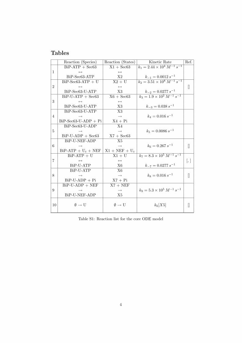
Tables
Reaction (Species) Reaction (States) Kinetic Rate Ref.
1BiP-ATP + Sec63 X1 + Sec63 k1 = 2.44× 10⁴ M−1 s−1
↔ ↔BiP-Sec63-ATP X2 k−1 = 0.0012 s−1
2BiP-Sec63-ATP + U X2 + U k2 = 3.51× 10⁸ M−1 s−1
↔ ↔ []BiP-Sec63-U-ATP X3 k−2 = 0.0277 s−1
3BiP-U-ATP + Sec63 X6 + Sec63 k3 = 1.9× 10² M−1 s−1
↔ ↔BiP-Sec63-U-ATP X3 k−3 = 0.038 s−1
4BiP-Sec63-U-ATP X3
→ → k4 = 0.016 s−1
BiP-Sec63-U-ADP + Pi X4 + Pi
5BiP-Sec63-U-ADP X4
→ → k5 = 0.0086 s−1
BiP-U-ADP + Sec63 X7 + Sec63
6BiP-U-NEF-ADP X5
→ → k6 = 0.267 s−1 []BiP-ATP + Ut + NEF X1 + NEF + Ut
7BiP-ATP + U X1 + U k7 = 8.3× 10⁵ M−1 s−1
↔ ↔ [, ]BiP-U-ATP X6 k−7 = 0.0277 s−1
8BiP-U-ATP X6
→ → k8 = 0.016 s−1 []BiP-U-ADP + Pi X7 + Pi
9BiP-U-ADP + NEF X7 + NEF
→ → k9 = 5.3× 10⁵ M−1 s−1
BiP-U-NEF-ADP X5
10 ∅ → U ∅ → U k6[X5] []
Table S1: Reaction list for the core ODE model
4
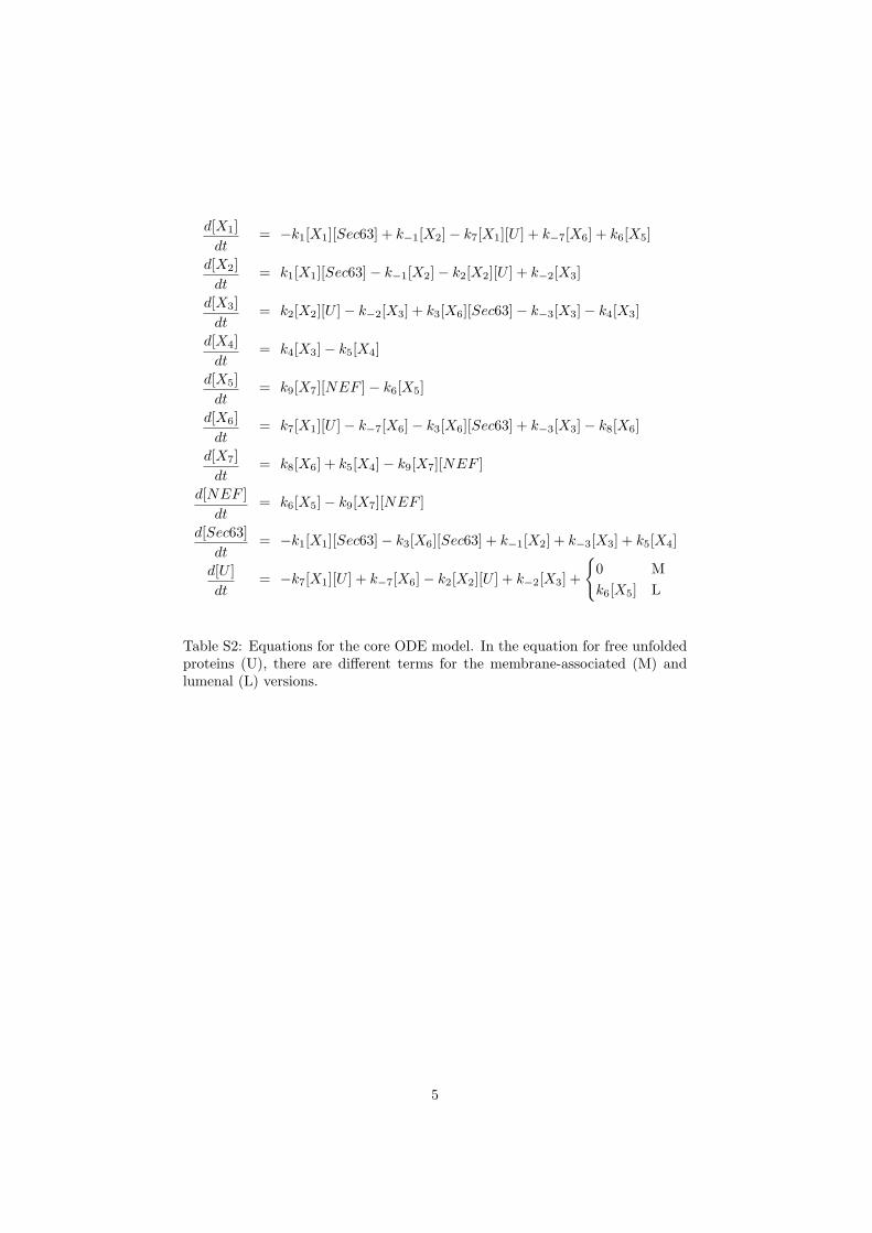
d[X1]dt
= −k1[X1][Sec63] + k−1[X2]− k7[X1][U ] + k−7[X6] + k6[X5]
d[X2]dt
= k1[X1][Sec63]− k−1[X2]− k2[X2][U ] + k−2[X3]
d[X3]dt
= k2[X2][U ]− k−2[X3] + k3[X6][Sec63]− k−3[X3]− k4[X3]
d[X4]dt
= k4[X3]− k5[X4]
d[X5]dt
= k9[X7][NEF ]− k6[X5]
d[X6]dt
= k7[X1][U ]− k−7[X6]− k3[X6][Sec63] + k−3[X3]− k8[X6]
d[X7]dt
= k8[X6] + k5[X4]− k9[X7][NEF ]
d[NEF ]dt
= k6[X5]− k9[X7][NEF ]
d[Sec63]dt
= −k1[X1][Sec63]− k3[X6][Sec63] + k−1[X2] + k−3[X3] + k5[X4]
d[U ]dt
= −k7[X1][U ] + k−7[X6]− k2[X2][U ] + k−2[X3] +
{0 Mk6[X5] L
Table S2: Equations for the core ODE model. In the equation for free unfoldedproteins (U), there are different terms for the membrane-associated (M) andlumenal (L) versions.
5

![Targeted Endoplasmic Reticulum Localization of Storage Protein … · Targeted Endoplasmic Reticulum Localization of Storage Protein mRNAs Requires the RNA-Binding Protein RBP-L1[OPEN]](https://img.pdfslide.us/doc/110x75/5cd3864e88c99315538d9990/targeted-endoplasmic-reticulum-localization-of-storage-protein-targeted-endoplasmic.jpg)


![Endoplasmic reticulum[1]](https://img.pdfslide.us/doc/110x75/58ed5fc71a28aba1678b4611/endoplasmic-reticulum1.jpg)
