Embed Size (px)
Citation preview

APPLIED AND ENVIRONMENTAL MICROBIOLOGY,0099-2240/01/$04.0010 DOI: 10.1128/AEM.67.3.1343–1350.2001
Mar. 2001, p. 1343–1350 Vol. 67, No. 3
Copyright © 2001, American Society for Microbiology. All Rights Reserved.
Spatial Arrangements and Associative Behavior of Speciesin an In Vitro Oral Biofilm Model
M. GUGGENHEIM, S. SHAPIRO, R. GMUR, AND B. GUGGENHEIM*
Institute for Oral Microbiology and General Immunology, Center for Dental and Oral Medicine andMaxillofacial Surgery, University of Zurich, CH-8028 Zurich, Switzerland
Received 5 September 2000/Accepted 30 November 2000
The spatial arrangements and associative behavior of Actinomyces naeslundii, Veillonella dispar, Fusobacteriumnucleatum, Streptococcus sobrinus, and Streptococcus oralis strains in an in vitro model of supragingival plaquewere determined. Using species-specific fluorescence-labeled antibodies in conjunction with confocal laserscanning microscopy, the volumes and distribution of the five strains were assessed during biofilm formation.The volume-derived cell numbers of each strain correlated well with respective culture data. Between 15 minand 64 h, populations of each strain increased in a manner reminiscent of batch growth. The microcolonymorphologies of all members of the consortium and their distributions within the biofilm were characterized,as were interspecies associations. Biofilms formed 15 min after inoculation consisted principally of singlenonaggregated cells. All five strains adhered strongly to the saliva-conditioned substratum, and therefore,coadhesion played no role during the initial phase of biofilm formation. This observation does not reflect theresults of in vitro coaggregation of the five strains, which depended upon the nature of the suspension medium.While the possibility cannot be excluded that some interspecies associations observed at later stages of biofilmformation were initiated by coadhesion, increase in bacterial numbers appeared to be largely a growthphenomenon regulated by the prevailing cultivation conditions.
Polyspecies microbial consortia typically consist of cells andmicrocolonies embedded in exopolymer matrices perforatedwith channels through which contact with the milieu exterieuris maintained (50). Dental plaque is a clinically relevant exam-ple of such a consortium which mediates oral diseases of mi-crobial etiology. The resistance or resilience of biofilms toantimicrobial agents appears to be related to their distinctivearchitectures (12, 17, 45), in which case an understanding ofthe fine structure of oral biofilms may lead to new or improvedstrategies for plaque control.
Efforts have been directed towards defining the temporaldevelopment and spatial organization of an in vitro model ofsupragingival plaque whose responses to various antimicrobialagents and proprietary oral hygiene products (15) mimic clin-ical observations. At the same time, information was sought onthe importance of intraspecies aggregation, interspecies coag-gregation, and coadhesion on surface attachment during theinitial stages of biofilm formation.
MATERIALS AND METHODS
Experimental design. Biofilms containing Actinomyces naeslundii OMZ 745,Veillonella dispar ATCC 17748T (OMZ 493), Fusobacterium nucleatum KP-F2(OMZ 596), Streptococcus sobrinus OMZ 176, and Streptococcus oralis SK248(OMZ 607) were formed on hydroxyapatite disks as previously described (15).Three independent trials were run, in each of which six or seven biofilms wererecovered per time point (Fig. 1). At every time point in each trial, three diskswere dip-washed to remove loosely adherent cells and vortexed, and the elutedcells were sonified, while the remaining disks were labeled with dye-conjugatedantibodies (Abs) and examined by confocal laser scanning microscopy (CLSM).
Quantification of eluted cells. Suspensions (25 ml) of eluted cells were incu-bated on microscope slides in the dark with LIVE/DEAD BacLight BacterialViability Kit solutions (0.25 ml each; Molecular Probes B. V., Leiden, TheNetherlands) for 15 min at room temperature. Three counts of 100 bacteria atdifferent sites on the slides were made using a Leitz Dialux 22 fluorescencemicroscope (Leica Mikroskopie Systeme AG, Glattbrugg, Switzerland) equippedwith an Osram 50-W high-pressure Hg lamp and an I2/3 filter block for fluores-cein isothiocyanate fluorescence. Serial dilutions of eluted cells were prepared inphysiological saline, and aliquots (50 ml) were plated onto Columbia blood agar,mitis salivarius agar, and a selective medium for fusobacteria based on FastidiousAnaerobe Agar (15). After anaerobic incubation for 72 h, differentiation of thefive species was achieved by observation of colony gross morphology in conjunc-tion with microscopic examination of cells from selected colonies. Nine indepen-dent CFU values per species per time point were averaged.
Influence of medium on in vitro coaggregation. Tubes (9 ml) of each specieswere grown overnight in mFUM (15) at 37°C. Following examination of eachliquid culture by phase-contrast microscopy, the cells were pelleted and washedtwice in cold physiological saline, and each pellet was resuspended either in a 1:1(vol/vol) mixture of mFUM plus saliva (five tubes) or in buffered KCl (7) (fivetubes) such that the final optical density at 550 nm of each cell suspension was0.5 6 0.05. All 10 nonredundant pairwise combinations (NRPCs) were generatedfor each resuspension medium by mixing 1 ml of one species with 1 ml of anotherin a 15-ml polystyrene tube, vortexing the pairs (5 s), and incubating them atroom temperature. After standing for 1 h, the tubes were viewed by eye for thepresence or absence of a sediment and then vortexed and examined for macro-scopic coaggregates by using a loupe and for minute coaggregates by using amicroscope. Two hours later, the tubes were vortexed and viewed with a loupe.Macroscopic and microscopic coaggregation were scored semiquantitatively on ascale ranging from no coaggregation to profuse coaggregation.
Labeling, embedment, and viewing of biofilms. A. naeslundii was detected withimmunoglobulin M (IgM) monoclonal Ab (MAb) 396AN1 (51), V. dispar wasdetected with IgG3 MAb 349VP1.1 (14), F. nucleatum was detected with IgG3MAb 395FN1 (52), and S. oralis was detected with IgM MAb 493SO1 (R. Gmurand T. Thurnheer, unpublished work). Culture supernatants with high MAbconcentrations were produced in MiniPerm cell culture vessels (Heraeus Instru-ments GmbH & Co. KG, Hanau, Germany) using serum-free HP-1 medium(Cell Culture Technologies, Zurich, Switzerland). S. sobrinus was labeled withpolyclonal rabbit anti-OMZ 176 Abs. Immunoglobulins were purified by proteinA affinity chromatography (Affi-Gel protein A gel; Bio-Rad Laboratories AG,Glattbrugg, Switzerland) and coupled with Alexa 594 or Oregon Green 488according to the manufacturer’s guidelines (Molecular Probes B. V.).
* Corresponding author. Mailing address: Institut fur Orale Mikro-biologie und Allgemeine Immunologie, Zentrum fur Zahn-, Mund-und Kieferheilkunde der Universitat Zurich, Plattenstrasse 11, Post-fach, CH-8028 Zurich, Switzerland. Phone: (41 1) 634 32 78. Fax: (411) 634 43 10. E-mail: [email protected].
1343
on July 4, 2018 by guesthttp://aem
.asm.org/
Dow
nloaded from

Disks destined for CLSM were dipped three times in sterile physiologicalsaline (room temperature) and then incubated in an opaque box at room tem-perature with appropriately diluted Abs. The box was agitated gently for 30 min(15-min biofilms) or 90 min (16-, 40-, and 64-h biofilms). Thereafter, Ab solu-tions were aspirated, and the disks were washed by immersion (5 min for 15-minbiofilms; 10 min for 16-, 40-, and 64-h biofilms) in three changes of physiologicalsaline (2 ml). Since Abs conjugated with either Alexa 594 or Oregon Green 488were available for each species, two species at a time were viewed by red andgreen fluorescence. All species pairings in the biofilms were visualized using 10biofilms (Fig. 1) with the 10 NRPCs of Abs, producing four sets of data perspecies per time point. The bottom of each stained disk was pressed firmly ontoa small wad of plasticine affixed to a glass microscope slide, and the uppersurfaces of the disks were covered immediately with Mowiol (8 ml) and toppedwith glass coverslips. Mowiol, a semipermeable mounting medium compatiblewith immunostaining (46), was prepared by mixing Mowiol 4-88 (2.4 g; Calbio-chem-Novabiochem Corp., San Diego, Calif.) with 50% (vol/vol) aqueous glyc-erol (12 ml); Tris-HCl (12 ml; 0.2 M; pH 8.5) was added, and the mixture wasstirred for 5 h and then left undisturbed for 2 h. 1,4-Diazabicyclo[2.2.2]octane (24mg; Fluka Chemie AG, Buchs, Switzerland) was added to retard fading, and thesuspension was incubated for 10 min at 50°C. The Mowiol suspension wasclarified by centrifugation (15 min; room temperature; 5,000 3 g), and thesupernatant was removed and frozen at 220°C; aliquots were thawed as needed.Mowiol-mounted discs were incubated in opaque containers at room tempera-ture for 6 h and then stored in the dark at 4°C. The stained biofilms wereexamined using a DM IRB E inverted microscope (Leica Mikroskopie undSysteme GmbH, Wetzlar, Germany) fitted with an Ar-Kr laser (model 543;Omnichrome, Inc., Chino, Calif.) and a TCS 4D computer-operated confocalscanning system (Leica Lasertechnik GmbH, Heidelberg, Germany). Confocalimages were obtained with oil immersion objectives (3100 for 15-min biofilms;340 for 16-, 40-, and 64-h biofilms). Cells labeled with Oregon Green 488 wereviewed with a 522-nm bandpass filter, whereas cells labeled with Alexa 594 wereviewed with a 590-nm longpass filter. The microscope pinhole size was set to100/255, with resulting radii of 636 nm (340 objective) or 254 nm (3100 objec-tive). Image acquisition was done in 83 line average mode.
Image analysis. Nine randomly selected square spots were examined perbiofilm disk. Z-series were generated by vertical optical sectioning of each spotinto 20 equispaced xy focal planes (lateral strata) spanning the height (z) ofimmunolabeled biofilms, and each optodigital thin section was scanned once forgreen fluorescence and once for red fluorescence. The data were processed on anIndigo 2ex workstation (Silicon Graphics, Inc., Mountain View, Calif.). The areaof each spot was transformed into a digital image containing 512 by 512 pixels;the side of each pixel represented 0.49 mm (340 objective) or 0.2 mm (3100objective). The ratio of total disk surface area to spot surface area was 1,299 forthe 340 objective and 7,781 for the 3100 objective.
The 40 scans per 15-min spot were recombined using Imaris 3.0 software(Bitplane AG, Zurich, Switzerland), and the bacterial cells in each reconstructedspot were counted manually. For 16-, 40-, and 64-h biofilms, where the extent ofdisk colonization precluded manual counting, VoxelShop Pro software (BitplaneAG) was used to calculate the volume occupied per species per reconstructedspot. The volume values for each species at 16, 40, and 64 h were averaged andscaled by the appropriate proportionality factor to obtain average volumes oc-cupied per species per time point per disk. Dividing the average species volume
per disk by the average volume per cell (Table 1) yielded the number of bacterialcells per species per disk at these time points. To study microcolony developmentwithin the biofilm and to characterize the associative behavior of the differentspecies, the 40 scans per NRPC per time point were recombined and analyzedusing Imaris 3.0, resulting in thousands of CLSM images, the analysis of whichprovided a description of the development of the consortium as a function oftime.
RESULTS
Quantification of species within biofilms by culture andimage analysis. High numbers (104 to 105 CFU) of each spe-cies were adherent to the substratum within 15 min of exposureto the inoculum, achieving attachment densities of $102 CFUper mm2 of disk surface per species. Average population pro-files per biofilm as a function of time for each species asdetermined from CFU counts are shown in Fig. 2A. Fifteenminutes after inoculation, the number of F. nucleatum KP-F2CFU adherent to saliva-coated disks was at least an order ofmagnitude lower than that of any other species, though thesubsequent growth of this strain was such that by 16 h itspopulation slightly exceeded that of A. naeslundii OMZ 745.Only modest changes in CFU counts occurred between 16 and64 h. Comparison of time-dependent population profiles fromCFU counts (Fig. 2A) with those calculated from images ofimmunolabeled cells (Fig. 2B) demonstrated a reasonable cor-respondence between cell numbers inferred from plating andcell numbers calculated from image analysis, though F. nuclea-tum plate counts were consistently lower than CLSM cellcounts. Live/dead staining of cells eluted from biofilms be-tween 15 min and 64 h showed a slight decrease in percentageof viability as the biofilm aged, but the mean proportion of livecells never fell below 80%.
Interspecies coaggregation of planktonic cells. Intraspeciesaggregation and interspecies coaggregation of planktonic bac-teria are strongly influenced by environmental conditions, es-pecially the growth or resuspension medium (6, 33). The effectsof the medium on coaggregation among the bacteria compris-ing the biofilm model were gauged by resuspending cells ineither the substrate used for biofilm formation (mFUM plussaliva) or buffered KCl (Table 2). In buffered KCl medium butnot in mFUM plus saliva, coaggregation between F. nucleatumKP-F2 and all partners was seen; in mFUM plus saliva, how-ever, it was S. sobrinus OMZ 176 which coaggregated with allpartners. Other pairings in either medium did not lead tocoaggregation. These results were independent of the timepoint and mode of observation.
FIG. 1. Experimental design for analysis of hydroxyapatite disks.The first, second, and third trials represent experiments done on dif-ferent occasions as checks for repeatability. Solid circles, disks used forCLSM; open circles, disks from which biofilms were eluted and ana-lyzed by conventional microscopy and plate counting. Tetrads ratherthan trios of disks were used in the third trial in order to obtain thetotal of 10 disks per time point needed for all NRPCs in CLSManalysis.
TABLE 1. Volumes of bacterial cells
Species Idealized shapeCalculated cell
vol (mm3)(avg 6 SD)a
A. naeslundii Cylinder 1.31 6 0.39V. dispar Sphere 0.41 6 0.35F. nucleatum Cylinder 0.86 6 0.19S. sobrinus Sphere 0.82 6 1.23S. oralis Sphere 0.74 6 0.46
a Lengths (L) and cross-sectional diameters (D) were measured for 10 cells ofeach species, and volumes were calculated as follows: spherical cells, V 5 pD3/6;cylindrical cells, V 5 pD2L/4. Approximately normal distributions of volumevalues per species were confirmed using the Ryan-Joiner test (47). SD, standarddeviation at the 0.05 significance level.
1344 GUGGENHEIM ET AL. APPL. ENVIRON. MICROBIOL.
on July 4, 2018 by guesthttp://aem
.asm.org/
Dow
nloaded from

Structural features of species within 15-min biofilms. At 15min, very few F. nucleatum cells participated in intraspeciesaggregates. While F. nucleatum formed interspecies coaggre-gates (particularly with streptococci) more frequently than anyother species in the biofilm (Fig. 3A), aggregation and coag-gregation clearly were not dominant modes of bacterial adhe-sion to the salivary pellicle of any of the five species, since at 15min, single nonaggregated cells (constituting $96% of thetotal cell population) attached directly to the salivary pellicle.
Structural features of species in biofilms between 16 and64 h. Live/dead stains of entire biofilms revealed that at 16 hthese consisted mostly of discrete microcolonies with minimalintercolony association. Images at 40 and 64 h showed moredensely populated biofilms containing large numbers of micro-colonies with few unstained regions between them.
(i) A. naeslundii. Microcolonies of A. naeslundii OMZ 745consisted mainly of small spheroid structures interspersed with
columnar microcolonies, few of which spanned the height ofthe biofilm. By 16 h (Fig. 3B), spheroid microcolonies werescattered throughout the biofilm, with a slight preference forthe lower and central strata; individual cells were noted nearthe disk surface. By 40 h, there had been a slight shift inmicrocolony distribution towards the central strata, and lonecells were absent. Microcolonies in the upper strata often con-tacted the roof of the biofilm. Loose associations of smallspheroid microcolonies stacked so as to resemble uprightDelaunay unduloids were seen in sagittal (xz; yz) views (Fig.3C). By 64 h, these unduloids were plentiful in the centralstrata.
(ii) V. dispar. Of the five strains comprising the biofilm, V.dispar ATCC 17748T displayed the greatest morphologic tran-sition between 16 and 40 h. By 16 h, single cocci and mostlysmall spheroid microcolonies were scattered sparsely through-out the biofilm (Fig. 3B) and were slightly concentrated withinthe central strata; few columnar or mushroom-shaped micro-colonies were observed, and the few large colonies seen wereoriented horizontally. Some microcolonies in the upper strataextended to the roof of the biofilm. By 40 h (Fig. 3E), singlecells were absent, and most microcolonies were large oblatestructures linked by slender cellular bridges.
(iii) F. nucleatum. By 16 h, F. nucleatum KP-F2 existedmostly as small oblate microcolonies in the lower strata,though some single cells were seen in the central strata. A fewcolumnar microcolonies spanned the height of the biofilm. By40 h (Fig. 3D), only oblate microcolonies were present, equi-distributed throughout the biofilm and often stretching fromthe disk surface to the roof of the biofilm.
(iv) S. sobrinus. S. sobrinus OMZ 176 formed mainly spher-oid microcolonies residing in the lower and middle strata; thefew microcolonies stretching from the disk surface to the roofof the biofilm were columnar or mushroom shaped. Microcolo-nies bordering the biofilm roof rarely extended below the cen-tral strata. By 16 h, spheroid microcolonies consisted of threeor four short, usually intertwined chains localized to the upperhalf of the biofilm, while independent cells and chains clus-tered near the surface of the disk. By 40 h (Fig. 3E), a fewlarge, elongate microcolonies lying parallel to the disk surfacehad formed, though some lone cells and chains were seenprincipally in the central strata. By 64 h (Fig. 3D), the propor-tion of large horizontal microcolonies had increased.
(v) S. oralis. S. oralis SK248 formed spheroid or ovoid mi-crocolonies which expanded laterally. A greater proportion ofmicrocolonies extended from the disk surface to the roof of thebiofilm than had been observed for S. sobrinus. By 16 h, mi-
FIG. 2. Population profiles for each species, determined by cultureor image analysis. (A) Average number of CFU recovered per speciesper time point per disk. (B) Average number of cells imaged perspecies per time point per disk. 3, A. naeslundii; F, V. dispar; Œ, F.nucleatum; {, S. sobrinus; h, S. oralis.
TABLE 2. In vitro coaggregation of NRPCs in mFUM plus saliva and in buffered KCl
Species
Coaggregationa
S. oralis V. dispar F. nucleatum A. naeslundii
mFUM 1 saliva Buffered KCl mFUM 1 saliva Buffered KCl mFUM 1 saliva Buffered KCl mFUM 1 saliva Buffered KCl
S. sobrinus 11 2 1 2 111 11 111 2S. oralis 2 2 2 111 2 2V. dispar 2 1 2 2F. nucleatum 2 11
a 2, none; 1, minor; 11, moderate; 111, profuse.
VOL. 67, 2001 SPATIAL ARRANGEMENTS IN AN ORAL BIOFILM MODEL 1345
on July 4, 2018 by guesthttp://aem
.asm.org/
Dow
nloaded from
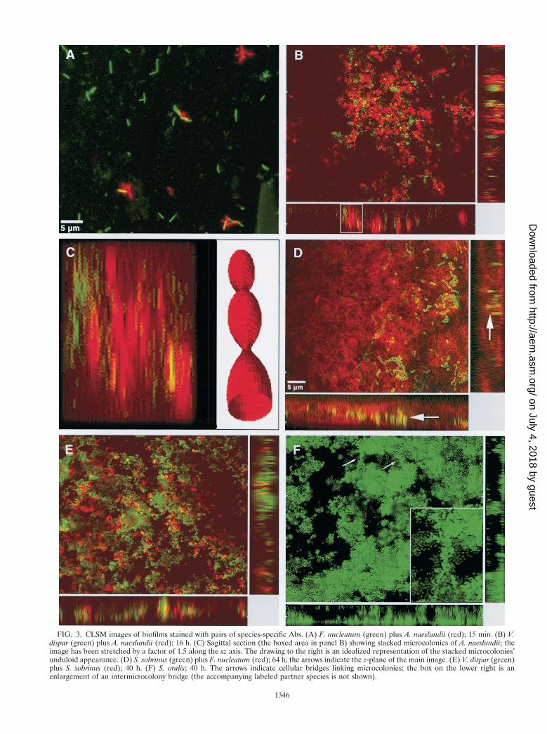
FIG. 3. CLSM images of biofilms stained with pairs of species-specific Abs. (A) F. nucleatum (green) plus A. naeslundii (red); 15 min. (B) V.dispar (green) plus A. naeslundii (red); 16 h. (C) Sagittal section (the boxed area in panel B) showing stacked microcolonies of A. naeslundii; theimage has been stretched by a factor of 1.5 along the xz axis. The drawing to the right is an idealized representation of the stacked microcolonies’unduloid appearance. (D) S. sobrinus (green) plus F. nucleatum (red); 64 h; the arrows indicate the z-plane of the main image. (E) V. dispar (green)plus S. sobrinus (red); 40 h. (F) S. oralis; 40 h. The arrows indicate cellular bridges linking microcolonies; the box on the lower right is anenlargement of an intermicrocolony bridge (the accompanying labeled partner species is not shown).
1346
on July 4, 2018 by guesthttp://aem
.asm.org/
Dow
nloaded from

crocolonies were equidistributed in size and dispersed evenlythroughout the biofilm; some small columnar microcoloniesand a few very large amorphous microcolonies were seen. Amass of single cells and short chains occurred near the surfacesof the disks. By 40 h, microcolonies, usually large oblate struc-tures linked by narrow cellular bridges (Fig. 3F) and anchoredto the disk surface by slender podia, had collected in thecentral strata of the biofilm. Between 40 and 64 h, the micro-colonies appeared to have drifted apart.
Associative behavior. The associative behavior of biofilmstrains was classified into the five general spatial types illus-trated in Fig. 4. At 16 h, the frequency of the modes of asso-ciation of the different species followed the order interlaced .interosculated . coated or embedded; by 40 h, however, therehad been a shift away from interlacing or interosculation to-wards embedment, possibly a consequence of growth obscuringthe other, more discrete forms of interspecies associations (Ta-ble 3). S. oralis and F. nucleatum dominated the images theyshared with other species (Fig. 3D and 4B). No special asso-ciations between the two streptococcal species were noted.A. naeslundii, and especially F. nucleatum, displayed a pro-nounced propensity to combine with all species except V. dis-par; indeed, V. dispar usually collected into autonomous mi-crocolonies, displaying minimal association with other strains(Fig. 3E and 4A). At 40 and 64 h, microcolonies of S. oralis andF. nucleatum formed a mosaic pattern devoid of extensiveintermingling (Fig. 4E).
DISCUSSION
Tagged antibodies are useful tools for probing the fine struc-ture of microbial ecosystems (3), though their use for thispurpose in conjunction with CLSM remains underexploited(44, 49, 50). Since methodologies for simultaneously visualiz-ing all five species in the biofilm are not yet generally available(5), recourse was made to pairwise application of fluorescence-labeled Abs followed by CLSM. Though no single image takenthrough a single plane of confocality can capture the complexspatial characteristics of a species during its growth in theconsortium, careful examination of the thousands of imagesrecorded showed enough interdisk consistencies per species
per time point to permit identification of the most salientmorphological features and spatial arrangements for eachmember of the biofilm.
After a 15-min exposure to the five-species inoculum, saliva-covered disks were coated with a shallow layer of cells, most ofwhich bound directly to the pellicle. Given the presumed im-portance of intraspecies aggregation and interspecies coaggre-gation in establishing dental plaque (20, 24, 25, 29), 15-minbiofilms were scrutinized for the presence of these cell com-plexes, but they were seldom encountered at this time. By 16 h,the adherent cells had given rise to microcolonies of varioussizes and shapes (predominantly spheroid or ovoid), while sin-gle cells occurred preferentially near the disk surface (A.naeslundii, S. sobrinus, and S. oralis) or the central strata (V.dispar and F. nucleatum). By 40 h, single cells either had van-ished (A. naeslundii, V. dispar, F. nucleatum, and S. oralis) orwere confined to the central strata in reduced numbers (S.sobrinus). There was an increase in the diversity of microcolonyform (columnar or mushroom shaped), including some large,quite distinctive morphologies (stacked spheroid microcolo-nies of A. naeslundii; oblate interbridged microcolonies of S.oralis). The population structures of only a few polymicrobialcommunities are known in sufficient detail for comparison withthat of our oral biofilm model. Cook et al. (8) described thesequential deposition of Streptococcus gordonii and Porphy-romonas gingivalis onto saliva-coated glass surfaces; after 2 h,this accretive biofilm consisted of obconical porphyromonadmicrocolonies anchored to a confluent streptococcal mono-layer. Most bacterial consortia whose microanatomies havebeen described were recovered from environments unrelatedto the oral cavity (1, 30, 34, 39, 48, 54), and therefore theymight be expected to differ substantially from our plaquemodel. Nonetheless, these divergent biofilms share numerousstructural features.
Dosani (10) reported that biofilms begin as an assemblage ofdiscrete microcolonies which eventually grow together and overone another to form a continuous mass; our live/dead-stainedbiofilms revealed such a coalescence to have taken place between16 and 40 h. James et al. (G. A. James, D. E. Caldwell, and J. W.Costerton, Abstr. Can. Soc. Microbiol./Soc. Ind. Microbiol. Annu.Meet., abstr. P138, 1993), Amann et al. (2), and Møller et al. (35)
TABLE 3. Spatial arrangements of pairs of species in the biofilm
Species
Arrangementa
16 h 40 and 64 h
V. dispar F. nucleatum S. sobrinus S. oralis V. dispar F. nucleatum S. sobrinus S. oralis
A. naeslundii 0 3, 4 4 . 2, 3b 0 . 3, 4 1 (An . Vd) 1 (Fn . An) 3, 4 1 (So . An)V. dispar 3, 4 (Fn . Vd) 0 @ 4 1 (So . Vd) 1 (Fn . Vd) 1, 3 1 (So . Vd)F. nucleatum 2,c 4 3, 4 2, 4 (Fn . Ss) 4d
S. sobrinus 1 (So . Ss) 1 (So . Ss)
a Code numbers are defined as follows: 0, unassociated (no conspicuous association between species [Fig. 4A]); 1, embedded (discrete microcolonies of one speciesdispersed or sprinkled within a much denser background of microcolonies of a different species [Fig. 4B]); 2, coated (surface of intraspecies aggregates encased orcoated in whole or in part by a mono- or bilayer of cells of a different species [Fig. 4C]); 3, interlaced (discrete, contacting intraspecies aggregates, in which contactbetween aggregates may be tangential or extensive but usually without a filigreed appearance [Fig. 4D]); 4, interosculated (interspecies complexes or, more commonly,aggregates of various shapes and sizes [Fig. 4E]). While the relative spatial arrangements of pairs of species were similar at 40 and 64 h, the 64-h images typically showedhigher cell densities than the 40-h images. Where particular species or arrangements predominate, these are indicated (., predominates over; @, greatly predominatesover). An, A. naeslundii; Fn, F. nucleatum; Vd, V. dispar; So, S. oralis; Ss, S. sobrinus.
b S. sobrinus cells coating A. naeslundii microcolonies.c S. sobrinus cells coating F. nucleatum microcolonies.d Each species nestled within microcolony surface pockets of the other species, contacting only at microcolony boundaries without extensive intermingling.
VOL. 67, 2001 SPATIAL ARRANGEMENTS IN AN ORAL BIOFILM MODEL 1347
on July 4, 2018 by guesthttp://aem
.asm.org/
Dow
nloaded from

FIG. 4. Categories of spatial arrangements between different species in the biofilm. (A) Code 0 (unassociated): V. dispar (green) plus A.naeslundii (red); 16 h. (B) Code 1 (embedded): F. nucleatum (green) plus A. naeslundii (red); 64 h. (C) Code 2 (coated): S. sobrinus (green) plusF. nucleatum (red); 16 h. (D) Code 3 (interlaced): S. sobrinus (green) plus A. naeslundii (red); 16 h. (E) Code 4 (interosculated): S. oralis (green)plus F. nucleatum (red); 40 h.
1348
on July 4, 2018 by guesthttp://aem
.asm.org/
Dow
nloaded from

each observed species-specific spatial heterogeneity within bio-films (also see reference 31). Formation of palisades and othervertical structures from initially adherent cells and the presence offree-floating grapelike clusters have been noted (23, 28, 38, 54).Zhang et al. (56) suggested that mushroom-shaped microcoloniesmight be gravitational artifacts of biofilms cultivated in an in-verted position, though mushroom-shaped microcolonies of V.dispar and S. sobrinus occurred in biofilms cultivated and stainedin upright positions, concordant with predictions by three-dimen-sional models of substrate-limited biofilms (41, 42). Neu andLawrence (38) demonstrated that some structural features in bio-films are artifacts of fluid flow. Our plaque model was not subjectto continuous drag forces, and lift forces were exerted only duringbrief episodes of disk dip-washing, punctuated by long periods ofstagnant incubation. Shear-induced perturbation in our biofilms,therefore, will have been slight, so that the microcolony morphol-ogies and associative (or partitive) behaviors adopted by the cellswill have arisen principally from physiological and physicochem-ical interactions among themselves.
The precise manner in which surface-adherent cells at 15min gave rise to free-floating microcolonies in the biofilm wasnot determined. They may have formed de novo by growth ofdetached single cells or small clusters, by exfoliation of upperportions of microcolonies, or by detachment of whole micro-colonies from the substratum. A shift away from free-floatingsingle cells and towards free-floating microcolonies between 16and 40 h is consistent with the formation of suspended micro-colonies by the growth of detached cells, while the large oblatemicrocolonies of S. oralis hovering over the substratum but stillattached to it by slender cellular threads might represent tran-sitional forms in the process of detachment. Such transitionalstructures may have been observed by de Beer et al. (9), whofound that as biofilms aged the substratal base of cell clustersbecame increasingly lacunose.
Besides CLSM, cultivation on Columbia blood agar and twoselective media for enumeration of fusobacteria and strepto-cocci was used for species quantification. For four of the fivespecies, image analysis counts were in agreement with CFUcounts of eluted cells; however, microscopic cell counts of F.nucleatum KP-F2 often exceeded CFU by a factor of $20. Thatmuch of the adherent population of F. nucleatum is in a viablebut noncultivable state is unlikely, since the F. nucleatum pop-ulation increases by some 3.5 orders of magnitude between 15min and 16 h, more than any other species in the biofilm,though this strain has a somewhat longer doubling time thanany other species comprising the biofilm (unpublished obser-vations). More likely, the cell numbers of F. nucleatum ob-tained from CFU data are underestimated. Discrepancies be-tween microscopic cell counts and CFU counts are encounteredcommonly for aggregatory microorganisms or for those specieswhose cells are not dispersed readily following mitosis (16, 19,21, 53). In addition, suboptimal recovery of F. nucleatum onthe selective Fastidious Anaerobe Agar medium may havecontributed to the low CFU counts of this species.
The predominance of F. nucleatum in our biofilm model—by64 h this species still represented .50% of the total cellload—is consistent with the report by Moore and Moore (36)that F. nucleatum figures among the taxa most frequently iso-lated from gingival-crevice plaque. It has been claimed that F.nucleatum binds poorly to the acquired pellicle (22), though in
fact the species has an affinity for salivary components, such asstatherin (55) and proline-rich glycoproteins (13); indeed, theadherence strength of F. nucleatum to saliva is comparable tothat of Streptococcus sanguis (43).
A. naeslundii and S. oralis are regarded as pioneer colonizersof tooth surfaces (26, 37, 40), the coaggregation of which withthe reputed secondary colonizers Fusobacterium and Veil-lonella are said to facilitate formation of dental plaque (18, 32).However, neither A. naeslundii nor S. oralis formed interspe-cies coaggregates with one another or with F. nucleatum or V.dispar, even after 3 h in a saliva-based medium promoting theformation and growth of oral biofilms (Table 2). The onlybacterium to coaggregate with the other four species in mFUMplus saliva was S. sobrinus (27). It would appear that, at least inour in vitro model of dental plaque, intraspecies aggregationand interspecies coaggregation are not crucial for biofilm for-mation (11). Coadhesion of planktonic cells to sessile cells hasbeen postulated to play a role in the formation of dentalplaque (4); however, careful examination of CLSM images of15-min polyspecies biofilms failed to identify any of the fivespecies acting as conspicuous anchors or nuclei for attachmentof any of the other species. Increase in bacterial numbers inthis biofilm appears to be largely a growth phenomenon reg-ulated by the prevailing cultivation conditions rather than theresult of specific or primary aggregation or coadhesion.
ACKNOWLEDGMENTS
We thank Andre Meier and Yvonne Helweg for excellent technicalassistance. The help of Matthias Hochli (ElektronenmikroskopischesZentrallaboratorium der Universitat Zurich) and Rene Fischer (Insti-tut fur Biochemie, ETH Zentrum, Zurich, Switzerland) with CLSMand hybridoma supernatant production, respectively, is gratefully ac-knowledged.
REFERENCES
1. Abella, C. A., X. P. Cristina, A. Martinez, I. Pibernat, and X. Vila. 1998. Twonew motile phototrophic consortia: “Chlorochromatium lunatum” and “Pe-lochromatium selenoides.” Arch. Microbiol. 169:452–459.
2. Amann, R. I., J. Stromley, R. Devereux, R. Key, and D. A. Stahl. 1992.Molecular and microscopic identification of sulfate-reducing bacteria in mul-tispecies biofilms. Appl. Environ. Microbiol. 58:614–623.
3. Bohlool, B., and E. Schmidt. 1980. The immunofluorescence approach tomicrobial ecology. Adv. Microb. Ecol. 4:203–236.
4. Bos, R., H. C. van der Mei, and H. J. Busscher. 1996. Co-adhesion of oralmicrobial pairs under flow in the presence of saliva and lactose. J. Dent. Res.75:809–815.
5. Castro, S. 1999. Fluorescent staining advances. Genet. Eng. News vol.19(17), 1 October.
6. Chisari, G., and M. R. Gismondo. 1986. Coaggregation between Actinomycesviscosus with Streptococcus pyogenes and Streptococcus agalactiae. Microbio-logica 9:393–398.
7. Clark, W. B., L. L. Bammann, and R. J. Gibbons. 1978. Comparative esti-mates of bacterial affinities and adsorption sites on hydroxyapatite surfaces.Infect. Immun. 19:846–853.
8. Cook, G. S., J. W. Costerton, and R. J. Lamont. 1998. Biofilm formation byPorphyromonas gingivalis and Streptococcus gordonii. J. Periodont. Res. 33:323–327.
9. de Beer, D., P. Stoodley, F. Roe, and Z. Lewandowski. 1994. Effects of biofilmstructures on oxygen distribution and mass transport. Biotechnol. Bioeng.43:1131–1138.
10. Dosani, R. 1991. An electron microscopic study of wastewater biofilm for-mation. M.S. thesis. The University of Cincinnati, Cincinnati, Ohio.
11. Ganeshkumar, N., C. V. Hughes, and E. I. Weiss. 1998. Co-aggregation indental plaque formation, p. 125–143. In H. J. Busscher and L. V. Evans (ed.),Oral biofilms and plaque control. Harwood Academic Publishers, Amster-dam, The Netherlands.
12. Gilbert, P., and D. G. Allison. 1999. Biofilms and their resistance towardsantimicrobial agents, p. 125–143. In H. N. Newman and M. Wilson (ed.),Dental plaque revisited: oral biofilms in health and disease. Bioline, Cardiff,United Kingdom.
VOL. 67, 2001 SPATIAL ARRANGEMENTS IN AN ORAL BIOFILM MODEL 1349
on July 4, 2018 by guesthttp://aem
.asm.org/
Dow
nloaded from

13. Gillece-Castro, B. L., A. Prakobphol, A. L. Burlingame, H. Leffler, and S. J.Fisher. 1991. Structure and bacterial receptor activity of a human salivaryproline-rich glycoprotein. J. Biol. Chem. 266:17358–17368.
14. Gmur, R., B. Guggenheim, E. Giertsen, and T. Thurnheer. 2000. Automatedimmunofluorescence for enumeration of selected taxa in supragingival den-tal plaque. Eur. J. Oral Sci. 108:393–402.
15. Guggenheim, B., E. Giertsen, P. Schupbach, and S. Shapiro. 2001. Valida-tion of an in vitro biofilm model of supragingival plaque. J. Dent. Res.80:363–370.
16. Harmsen, H. J. M., G. R. Gibson, P. Elfferich, G. C. Raangs, A. C. M.Wildeboer-Veloo, A. Argaiz, M. B. Roberfroid, and G. W. Welling. 2000.Comparison of viable cell counts and fluorescence in situ hybridization usingspecific rRNA-based probes for the quantification of human fecal bacteria.FEMS Microbiol. Lett. 183:125–129.
17. Helmerhorst, E. J., R. Hodgson, W. van’t Hof, E. C. I. Veerman, C. Allison,and A. V. Nieuw Amerongen. 1999. The effects of histatin-derived basicantimicrobial peptides on oral biofilms. J. Dent. Res. 78:1245–1250.
18. Jacquelin, L. F., L. Brisset, E. Le Magrex, J. Carquin, M. P. Gelle, and C.Choisy. 1995. Prevention de la plaque dentaire cariogene. Etude des struc-tures impliquees dans l’adhesion et la coagregation chez Streptococcus mu-tans et Streptococcus sobrinus. Pathol. Biol. 43:371–379.
19. Jannasch, H. W., and G. E. Jones. 1959. Bacterial populations in seawater asdetermined by different methods of enumeration. Limnol. Oceanogr. 4:128–139.
20. Jones, S. J. 1972. A special relationship between spherical and filamentousmicroorganisms in mature human dental plaque. Arch. Oral Biol. 17:613–616.
21. Karlsson, K., and P. Malmberg. 1989. Characterization of exposure to moldsand actinomycetes in agricultural dusts by scanning electron microscopy,fluorescence microscopy and the culture method. Scand. J. Work Environ.Health 15:353–359.
22. Kaufman, J., and J. M. DiRienzo. 1989. Isolation of a corncob (coaggrega-tion) receptor polypeptide from Fusobacterium nucleatum. Infect. Immun.57:331–337.
23. Keevil, C. W., and J. T. Walker. 1992. Nomarksi DIC microscopy and imageanalysis of biofilms. Binary-Comput. Microbiol. 4:93–95.
24. Kohlenbrander, P. E., and J. London. 1992. Ecological significance of coag-gregation among oral bacteria. Adv. Microb. Ecol. 12:183–217.
25. Kohlenbrander, P. E., and J. London. 1993. Adhere today, here tomorrow:oral bacterial adherence. J. Bacteriol. 175:3247–3252.
26. Kohlenbrander, P. E., R. N. Andersen, D. L. Clemans, C. J. Whittaker, andC. M. Klier. 1999. Potential role of functionally similar coaggregation me-diators in bacterial succession, p. 171–186. In H. N. Newman and M. Wilson(ed.), Dental plaque revisited: oral biofilms in health and disease. Bioline,Cardiff, United Kingdom.
27. Lamont, R. J., and B. Rosan. 1990. Adherence of mutans streptococci toother oral bacteria. Infect. Immun. 58:1738–1743.
28. Lawrence, J. R., D. R. Korber, G. M. Wolfaardt, and D. E. Caldwell. 1995.Behavioral strategies of surface-colonizing bacteria. Adv. Microb. Ecol. 14:1–75.
29. Listgarten, M. A., H. E. Mayo, and R. Tremblay. 1975. Development ofdental plaque on epoxy resin crowns in man. A light and electron micro-scopic study. J. Periodontol. 46:10–26.
30. Lunsdorf, H., I. Brummer, K. N. Timmis, and I. Wagner-Dobler. 1997. Metalselectivity of in situ microcolonies in biofilms of the Elbe River. J. Bacteriol.179:31–40.
31. Manz, W., G. Arp, G. Schumann-Kindel, U. Szewzyk, and J. Reitner. 2000.Widefield deconvolution epifluorescence microscopy combined with fluores-cence in situ hybridization reveals the spatial arrangement of bacteria insponge tissue. J. Microbiol. Methods 40:125–134.
32. Marsh, P., and M. Martin. 1992. Oral microbiology, 3rd ed., p. 109–110.Chapman & Hall, London, United Kingdom.
33. McIntire, F. C., A. E. Vatter, J. Baros, and J. Arnold. 1978. Mechanism ofcoaggregation between Actinomyces viscosus T14V and Streptococcus sanguis34. Infect. Immun. 21:978–988.
34. Møller, S., A. R. Pedersen, L. K. Poulsen, E. Arvin, and S. Molin. 1996.Activity and three-dimensional distribution of toluene-degrading Pseudomo-nas putida in a multispecies biofilm assessed by quantitative in situ hybrid-ization and scanning confocal laser microscopy. Appl. Environ. Microbiol.62:4632–4640.
35. Møller, S., C. Sternberg, J. B. Andersen, B. B. Christensen, J. L. Ramos, M.Givskov, and S. Molin. 1998. In situ gene expression in mixed-culture bio-films: evidence of metabolic interactions between community members.Appl. Environ. Microbiol. 64:721–732.
36. Moore, W. E. C., and L. V. H. Moore. 1994. The bacteria of periodontaldiseases. Periodontol. 2000 5:66–77.
37. Morou-Bermudez, E., and R. A. Burne. 1999. Genetic and physiologic char-acterization of urease of Actinomyces naeslundii. Infect. Immun. 67:504–512.
38. Neu, T., and J. R. Lawrence. 1997. Development and structure of microbialbiofilms in river water studied by confocal laser scanning microscopy. FEMSMicrobiol. Ecol. 24:11–25.
39. Nielsen, A. T., T. Tolker-Nielsen, K. B. Barken, and S. Molin. 2000. Role ofcommensal relationships on the spatial structure of a surface-attached mi-crobial consortium. Environ. Microbiol. 2:59–68.
40. Pearce, C., G. H. Bowden, M. Evans, S. P. Fitzsimmons, J. Johnson, M. J.Sheridan, R. Wientzen, and M. F. Cole. 1995. Identification of pioneerviridans streptococci in the oral cavity of human neonates. J. Med. Microbiol.42:67–72.
41. Picioreanu, C., M. C. M. van Loosdrecht, and J. J. Heijnen. 1998. Mathe-matical modeling of biofilm structure with a hybrid differential-discrete cel-lular automaton approach. Biotechnol. Bioeng. 58:101–116.
42. Picioreanu, C., M. C. M. van Loosdrecht, and J. J. Heijnen. 1999. Discrete-differential modelling of biofilm structure. Water Sci. Technol. 39:115–122.
43. Prakobphol, A., C. A. Burdsal, and S. J. Fisher. 1995. Quantifying thestrength of bacterial adhesive interactions with salivary glycoproteins. J.Dent. Res. 74:1212–1218.
44. Prensier, G., H. C. Dubourguier, I. Thomas, G. Albagnac, and M. N. Buis-son. 1988. Specific immunological probes for studying the bacterial associa-tions in granules and biofilms, p. 55–61. In G. Lettinga, A. J. B. Zehnder,J. T. C. Grotenhuis, and L. W. Hulshoff Pol (ed.), Granular anaerobicsludge: microbiology and technology. Centre for Agricultural Publishing andDocumentation, Wageningen, The Netherlands.
45. Reid, G. 1999. Biofilms in infectious disease and on medical devices. Int. J.Antimicrob. Agents 11:223–226.
46. Rodriguez, J., and F. Deinhardt. 1960. Preparation of a semipermanentmounting medium for fluorescent antibody studies. Virology 12:316–317.
47. Ryan, T. A., Jr., and B. L. Joiner. 1976. Normal probability plots and tests fornormality. Technical report, Department of Statistics. The PennsylvaniaState University, University Park.
48. Schwarzer, C., B. Auer, J. Klima, and K. Haselwandter. 1998. Physiologicaland electron microscopical investigations on syntrophic dicyandiamide deg-radation by soil bacteria. Soil Biol. Biochem. 30:385–391.
49. Singleton, S., L. Albiston, R. Treloar, E. Mahers, R. Hodgson, K. Watson, K.Schilling, and C. Allison. 1995. Optical imaging and characterisation of oralbiofilm structures using viral stains and specific antibody probes, p. 33–36. InJ. Wimpenny, P. Handley, P. Gilbert, and H. Lappin-Scott (ed.), The life anddeath of biofilm. Bioline, Cardiff, United Kingdom.
50. Singleton, S., R. Treloar, P. Warren, G. K. Watson, R. Hodgson, and C.Allison. 1997. Methods for microscopic characterization of oral biofilms:analysis of colonization, microstructure, and molecular transport phenom-ena. Adv. Dent. Res. 11:133–149.
51. Thurnheer, T., B. Guggenheim, and R. Gmur. 1997. Characterization ofmonoclonal antibodies for rapid identification of Actinomyces naeslundii inclinical samples. FEMS Microbiol. Lett. 150:255–262.
52. Thurnheer, T., B. Guggenheim, B. Gruica, and R. Gmur. 1999. Infiniteserovar and ribotype heterogeneity among oral Fusobacterium nucleatumstrains? Anaerobe 5:79–92.
53. Wagner, M., R. Amann, H. Lemmer, and K.-H. Schleifer. 1993. Probingactivated sludge with oligonucleotides specific for proteobacteria: inade-quacy of culture-dependent methods for describing microbial communitystructure. Appl. Environ. Microbiol. 59:1520–1525.
54. Wolfaardt, G. M., J. R. Lawrence, R. D. Robarts, S. J. Caldwell, and D. E.Caldwell. 1994. Multicellular organization in a degradative biofilm commu-nity. Appl. Environ. Microbiol. 60:434–446.
55. Xie, H., R. J. Gibbons, and D. I. Hay. 1991. Adhesive properties of strains ofFusobacterium nucleatum of the subspecies nucleatum, vincentii and polymor-phum. Oral Microbiol. Immunol. 6:257–263.
56. Zhang, T. C., Y.-C. Fu, and P. L. Bishop. 1995. Competition for substrateand space in biofilms. Water Environ. Res. 67:992–1003.
1350 GUGGENHEIM ET AL. APPL. ENVIRON. MICROBIOL.
on July 4, 2018 by guesthttp://aem
.asm.org/
Dow
nloaded from