Embed Size (px)
Citation preview

1Souvenir / Abstract Book
Souveneir/Abstract Book

Souvenir / Abstract Book2
WEL
COME
TO
FERT
IVIS
ION
2018
thth
thth
Welc
ome t
o Koc
hi, Ke
rala
(India
), the
God
's ow
n cou
ntry f
or Fe
rtivis
ion 2
018,
the 14
annu
al co
ngre
ss of
India
n Fer
tility
Socie
ty, sc
hedu
led to
be he
ld on
14,15
and 1
6 D
ecem
ber 2
018.
The
annu
al co
ngre
ss of
India
n Fer
tility
Socie
ty ha
ve ev
olved
over
the l
ast o
ne an
d half
deca
des a
s one
of th
e mos
t sou
ght a
fter c
ongr
ess i
n the
field
of re
prod
uctiv
e med
icine
and a
ttrac
ts de
legate
s be
yond
geog
raph
ical b
orde
rs.
We h
ave d
esign
ed 7
cons
ecuti
ve w
orks
hops
on fir
st da
y of th
e con
feren
ce. T
hese
are n
amely
- IF
S W
orks
hop o
n Do’s
and D
on’ts
in O
varia
n Stim
ulatio
n, Ev
idenc
e bas
ed In
fertili
ty Pr
actic
e, Re
prod
uctiv
e Sur
gery,
Ultra
sono
grap
hy Im
aging
in In
fertili
ty Ma
nage
ment,
And
rolog
y & S
emen
ology
, Tec
hnolo
gical
Upda
tes. T
he O
varia
n Stim
ulatio
n Wor
ksho
p will
be
cond
ucted
in as
socia
tion w
ith IF
S. T
he E
mbryo
logy W
orks
hop i
s bein
g con
ducte
d in c
ollab
orati
on w
ith A
CE, fo
r the
first
time.O
ne of
thes
e wor
ksho
ps w
ill be
on fu
ll tim
e floa
ting
back
water
cruis
e boa
t, the
first
of its
kind
ever
cond
ucted
durin
g any
Fer
tivisi
on so
far.
The l
ocal
orga
nising
comm
ittee,
Kera
la Ch
apter
of IF
S ha
s cho
sen '
Beyo
nd A
ll Lim
its- B
reak
thro
ugh
In E
xcell
ence
' as t
he th
eme f
or th
is ye
ar's
Fertiv
ision
. Ass
isted
repr
oduc
tive
techn
ologie
s are
abou
t mor
e tha
n ach
ieving
the h
ighes
t suc
cess
rates
. For
man
y pati
ents,
the w
ay in
whic
h the
ir dre
am is
fulfil
led is
anoth
er ve
ry im
porta
nt de
termi
nant
of su
cces
s. W
ith
the pr
opos
ed th
eme i
n mind
, we h
ave p
ut tog
ether
wha
t we h
ope w
ill be
an ex
citing
prog
ramm
e.
This
confe
renc
e blen
ds to
gethe
r an e
mine
nt gr
oup o
f 25 i
ntern
ation
ally-r
enow
ned f
acult
y and
a lar
ge gr
oup o
f exp
erien
ced I
ndian
facu
lty to
pres
ent th
e late
st de
velop
ments
in ev
ery
phas
e of A
RT. It
addr
esse
s the
need
s of p
racti
cing s
cienti
sts, g
ynae
colog
ists,
repr
oduc
tive e
ndoc
rinolo
gists,
resid
ents,
and f
ellow
s who
wish
to re
view
the sp
ecial
ity an
d upd
ate th
eir
know
ledge
in th
is ra
pidly
chan
ging a
nd ex
pand
ing fie
ld.
Look
ing fo
rwar
d to w
elcom
ing yo
u all!
The c
onfer
ence
settin
g pro
motes
exten
sive c
ontac
t amo
ng sp
eake
rs an
d par
ticipa
nts w
ith qu
estio
n per
iods,
pane
ls, an
d man
y opp
ortun
ities f
or in
forma
l inter
actio
n. Ca
re ha
s bee
n tak
en
to en
sure
the p
ostgr
adua
tes an
d fell
ows g
et am
ple op
portu
nities
to in
terac
t and
clea
r the
ir dou
bts w
ith fa
culty
of em
inenc
e. An
other
spec
ial fe
ature
of th
is co
nfere
nce i
s a un
ique
solid
arity
sess
ion to
be co
nduc
ted in
asso
ciatio
n with
ISAR
.
Welc
ome t
o Fer
tivisi
on 20
18
Pres
ident
IFS
Chair
perso
n
Dr. M
. Gou
ri De
viDr
. K. U
. Kun
jimoid
een
Orga
nising
Sec
retar
yKe
rala
Chap
ter
Dr. M
. Ven
ugop
alSc
ientifi
c Com
mitte
e Ch
airpe
rson
Kera
la Ch
apter
Dr. P
aras
uram
G.
Joint
Sec
retar
y &
Trea
sure
rSe
cretar
y Gen
eral,
IFS
Orga
nizing
Sec
retar
y
Dr. P
anka
j Talw
arPr
eside
nt El
ect, I
FSSc
ientifi
c Cha
irper
son
Dr. S
udha
Pra
sad
Dr. F
essy
Lou
isVi
ce C
hairp
erso
n

3Souvenir / Abstract Book
INDI
AN F
ERTI
LITY
SOC
IETY
- OF
FICE
BEA
RERS
- (2
018 -
2020
)
Dr. M
. Gou
ri De
viPr
eside
nt IF
S20
18-2
020
Dr. M
. Koc
hhar
Patro
nDr
. M. T
elang
Foun
der P
resid
ent
Dr. S
udha
Pra
sad
Pres
ident
Elec
tDr
. Soh
ani V
erm
aPa
st Pr
eside
ntDr
. Son
ia Ma
likPa
st Pr
eside
ntDr
. Kul
deep
Jain
Past
Pres
ident
Dr. A
bha M
ajum
dar
Past
Pres
ident
Dr. N
alini
Mah
ajan
Past
Pres
ident
Dr. S
ushm
a P. S
inha
Past
Vice
Pre
siden
tDr
. K. D
. Nay
arSr
. Vice
Pre
siden
tDr
. Gee
ta R
adha
krish
nan
Vice
Pre
siden
tDr
. Pan
kaj T
alwar
Gene
ral S
ecre
tary
Dr. R
ashm
i Sha
rma
Joint
Sec
retar
y
Brig
. Dr.
R.K.
Sha
rma
Co-o
pted M
embe
rDr
. Alka
Krip
lani
Co-o
pted M
embe
rDr
. Nym
phae
a Wale
cha
Exec
utive
Mem
ber
Dr. J
ayan
t Meh
taCo
-opte
d Mem
ber
Dr. R
ama R
ajuCo
-opte
d Mem
ber
Dr. T
anya
Buc
kshe
eEx
ecuti
ve M
embe
rDr
. Urv
ashi
Jha
Co-o
pted M
embe
r
Dr. N
eena
Malh
otra
Trea
sure
rDr
. Ritu
Kha
nna
Joint
Trea
sure
rDr
. Sur
veen
Ghu
mm
anEd
itor
Dr. S
hwet
a Mitt
alJo
int E
ditor
Dr. L
eena
Wad
hwa
Web
Edit
orDr
. S. N
Bas
uEx
ecuti
ve A
dviso
r Dr
. Um
esh
Jinda
lEx
ecuti
ve A
dviso
r
Dr. R
itu Ja
inEx
ecuti
ve M
embe
rDr
. Swe
ta G
upta
Exec
utive
Mem
ber
Dr. R
upali
Bas
si Go
yal
Exec
utive
Mem
ber
Dr. R
enu
Misr
aEx
ecuti
ve M
embe
rDr
. Gau
rav M
ajum
dar
Exec
utive
Mem
ber
Exec
utive
Mem
ber
Dr. V
anda
na B
hatia
Dr. S
ande
ep Ta
lwar
Exec
utive
Adv
isor

Souvenir / Abstract Book4
E: [email protected]. Mujibur Rehman (North East)M: 9435070660E: [email protected]. Uma Shrivastava (Nepal)M: 977-9851074477 E: [email protected] Jayesh S.Amin ( Gujarat)M: 9824302671E: [email protected]. Papa Dasari (Puducherry)M: 9442566883E: [email protected]. JK Goel (UP West)M: 9458702304 E: [email protected]. Anupama Bahadur (Uttarakhand)M: 9810326959 E: [email protected]. Roya Rozati (Telangana)E: [email protected]. Archana Kumari (Jharkhand)E: [email protected]
Dr Gouri DeviPresident9810023111
Dr M KochharPatron
9810018277 / [email protected]
Dr Nalini MahajanPast President
Dr M TelangFounder President
9811030476/[email protected]
Dr Kuldeep JainPast President
9810018951, [email protected]
Dr Abha MajumdarPast President
Dr Sonia MalikPast President
Dr Surveen GhummanEditor
Dr Rashmi SharmaJt. Seretary
Dr KD NayarSr. Vice President
Dr Sohani VermaImmediate Past President
Dr Shweta MittalJt. editor9910303056
Dr Neena MalhotraTreasurer9891557707
malhotraneena@yahoo.
Dr Gita RadhakrishnanVice President
Dr Sudha PrasadPresident Elect
Dr Leena WadhwaWeb Editor
9910933447, 9818145296 [email protected]
Dr Ritu KhannaJt. Treasurer
Dr Pankaj TalwarSecretary General
Dr Ritu Jain9873183030 / [email protected]
EXECUTIVE MEMBERS
Dr Sweta Gupta8130140007
Dr Vandana Bhatia9891967417
Dr Renu Mishra9811147217
Dr Tanya Buckshee9910003731
Dr Rupali Bassi Goyal9818331760
Dr Nymphaea Walecha9873855738
CHAPTER SECRETARIESDr. Renu Makkar (UP)M: 9415002674 E: [email protected] Dr. (Mrs) Harinder Kaur Oberoi (Punjab)M: 9888030729 E: [email protected]. Neeru Thakral (Haryana)M: 9810569387E: [email protected] Dr. Sangeeta Sinha (Chattisgarh)M: 9752595605 E: [email protected] Dr. Dr Mamta Dighe (Maharashtra)M: 9881125250 E: [email protected]. Sangita Sharma (Rajasthan)M: 9549500137 E: [email protected]. Swati Verma (Greater Chandigarh)M: 9646004459 E: [email protected]
EXECUTIVE ADVISORSDr. Umesh JindalM: 9876130501, 0172-2703222E: [email protected]. S.N. BasuM: 9810119072 E: [email protected] Dr Sandeep TalwarM: 9810306455 E: [email protected] MEMBERSDr. Alka KriplaniM: 9810828717 E: [email protected]. Urvashi JhaM: 9811029310 / 9350550669E: [email protected]. R.K. SharmaM: 9810442301 E: [email protected]. Jayant MehtaE: [email protected]. Rama RajuM: 9849110004E: [email protected]
Dr. Shilpi Sud (Vidharba )M: 9923737304 E: [email protected]. Monica Singh (MP)M: 9200002833 E: [email protected] Dr. Surender Kumar (Jammu)M: 09419188392 E: [email protected]. Anita Singh (Bihar)M: 9334111925 E: [email protected]. Alok Sharma (Himachal)M: 9418477725 E: [email protected] Dr. Suparna Banerjee (West Bengal)M: 8697475255E: [email protected] K.U.Kunjumoideen (Kerala)T: 9895983376E: [email protected]. P.M. Gopinath (Tamil Nadu)M: 04426163884, 9840888878
Dr Gaurav Majumdar9810794610
Team ifsGoverninG council

5Souvenir / Abstract Book
ORGA
NIZI
NG C
OMMI
TTEE
Chair
perso
n:Or
ganis
ing Se
cretar
ies:
Dr. M
. Gou
ri Dev
i | D
r. Pan
kaj T
alwar,
Dr. K
. U. K
unjim
oidee
nVic
e Cha
irpers
on:
Joint
Secre
taries
: D
r. Fes
sy Lo
uis |
Dr. M
. Ven
ugop
al, D
r. Para
suram
Gop
inath
Scien
tific C
hair:
Kera
la Ch
apter
Scien
tific C
ommi
ttee C
hairp
erson
: D
r. Sud
ha Pr
asad
(Pres
ident
Elect)
|Dr
. M. V
enug
opal
IFS In
terna
tiona
l Coo
rdina
tor:
Ferti
vision
2018
Inter
natio
nal C
oord
inator
: D
r. Kuld
eep J
ain |
Dr. K
. Jay
aprak
asan
(U.K.
)
Dr. N
.P. Vi
jayala
kshm
y D
r. K.K.
Gop
inatha
nDr
. Moh
d. As
hraf
PATR
ONS
CHIEF
PATR
ONS
Dr. S
onia
Malik
Dr. N
alini
Maha
janDr
. Alka
Kripl
ani
Dr. S
ohan
i Verm
aDr
. K. K
rishn
anku
ttyDr
. Sath
y M. P
illai
Scien
tific C
omm
ittee
Dr. S
udha
Pra
sad (
Chair
)Dr
. M. V
enug
opal
(Cha
ir, LO
C)Dr
. Kuld
eep J
ainDr
. K.D
. Nay
arDr
. K. J
ayap
raka
san (
UK)
Dr. M
ohan
S. K
amath
Hall M
anag
emen
tDr
. Ram
gopa
l Pilla
iDr
. Gita
Kha
nna
Dr. A
mar R
amac
hand
ran
Dr. S
usan
Pra
deep
Dr. S
ande
ep D
atta R
oy
Inau
gura
tion
& Va
ledict
ory
Dr. R
ashm
i Sha
rma
Dr. S
hame
ema A
nwar
Dr. N
oshin
Ash
raf
Dr. S
wati S
ingh
Mrs .
Thas
ni Ma
riyam
Dr. R
enu M
isra
Wor
ksho
p Dr
. Ash
ok K
hura
naDr
. P. G
. Pau
lDr
. Alex
C. V
argh
ese
Dr. A
zif K
han
Dr. B
aiju A
hmed
Dr. D
harm
araj
Dr. F
esee
naDr
. Gau
rav
Dr. N
irmal
K.Dr
. P.M
. Gop
inath
Dr. R
agha
vend
raDr
. Raju
Nair
Dr. S
anka
lp Si
ngh
Dr. S
heila
Bala
krish
nan
Dr. S
unil G
. Nay
arBa
nque
t & C
ultur
al Pr
ogra
mDr
. Shy
jus N
airDr
. Swe
ta Gu
ptaDr
. Sre
eja S
ajith
Dr. J
ayala
kshm
yDr
. Vive
k Pau
l
Rece
ptio
n Co
mm
ittee
Dr. P
oona
m Na
yyar
Dr. V
ijaya
laksh
my P
illai
Dr. R
ajina
Mun
eer
Dr. S
andh
ya K
rishn
an
Quiz
Com
mitt
eeDr
. Shw
eta M
ittal
Dr. A
swath
Kum
arDr
. Anu
pama
Dr. C
yriac
Pap
acha
n
Souv
enir &
Abstr
act B
ook
Dr. S
urve
en G
humm
anDr
. Bav
in Ba
lakris
hnan
Dr. V
asun
dhar
aDr
. Van
dana
Bha
tiaDr
. Tan
ya B
aksh
ee
Free
Pap
ers &
Pos
ters
Dr. U
mesh
Jind
alDr
. San
deep
Talw
arDr
. Pun
eet K
. Aro
raDr
. K. J
ayak
rishn
anDr
. Raju
Nair
Dr. A
by K
oshy
Facu
lty C
oord
inat
ion
Dr. N
eena
Malh
otra
Dr. R
ames
hDr
. Sim
i Fab
ianDr
. Jith
a Vine
ethDr
. Smi
tha B
hask
er
Trav
el &
Hosp
italit
yDr
. Jee
tendr
a Beh
ara
Dr. A
bishe
skDr
. Geo
rge P
aul
Dr. J
ayan
thi R
agha
van
Mr. S
hahin
Abo
obac
ker
Fina
nce
Dr. S
onia
Malik
Dr. N
eena
Malh
otra
Dr. K
. U. K
unjim
oidee
nDr
. Par
asur
am G
opina
th
Regi
stra
tion
Dr. R
upali
Goy
alDr
. Nirm
al Kr
ishna
nDr
. Siva
das V
.KDr
. Pre
mu Jo
hnso
nDr
. Nira
njana
Dr. R
itu Ja
in
Offic
e Coo
rdin
atio
nDr
. Jee
tendr
a Beh
ara
Dr. P
iyush
i Sha
rma
Dr. R
achit
a Cha
wla
Mr. M
oham
med S
hahid
Scien
tific E
xhib
ition
Dr. A
by K
. Kos
hyDr
. Reji
Moh
anDr
. Sab
ine S
.Dr
. Sub
ash M
allya
Publ
icity
and
Medi
aSo
uth
Zone
Dr. P
aras
uram
G.
Dr. S
hyjus
Dr. S
wami
natha
nDr
. Jay
aran
i Kam
araj
Dr. P
riya K
anna
nNo
rth Zo
neDr
. Ved
praka
shDr
. Gau
rav K
ant
Dr. N
ymph
eaCe
ntra
lDr
. Ran
dhee
r Sing
hW
est
Dr. J
ayes
h Ami
nEa
stDr
. Muje
eb R
ahma
nAw
ards
& M
emen
tos
Dr. R
itu K
hann
a | D
r. Lee
na W
adhw
aDr
. Fes
eena
Kun
jimoid
een |
Dr.
Gaur
av M
ajumd
ar
Dr. A
bha M
ajumd
arDr
. Kuld
eep J
ain
Souvenir / Abstract Book6
INTE
RNAT
IONA
L FA
CULT
Y
Adria
n Sh
ulm
anIsr
ael
Arne
Sun
deUK
Bala
Bhag
avat
hUS
ABa
sil Ta
rlatzi
sSp
ainBe
n Mo
lAu
stra
liaCa
rmen
Mor
ales
Spain
Sam
uel d
os
Sant
os R
ibeir
oPo
rtuga
l
E. B
alaji
Sin
gapo
reJa
yant
Meh
taUK
Gaut
am A
llahb
adia
UAE
K. Ja
yapr
akas
an U
KJo
hann
es E
vers
Neth
erlan
ds
Osam
a Sha
wki
Egyp
t
Niko
laos P
olyz
osSp
ain
Sesh
Kam
al Su
nkar
a U
KPa
ul D
evro
ey B
elgiu
mSt
uart
Long
UK
Yaco
ub K
halaf
UK
Willi
am L
edge
rAu
stra
lia
Moha
med
Sale
emMa
laysia
7Souvenir / Abstract Book
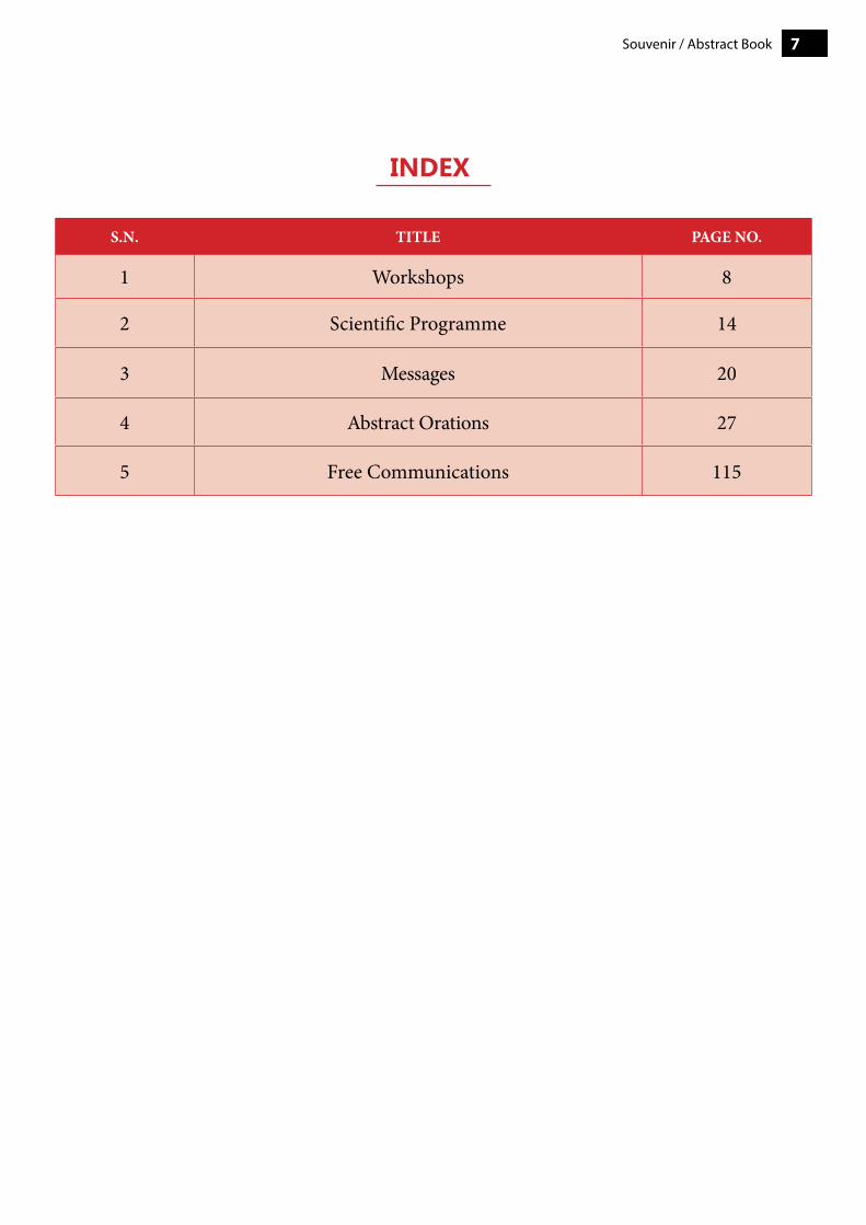
S.N. TITLE PAGE NO.
1 Workshops 8
2 Scientific Programme 14
3 Messages 20
4 Abstract Orations 27
5 Free Communications 115
inDeX

Souvenir / Abstract Book8
Workshops14 December, 2018

9Souvenir / Abstract Book
Tim
eTo
pic
Spea
ker
08:15
-08:3
0Int
rodu
ction
to th
e Wor
ksho
pM
GOU
RI D
EVI
08:30
-08:5
0Ph
ysiol
ogy o
f Fina
l Ooc
yte M
atura
tion:
Role
of H
CG/G
nRHa
/Kiss
pepti
nSA
MUE
L DOS
SAN
TOS
RIBE
IRO
08:50
-09:1
0Ov
arian
Res
erve
Mar
kers
- Sele
cting
the A
ppro
priat
e Star
ting D
ose a
nd T
ype o
f Go
nado
tropin
sBA
SIL T
ARLA
TZIS
09:10
-09:3
0Ke
eping
the S
timula
tion M
ild - I
n W
hom
?
PAUL
DEV
ROEY
09:30
-09:5
0Ef
fect o
f Ova
rian S
timula
tion o
n Ooc
yte Q
uality
and E
ndom
etrium
PURN
IMA N
ADKA
RNI
09:50
-10:1
0St
imula
ting C
ance
r Pati
ents
K JA
YAPR
AKAS
AN10
:10-1
0:30
10:30
-11:0
011
:00-1
1:30
Unex
pecte
d Poo
r Res
pons
e in Y
oung
Pati
ents-
Pos
sible
Caus
es an
d Res
cue
SAM
UEL D
OS S
ANTO
S RI
BEIR
O11
:30-1
2:00
New
Stim
ulatio
n Reg
imen
sBA
SIL T
ARLA
TZIS
12:00
-12:3
0To
ward
s OHS
S Fr
ee C
linic
PAUL
DEV
ROEY
12:30
-13:0
013
:00-1
4:00
14:00
-14:3
0Un
derst
andin
g POS
EIDO
N Cl
assif
icatio
nSA
MUE
L DOS
SAN
TOS
RIBE
IRO
14:30
-15:0
0Lu
teal P
hase
PAUL
DEV
ROEY
15:00
-15:3
0Ne
ver E
nding
Stra
tegies
for P
oor R
espo
nder
sBA
SIL T
ARLA
TZIS
15:30
-16:0
0Co
mm
ents
and D
iscus
sions
K JA
YAPR
AKAS
AN, P
AUL D
EVRO
EY,
BASI
L TAR
LATZ
IS
16:00
-17:0
0PA
NEL
DISC
USSI
ON: C
ase S
cena
rios
MOD
ERAT
ORS:
NEE
RU T
HAKR
AL,
GAUR
AV G
UJAR
ATHI
PANE
LIST
S: K
JAYA
PRAK
ASAN
, SA
MUE
L DOS
SAN
TOS
RIBE
IRO,
AS
HA R
AO, B
HARA
TI D
HORE
PATI
L, SH
EILA
BAL
AKRI
SHNA
N, M
ANOJ
CH
ELLA
NI, R
IMM
Y SIN
GALA
, REN
U M
AKKA
R, F
ESSY
LOUI
S, AS
HWIN
I N,
SHE
ETAL
JIND
AL, R
HYTH
M
AHUJ
A
Com
men
ts an
d Disc
ussio
nsTe
a Bre
ak
Lunc
h
BASI
CS
STRA
TEGI
ES
Com
men
ts an
d Disc
ussio
ns
Tim
eTo
pic
Spea
ker
09:00
-09:3
0Int
rodu
ction
to th
e Wor
ksho
pM
OHAN
S. K
AMAT
H09
:30-0
9:55
Inves
tigati
ng S
ubfer
tile F
emale
Par
tner -
How
Muc
h to I
nves
tigate
?SU
PARN
A BAN
ERJE
E09
:55-1
0:20
DNA F
ragm
entat
ion As
say i
n Clin
ical P
racti
ceSA
NGIT
A SHA
RMA
10:20
-10:4
5Im
prov
ing IU
I Suc
cess
APOO
RVA P
ALLA
M R
EDDY
10:45
-11:1
0Pr
e IVF
wor
kup -
Wha
t is in
? Wha
t is ou
t?RE
NU T
ANW
AR11
:10-1
1:30
11:30
-11:5
5Ad
enom
yosis
& In
fertili
tyKO
RULA
GEO
RGE
11:55
-12:2
0Go
nado
tropin
s In I
VF- H
ow M
uch a
nd W
hich O
ne?
YACO
UB K
HALA
F12
:20-1
2:45
Ovula
tion T
rigge
r- An
Upd
ateM
OHAN
S. K
AMAT
H
12:45
-13:3
0PA
NEL
DISC
USSI
ON: D
ifficu
lt Infe
rtility
Sce
nario
s
MOD
ERAT
ORS:
RAJ
U NA
IR, R
EJI
MOH
AN
PANE
LIST
S: S
ESH
K SU
NKAR
A,GUN
JAN
GUPT
A, SH
ILPI
SUD,
PRA
MYA
, KOR
ULA G
EORG
E,
KOKI
LA D
ESAI,
SM
ITHA
BHA
SKER
, RA
MGO
PAL P
ILLAI
13:30
-14:0
014
:00-1
4:25
Proto
cols
for F
roze
n Em
bryo
Tra
nsfer
sKU
NDAN
INGA
LE
14:25
-15:2
0PA
NEL
DISC
USSI
ON: D
ifficu
lt IVF
Sce
nario
s
MOD
ERAT
ORS:
MOH
AN S
. KA
MAT
H, K
ORUL
A GEO
RGE
PANE
LIST
S: S
ESH
K SU
NKAR
A, M
OHAM
MED
ASHR
AF, S
ATHY
PI
LLAI,
KRI
SHNA
NKUT
TY, B
AVIN
BA
LAKR
ISHN
AN, K
RISH
NALE
ELA
M, S
WET
HA T
UMM
ALA,
CHAIT
ANYA
GAN
PULE
15:20
-15:4
5Si
mpli
fying
Lutea
l Sup
port i
n IVF
LEEN
A WAD
HWA
15:45
-16:1
0El
evate
d Pro
geste
rone
in IV
F- S
hould
We F
reez
e All?
SESH
K S
UNKA
RA16
:10-1
6:35
Recu
rrent
Impla
ntatio
n Fail
ure
DEVIK
A GUN
ASHE
ELA
16:35
-17:0
0PG
S - W
here
Are W
e?M
OHAM
ED S
ALEE
M
Tea B
reak
Lunc
h

Souvenir / Abstract Book10
Tim
eTo
pic
Spea
ker
08:30
-08:4
5Int
rodu
ction
to th
e Wor
ksho
pKD
NAY
AR08
:45-0
9:00
Inves
tigati
ons O
f Rele
vanc
e In M
ale In
fertili
tyKR
ISHN
A DAS
09:00
-09:1
5Do
ing S
emen
Analy
sis T
he R
ight W
ayNI
RMAL
KRI
SHNA
N09
:15-0
9:30
Sper
m M
orph
ology
And R
epro
ducti
ve C
halle
nges
VIJES
H VE
D09
:30-0
9:45
09:45
-10:0
0Lif
e Styl
e Mod
ificati
ons a
nd An
tioxid
ants
in Idi
opath
ic M
ale In
fertili
ty - E
viden
ce
Base
d Rec
omm
enda
tions
BHAV
ATEJ
10:00
-10:1
5Ta
cklin
g Male
Acce
ssor
y Glan
d Infe
ction
(MAG
I)VA
SAN
SS10
:15-1
0:30
Deali
ng W
ith M
ale S
exua
l Dys
functi
onRA
GUL R
EDDY
10:30
-10:4
510
:45-1
1:15
11:15
-11:4
0Qu
ality
Man
agem
ent S
ystem
(QM
S) in
Andr
ology
Lab
Lt.Co
l. NIK
ITA N
ARED
I11
:40-1
2:05
Surg
ical S
perm
Retr
ieval
-Tec
hnica
l Minu
tiae
VASA
N SS
12:05
-12:1
5
12:15
-13:0
0De
bate
- Var
icoce
le- T
o Ope
rate
or N
ot?FO
R - D
HARM
ARAJ
AGAI
NST
- RAG
HAVE
NDRA
PR
ASAD
13:00
-14:0
0
14:00
-14:4
5PA
NEL
DISC
USSI
ON: T
ackli
ng Az
oosp
erm
ia- C
ase S
cena
rios
MOD
ERAT
OR: K
K GO
PINA
THAN
PANE
LIST
S:, V
ASAN
SS,
DH
ARM
ARAJ
, P.M
. GOP
INAT
H,
SRIN
IVAS
MS,
SAN
JAY D
ESAI,
SU
JATA
AGAR
WAL
, MAN
ISHA
VA
JPEY
EE, A
RUN
MUT
HUVE
L14
:45-1
5:05
Role
of Te
sticu
lar S
perm
s In N
on Az
oosp
erm
ic P
atien
tsST
UART
LONG
15:05
-15:2
5Sp
erm
Pre
para
tion A
nd S
electi
on T
echn
iques
RAJE
EV S
HARM
A15
:25-1
5:45
Nove
l Spe
rm Vi
trifica
tion T
echn
iques
SUJA
THA S
URES
H15
:45-1
6:00
Take
Hom
e Mes
sage
sVA
SAN
SS, P
.M. G
OPIN
ATH
Lunc
h
Tea B
reak
Sess
ion
1
Sess
ion
2
Sess
ion
3
Com
men
ts an
d Disc
ussio
ns
Com
men
ts an
d Disc
ussio
ns
Com
men
ts an
d Disc
ussio
ns
Tim
eTo
pic
Spea
ker
08:45
-09:0
0Int
rodu
ction
to th
e Wor
ksho
pKU
LDEE
P JA
IN09
:00-0
9:20
Fertil
ity E
nhan
cing L
apar
osco
pic S
urge
ry - W
hat's
the F
uture
?SA
UMYA
PRA
SAD
09:20
-09:4
0St
rateg
ies to
Pre
serve
Ova
rian R
eser
ve in
End
ometr
ioma S
urge
ryM
OHD
ASHR
AF09
:40-1
0:00
Step
s in U
terine
Tra
nspla
ntatio
n Sur
gery
MILI
ND T
ELAN
G10
:00-1
0:20
Small
End
ometr
ioma-
Lapa
rosc
opy o
r ART
: Wha
t is th
e Evid
ence
?SA
NJAY
MAK
WAN
A10
:20-1
1:00
11:00
-11:3
011
:30-1
1:50
Sim
plifyi
ng M
yom
ectom
y for
Larg
e Fibr
oids
BALA
BHA
GAVA
TH11
:50-1
2:10
Robo
tic S
urge
ry an
d its
Appli
catio
n in I
nfertil
ity M
anag
emen
tSH
AMEE
MA
12:10
-13:0
0PA
NEL
DISC
USSI
ON: D
ecisi
on M
aking
Dur
ing La
paro
scop
y for
Infer
tility
MOD
ERAT
ORS:
PG
PAUL
, AB
Y KOS
HY
PANE
LIST
S: S
HWET
A M
ITTA
L, SI
VADA
S VK
, MUM
TAZ
P, S
UBAS
H M
ALLY
A, SA
NDEE
P DA
TA R
OY,
ARAV
IND
CHAN
DER,
MILI
ND
DUGG
AD, M
ALVIK
A M
ISHR
A, W
g.Cdr
. ABH
A KHU
RANA
13:00
-14:0
0
14.00
-15.0
0PA
NEL
DISC
USSI
ON: T
roub
le Sh
ootin
g In L
apar
osco
py in
Infer
tility
MOD
ERAT
OR: S
HAILE
SH G
OKAV
I, SO
WJA
NYA A
GGAR
WAL
PANE
LIST
S: A
JITH
S, AM
ITI
AGAR
WAL
, PG
PAUL
, SUN
ITA
SAM
AL, J
AYAL
AKSH
MI S
URAJ
, GN
ANA S
ANKE
R NA
TESA
N,
NAZA
R T
15.00
-16.0
0HA
NDS
ON S
ESSI
ON - E
NDOS
UTUR
ING
FACU
LTY:
SHA
ILESH
GOK
AVI,
SUNI
TA S
AMAL
, GNA
NA S
ANKE
R NA
TESA
N
16:00
-17:0
0PA
NEL
DISC
USSI
ON: D
ilem
mas
in H
yster
osco
py
MOD
ERAT
OR: K
JAYA
KRIS
HNAN
PA
NELI
STS:
CYR
IAC P
APPA
CHAN
, BI
MAL
JOHN
, PRA
VEEN
R, V
IVEK
PAUL
, NIR
ANJA
NA J,
GEO
RGE
PAUL
, LAK
SHM
I CHI
RUM
AMILL
A, PH
ANI M
ADHU
RI
Com
men
ts an
d Disc
ussio
ns
Lunc
h
Tea B
reak
11Souvenir / Abstract Book
Tim
eTo
pic
Spea
ker
09:00
-09:0
5Ina
ugur
ation
of th
e Wor
ksho
pSU
DHA P
RASA
D09
:05-0
9:10
Intro
ducti
on to
the C
once
ptAS
HOK
KHUR
ANA
09:10
-09:3
5Th
e Day
2 Sc
anAS
HOK
KHUR
ANA
09:40
-10:0
5Fo
llicle
and E
ndom
etrial
Mon
itorin
g SO
NAL P
ANCH
AL10
:10-1
0:35
Tuba
l Pate
ncy i
n 201
8BE
LA B
HATT
10:35
-11:0
0
11:00
-11:2
5Ut
erine
Cav
ity E
valua
tion b
y Ultra
soun
dM
ANJU
LA H
ANDA
VIR
MAN
I11
:30-1
1:55
Myo
metr
ial F
actor
s in I
nfertil
ity: A
sses
smen
t by U
ltraso
und
SONA
L PAN
CHAL
12:00
-12:2
5Pe
ritone
al &
Tuba
l Mor
pholo
gy an
d Path
ology
by U
ltraso
und
SONA
L PAN
CHAL
12:30
-13:0
0Ti
ps an
d Tric
ks in
Infer
tility
Scan
ning
ASHO
K KH
URAN
A13
:00-1
4:00
14:00
-14:2
5Ul
traso
und G
uided
Inter
venti
onal
Proc
edur
es in
Infer
tility
BHAR
ATI J
AIN14
:30-1
4:55
Tech
nolog
ical A
dvan
ces i
n Infe
rtility
Ultra
soun
dAS
HOK
KHUR
ANA
15:00
-15:3
0Im
ages
in In
fertili
tyRI
TU K
HANN
A
15:30
-17:0
03D
/ 4D
Sim
ulator
Tra
ining
Mas
ter C
lass -
Kind
ly Ca
rry Yo
ur O
wn W
indow
s Lap
top
for T
his S
essio
n
ASHO
K KH
URAN
A, M
ANJU
LA
HAND
A VIR
MAN
I, SON
AL P
ANCH
AL,
MEE
NU B
ATRA
, ZEE
NAT
CHAU
HAN
Sess
ion
3
Sess
ion
4 - Q
uiz
Sess
ion
5 - S
imul
ator
Tra
inin
g
Lunc
h
Sess
ion
1
Sess
ion
2Te
a Bre
ak
Tim
eTo
pic
Spea
ker
08:30
- 08:4
5Int
rodu
ction
to th
e Wor
ksho
p PA
NKAJ
TAL
WAR
08:4
5-10
:30
08:45
- 09:0
0 Ch
allen
ges i
n Air Q
uality
Man
agem
ent
SRIN
IVAS
MS
09:00
- 09:1
5Inc
ubato
r Malf
uncti
on - D
etecti
on &
Cor
recti
onKA
LYAN
I INGA
LE
09:15
- 09:3
0M
icrom
anipu
lation
AZIF
KHA
N09
:30 - 0
9:45
Powe
r Man
agem
ent in
ART
Lab
GAUR
AV K
ANT
09:45
- 10:0
0Te
chnic
al Ch
allen
ges i
n Gas
Sup
ply
SWAM
INAT
HAN
D10
:00-1
0:15
10:15
-10:3
010
:30-
12:0
010
:30-1
0:50
Fertil
izatio
n and
Clea
vage
Fail
ures
SA
NDEE
P KA
RUNA
KARA
N10
:50-1
1:10
Chem
ical &
Micr
obial
Con
tamina
tion i
n ART
Lab
PANK
AJ T
ALW
AR11
:10-1
1:30
pH &
Tem
pera
ture M
anag
emen
t from
OPU
to E
TAL
EX VA
RGHE
SE11
:30-1
1:50
Identi
ficati
on, C
omm
unica
tion &
Doc
umen
tation
KR
ISHN
A CHA
ITAN
YA11
:50 -1
2:00
12:00
-13:0
0 PA
NEL
DISC
USSI
ON: C
risis
Man
agem
ent -
Case
Sce
nario
s
MOD
ERAT
OR: P
RIYA
KAN
NAN
PA
NELI
STS:
ARNE
SUN
DE,
JAYA
NT M
EHTA
, KRI
SHNA
CH
AITAN
YA, P
RASH
ANTH
CP,
AZIF
KH
AN, Y
ATIN
DRA V
ARM
A13
:00-1
4:00
14:00
-15:0
0 PA
NEL
DISC
USSI
ON: T
echn
ical C
halle
nges
in P
GT (P
reim
planta
tion G
eneti
c Te
sting
)
MOD
ERAT
OR: G
AURA
V M
AJUM
DAR
PA
NELI
STS:
SON
U T
LUKO
SE,
ALEX
VARG
HESE
, SUJ
ATHA
RA
MAK
RISH
NAN,
RAM
PRA
KASH
, PR
ANAY
GHO
SH, P
ARES
H M
AKW
ANA
15:00
-16:3
015
:00-1
5:20
IVF La
b Erro
rs - H
ow to
Man
age T
hem
?JA
YANT
MEH
TA15
:20-1
5:40
Disa
ster M
anag
emen
t in IV
F La
bET
HIRA
J BAL
AJI
15:40
-16:0
0Te
chnic
al Ch
allen
ges i
n Vitri
ficati
on &
Stor
age
SUJA
THA R
AMAK
RISH
NAN
16:00
-16:2
0Te
chnic
al Ch
allen
ges i
n Em
bryo
Tra
nsfer
KA
RTHI
KA D
KUM
AR
16:20
-16:3
0Ta
ke H
ome M
essa
ges
PANK
AJ T
ALW
AR,
ALEX
VARG
HESE
Com
men
ts an
d Disc
ussio
ns
Com
men
ts an
d Disc
ussio
ns
Sess
ion
2 - T
echn
ical
Cha
lleng
es in
Cul
ture
Sys
tem
Man
agem
ent
Sess
ion
1 -Te
chni
cal C
halle
nges
in L
ab M
anag
emen
t
Sess
ion
3 Lunc
h
Tea B
reak
Sess
ion
4
Sess
ion
5- T
echn
ical
Cha
lleng
es
Souvenir / Abstract Book12
Tim
eTo
pic
Spea
ker
08:30
-08:4
5Int
rodu
ction
to th
e Wor
ksho
pSO
NIA M
ALIK
08:45
- 09:0
0Em
bryo
Mor
phok
inetic
s -Th
e Way
Ahea
d Or T
ime F
or O
bitua
ryET
HIRA
J BAL
AJI
09:00
-09:1
5No
vel S
perm
Sele
ction
Meth
ods-
Do T
hey I
mpr
ove R
epro
ducti
ve O
utcom
es?
STUA
RT LO
NG
09:15
-09:3
0Hi
gh E
nd U
ltraso
und S
ystem
s And
Autom
ation
And T
heir P
lace I
n Infe
rtility
M
anag
emen
tSU
MIT
A SOF
AT
09:30
-09:4
509
:45-1
0:00
Plate
let R
ich P
lasm
a - T
he N
ew M
aster
Key
To A
ll Ridd
les In
ART?
ROHI
T GU
TGUT
IA10
:00-1
0:15
Epige
netic
Effe
cts of
Em
bryo
Cult
ure,
Poten
tial E
ffects
on th
e Offs
pring
?AR
NE S
UNDE
10:15
-10:3
0St
em C
ell T
hera
py - D
awn O
f New
Era
Or F
alse H
opes
SANK
ALP
SING
H10
:30-1
0:45
10:45
-11:1
5
11:15
-11:3
0M
itoch
ondr
ial R
eplac
emen
t The
rapy
: Is It
Here
To S
tay?
NYM
PHAE
A WAL
ECHA
11:30
-11:4
5No
n Inv
asive
Asse
ssm
ent F
or Vi
abilit
y Of E
mbr
yos-
The W
ay Ah
ead?
GAUR
AV M
AJUM
DAR
11:45
-12:0
0
12:00
-13:0
0 PA
NEL
DISC
USSI
ON: In
tegra
tion o
f Tec
hnolo
gies i
n Day
to da
y Pra
ctice
MODE
RATO
R : P
ARAS
URAM
GPA
NELI
STS:
SON
IA M
ALIK
, M
IRUD
HUBA
SHIN
I G, G
AURA
V M
AJUM
DAR,
SAN
KALP
SIN
GH,
RAM
ESH
P, U
MA R
AMES
H,
SHYL
AJA G
OWDA
13:00
-14:0
0
14:00
-14:4
5De
bate
- PGS
2:0 -
Is T
he W
ay F
orwa
rd?
FOR-
CAR
MEN
MOR
ALES
AGAI
NST-
PRA
TAP
KUM
AR
14:45
-15:0
0Ar
tificia
l Intel
ligen
ce An
d Auto
mati
on In
IVF
Lab:
Will
It Cha
nge T
he W
ay W
e Pr
actic
e?KR
ISHN
A CHA
ITAN
YA
15:00
-15:1
5Th
e Arri
val O
f Gen
e Edit
ing T
ools-
Like
CRI
SPR/
Cas -
-W
hat It
Mea
ns?
RITU
HAR
I
15:15
-15:3
0
15:30
-16:0
0Ta
ke H
ome M
essa
ges
MIR
UDHU
BASH
INI G
, CA
RMEN
MOR
ALES
In T
he H
orizo
nLu
nch
Curre
nt S
tars
At T
he D
oor S
tep
Tea B
reak
Com
men
ts an
d Disc
ussio
n
Com
men
ts an
d Disc
ussio
ns
Com
men
ts an
d Disc
ussio
ns
Com
men
ts an
d Disc
ussio
ns
17:30
-18:0
0
18:00
-18:3
0
18:30
-19:0
0
Rum
inatio
ns of
a Re
stles
s Reti
ree :
GIT
A AR
JUN
Ferti
visi
on 20
18 O
ratio
n in
Clin
ical
Em
bryo
logy
CH
AIR
- M. G
OURI
DEV
I, KUL
DEEP
JAIN
, PRI
YA K
ANNA
N
The A
ssist
ed R
epro
ducti
ve T
echn
ology
Lab -
Tow
ards
Evid
ence
Bas
ed P
racti
ce: A
RNE
SUND
E
19:3
0 hrs
- Ina
ugur
atio
n fo
llowe
d by
Net
work
nig
Dinn
er
PLEN
ARY
LECT
URES
CHAI
R - K
.D. N
AYAR
, SON
IA M
ALIK
, SUD
HA P
RASA
D
Med
ico Le
gal A
spec
ts In
Assis
ted R
epro
ducti
on : H
ITES
H BH
ATT

13Souvenir / Abstract Book
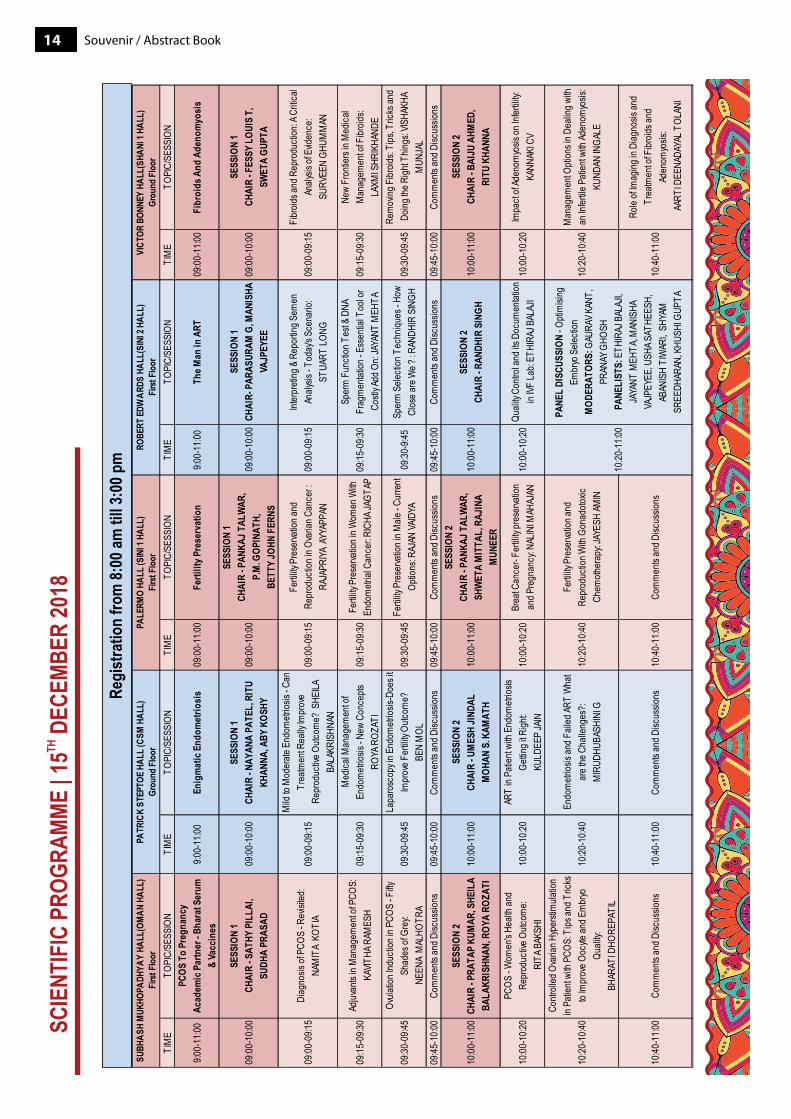
scienTificprogramme
Souvenir / Abstract Book14
11:00
-11:3
011
:30-1
2:30
11:30
-11:5
011
:50-1
2:10
12:10
-12:3
0
12:30
-13:0
0
13:00
-14:0
0
14:00
-14:3
0
Lunc
h an
d Po
ster
Ses
sion
(Lob
by L
evel
) F
ertiv
isio
n 20
18 O
ratio
n on
Clin
ical
Rep
rodu
ctiv
e Med
icin
e Ov
aria
n Bi
omar
kers
: WIL
LIAM
LED
GER
CH
AIR
- MIR
UDHU
BASH
INI G
., BHA
RTI D
HORE
PATI
L
Tea /
Cof
fee B
reak
SESS
ION
3 - P
LENA
RY L
ECTU
RES
| CHA
IR: K
.K. G
OPIN
ATHA
N, JA
IDEE
P M
ALHO
TRA,
NEE
NA M
ALHO
TRA
Does
End
ometr
ial T
hickn
ess M
atter
? JL
H EV
ERS
Prac
ticing
Evid
ence
Bas
ed M
edici
ne in
India
: MOH
AN S
. KAM
ATH
Role
of Fr
eeze
All in
Con
tempo
rary
ART
Prac
tice -
BAS
IL TA
RLAT
ZIS
SESS
ION
4: P
RESI
DENT
'S O
RATI
ON | G
azin
g at
The
Hor
izon
: M. G
OURI
DEV
ICH
AIR
: PAN
KAJ T
ALW
AR, K
.U. K
UNJI
MOI
DEEN
, M. V
ENUG
OPAL
14:30
-16:0
0
SESS
ION
5 INV
ITED
LEC
TURE
S CH
AIR-
K. J
AYAP
RAKA
SAN,
M
OHAM
MED
ASH
RAF,
RIT
A BA
KSHI
14:00
-16:0
0
SESS
ION
5 INV
ITED
LEC
TURE
S CH
AIR
- SAN
JEEV
A RE
DDY,
PK
SEK
HARA
N, K
.U.
KUNJ
IMOI
DEEN
14:30
-16:0
0SE
SSIO
N 5:
INVI
TED
LECT
URES
: CH
AIR-
M. G
OURI
DEV
I, PAN
KAJ
TALW
AR14
:00-1
6:00
SESS
ION
5: IN
VITE
D LE
CTUR
ES:
CHAI
R - P
RABH
AKAR
SIN
GH, A
NU
MAT
HEW
S
14:30
-14:5
0Ad
vanc
ed M
atern
al Ag
e and
its
Chall
enge
s: YA
COUB
KHA
LAF
14:30
-14:5
0AR
T An
d Risk
of E
xtrau
terine
Ge
statio
ns:
BALA
BHA
GAVA
TH14
:30-1
4:50
PCPN
DT Ac
t and
Infer
tility
Prac
tice:
RAJN
IKAN
T CO
NTRA
CTOR
14:30
-14:5
0Lo
w Ox
ygen
Em
bryo
Cult
ure -
Do W
e All
Nee
d it ?
JA
YANT
MEH
TA
14:50
-15:1
0Un
derst
andin
g Con
cept
of Ide
al Ov
arian
Stim
ulatio
n:SE
SH K
. SUN
KARA
14:50
-15:1
0Th
e role
of th
e end
ometr
ium in
IVF:
SAM
UEL D
OS S
ANTO
S RI
BEIR
O14
:50-1
5:10
ICSI
for A
ll. Is
it Jus
tified
? P
AUL D
EVRO
EY14
:50-1
5:10
Are W
e Rea
dy fo
r Blas
tocys
t Cult
ure ?
Br
ig. D
r. R.K
. SHA
RMA
15:10
-15:3
0En
dom
etrial
Scr
atch I
njury
in Pa
tients
W
ith R
IF - W
hat is
the E
viden
ce?
NIKO
LAOS
POL
YZOS
15:10
-15:3
0St
imula
ting P
atien
ts W
ith
Hypo
gona
dotro
pic H
ypog
onad
ism:
PRAT
AP K
UMAR
15:10
-15:3
0Lu
teal S
uppo
rt in A
RT: K
.D. N
AYAR
15:10
-15:3
0Pr
e-Im
planta
tion G
eneti
c Tes
ts -
Curre
nt Co
nsen
sus :
CAR
MEN
MOR
ALES
15:30
-16:0
0Co
mm
ents
and D
iscus
sions
15:30
-16:0
0Co
mm
ents
and D
iscus
sions
15:30
-16:1
5
PANE
L DI
SCUS
SION
- New
Int
erna
tiona
l Guid
eline
s in
Man
agem
ent o
f PCO
SM
ODER
ATOR
S : S
ONIA
MAL
IK,
VAND
ANA B
HATI
APA
NELI
STS:
PAU
L DEV
ROEY
, BAS
IL TA
RLAT
ZIS,
SES
H K
SUNK
ARA,
PRAT
AP K
UMAR
, KAN
NAKI
CV,
M
OHAN
S K
AMAT
H, S
ONAL
PA
NCHA
L, JY
OTHI
PAT
IL, AA
RATH
Y PA
ARI
15:30
-15:4
0Co
mm
ents
and D
iscus
sions
14:30
-16:0
0SE
SSIO
N 5:
INVI
TED
LECT
URES
: CH
AIR
- SHW
ETA
MIT
TAL,
RAS
HMI
SHAR
MA
14:30
-14:5
0M
anag
emen
t of O
varia
n Cys
ts in
Infer
tile P
atien
ts:
GITA
RAD
HAKR
ISHN
AN
14:50
-15:1
0Or
al An
tioxid
ant T
hera
py an
d Im
pact
on S
emen
Par
amete
rs:
SAYA
LI KA
NDAR
I
15:10
-15:3
0IU
I in T
he E
ra of
Assis
ted
Repr
oduc
tion:
GEET
A KHA
NNA
15:30
-16:0
0Co
mm
ents
and D
is cus
sions
TIM
ETO
PIC/
SESS
ION
TIM
ETO
PIC/
SESS
ION
TIM
ETO
PIC/
SESS
ION
TIM
ETO
PIC/
SESS
ION
9:00-
11:00
PCOS
To
Preg
nanc
yAc
adem
ic P
artn
er - B
hara
t Ser
um
& Va
ccin
es9:0
0-11
:00En
igm
atic
End
omet
riosi
s09
:00-1
1:00
Ferti
lity P
rese
rvat
ion
9:00-
11:00
The M
an in
ART
09:00
-10:0
0SE
SSIO
N 1
CHAI
R - S
ATHY
PIL
LAI,
SUDH
A PR
ASAD
09:00
-10:0
0SE
SSIO
N 1
CHAI
R - N
AYAN
A PA
TEL,
RIT
U KH
ANNA
, ABY
KOS
HY09
:00-1
0:00
SESS
ION
1CH
AIR
- PAN
KAJ T
ALW
AR,
P.M
. GOP
INAT
H,
BETT
Y JO
HN F
ERNS
09:00
-10:0
0SE
SSIO
N 1
CHAI
R- P
ARAS
URAM
G, M
ANIS
HA
VAJP
EYEE
09:00
-09:1
5Di
agno
sis of
PCO
S - R
evisi
ted:
NAM
ITA
KOTI
A
09:00
-09:1
5
Mild
to M
oder
ate E
ndom
etrios
is - C
an
Trea
tmen
t Rea
lly Im
prov
e Re
prod
uctiv
e Outc
ome?
SHE
ILA
BALA
KRIS
HNAN
09:00
-09:1
5Fe
rtility
Pre
serva
tion a
nd
Repr
oduc
tion i
n Ova
rian C
ance
r : RA
JAPR
IYA A
YYAP
PAN
09:00
-09:1
5Int
erpr
eting
& R
epor
ting S
emen
An
alysis
- Tod
ay's
Scen
ario:
ST
UART
LONG
09:15
-09:3
0Ad
juvan
ts in
Man
agem
ent o
f PCO
S:KA
VITHA
RAM
ESH
09:15
-09:3
0M
edica
l Man
agem
ent o
f En
dom
etrios
is - N
ew C
once
pts
ROYA
ROZ
ATI
09:15
-09:3
0Fe
rtility
Pre
serva
tion i
n Wom
en W
ith
Endo
metr
ial C
ance
r: RIC
HA JA
GTAP
09:15
-09:3
0Sp
erm
Fun
ction
Tes
t & D
NA
Frag
men
tation
- Ess
entia
l Too
l or
Costl
y Add
On:
JAYA
NT M
EHTA
09:30
-09:4
5Ov
ulatio
n Ind
uctio
n in P
COS
- Fifty
Sh
ades
of G
rey:
NEEN
A M
ALHO
TRA
09:30
-09:4
5La
paro
scop
y in E
ndom
etrios
is-Do
es it
Impr
ove F
ertili
ty Ou
tcom
e?
BEN
MOL
09:30
-09:4
5Fe
rtility
Pre
serva
tion i
n Male
- Cur
rent
Optio
ns: R
AJAN
VAID
YA09
:30-9
:45Sp
erm
Sele
ction
Tec
hniqu
es - H
ow
Clos
e are
We ?
: RAN
DHIR
SIN
GH
09:45
-10:0
0Co
mm
ents
and D
iscus
sions
09:45
-10:0
0Co
mm
ents
and D
iscus
sions
09:45
-10:0
0Co
mm
ents
and D
iscus
sions
09:45
-10:0
0Co
mm
ents
and D
iscus
sions
10:00
-11:0
0SE
SSIO
N 2
CHAI
R - P
RATA
P KU
MAR
, SHE
ILA
BALA
KRIS
HNAN
, ROY
A RO
ZATI
10:00
-11:0
0SE
SSIO
N 2
CHAI
R - U
MES
H JI
NDAL
MOH
AN S
. KAM
ATH
10:00
-11:0
0
SESS
ION
2CH
AIR
- PAN
KAJ T
ALW
AR,
SHW
ETA
MIT
TAL,
RAJ
INA
MUN
EER
10:00
-11:0
0SE
SSIO
N 2
CHAI
R - R
ANDH
IR S
INGH
10:00
-10:2
0PC
OS - W
omen
's He
alth a
nd
Repr
oduc
tive O
utcom
e: R
ITA B
AKSH
I10
:00-1
0:20
ART
in Pa
tient
with
Endo
metr
iosis
Gettin
g it R
ight:
KULD
EEP
JAIN
10:00
-10:2
0Br
eat C
ance
r- Fe
rtility
pres
erva
tion
and P
regn
ancy
: NAL
INI M
AHAJ
AN10
:00-1
0:20
Quali
ty Co
ntrol
and i
ts Do
cum
entat
ion
in IVF
Lab:
ETHI
RAJ B
ALAJ
I
10:20
-10:4
0
Contr
olled
Ova
rian H
yper
stim
ulatio
n in
Patie
nt wi
th PC
OS: T
ips an
d Tric
ks
to Im
prov
e Ooc
yte an
d Em
bryo
Qu
ality:
BHAR
ATI D
HORE
PATI
L
10:20
-10:4
0En
dom
etrios
is an
d Fail
ed AR
T W
hat
are t
he C
halle
nges
?: M
IRUD
HUBA
SHIN
I G10
:20-1
0:40
Fertil
ity P
rese
rvatio
n and
Re
prod
uctio
n With
Gon
adoto
xic
Chem
other
apy:
JAYE
SH AM
IN
10:40
-11:0
0Co
mm
ents
and D
iscus
sions
10:40
-11:0
0Co
mm
ents
and D
iscus
sions
10:40
-11:0
0Co
mm
ents
and D
iscus
sions
PATR
ICK
STEP
TOE
HALL
(CSM
HAL
L)Gr
ound
Flo
or
SUBH
ASH
MUK
HOPA
DHYA
Y HA
LL(O
MAN
HAL
L)Fi
rst F
loor
ROBE
RT E
DWAR
DS H
ALL(
SINI
2 HA
LL)
Firs
t Flo
or
10:20
-11:0
0
PANE
L DI
SCUS
SION
- Opti
misi
ng
Embr
yo S
electi
onM
ODER
ATOR
S: G
AURA
V KAN
T,
PRAN
AY G
HOSH
PANE
LIST
S: E
THIR
AJ B
ALAJ
I, JA
YANT
MEH
TA, M
ANIS
HA
VAJP
EYEE
, USH
A SAT
HEES
H,
ABAN
ISH
TIW
ARI,
SHYA
M
SREE
DHAR
AN, K
HUSH
I GUP
TA
PALE
RMO
HALL
(SIN
I 1 H
ALL)
Firs
t Flo
orTI
ME
TOPI
C/SE
SSIO
N
09:00
-11:0
0Fi
broi
ds A
nd A
deno
myo
sis
09:00
-10:0
0SE
SSIO
N 1
CHAI
R - F
ESSY
LOU
IS T
, SW
ETA
GUPT
A
09:00
-09:1
5Fi
broid
s and
Rep
rodu
ction
: A C
ritica
l An
alysis
of E
viden
ce:
SURV
EEN
GHUM
MAN
09:15
-09:3
0Ne
w Fr
ontie
rs in
Med
ical
Man
agem
ent o
f Fibr
oids:
LAXM
I SHR
IKHA
NDE
09:30
-09:4
5Re
mov
ing F
ibroid
s: Ti
ps, T
ricks
and
Doing
the R
ight T
hings
: VIS
HAKH
A M
UNJA
L09
:45-1
0:00
Com
men
ts an
d Disc
ussio
ns
10:00
-11:0
0SE
SSIO
N 2
CHAI
R - B
AIJU
AHM
ED,
RITU
KHA
NNA
10:00
-10:2
0Im
pact
of Ad
enom
yosis
on In
fertili
ty:
KANN
AKI C
V
10:20
-10:4
0M
anag
emen
t Opti
ons i
n Dea
ling w
ith
an In
fertile
Pati
ent w
ith Ad
enom
yosis
: KU
NDAN
INGA
LE
10:40
-11:0
0
Role
of Im
aging
in D
iagno
sis an
d Tr
eatm
ent o
f Fibr
oids a
nd
Aden
omyo
sis:
AART
I DEE
NADA
YAL T
OLAN
I
VICT
OR B
ONNE
Y HA
LL(S
HANI
1 HA
LL)
Grou
nd Fl
oor

15Souvenir / Abstract Book
11:00
-11:3
011
:30-1
2:30
11:30
-11:5
011
:50-1
2:10
12:10
-12:3
0
12:30
-13:0
0
13:00
-14:0
0
14:00
-14:3
0
Lunc
h an
d Po
ster
Ses
sion
(Lob
by L
evel
) F
ertiv
isio
n 20
18 O
ratio
n on
Clin
ical
Rep
rodu
ctiv
e Med
icin
e Ov
aria
n Bi
omar
kers
: WIL
LIAM
LED
GER
CH
AIR
- MIR
UDHU
BASH
INI G
., BHA
RTI D
HORE
PATI
L
Tea /
Cof
fee B
reak
SESS
ION
3 - P
LENA
RY L
ECTU
RES
| CHA
IR: K
.K. G
OPIN
ATHA
N, JA
IDEE
P M
ALHO
TRA,
NEE
NA M
ALHO
TRA
Does
End
ometr
ial T
hickn
ess M
atter
? JL
H EV
ERS
Prac
ticing
Evid
ence
Bas
ed M
edici
ne in
India
: MOH
AN S
. KAM
ATH
Role
of Fr
eeze
All in
Con
tempo
rary
ART
Prac
tice -
BAS
IL TA
RLAT
ZIS
SESS
ION
4: P
RESI
DENT
'S O
RATI
ON | G
azin
g at
The
Hor
izon
: M. G
OURI
DEV
ICH
AIR
: PAN
KAJ T
ALW
AR, K
.U. K
UNJI
MOI
DEEN
, M. V
ENUG
OPAL
14:30
-16:0
0
SESS
ION
5 INV
ITED
LEC
TURE
S CH
AIR-
K. J
AYAP
RAKA
SAN,
M
OHAM
MED
ASH
RAF,
RIT
A BA
KSHI
14:00
-16:0
0
SESS
ION
5 INV
ITED
LEC
TURE
S CH
AIR
- SAN
JEEV
A RE
DDY,
PK
SEK
HARA
N, K
.U.
KUNJ
IMOI
DEEN
14:30
-16:0
0SE
SSIO
N 5:
INVI
TED
LECT
URES
: CH
AIR-
M. G
OURI
DEV
I, PAN
KAJ
TALW
AR14
:00-1
6:00
SESS
ION
5: IN
VITE
D LE
CTUR
ES:
CHAI
R - P
RABH
AKAR
SIN
GH, A
NU
MAT
HEW
S
14:30
-14:5
0Ad
vanc
ed M
atern
al Ag
e and
its
Chall
enge
s: YA
COUB
KHA
LAF
14:30
-14:5
0AR
T An
d Risk
of E
xtrau
terine
Ge
statio
ns:
BALA
BHA
GAVA
TH14
:30-1
4:50
PCPN
DT Ac
t and
Infer
tility
Prac
tice:
RAJN
IKAN
T CO
NTRA
CTOR
14:30
-14:5
0Lo
w Ox
ygen
Em
bryo
Cult
ure -
Do W
e All
Nee
d it ?
JA
YANT
MEH
TA
14:50
-15:1
0Un
derst
andin
g Con
cept
of Ide
al Ov
arian
Stim
ulatio
n:SE
SH K
. SUN
KARA
14:50
-15:1
0Th
e role
of th
e end
ometr
ium in
IVF:
SAM
UEL D
OS S
ANTO
S RI
BEIR
O14
:50-1
5:10
ICSI
for A
ll. Is
it Jus
tified
? P
AUL D
EVRO
EY14
:50-1
5:10
Are W
e Rea
dy fo
r Blas
tocys
t Cult
ure ?
Br
ig. D
r. R.K
. SHA
RMA
15:10
-15:3
0En
dom
etrial
Scr
atch I
njury
in Pa
tients
W
ith R
IF - W
hat is
the E
viden
ce?
NIKO
LAOS
POL
YZOS
15:10
-15:3
0St
imula
ting P
atien
ts W
ith
Hypo
gona
dotro
pic H
ypog
onad
ism:
PRAT
AP K
UMAR
15:10
-15:3
0Lu
teal S
uppo
rt in A
RT: K
.D. N
AYAR
15:10
-15:3
0Pr
e-Im
planta
tion G
eneti
c Tes
ts -
Curre
nt Co
nsen
sus :
CAR
MEN
MOR
ALES
15:30
-16:0
0Co
mm
ents
and D
iscus
sions
15:30
-16:0
0Co
mm
ents
and D
iscus
sions
15:30
-16:1
5
PANE
L DI
SCUS
SION
- New
Int
erna
tiona
l Guid
eline
s in
Man
agem
ent o
f PCO
SM
ODER
ATOR
S : S
ONIA
MAL
IK,
VAND
ANA B
HATI
APA
NELI
STS:
PAU
L DEV
ROEY
, BAS
IL TA
RLAT
ZIS,
SES
H K
SUNK
ARA,
PRAT
AP K
UMAR
, KAN
NAKI
CV,
M
OHAN
S K
AMAT
H, S
ONAL
PA
NCHA
L, JY
OTHI
PAT
IL, AA
RATH
Y PA
ARI
15:30
-15:4
0Co
mm
ents
and D
iscus
sions
14:30
-16:0
0SE
SSIO
N 5:
INVI
TED
LECT
URES
: CH
AIR
- SHW
ETA
MIT
TAL,
RAS
HMI
SHAR
MA
14:30
-14:5
0M
anag
emen
t of O
varia
n Cys
ts in
Infer
tile P
atien
ts:
GITA
RAD
HAKR
ISHN
AN
14:50
-15:1
0Or
al An
tioxid
ant T
hera
py an
d Im
pact
on S
emen
Par
amete
rs:
SAYA
LI KA
NDAR
I
15:10
-15:3
0IU
I in T
he E
ra of
Assis
ted
Repr
oduc
tion:
GEET
A KHA
NNA
15:30
-16:0
0Co
mm
ents
and D
iscus
sions

Souvenir / Abstract Book16
16:00
-17:3
0SE
SSIO
N 6
15:30
-16:1
5
PANE
L DI
SCUS
SION
- Cas
e Sc
enar
ios: E
thica
l Dile
mm
a in A
RTM
ODER
ATOR
: FAL
GUNI
BAV
ISHI
PANE
LIST
S : H
IMAN
SHU
BAVIS
HI
MAN
ISHA
VAJP
EYEE
, NYM
PHAE
A W
ALEC
HA, V
ANI P
UJAR
I, AAR
TI
DEEN
ADAY
AL T
OLAN
I, SOU
MYA
NA
IR, R
AJIN
A MUN
EER,
PRE
RNA
KESH
AN
16:15
-17:3
0
IFS-
AMIT
Y FEL
LOW
S SE
SSIO
N
Po
st Gr
adua
te M
entor
Inter
actio
n -
Te
ll Me W
hy? T
ell M
e Wha
t Nex
t?:Fa
culty
: BAS
IL TA
RLAT
ZIS,
BALA
BHA
GAVA
TH,P
ANKA
J TAL
WAR
, AR
NE S
UNDE
16:00
-17:3
0SE
SSIO
N 6
CHAI
R- K
.U. K
UNJI
MOI
DEEN
, M
. VEN
UGOP
AL16
:00-1
7:00
SESS
ION
6CH
AIR
- KUL
DEEP
JAIN
, FE
SSY
LOUI
S T
16:15
-17:0
0SE
SSIO
N 6
15:40
-17:0
0SE
SSIO
N 6
CHAI
R - A
ZIF
KHAN
,PR
ASHA
NTH
C.P
16:00
-16:4
5
Video
Inter
activ
e Ses
sion:
Hy
stero
scop
y in I
nfertil
ityOS
AMA S
HAW
KI15
:30-1
6:15
DEBA
TE: E
ndom
etrios
is - S
urge
ry Ve
rsus A
RT
FOR
: PG
PAUL
AGAI
NST:
JLH
EVER
S
15:40
-16:2
0
DEBA
TE : S
INGL
E ST
EP V/
S SE
QUEN
TIAL
CUL
TURE
RITE
SH AG
RAW
AL ( S
INGL
E ST
EP
CULT
URE
) V/S
SAR
ABPR
EET
SING
H ( S
EQUE
NTIAL
CUL
TURE
)
16:45
-17:3
0
PANE
L DI
SCUS
SION
- Cas
e Sc
enar
ios - C
ontro
versi
es In
Assis
ted
Repr
oduc
tion
MOD
ERAT
ORS:
NAY
ANA P
ATEL
, LA
KSHM
I CHI
RUM
AMILL
APA
NELI
STS:
RAJ
ESH
S KO
RADI
A, IN
DU LA
TA, S
WET
A AGA
RWAL
, RA
CHIT
A CHA
WLA
, VER
ONIC
A YU
EL, V
IDHY
A PRA
BHAK
AR, P
RIYA
N
16:15
-17:0
0
PANE
L DI
SCUS
SION
- Ultra
soun
d In
Infer
tility
MOD
ERAT
ORS:
SON
AL P
ANCH
AL,
BAND
ANA S
ODHI
PANE
LIST
S: B
HARA
TI JA
IN,
RAGH
AVEN
DRA P
RASA
D, P
IYUSH
I SH
ARM
A, G
ARIM
A SHA
RMA,
VIJI
PRAV
EEN,
PRA
KRIT
I VAR
MA,
VANI
THA D
EVI, B
HAVN
A BAN
GA
16:20
-17:0
0
PANE
L DI
SCUS
SION
- Opti
misi
ng
the IV
F La
bM
ODER
ATOR
S: S
UDES
H KA
MAT
, SE
EMA N
AIR
PANE
LIST
S: S
UJAT
HA
RAM
AKRI
SHNA
N, R
AJEE
V SHA
RMA,
RK S
HARM
A, AN
U M
ATHE
WS,
PO
OJA A
WAS
THI, T
HASN
I MAR
IYAM
, SA
NJEE
V MAH
ESHW
ARI, K
ERSI
AV
ARI
17:3
0-18
:00
IFS
GENE
RAL
BODY
MEE
TING
17:00
-17:2
0De
letion
Analy
sis In
Infer
tility
Clini
cs:
RAJE
NDER
SIN
GH19
:30 h
rs
16:15
-17:0
0
Post
Grad
uate
Men
tor In
terac
tion
Jour
nal A
rticle
Disc
ussio
n:FA
CULT
Y: S
ANJE
EVA R
EDDY
,
NI
KOLA
OS P
OLYZ
OS,
K JA
YAPR
AKAS
AN
COOR
DINA
TOR:
RAM
ESH
P IYE
R
Banq
uet a
nd E
nter
tain
men
t

17Souvenir / Abstract Book
TIM
ETO
PIC/
SESS
ION
9:00-
11:00
Infe
ctio
ns an
d Th
eir I
mpa
ct O
n Fe
rtilit
y
09:00
-10:0
0SE
SSIO
N 1
CHAI
R - R
ADHI
KA R
EDDY
P,
MOV
VA M
ADHU
RI
09:00
-09:1
5Ch
ronic
End
ometr
itis an
d Re
prod
uctiv
e Outc
ome :
CHI
TRA
TYAG
ARAJ
U
09:15
-09:3
0Ho
w Tu
berc
ulosis
Affec
ts Inf
ertili
ty:
RITU
KHA
NNA
09:30
-09:4
5Ro
le of
Geno
mics
in D
iagno
sis of
Inf
ectio
ns in
Infer
tility:
SH
WET
HA G
UPTA
10:00
-11:0
0SE
SSIO
N 2
CHAI
R- R
EVAT
HY S
ADAS
IVAM
10:00
-10:1
5Ro
le of
Prob
iotics
in In
fertili
ty M
anag
emen
t: NIM
ISH
SHEL
AT
10:15
-10:3
0Tu
bal fl
ushin
g in t
he in
fertile
wom
an :
BE
N M
OL
10:30
-10:4
5M
ale Ac
cess
ory G
land I
nfecti
on :
MUJ
IBUR
RAH
MAN
10:45
-11:0
0Co
mm
ents
and D
iscus
sions
VICT
OR B
ONNE
Y HA
LL(S
HANI
1 HA
LL)
Grou
nd F
loor
09:45
-10:0
0Co
mm
ents
and D
iscus
sions
TIM
ETO
PIC/
SESS
ION
TIM
ETO
PIC/
SESS
ION
TIM
ETO
PIC/
SESS
ION
TIM
ETO
PIC/
SESS
ION
9:00-
11:00
Clin
ical
Dile
mm
as In
ART
09:00
-11:0
0Ge
tting
The
Med
icat
ion
Righ
t09
:00-1
1:00
Diffi
cult
Situ
atio
ns in
ART
09:00
-11:0
0In
side
the A
RT L
abor
ator
y
09:00
-10:0
0SE
SSIO
N 1
CHAI
R - P
K SE
KHAR
AN,
SWET
A GU
PTA,
VANI
PUJ
ARI
09:00
-10:0
0SE
SSIO
N 1
CHAI
R- VI
JAYA
STE
PHEN
, K
.D. N
AYAR
09:00
-10:0
0SE
SSIO
N 1
CHAI
R- S
ESH
K SU
NKAR
A,
ANUR
EKHA
JP09
:00-1
0:00
SESS
ION
1CH
AIR-
NIM
MI N
IRM
AL, S
UNIL
NA
YAR,
JAYA
LAKS
HMI S
HARO
FF
09:00
-09:1
5Ps
ycho
socia
l Car
e Dur
ing As
sisted
Re
prod
uctio
n: PO
ONAM
NAY
AR09
:00-0
9:15
Impa
ct of
Oral
Ovulo
gens
on C
OS
Outco
me:
RAJE
NDRA
BOL
DANE
09:00
-09:2
0Pr
oblem
s in O
ocyte
Aspir
ation
: KO
KILA
DES
AI09
:00-0
9:15
ART
Lab -
Dail
y Che
ck lis
t : PR
IYA K
ANNA
N
09:15
-09:3
0Ov
arian
rese
rve te
sting
and
stim
ulatio
n in I
VF: B
EN M
OL09
:15-0
9:30
Reco
mbin
ant V
s Urin
ary -
The
Nev
er
Endin
g Deb
ate:
HIM
ANSH
U BA
VISHI
09:20
-09:4
0De
aling
with
Diffc
ult E
mbr
yo T
rans
fers
GAUT
AM AL
LAHB
ADIA
09:15
-09:3
0pH
Mon
itorin
g - T
he W
ay F
orwa
rd !:
CHAR
ULAT
A CHA
TTER
JEE
09:30
-09:4
5Th
e Tru
th Ab
out M
ild S
timula
tion:
WILL
IAM LE
DGER
09:30
-09:4
5LH
Sup
plem
entat
ion in
Ova
rian
Stim
ulatio
n - Is
it Be
nefic
ial?
RU
PALI
BASS
I GOY
AL09
:40-1
0:00
Empty
Foll
icle S
yndr
ome:
KAVIT
HA G
AUTH
AM09
:30-0
9:45
Fertil
izatio
n Fail
ure
- Ooc
yte
Activ
ation
& M
ore:
FESE
ENA
KUNJ
IMOI
DEEN
09:45
-10:0
0Em
bryo
Biop
sy T
echn
iques
: HA
RSHA
BAN
DARK
A10
:00-1
0:20
Com
men
ts an
d Disc
ussio
ns
10:00
-11:0
0SE
SSIO
N 2 I
SAR
SESS
ION
CHAI
R- M
. GOU
RI D
EVI, P
ANKA
J TA
LWAR
, SUD
HA P
RASA
D10
:00-1
1:00
SESS
ION
2CH
AIR-
BHA
RATI
DHO
REPA
TIL,
SA
BINE
S, A
NURE
KHA
JP10
:00-1
1:00
SESS
ION
2CH
AIR-
NEE
NA M
ALHO
TRA,
RA
SHM
I SHA
RMA
10:20
-11:0
0SE
SSIO
N 2
10:00
-10:2
0
Emer
ging E
viden
ce an
d New
Ap
proa
ches
to M
anag
emen
t –
Prem
ature
Ova
rian I
nsuff
icien
cyM
ADHU
RI P
ATIL
10:00
-10:1
5Nu
mbe
r of o
ocyte
s and
cum
ulativ
e liv
e birth
rates
- Doe
s it h
ave a
ny
corre
lation
? : N
IKOL
AOS
POLY
ZOS
10:00
-10:1
5M
inim
ising
Ear
ly Pr
egna
ncy L
oss A
fter
ART:
S C
HITR
A
10:20
-10:4
0Po
or R
espo
nder
s - Is
The
re S
till
Hope
? RI
SHM
A D P
AI10
:15-1
0:30
Ovula
tion T
rigge
r - W
hat is
the R
ight
Recip
e?M
ONIC
A SIN
GH10
:15-1
0:30
Prev
entin
g High
er O
rder
Pre
gnan
cies
and O
fferin
g Nec
essa
ry Re
med
ies:
KS JE
YARA
NI K
AMAR
AJ
10:30
-10:4
5Im
mun
ology
and E
ndom
etrios
is:
DHIR
AJ G
ADA
10:30
-10:4
5Ta
king C
are o
f ART
Pre
gnan
cies:
SARE
ENA G
ILWAZ
10:45
-11:0
0Co
mm
ents
and D
iscus
sions
10:45
-11:0
0Co
mm
ents
and D
iscus
sions
PALE
RMO
HALL
(SIN
I 1 H
ALL)
Firs
t Flo
orRO
BERT
EDW
ARDS
HAL
L(SI
NI 2
HALL
)Fi
rst F
loor
09:45
-10:0
0Co
mm
ents
and D
iscus
sions
09:45
-10:0
0Co
mm
ents
and D
iscus
sions
09:45
-10:0
0Co
mm
ents
and D
iscus
sions
PANE
L DI
SCUS
SION
: PGS
for A
ll M
ODER
ATOR
S: S
UJAT
HA R
AMAK
RISH
NAN,
JA
YALA
KSHM
I SHA
ROFF
PA
NELI
STS:
CAR
MEN
MOR
ALES
, HAR
SHA
BAND
ARKA
, CHA
RULA
TA C
HATT
ERJE
E, G
AURA
V KA
NT, R
ITU
HARI
10:40
-11:0
0Hy
stero
scop
y in I
nfertil
ity:
KRI
SHNA
KUM
AR
SUBH
ASH
MUK
HOPA
DHYA
Y HA
LL(O
MAN
HAL
L)Fi
rst F
loor
PATR
ICK
STEP
TOE
HALL
(CSM
HAL
L)Gr
ound
Flo
or

Souvenir / Abstract Book1811
:00-1
1:30
11:30
-12:3
011
:30-1
1:50
11:50
-12:1
012
:10-1
2:30
12:30
-13:0
013
:00-1
4:00
Tea/C
offe
e Bre
akSE
SSIO
N 3 -
PLE
NARY
LEC
TURE
S | C
HAIR
- BAL
A BH
AGAV
ATH,
ARN
E SU
NDE,
PAR
ASUR
AM G
.Re
prod
uctiv
e Outc
omes
in C
onge
nital
Uter
ine An
omali
es - K
JAYA
PRAK
ASAN
Has U
ltraso
und R
evolu
tionis
ed AR
T P
racti
ce?
MAM
ATA D
EENA
DAYA
LM
itoch
ondr
ia An
d Its
Role
In AR
T - K
AMIN
I RAO
SESS
ION
4: P
RESI
DENT
ELE
CT'S
ORA
TION
| CHA
IR - J
LH E
VERS
, K.D
. NAY
AR, G
ITA
RADH
AKRI
SHNA
NLu
nch
14:00
-15:0
0SE
SSIO
N 5 -
INVI
TED
LECT
URES
CHAI
R - S
ONAL
PAN
CHAL
, K
JAYA
PRAK
ASAN
14
:00-1
5:00
SESS
ION
5- IN
VITE
D LE
CTUR
ESCH
AIR-
SAM
P A
BRAH
AM,
RUPA
LI B
ASSI
GOY
AL14
:00-1
5:00
SESS
ION
5- IN
VITE
D LE
CTUR
ESCH
AIR-
K.U
. KUN
JIM
OIDE
EN,
RITU
JAIN
14:00
-15:0
0
SESS
ION
5- IN
VITE
D LE
CTUR
ESCH
AIR
- CHA
RULA
TA
CHAT
TERJ
EE, G
AURA
V KAN
T,
HARS
HA B
ANDA
RKA
14:00
-14:2
5Im
mun
other
apy i
n Rec
urre
nt Pr
egna
ncy L
oss:
MOH
AN R
AUT
14:00
-14:2
0Int
ricac
ies of
Antag
onist
Pro
tocol
in AR
T: P
M G
OPIN
ATH
14:00
-14:2
0Alt
erna
tive T
hera
pies f
or In
fertili
ty:
VAND
ANA N
ARUL
A14
:00-1
4:20
Embr
yo G
lue - C
urre
nt Co
nsen
sus:
GURU
PRAS
AD K
ALTH
UR
14:25
-14:5
0Fi
broid
s with
Infer
tility
- ART
Versu
s La
paro
scop
y: : B
ALA B
HAGA
VATH
14:20
-14:4
0
Prog
ester
one A
ssay
Con
centr
ation
s to
Guide
Whe
ther to
Tra
nsfer
or F
reez
e Em
bryo
s in F
resh
Cyc
le:
UMES
H JIN
DAL
14:20
-14:4
0AR
T an
d Risk
of C
onge
nital
Malf
orm
ation
s: GK
TRI
PATH
I14
:20-1
4:40
Assis
ted H
atchin
g - C
urre
nt Co
nsen
sus:
CHAN
DAN
N
14:50
-15:0
0Co
mm
ents
and D
iscus
sions
14:40
-15:0
0As
herm
ann S
yndr
ome-
Wha
t is N
ew?:
SHW
ETA
MIT
TAL
14:40
-15:0
0Fe
tal R
educ
tion i
n Mult
ifetal
Ge
statio
n: SW
ETA G
UPTA
14:40
-15:0
0Oo
cyte
Vitrifi
catio
n - P
ast, P
rese
nt &
Futur
e: SU
NIL N
AYAR
15:00
-15:3
0Co
mm
ents
and D
iscus
sions
15:00
-15:3
0Co
mm
ents
and D
iscus
sions
15:00
-15:2
0M
anag
ing S
erop
ositiv
e Cas
es in
ART
Lab:
NIM
MI N
IRM
AL15
:30-1
6:15
SESS
ION
615
:30-1
6:15
SESS
ION
615
:20-1
5:30
Com
men
ts an
d Disc
ussio
ns
15:30
-16:0
0
Deba
te - Y
oung
ster
s Vie
w Po
int
PreIV
F Hy
stero
scop
y Sho
uld be
M
anda
tory
For -
MAA
NSI J
AINAg
ainst
- SH
UCHI
LAKH
ANPA
L
16:00
-16:3
0
Ultra
Long
Pro
tocol
is Ide
al Pr
otoco
l for
ART
in En
dom
etrios
isFo
r - A
NEES
HA M
INOC
HA G
ROVE
RAg
ainst
- TEJ
ASHR
I MUR
LIDHA
R
15:00
-16:1
5QU
IZQU
IZ M
ASTE
R - A
SHW
ATH
KUM
AR
PANE
L DI
SCUS
SION
: Cas
e Sce
nario
s - R
ecur
rent
Preg
nanc
y Los
sM
ODER
ATOR
:SA
NJEE
VA R
EDDY
PANE
LIST
S: S
HYJU
S, JE
ETEN
DRA,
SIM
I FAB
IAN,
NOUS
HIN
ABDU
L MAJ
IYD, A
SWAT
HY K
UMAR
AN,
PRAB
HUJO
T KA
UR, S
EEM
A MIT
TAL,
PUNE
ET
AROR
A, NE
ELA B
AHET
I
PANE
L DI
SCUS
SION
- Clin
ical S
cena
rios I
n Inf
ertili
ty: W
hat N
ext?
MOD
ERAT
ORS
: RA
SHM
I SHA
RMA,
TITY
CHA
CKO
PANE
LIST
S : R
AJUL
TYA
GI, S
WAT
I SIN
GH,
GAUR
AV G
UJAR
ATHI
, VIJA
Y PAW
AR, C
HINM
AY
KULK
ARNI
, POO
NGOT
HAI S
ELVA
RAJ,
NARM
ADA
VENU
GOPA
L, HA
RIKU
MAR
14:00
-15:0
0
SESS
ION
5- IN
VITE
D LE
CTUR
ESCH
AIR-
ANA
D CH
AUDH
ARI,
PALA
K GA
URI,
SRIL
ATHA
GOR
THI
14:00
-14:2
0La
paro
scop
y in I
nfertil
ity S
urge
ry for
En
dom
etrios
is:
KISH
ORE
PAND
IT
14:20
-14:4
0Fe
rtility
Pre
serva
tion S
trateg
ies in
On
cosu
rgica
l Pro
cedu
res:
HAFE
EZ R
AHM
AN
14:40
-15:0
0La
paro
scop
ic Tu
bopla
sty:
URM
ILA S
OMAN
15:00
-15:3
0Co
mm
ents
and D
iscus
sions
15:30
-16:1
5SE
SSIO
N 6
PANE
L DI
SCUS
SION
: Cas
e Sce
nario
s Dec
ision
M
aking
In E
ndos
copy
In In
fertile
Pati
ents
MOD
ERAT
ORS
: FES
SY LO
UIS
T, AB
HISH
EK
RADH
AKRI
SHNA
NPA
NELI
STS
: KAP
IL LA
L, PO
ONAM
GAR
G,
RAJA
LAXM
I WAL
AVAL
KAR,
ABDU
L VAH
AB, A
MAR
RA
MAC
HAND
RAN,
PRA
KASH
KOT
HAW
ALE,
AB
HISH
EKH
SING
H PA
RIHA
R, R
AMYA
RA
MAL
INGA
M

19Souvenir / Abstract Book
messaGes

Souvenir / Abstract Book20
messaGe from ThepreSIDeNT - IFS
The much awaited annual event of Indian Fertility Society is here - Fertivision 2018. This year it is in Kochi, Kerala - The God’s own country. The team Kerala under the dynamic leadership of secretary and organizing chairperson Dr. Kunji-moideen has organised 7 workshops and 2 full days of conference with deliberations on the latest topics in the form of lectures, plenary sessions, orations,debates,panel discussions etc for the clinicians as well as embryologists.
We have delegates coming in from Africa, Srilanka, Middle eastern countries as well as from all over India. The faculty are very learned and are from all over the globe as well as from all over India.
We have tried to collect all the contents of the lectures and put it into the souvenir so that the delegates can at leisure sit and listen to them. The editorial team under the able guidance of Dr. Surveen Ghumman has worked hard towards this.
Wishing all of you a very Happy New Year!
Dr. M. Gouri DeviPresident, Indian Fertility Society
Dr Gouri DeviDirectorRidge IVF &Gouri HospitalsDelhi
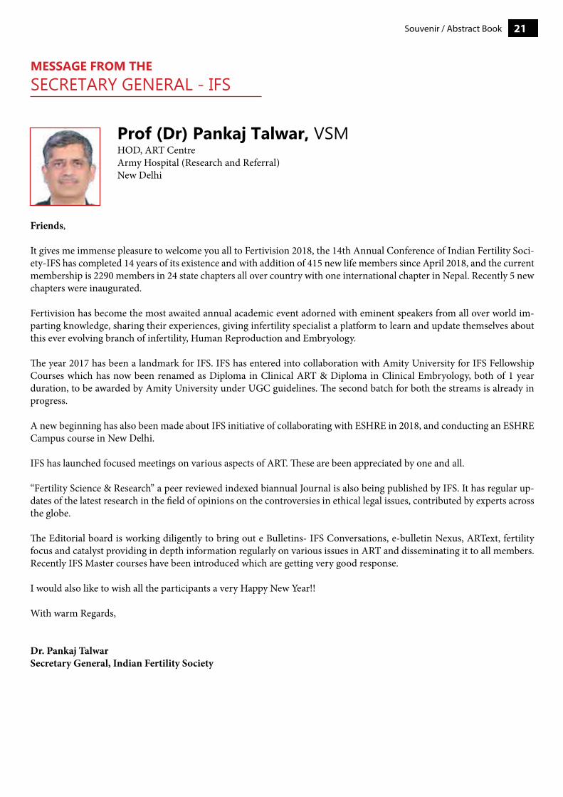
21Souvenir / Abstract Book
messaGe from TheSecreTarY geNeraL - IFS
Friends,
It gives me immense pleasure to welcome you all to Fertivision 2018, the 14th Annual Conference of Indian Fertility Soci-ety-IFS has completed 14 years of its existence and with addition of 415 new life members since April 2018, and the current membership is 2290 members in 24 state chapters all over country with one international chapter in Nepal. Recently 5 new chapters were inaugurated.
Fertivision has become the most awaited annual academic event adorned with eminent speakers from all over world im-parting knowledge, sharing their experiences, giving infertility specialist a platform to learn and update themselves about this ever evolving branch of infertility, Human Reproduction and Embryology.
The year 2017 has been a landmark for IFS. IFS has entered into collaboration with Amity University for IFS Fellowship Courses which has now been renamed as Diploma in Clinical ART & Diploma in Clinical Embryology, both of 1 year duration, to be awarded by Amity University under UGC guidelines. The second batch for both the streams is already in progress.
A new beginning has also been made about IFS initiative of collaborating with ESHRE in 2018, and conducting an ESHRE Campus course in New Delhi.
IFS has launched focused meetings on various aspects of ART. These are been appreciated by one and all.
“Fertility Science & Research” a peer reviewed indexed biannual Journal is also being published by IFS. It has regular up-dates of the latest research in the field of opinions on the controversies in ethical legal issues, contributed by experts across the globe.
The Editorial board is working diligently to bring out e Bulletins- IFS Conversations, e-bulletin Nexus, ARText, fertility focus and catalyst providing in depth information regularly on various issues in ART and disseminating it to all members. Recently IFS Master courses have been introduced which are getting very good response.
I would also like to wish all the participants a very Happy New Year!!
With warm Regards,
Dr. Pankaj TalwarSecretary General, Indian Fertility Society
prof (Dr) pankaj Talwar, VSmHOD, ART CentreArmy Hospital (Research and Referral)New Delhi

Souvenir / Abstract Book22
messaGe from TheScIeNTIFIc cHaIrperSoN
It is my immense pleasure to give to you all “FERTIVISION 2018”, the 14th annual conference of Indian Fertility Society. Since the establishment of the Indian fertility society in 2005, it has aimed to provide a platform for interested professionals to interact, share knowledge and keep up with the vast advances in the field of reproductive medicine.
Now in 2018, it has become a remarkable academic society with membership exceeding two thousand members with twen-ty three state chapters and affiliation to International Federation of Fertility Societies (IFFS) since 2007.
IFS believes that its progress lies in training and has been instrumental in holding regular CMEs and workshops in the field of reproductive techniques to educate about the latest techniques and methods. Research has always been an integral part of IFS and the IFS ART fellowship was started in 2014 at various centres of Excellence across the country.
The theme of the conference this year is “Beyond all limits..Breakthrough to Excellence…”.It will be an academic fiesta adorned with eminent speakers from all over the world, sharing their knowledge, experiences, giving infertility specialist a platform to learn and update themselves in branch of infertility, human reproduction and embryology.
So I welcome you all and hope that this conference will help you to update your current standards in clinical practice.
With Warm Regards,
Dr Sudha PrasadScientific Chairperson, FertivisionPresident Elect, Indian Fertility Society
Dr sudha prasadDir Prof and IVF CoordinatorIVF & Reproductive Biology CentreDepartment of Obstetrics and GynecologyMaulana Azad Medical CollegeNew Delhi
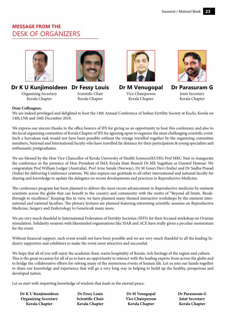
23Souvenir / Abstract Book
messaGe from TheDeSK oF orgaNIZerS
Dear Colleagues, We are indeed privileged and delighted to host the 14th Annual Conference of Indian Fertility Society at Kochi, Kerala on 14th,15th and 16th December 2018.
We express our sincere thanks to the office bearers of IFS for giving us an opportunity to host this conference and also to the local organising committee of Kerala Chapter of IFS for agreeing upon to organize the most challenging scientific event. Such a herculean task would not have been possible without the voyage travelled together by the organizing committee members, National and International faculty who have travelled far distance for their participation & young specialists and enthusiastic postgraduates.
We are blessed by the Hon Vice Chancellor of Kerala University of Health Sciences(KUHS) Prof MKC Nair to inaugurate the conference in the presence of Hon President of IMA Kerala State Branch Dr ME Sugathan as Guestof Honour. We congratulate Prof William Ledger (Australia). Prof Arne Sunde (Norway), Dr M Gouri Devi (India) and Dr Sudha Prasad (India) for delivering Conference orations. We also express our gratitude to all other international and national faculty for sharing and knowledge to update the delegates on recent developments and practices in Reproductive Medicine.
The conference program has been planned to deliver the most recent advancement in Reproductive medicine by eminent scientists across the globe that can benefit to the country and community with the motto of “Beyond all limits. Break-through to excellence”. Keeping this in view, we have planned many themed interactive workshops by the eminent inter-national and national faculties. The plenary lectures are planned featuring interesting scientific sessions on Reproductive Medicine, Surgery and Embryology to Genetics& many more.
We are very much thankful to International Federation of Fertility Societies (IFFS) for their focused workshop on Ovarian stimulation. Solidarity sessions with likeminded organisations like ISAR and ACE have really given a peculiar momentum for the event.
Without financial support, such event would not have been possible and we are very much thankful to all the leading In-dustry supporters and exhibitors to make the event most attractive and successful.
We hope that all of you will enjoy the academic feast, warm hospitality of Kerala, rich heritage of the region and culture. This is the great occasion for all of us to have an opportunity to interact with the leading experts from across the globe and to bridge the collaborative efforts for solving many of the mysterious events of human life. Let us join our hands together to share our knowledge and experience that will go a very long way in helping to build up the healthy, prosperous and developed nation.
Let us start with imparting knowledge of wisdom that leads to the eternal peace.
Dr k u kunjimoideenOrganizing Secretary
Kerala Chapter
Dr K U KunjimoideenOrganizing Secretary
Kerala Chapter
Dr fessy louisScientific ChairKerala Chapter
Dr Fessy LouisScientific ChairKerala Chapter
Dr m venugopalVice Chairperson
Kerala Chapter
Dr M VenugopalVice Chairperson
Kerala Chapter
Dr parasuram GJoint SecretaryKerala Chapter
Dr Parasuram GJoint SecretaryKerala Chapter

Souvenir / Abstract Book24
messaGe from ThepaTroN - IFS
Fertivision has become the most awaited annual academic event in the field of infertility as it is associated with eminent speakers from all over the world imparting knowledge, sharing their experiences, and giving infertility specialist a platform to learn and update themselves. The organizing committee has worked relentlessly this year with great passion and enthu-siasm to put together an exhaustive scientific program comprising of different workshops and two days of main conference at Kochi. The committee has made conscious efforts to cover a wide range of topics enabling each participant to tailor the program specifically to his / her area of interest and practice.
It is indeed a matter of great pride to see the growth of our society from a humble beginning in 2005 to its present level with 24 chapters nationwide. This has been possible only through a succession of very able and visionary past presidents and highly dedicated efforts of executive team members over the years.
Being a founder member I have been an integral part of it. I have had the chance of guiding the society through these years. I am proud to see good work being done in the field of assisted reproduction
With Best Wishes,
Dr Mohinder KochherPatron, Indian Fertility Society
Dr mohinder kochherMBBS, DGO, MD (DELHI), FACOG (USA) Obstetrician & Gynaecologist Specialist in Reproductive Endocrinolgy & IVFDelhi

25Souvenir / Abstract Book
messaGe from TheFoUNDer preSIDeNT - IFS
I am honoured and delighted to write this message for the 14th Annual conference of the Indian Fertility Society though I will not be attend the meeting due to personal problems.
The Kerala chapter is extremely dynamic and has a pride of place among our society members. The energy which went into organizing this conference was visible at every step. The content, the renowned faculty, the workshops, the topics are all very contemporary and progressive and there is a lot to learn.
Indian Fertility Society is making strides in all aspects of education, research and has made its presence felt nationally and internationally.
This conference is specially remarkable as it shows the resilience shown by the organizing members in getting over the natural disaster it had to face and make perfect arrangements. I am sure this conference will be remembered by all for not only its educational value but also for the natural beauty and famous hospitality of Kerala.
Wishing you a very pleasant and fruitful conference.
Dr Mangala TelangFounder President, Indian Fertility Society
Dr mangala TelangDirectorFertility Research & IVF CentreNew Delhi

Souvenir / Abstract Book26
messaGe from ThecHaIrperSoN SoUVeNIr commITTee
Fertivision has been, the much looked forward to, annual event of the Indian Fertility Society for the last 14 years. Besides, giving a platform for discussion on recent advances, global research and basic sciences, it provides a platform for young scholars, to present their work. Fertivision has earned a distinction in addressing the needs of all sections – clinicians, embryologists, urologist, stalwarts as well as young researcher in the field of infertility.
The conference theme of ‘Beyond all limits – Breakthrough to Excellence” is distinctive. Besides the main conference there are numerous workshops on various aspects of Reproductive Medicine. This conference blends together an eminent group of internationally-renowned faculty and a large group of experienced Indian faculty to present the latest developments in every aspect of ART.
The conference has initiated the ‘Go green” mottoand is a paperless conference.A mobile app will be utilized to access the material being presented, and electronic polling through the mobile app will be used to promote interaction with the speakers.
There are over 90 research papers being presented by young researchers and this souvenir contains a brief write up on all. It is a conglomeration of research and conclusions being presented at Fertivision 2018, both by stalwarts and young researcher, with the aim of going beyond limits and reaching a breakthrough in excellence in the field of ART.
I would like to acknowledge the invaluable contribution of DrVandana Bhatia in editing the free papers for the souvenir. I thank all the contributors for the timely submission of their abstracts.
Hope to see you all at Fertivision 2018!
Warm Regards,
Dr Surveen GhummanChairperson, Souvenir Committee Editor, Indian Fertility Society
Dr surveen Ghumman sinDhu mDEditor, Indian Fertility SocietyDirector & Head, IVF & Reproductive MedicineMAX Superspeciality Hospitals,Panchsheel, Saket & Patparganj, New Delhi

27Souvenir / Abstract Book
aBsTracToraTIoNS
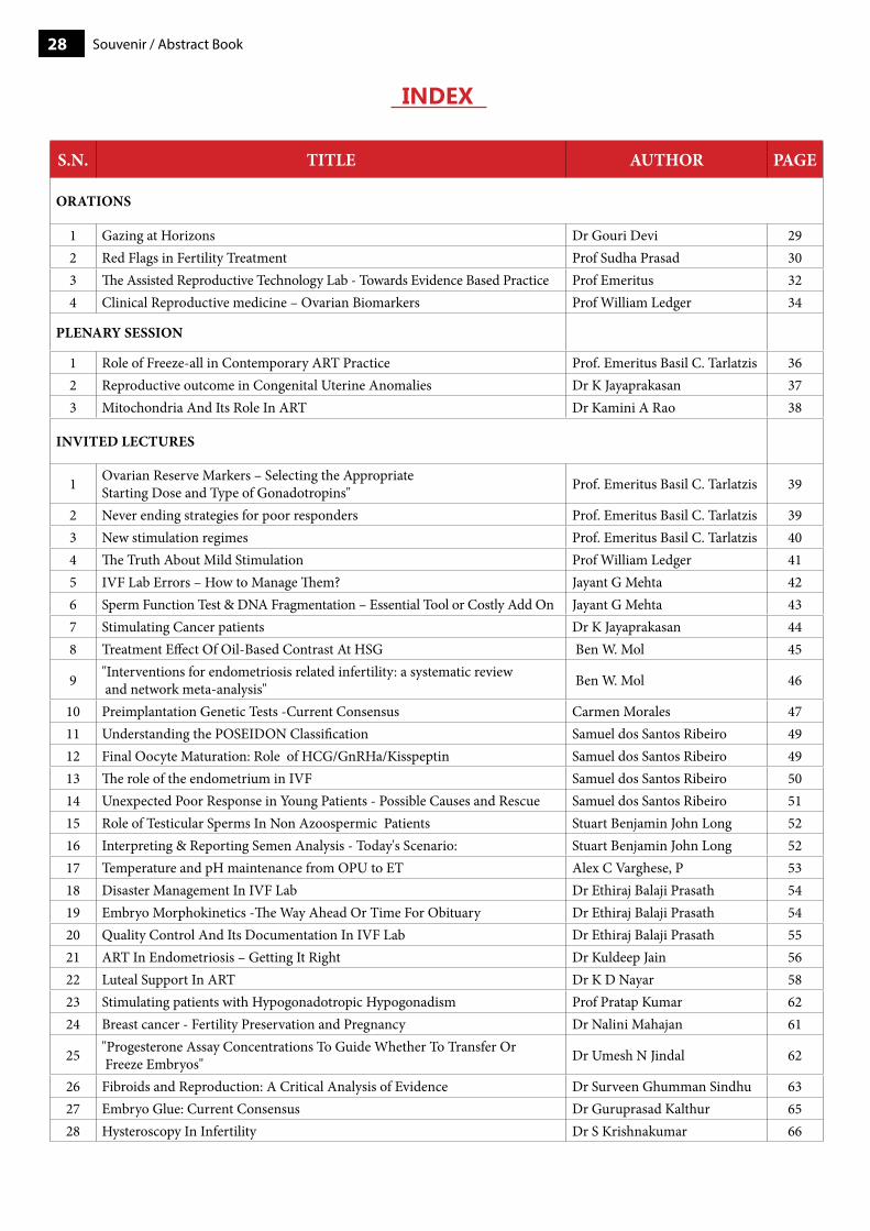
Souvenir / Abstract Book28
inDeX
S.N. TITLE AUTHOR PAGE
ORATIONS
1 Gazing at Horizons Dr Gouri Devi 292 Red Flags in Fertility Treatment Prof Sudha Prasad 303 The Assisted Reproductive Technology Lab - Towards Evidence Based Practice Prof Emeritus 324 Clinical Reproductive medicine – Ovarian Biomarkers Prof William Ledger 34
PLENARy SESSION
1 Role of Freeze-all in Contemporary ART Practice Prof. Emeritus Basil C. Tarlatzis 362 Reproductive outcome in Congenital Uterine Anomalies Dr K Jayaprakasan 373 Mitochondria And Its Role In ART Dr Kamini A Rao 38
INVITED LECTURES
1 Ovarian Reserve Markers – Selecting the Appropriate Starting Dose and Type of Gonadotropins" Prof. Emeritus Basil C. Tarlatzis 39
2 Never ending strategies for poor responders Prof. Emeritus Basil C. Tarlatzis 393 New stimulation regimes Prof. Emeritus Basil C. Tarlatzis 404 The Truth About Mild Stimulation Prof William Ledger 415 IVF Lab Errors – How to Manage Them? Jayant G Mehta 426 Sperm Function Test & DNA Fragmentation – Essential Tool or Costly Add On Jayant G Mehta 437 Stimulating Cancer patients Dr K Jayaprakasan 448 Treatment Effect Of Oil-Based Contrast At HSG Ben W. Mol 45
9 "Interventions for endometriosis related infertility: a systematic review and network meta-analysis" Ben W. Mol 46
10 Preimplantation Genetic Tests -Current Consensus Carmen Morales 4711 Understanding the POSEIDON Classification Samuel dos Santos Ribeiro 4912 Final Oocyte Maturation: Role of HCG/GnRHa/Kisspeptin Samuel dos Santos Ribeiro 4913 The role of the endometrium in IVF Samuel dos Santos Ribeiro 5014 Unexpected Poor Response in Young Patients - Possible Causes and Rescue Samuel dos Santos Ribeiro 5115 Role of Testicular Sperms In Non Azoospermic Patients Stuart Benjamin John Long 5216 Interpreting & Reporting Semen Analysis - Today's Scenario: Stuart Benjamin John Long 5217 Temperature and pH maintenance from OPU to ET Alex C Varghese, P 5318 Disaster Management In IVF Lab Dr Ethiraj Balaji Prasath 5419 Embryo Morphokinetics -The Way Ahead Or Time For Obituary Dr Ethiraj Balaji Prasath 5420 Quality Control And Its Documentation In IVF Lab Dr Ethiraj Balaji Prasath 5521 ART In Endometriosis – Getting It Right Dr Kuldeep Jain 5622 Luteal Support In ART Dr K D Nayar 5823 Stimulating patients with Hypogonadotropic Hypogonadism Prof Pratap Kumar 6224 Breast cancer - Fertility Preservation and Pregnancy Dr Nalini Mahajan 61
25 "Progesterone Assay Concentrations To Guide Whether To Transfer Or Freeze Embryos" Dr Umesh N Jindal 62
26 Fibroids and Reproduction: A Critical Analysis of Evidence Dr Surveen Ghumman Sindhu 6327 Embryo Glue: Current Consensus Dr Guruprasad Kalthur 6528 Hysteroscopy In Infertility Dr S Krishnakumar 66
29Souvenir / Abstract Book
29 Asherman’s Syndrome: What is New ? Dr.Shweta Mittal Gupta 6730 Psychosocial Care During Assisted Reproduction Dr Poonam Nayar 6831 Intricacies Of Antagonist Protocol In ART Dr. P.M. Gopinath 69
32 Emerging Evidence and New Approaches to Management – Premature Ovari-an Insufficiency Dr. Madhuri Patil 71
33 Simplifying Luteal Support in IVF Dr. Leena Wadhwa 7334 Fertility enhancing laparoscopic surgery- what’s the future? Dr. Saumya Prasad 7535 Ovulation Trigger - What Is The Right Recipe? Dr Monica Singh 7636 Role Of Genomics In Diagnosing Infections In Infertility Dr Shweta Gupta 7837 Sperm Selection Techniques - How Close Are We? Dr Randhir Singh 8038 Diagnosis Of PCOS Revisited Dr Namita Kotia 8239 Urinary vs Recombinant Gonadotropins Dr Himanshu Bhavishi 8340 Mitochondrial Replacement Therapy: Is it here to stay? Dr Nymphea Walecha 8441 DNA Fragmentation Assay In Clinical Practice Dr Sangita Sharma 8542 Peritoneal and tubal morphology and pathology by ultrasound Dr Sonal Panchal 8843 Ultrasound monitoring of ovarian stimulation: from basic to advanced Dr Sonal Panchal 8844 Medical Management of Endometriosis-New Concepts Dr Roya Rozati 9045 New Frontiers in Medical Management of Fibroids Dr Laxmi Shrikhande 9246 Problems in oocyte Aspiration Dr Kokila Desai 9347 Role of Imaging in Diagnosis and Treatment of Fibroids and Adenomyosis Dr Aarti Deenadayal Tolani 9448 pH monitoring - The way forward! Dr Charulata Chatterjee 9649 Role of alternate therapies in infertility Dr Vandana Narula 9750 Embryo Biopsy Techniques Harsha Bhadarka 9851 Tubal Patency in 2018 Dr Bela Bhatt 101
52 "Life Style Modifications and Antioxidants in Idiopathic Male Infertility - Evidence Based Recommendations" Dr Bhavatej Enganti 103
53 The Assisted Hatching Procedure Dr Chandan 10454 Identification, Communication & Documentation Dr Krishna Chaitanya 105
55 "Artificial Intelligence And Automation In IVF Lab: Will It Change The Way We Practice?" Dr Krishna Chaitanya 105
56 Technical Challenges In Embryo Transfer Dr Karthika D Kumar 10657 Adjuvants In Management Of PCOS Dr Kavitha Ramesh 10758 Management of Seropositive cases in ART lab Dr Nimmi Nirmal 10959 Chronic Endometritis and Reproductive outcome Dr Chitra Thyagaraju 11060 Ovarian Stimulation – Do’s And Don’ts Dr Neeru Thakral 11161 Novel Sperm Vitrification Techniques Dr M Sujatha Suresh 114

Souvenir / Abstract Book30
Dr Gouri Devipresident, IFSDirector, ridge IVFDelhi
Gazing at horizons
Advances in assisted reproductive technology for infertile couples were among the great medical successes of the last century. ART has made huge strides and fast progress towards finding suitable treatment options for each infertile couple.Individualising the treatment for each patient for better results are what we are aiming at.
History shows that From Clomiphene citrate in 1960s to Gonadotropins in 1970s to Agonists in 1980s to Antaginists in 1990 and long acting FSH 2000 ,we have come a long way. From the first test tube baby in an unstimulated cycle to stimulated cycles with Gonadotropins, ICSI in1990s, Vitrification of embryos and gametes in 2000s are some of the land mark discoveries in Reproductive medicine.
When we gaze at horizons, the issues are Newer drugs, newer protocols and updated lab conditions.
DRUGS: 1. Corifollitropin, a long acting Gonadotropin, has yet to come to India. One injection on Day 2 is enough for 8 days
and might recquire 2 or 3 additional daily gonadotropin injection ,with comparable results compared to conventional protocol.
2. Recovelle –Follitropin delta Has finished the phase III trial ,for individualised dosing based on S.AMH levels and body weight and the results are comparable to other gonadotropins.
3. Kisspeptin-Kisspeptin was first discovered in 1996 as a metastasis inhibitor.It is used as a ovulation trigger.Exogenous kisspeptin-54 has been successfully administered as a promising method of triggering oocyte maturation following COH in women undergoing IVF, due to its efficacy in achieved pregnancy rates compared to hCG and GnRH agonists. Also, its safety in patients at high risk of developing OHSS is noteworthy.
NEWER PROTOCOLS:• Stair Step Protocols : Stair Step protocols with both Clomiphene and letrazole have been tried. Though the time to
ovulation is shorter, the pregnancy rate is not higher.• Progestin-primed ovarian stimulation (PPOS) Progestin-primed ovarian stimulation (PPOS) using 4 or 10 mg of
MPA per day was comparable in terms of the number of oocytes retrieved and pregnancy outcome after FET. The administration of 4 mg of MPA per day was sufficient to prevent an untimely LH rise in women undergoing IVF/ICSI treatment.
Time constraints associated with emergency fertility preservation have led to shorter protocols.• Dual stimulation• Random-start protocols
LAB ENVIRONMENT : Bench top incubators: This has also has changed considerably.From Large box incubators to bench top and individual bench top incubators. Time lapse imaging: Time lapse imaging has made it easy for the embryologists to check on the embryo and select the best one. Culture medias are readymade and fortified and from single step to sequential.RFID tags: With RFID tags , the errors in the lab are almost nil.Microfluidics: The future is for Microfluidics : From semen preparation, and to separate the motile healthy sperm it is very useful. Microfluidic semen processing could provide sperm with high motility, enhanced percent normal morphology and significantly reduced percentage of sperm with DNA damage in comparison with simple wash and density gradient centrifugation (Schulte et al., 2007).Lab-on-a-chip: Lately, lab-on-a-chip (LOC) has proven to be a useful tool for enhancing non-invasive assisted reproductive technology (ART).
The advancements are many like mitochondrial transfer, 3 parent baby, cloning( though not allowed except for therapeutic purposes),oocyte activation,gametes from stem cells etc.
The scope of recent advances in the field of ART is staggering. Technologies that are coming of age now and are visible on the horizon have the potential to expand the utilization of ART to broad portions of society with and without an infertility diagnosis. Moral and Ethical responsibility may prove to be one of the largest challenges surrounding ART moving forward.
presiDenTial oraTion
31Souvenir / Abstract Book
prof suDha prasaDpresident elect, IFSDir prof and IVF coordinatorIVF & reproductive biology centreDepartment of obstetrics and gynecologymaulana azad medical college, New Delhi
red flags in fertility Treatment
Fertility treatment revolutionized by Assisted reproductive technology (ART) is a safe and effective for people with fertility problems wanting to conceive. Like any other area in medicine, fertility management especially ART is not without risks, some associated with the treatment and the others associated with the outcome.
The problem areas in fertility management may be categorized as those associated with treatment, those associated with outcomes, or those areas where further research is required to decide regarding what management techniques are ideal [1]. Red flag zones in treatment for fertility problems include those in-patient selection, choosing the treatment technique is most appropriate, side effects of medicines used, differences in patient response to treatment, Ovarian hyperstimulation syndrome, risks associated with sedation or anesthetic, risks associated with oocyte retrieval – hemorrhage and infection, surgical sperm recovery risks – infection, bleeding. The costs incurred, patient grievances and psychological adjustments to treatment as well as facing a negative result are other problematic areas.
Risks associated with outcomes include multiple pregnancy, ectopic and heterotopic pregnancy and higher incidence of obstetric complication in fertility treated conceptions. These also include the potential risk of increased developmental and congenital abnormalities in fertility treated children.Further research is needed to assess risk for borderline ovarian tumor in women and childhood malignancy and imprinting errors in children conceived of fertility treatment.
Risks from treatmentOvarian hyperstimulation syndromeMild OHSS occurs in up to one third of the women undergoing ART treatment. The severeform of OHSS is reported in 1–2% of ART cycles. Although a self-limitingcondition, it may worsen and be accompanied by ascites, pleural and pericardial effusions, renal dysfunction and, sometimes, abnormal liver function test results. Recognized complications of OHSS are renal failure, venous and arterial thromboembolism, adult respiratory distress syndrome, hemorrhage from ovarian rupture and, very rarely, death. OHSS is best prevented than managed hence an antagonist stimulation protocol and agonist or dual trigger and freeze all strategy can prevent OHSS. Use of metformin and lower doses of gonadotropin during ovarian stimulation reduces the risk in high risk women. Mild to moderate OHSS can be managed on an outpatient basis and spontaneous resolution of symptoms Patients with severe OHSS and those with significant pain or nausea that limits oral intake are usually managed as inpatients and more severe cases (critical OHSS) are admitted to an intensive care unit for management under a multidisciplinary team
Multiple pregnancyThe chance of a multiple pregnancy is almost 20 times higher with ART treatment than withspontaneous conception. UK data compiled in 2006 showed a staggering 24% multiple pregnancy rate. This led to promoting efforts in reducing the number of embryos transferred and adoption of the elective single embryo transfer (eSET) strategy has led to fall in the number of multiple pregnancies. Elective SET does not mitigate the increased risk of monozygotic twining (0.7–3.1%) compared to natural conceptions (0.4%) [2]. Ectopic pregnancy and heterotopic pregnancyPelvic inflammatory disease, previous ectopic pregnancy, endometriosis, and previous tubal surgery and impaired tubal function increase the risk of ectopic pregnancy. The prevalence of ectopic pregnancy is as high as 8.6% in women undergoing ART treatment compared to 1–2% following natural conception. The incidence of heterotopic pregnancy is rare and is 1 in 30,000 in the general population, increases to 8 in 1000 following ART treatment. Multiple embryo transfers, transfer techniques, alteration in the implantation environment in a stimulated cycle all may increase the risk of ectopic/heterotopic pregnancy [3].
presiDenT elecT oraTion
Souvenir / Abstract Book32
Complications of the oocyte retrieval procedureTransvaginal oocyte retrieval is generally very safe but may be rarely complicated by minor vaginal bleeding (1.4–18.4%), pelvic infection (0.1–0.6%) or severe intraabdominal bleeding (0.05–0.2%). Though these complications occur very rarely but we must take all precautions to avoid them as the affected woman suffers from significant morbidity.
Cancer riskCurrent evidence showed there is no increased risk in hormone independent cervical cancer [4].
The breast cancer risk is similar in general population and women undergoing fertility treatment [5].Pooled evidence reveals that, after removing the confounding effect of infertility, the risk of ovarian cancer in general population and those undergoing controlled ovarian stimulation is the same. But there is a small increased risk for borderline ovarian tumors [6].
Risk of Early menopauseMany are concerned whether undergoing COS will lead to depletion of ovarian follicular cohort quickly culminating in early menopause. This because COS do not act on primordial follicles but on FSH sensitive antral follicles which would have undergone atresia unless recruited.
Perinatal outcomesFertility treated pregnancy is associated with increased risk adverse pregnancy outcomes compared to pregnancies in fertile women. The present evidence points to the fact that it is the subfertility causing factors rather than the fertility treatment that accounts for the higher morbidity associated with singleton pregnancy. Fertility treatment predisposes to multiple pregnancy and higher risk due to this. Hence the goal of fertility treatment should be to achieve singleton pregnancy.
Risks to offspringAnalysis of available evidence suggests that there are a 30-40% increased risk congenital anomalies in ART conceived pregnancies, although it is not very clear whether the higher risk is due to factors relating to subfertility or the treatment. Compared to a background prevalence of 5% the absolute risk of anomalous birth is low 6.5-7%. There is an increased risk of offspring with imprinting errors. Young ICSI conceived males have a higher prevalence of male subfertility.
Risk for Childhood cancer Most of the evidence suggests possible increased risk in childhood cancer but again whether it is due to subfertility itself or fertility treatment is not clear. Ovarian stimulation does not seen to be associated with increased cancer risk, but exposure to maternal progesterone markedly increased the risk of acute lymphocytic leukemia and sympathetic nervous system tumors [1].
The affordability and psychological factorsAlways one should choose a patient tailored treatment strategy keeping their feasibility to undergo treatment. Couple evaluation and appropriate patient counselling is mandatory part of all fertility treatment programs.
Risk associated with treatment outcomesTreatment outcomes are dependent on the age of the woman, ovarian reserve, number of eggs retrieved, fertilization method, number of embryos transferred, Life style, body mass index, prior obstetric history and male factors.Interventions like endometrial scratching in women with prior IVF failures, Day 5 Blastocyst PGS, Time lapse assessment of embryogenesis and embyoselection, Hyaluronic acid, oocyte activation with calcium ionophore are interventions that are promising as per available evidence [1].
References1. Bhandari HM, Choudhary MK, Stewart JA. Complications of assisted reproductive technology treatment and the factors influencing
reproductive outcome. The Obstetrician & Gynaecologist. 2018.2. Osianlis T, Rombauts L, Gabbe M, Motteram C, Vollenhoven V. Incidence and zygosity of twin births following transfers using a
single fresh or frozenembryo. Hum Reprod 2014;29:1438–433. Refaat B, Dalton E, Ledger WL. Ectopic pregnancy secondary to in vitrofertilisation-embryo transfer: pathogenic mechanisms and
managementstrategies. Reprod Biol Endocrinol 2015;13:30.4. Siristatidis C, Sergentanis TN, Kanavidis P, Trivella M, Sotiraki M, Mavromatis I, et al. Controlled ovarian hyperstimulation for IVF:
impact on ovarian,endometrial and cervical cancer–a systematic review and meta-analysis.Hum Reprod Update 2013;19:105–23.5. Zreik TG, Mazloom A, Chen Y, Vannucci M, Pinnix CC, Fulton S, et al.Fertility drugs and the risk of breast cancer: a meta-analysis
and review.Breast Cancer Res Treat 2010;124:13–26.6. National Institute for Health and Care Excellence. Fertility: assessment andtreatment for people with fertility problems. Clinical
guideline CG11.London: NICE; 2013 [https://www.nice.org.uk/guidance/cg11].
33Souvenir / Abstract Book
prof arne sunDe
professor emeritus Department of clinical and molecular medicine, Norwegian University of Science and Technology,Trondheim, Norway
The assisted reproductive Technology lab - Towards evidence Based practice
Since the start of assisted reproduction, the clinical part of ART has seen a strong development towards evidence-based clinical practise. New drugs and new treatment protocols are usually tested in properly powered prospective controlled trials before being introduced in general clinical practise (Level 1 evidence). This has been the result of the introduction of evidence-based medicine in the 90-ties. Although one can still see widespread use of clinical practise, it is at least a consensus that the evidence gathered by large prospective randomised clinical trials have carried more weight than the subjective opinion of clinicians.
Concerning the ART-laboratory, the situation is different. Most of the procedures, medical devices and consumables have not been evaluated in large trials. Still products and procedures are introduced with marginal scientific evidence. As a result, there is a large variation between ART-laboratories concerning. Most often it is the senior embryologist that decides based on his or her best judgement. Even if the embryologist is experienced and knowledgeable, it is still considered low level evidence (level III)
The ART- laboratory task is in short to fertilize oocytes, culture and cryopreserve gametes and embryos and evaluate their biological potential. A weak point in the evaluation of the ART laboratory is that we do not agree on success criteria. The obvious goal of ART-treatment is the birth of a healthy baby. From the patient’s perspective the likelihood of obtaining at least one delivery per oocyte collection is the most relevant criteria. From the ART-clinic’s perspective this criteria is difficult so we use surrogate criteria such as fertilization rate, embryo development rate, proportion of “good quality embryos”, hCG rise and clinical pregnancies. Similarly, we need to agree upon the denominator when calculating rates: started cycle, oocyte recovery or embryo transfer.
Concerning fertilization, ICSI is used for non-male factor despite evidence that it does not increase delivery rates. The reason for this misuse of an invasive technique is that the ART-clinic’s focus is on surrogate markers such as fertilization rate ang failed fertilization rate
Evaluation of the biological potential of embryos should result in a ranking of the embryos available from high to medium and low implantation potential. The embryos with high implantation potential are usually selected for fresh transfer. Empirical evidence shows that embryo evaluation based on morphology, on morphokinetics, on preimplantation genetic testing (PGT-A) or on embryo metabolism all have the potential to rank embryos according to implantation potential. We do not have high quality data showing the difference in ranking potential between these different methods to evaluate embryo quality. A common misunderstanding is that embryo grading can increase the quality of the embryos available. This is not the case, you cannot enhance the biological potential of the embryo cohort you can only rank the ones available. The ideal embryo ranking system can therefore influence delivery rate after fresh transfer, time to pregnancy and potentially rates of early abortions. Cumulative delivery rate will be the same regardless of ranking method.
Concerning culture media, there is almost a complete absence of sufficiently large prospective trials comparing different media. We know that the composition of different commercially available culture media differs, but their precise composition is kept secret by the companies producing the media. We do not know which culture media is “the best” concerning delivery rates not to speak of cumulative delivery rates. This is due to lack of sufficiently large prospective clinical studies.What we do know from large prospective trials is that the composition of culture media will influence gene expression profile in embryos, the birth weight of children and postnatal growth curves. We also know that addition of Granulocyte colony stimulating factor (G-CSF) to the culture media does not enhance delivery rates.
Concerning culture conditions, we have evidence that reduced oxygen concentration (5%) is beneficial compared to
oraTion on clinical emBrYoloGY

Souvenir / Abstract Book34
atmospheric concentration (20%), we have some evidence suggesting that transfer of blastocysts might give a higher fresh pregnancy rate compared to transfer of cleavage stages and increased cumulative delivery rates. We know almost nothing about the effects of the myriad of different utensils we use for embryo culture. We do need to standardize thongs like culture dishes/tubes, pipettes, needles and catheters. We have literally a jungle different types of laboratory equipment used in the ART laboratory. Most of it not intended for ART. We have very little information concerning most of the equipment we use.
In order advance the ART-laboratory towards evidence-based practise we need to:Implement quality management systems in the lab(clinic) so we collect, record and evaluate critical parameters.Engage in prospective randomised trials to evaluate different laboratory parameters. The most effective way of doing this is to do multi-centre trials. The Dutch ART-clinics have shown the this is feasible, and we should learn from them.
35Souvenir / Abstract Book
prof William leDGer
Head, Discipline of obstetrics & gynaecologyFaculty of medicine, University of New South Wales,Director, reproductive medicineSenior Staff Specialist, royal Hospital for Womenchair of the research and Development committeeFertility specialist, IVFaustralia
ovarian Biomarkers
Oocyte quantity and quality are rate limiting to reproductive success. Current serum biomarkers of reproductive function, such as anti-Müllerian hormone (AMH), are secreted by granulosa cells and, as such, are surrogate markers of oocyte biology. AMH is useful clinically for estimation of ovarian reserve when programming gonadotropin dosing for superovulation and in prediction of menopause, but estimates based on AMH concentration are imprecise and have led to confusion amongst patients and clinicians.
There are currently no serum oocyte biomarkers available for clinical use. The oocyte-secreted growth factors, growth differentiation factor-9 (GDF9) and bone morphogenetic protein-15 (BMP15) are regulators of folliculogenesis, oocyte quality and fertility, and are essentially only produced by the gametes. We are now able to reliably measure these potential biomarkers of oocyte function in peripheral blood. We have developed novel immunoassays for the reliable detection of GDF9 and BMP15 and analysed serum samples from women having unstimulated or antagonist (FSH-stimulated) superovulated cycles.
GDF9 and BMP15 assays detected proteins in female serum with similar profiles between the two assays. Serum GDF9 and BMP15 levels varied markedly between individual women but within subjects were unchanged throughout the ovarian cycle. Serum levels were unchanged during antagonist controlled superovulation, regardless of FSH dose, and were similar across different treatment cycles. Serum levels of GDF9 positively correlated with the number of oocytes retrieved from non-PCO/PCOS women after superovulation. Serum levels of GDF9 and BMP15 did not correlate with age or baseline (day 2) endocrine measures of LH, oestradiol or progesterone. GDF9 was correlated with baseline FSH and with AMH.
The association of serum concentrations of GDF9 and BMP15 with oocyte yield during IVF supports their being secreted from the oocyte. Serum GDF9 and BMP15 may prove clinically valuable for diagnosing reproductive dysfunction and as predictors of fertility potential.
oraTion on clinical reproDucTive meDicine

Souvenir / Abstract Book36
plenarYSeSSIoN
37Souvenir / Abstract Book
prof emeriTus Basil c. TarlaTzis
mD, phD, Frcog (hon) School of medicinearistotle University of Thessaloniki, greece
role of freeze-all in contemporary arT practice
Ovarian stimulation represents an important part of the IVF procedure. However, the development of multiple follicles leads to abnormal estradiol (E2) and progesterone secretion, which may induce abnormal endometrial development in the early luteal phase and adversely affect the probability of embryo implantation. Thus, it has been hypothesized that freezing all embryos in a fresh IVF cycle and deferring embryo transfer in subsequent cycles may provide a more physiological endometrial milieau for embryo implantation when compared to fresh ΕΤ. Recently, several relevant studies have been published, evaluating the effectiveness of frozen ΕΤ (FET) using a freeze-all policy compared to fresh ΕΤ either in high or in normal responder patients.
The available evidence from randomized controlled trials (RCTs) and systematic reviews and meta-analyses indicate that overall live birth rates (LBR) were significantly higher after FET as compared to fresh ET. However, sub-group analysis showed that this difference was attributed to hyper-responders, in whom the LBR after FET was significantly higher than after fresh ET, while in normo-responders there was no difference. Concerning safety, the risk of moderate/severe OHSS was significantly lower in the FET cycles than in the fresh ET ones. On the other hand, the risk of pre-eclampsia was significantly increased in the group of FET as compared to fresh ET.
In conclusion, the freeze-all strategy with subsequent FET is at least as efficient in terms of LBR in the normo-responders and more efficient in the hyper-responders, with significantly lower risk for OHSS in both groups. Nevertheless, FET pregnancies, especially in the hyper-responder group, were associated with a higher risk for pre-eclampsia.
Souvenir / Abstract Book38
Dr k JaYaprakasan
Subspecialist and Hon. associate professorreproductive medicine & Surgeryconsultant gynaecologist/ Fertility Lead,royal Derby Hospital, Derby& University of Nottingham, Nottingham
reproductive outcome in congenital uterine anomalies
Congenital uterine anomalies (CUAs) result from embryological maldevelopment of the Müllerian ducts. While most CUAs are asymptomatic and are associated with normal reproductive outcome, some may be associated with adverse reproductive outcomes. Diagnosis of CUAs has become easier with the advent of three-dimensional (3D) ultrasound, which has the ability to demonstrate the internal and external contours of the uterus. 3D ultrasound makes the assessment of uterine morphology reproducible, and is less invasive than other commonly employed radiological and surgical diagnostic modalities. A recent meta-analysis estimated the overall prevalence of CUAs to be 5.5% in an unselected population, 8.0% in infertile women, 13.3% in those with a history of miscarriage and 24.5% in those with miscarriage and infertility. It is therefore not uncommon and clinicians will be regularly required to counsel women with a CUA.
CUA have been implicated as a potential cause of infertility, recurrent pregnancy loss, preterm delivery and fetal malpresentation. Various types of uterine anomaly are individually associated with varying degrees of adverse outcomes, with greater effects being evident in women with more significant defects. A systematic review incorporating 3805 patients have reported the impact of congenital uterine anomalies on reproductive outcome by including only studies that have appropriate control groups and that considered the effect of the sub-types of uterine anomaly. Women with canalization defects, such as septate and subseptate uteri, appear to have the poorest reproductive performance with a reduced conception rate and increased risk of first-trimester miscarriage, preterm birth and fetal malpresentation at delivery. When compared with women with subseptate (partial septate) uteri, women with septate uteri have poorer outcomes throughout the course of pregnancy. Unification defects, such as the bicornuate, unicornuate and didelphic uterus, do not appear to reduce fertility but are associated with increased risk of adverse outcome during pregnancy. The risks are, however, dependent on the type of anomaly. Women with bicornuate and unicornuate uteri have an increased risk of miscarriage, preterm birth and fetal malpresentation while women with uterus didelphys seem to have an increased risk of preterm labor and malpresentation. While there is evidence of a strong association between unification defects and canalization defects and adverse reproductive outcome, the effectiveness of treating such non-obstructed uterine anomalies surgically to improve the reproductive outcome, especially if they are incidentally diagnosed, is still debated.

39Souvenir / Abstract Book
Dr kamini a rao
Dgo, Dorcp, mrcog, Dch, Frcog (UK),mobg(UK), FIcog, pgDmLe (Law), FNamS phD, Frcog (hon)medical Director of milann
mitochondria and its role in arT
Introduction:Mitochondria, a small maternally inherited organelle, is the powerhouse of all cells and of special importance to our oocyte. Oocytes have the highest mitochondrial content among all cells. Mitochondria also functions as the key regulator of reproductive competence of oocyte. Understanding the story of how mitochondria functions within an oocyte is a basic pre-requisite for knowing oocyte physiology and the pathology that comes with oocyte ageing! This in-turn forms the crux for therapeutic interventions to combat oocyte ageing and improve ART outcome.
ATP production: The quest for fuel starts from the cell!Oocytes differ from somatic cells in energy production, in that, 88% of energy is derived from mitochondrial oxidative phosphorylation of glucose, lesser contributions from glycolysis and tricarboxylic acid cycle. Resumption of meiosis with LH surge to fertilization and embryogenesis are energy consuming processes that need an abundant endowment of capable mitochondria in the oocyte to start with. Even the surrounding granulosa cells should be well equipped with mitochondria to provide the nourishing support to developing oocyte within the follicle. Mitochondrial dysfunction can not only lead to arrest of development of oocyte/embryo but they can also cause abnormal meiosis resulting in aneuploid oocytes and embryos.
Mitochondrial DNA content per oocyte/embryo – how they differ!Mitochondria number and DNA content increases in an oocyte during its development, upto immediately before fertilization. After that, mitochondrial multiplication resumes only at blastocyst stage. Thus, naturally there is a dilution of the existing mitochondria number among blastomeres from the stage of oocyte to blastocyst. This leads us to appreciate the irony that even though a better quality oocyte has a higher mitochondrial number than its poorer counterpart, the same is not true with regard to an embryo.Aneuploid embryos and embryos that do not possess the competence to implant will have a higher mitochondrial number and mitochondrial DNA content. This enhanced mitochondrial content is a mechanism of adaptation to greater oxidative stress in these poorer quality embryos. However, quantity does not ensure quality and often, the increased number of mitochondria are functionally defective! This forms the basis of prediction of implantation competence of embryos by assessing the mito-score of trophectoderm cells obtained by blastocyst biopsy.
Therapeutic options of treating mitochondrial dysfunction – Is this the Midas touch to ageing?The pharmaceutical market is flooded with anti-oxidants that claim to cure ageing. But guidelines give a guarded stand on the benefits of anti-oxidant therapy, based on the evidence available today. Cochrane Review by Showell et al (2013) gives no benefit for anti-oxidant therapy in improving clinical pregnancy or live birth. However, Coenzyme Q10, resveratrol and alpha-lipoic acid have been showed to have benefits such as reducing aneuploidy rate, delaying oocyte reserve depletion, improving mitochondrial activity and reducing intracellular oxidative stress. The gap between studies on the benefit of these chemicals and the therapeutic impact, only aims at the distance that needs to go in improving the drug delivery and action.
Modern mitochondrial therapies – Taking things into our own hands!Mitochondrial replacement therapy is useful among two group of patients –1. Patients with mitochondrial diseases to prevent the inheritance of the same in the progeny.2. Patients with poor oocyte and embryo quality with previous failed IVF cycles, at an attempt to improve oocyte/embryo
competence and achieve pregnancy.
Meiotic spindle transfer, pronuclei transfer, cytoplasm transfer from donor oocyte and autologous germline mitochondrial energy transfer (AUGMENT) are the techniques in our armamentarium to boost oocyte/embryo mitochondrial endowment. The use of these techniques are still experimental and shrouded with ethical and political challenges as well.
Souvenir / Abstract Book40
prof emeriTus Basil c. TarlaTzis
mD, phD, Frcog (hon) School of medicinearistotle University of Thessaloniki, greece
ovarian reserve markers – selecting the appropriate starting Dose and Type of Gonadotropins
never ending strategies for poor responders
The assessment of ovarian reserve is very important prior to ovarian stimulation for IVF in order to identify potential poor/hypo- and hyper-responders and select the optimal dose accordingly. To that end, several markers have been proposed: woman’s age, body mass index (BMI), FSH, estradiol (E2), antimullerian hormone (AMH), antral follicle count (AFC), ovarian volume and perfusion (doppler score).
Several studies have indicated that the best prediction of hypo- or hyper-response is achieved by the combination of AMH and AFC with an AUC 0.877 (95% CI 0.827-0.928) and 0.939 (0.878-1.00), respectively. Age, serum FSH and BMI have more limited predictive value but can be taken in account for the calculation of the starting dose of FSH treatment.
Based on the aforementioned markers, algorithms have been developed for the calculation of the starting FSH dose. Recently, a model based solely on serum AMH levels (Elecsys, Roche Inc), in combination with a new recombinant human FSH preparation, was prospectively tested in an RCT, demonstrating that the incidence of poor, hypo- and hyper responders was reduced.
In conclusion, AMH and AFC seem to be the most helpful parameters in predicting individual ovarian response. Individualized dosing based solely on AMH seems to have similar efficacy and improved safety. Nevertheless, more biomarkers are needed in order to be able to choose the most appropriate protocol and dose for each individual patient, in the context of personalized medicine.
Background: Androgens are reported to play an important role in follicular growth by increasing the ovarian sensitivity to FSH and it is assumed that androgen pretreatment during the early phase of follicular recruitment might improve the number of small antral follicles. The purpose of this systematic review and meta-analysis was to evaluate the role of androgens or androgen modulating agents on the probability of pregnancy in poor responders undergoing IVF.
Methods: Medline, EMBASE, CENTRAL, Scopus and Web of Science databases were searched for the identification of randomized controlled trials (RCTs) evaluating the administration of testosterone, dehydroepiandrosterone (DHEA), aromatase inhibitors, recombinant luteinizing hormone (rLH) and recombinant human chorionic gonadotrophin (rhCG) before or during ovarian stimulation in poor responders.
Results: Pretreatment with transdermal testosterone was associated with an increase in clinical pregnancy [rate difference (RD): +15%, 95% CI: +3% to +26%] and live birth rates (RD: +11%, 95% CI: +0.3% to +22%) in poor responders undergoing ovarian stimulation for IVF. No significant differences in clinical pregnancy and live birth rates were observed between patients who received DHEA and those who did not. Similarly, i) the use of aromatase inhibitors, ii) addition of rLH and iii) addition of rhCG in poor responders stimulated with rFSH for IVF were not associated with clinical pregnancy rates. In the only eligible study that provided data, live birth rate was increased in patients who received rLH as compared to those who did not (RD: +19%, 95% CI: +1% to +36%).
Conclusions: Based on the best available evidence, transdermal testosterone pretreatment seems to increase clinical pregnancy and live birth rates in poor responders undergoing ovarian stimulation for IVF. There is insufficient data to support a beneficial role of rLH, hCG, DHEA or letrozole administration. The small number of the available RCTs limited our ability to draw substantial conclusions. Thus, further clinical studies are warranted to establish definite recommendations on the use of androgens during ovarian stimulation in poor responders.

41Souvenir / Abstract Book
new stimulation regimesAlthough the first IVF baby was born in a natural cycle, subsequent studies showed that ovarian stimulation using clomiphene and gonadotropins was associated with a significant increase in pregnancy rates. However, approximately 20% of these cycles had to be cancelled due to premature LH rises, necessitating the co-administration of GnRH agonists or antagonists. These protocols were very efficient but, also, introduced new problems: the ovarian hyperstimulation syndrome, follicular phase progesterone elevation, luteal phase defects and disturbed endometrial receptivity. Hence, after 30 years, our approach to ovarian stimulation needs to be reconsidered
In this respect, ovarian stimulation is shifting from universal to individualized regimens. The aim is to select the appropriate dose of gonadotropins for each individual woman, thus avoiding hyperstimulation and the other aforementioned problems. Several patient characteristics (age, BMI, FSH levels, AFC) have been examined in predicting the optimal dose. However, the most promising is AMH measurement (especially with the recent new assay, which seems to be more stable and precise), since it reflects more accurately the follicle pool.
At the same time, it is becoming apparent that it is far more meaningful to assess ART efficacy per whole treatment than per attempt. This would take into account cumulative pregnancy rates from fresh and frozen-thawed embryo transfers (FRET) or from several FRET cycles, when applying a freeze-all policy as done in Japan and elsewhere.In conclusion, these new trends need to be further explored, aiming to combine high efficacy with safety and patient convenience in ART.
Souvenir / Abstract Book42
prof William leDGer
Head, Discipline of obstetrics and gynaecologyFaculty of medicine, University of New South Wales,Director, reproductive medicineSenior Staff Specialist, royal Hospital for Womenchair of the research and Development committeeFertility specialist, IVFaustralia
The Truth about mild stimulation
Controlled ovarian hyperstimulation with gonadotropins has been used for over 30 years to induce multiple follicle development prior to egg collection. The conventional aim of this approach has been to try to maximise the number of oocytes collected during an IVF cycle. However, high dose FSH superovulation, usually in a GnRH agonist controlled “long protocol” is associated with a number of problems and complications for the patient and for the clinician. Long protocol downregulation carries side effects of hypo-oestrogenism, risk of ovarian cyst formation and, critically, significant risk of ovarian hyperstimulation syndrome (OHSS). High oocyte number after superovulation has also been implicated as a cause of embryonic aneuploidy, due to recruitment of poor quality oocytes. These problems, along with the adverse effects of multiple pregnancy after multiple embryo transfer, led to development of the concept of “mild” IVF. “Mild” IVF uses a low dose stimulation strategy, frequently with injectable gonadotropins but also with oral ovulation induction agents such as clomifene citrate or letrozole, to stimulate growth of only a small number of follicles. This minimizes side effects and risk of OHSS. In many cases “mild” stimulation is combined with GnRH antagonist suppression of premature LH rises, allowing the option of a GnRH agonist trigger if necessary. Definitions of what constitutes “mild” IVF vary, but all include single embryo transfer or, more recently, a “freeze all” approach.
The concept of “mild” IVF appeals to many patients, who are understandably anxious about the effects of large doses of injected gonadotropins on them and on the health of their child. Reports of success rates vary from centre to centre and it is difficult to see a consensus emerging amongst IVF specialists about the place of “mild” approaches. However the reality seems to be that there are groups of women for whom this may be the best approach, particularly those with polycystic ovary syndrome and high AMH, who are in danger of over stimulation and OHSS and, conversely, those nearing the end of their reproductive life who are only able to generate one or two follicles whatever doses of gonadotropin are used.
There have been no prospective randomized studies comparing rates of aneuploidy at day five of embryonic development using comprehensive chromosome screening techniques. However evidence from large retrospective databases suggests that there is a threshold number of oocytes collected above which there is no further increase in chance of livebirth. Hence “mild” IVF may also have a place in cases where PGT-A consistently shows high rates of aneuploidy.
43Souvenir / Abstract Book
prof JaYanT k mehTa
Sub-fertility Laboratory Director & Quality control managerSub-Fertility Unit. Queen’s Hospitalbarking, Havering and redbridge University Hospitals NHS Trustromfrod, essex, UK
ivf lab errors – how to manage Them?
The last four decades has seen introduction of several highly complex procedures in the treatment of infertility resulting in higher pregnancy rates. This has included cryopreservation of eggs and embryos, micromanipulation and pre-implantation genetic screening. A promise of a possible pregnancy has also broadened the population of patients requiring gamete donors and gestational surrogates. Unfortunately, sporadic reports in the press of a wrong infant to IVF couples has highlighted possible errors in the IVF laboratory. In the absence of a regulatory frame work of transparently reporting potential IVF errors, there remains a concern that errors may be ignored. The root cause analysis of the type and how often and how can these errors be minimised needs to be addressed by all clinics for the protection and safety of the unborn child and the parents. The need for an informed consent prior to any procedure should form the basis of transparency.
There are number of areas where IVF Lab errors can be observed, starting from egg collection, insemination, fertilisation check, embryo culture and scoring, embryo transfer and cryopreservation. There are also number of clinical errors that can impinge on the laboratory outcomes.In the UK, The Human Fertilisation and Embryology Authority (HFEA) classify incidents in clinics into three grades of severity: A, B and C. They also hold information about ‘near misses’ in which only luck prevented an incident from occurring.
Grade A: involves severe harm to one person (such as a death, being implanted with the wrong embryo or birth of an affected child following genetic testing) or major harm to many (such as the failure of a frozen storage unit containing the embryos of many patients).
Grade B: involves serious harm to one person (such as the loss or damage of embryos for one patient) or moderate harm to many (such as sensitive personal data about more than one patient being sent to the wrong recipient) and Grade C: involves minor harm, such as one of many eggs being rendered unusable in the laboratory.In 2016 according to HFEA, clinics in the UK reported a total of 540 (502 incidents and 38 near misses). This represents a slight increase from 497 in 2015. Although there was an increase in the number of incidents and near misses in 2016, they have increased only slightly as a proportion of all treatments carried out in the year. In 2016, 76,500 treatments were carried out – a 6% increase from 2015 – whilst the number of incidents increased by 8.5%. Whilst the overall number in real terms has increased slightly, there has been a change in the severity of incidents, with a lower number of grade B incidents and a higher number of the less serious grade C incidents. There was also a decrease in the number of severe or critical ovarian hyperstimulation syndrome (OHSS) cases reported. Categorised amongst the clinical incidents, the number of severe or critical OHSS cases in 2016 was 38, compared with 60 in 2015. HFEA Alerts, circulated to all licensed clinics address the need to be vigilant about certain procedures, media, including use of consumables that are not CE marked and not passed as Class Two Medical grade.
It is recognized that heavy clinical and laboratory work-loads and distraction, communication failures between the team and inadequacy of the labelling system, witnessing can lead to serious incidents. The potential sources of errors in other clinical laboratories, highlighting errors are least likely to be associated with the actual laboratory procedural or analytic event and more so with procedures that are pre-analytical (e.g. patient identification, communication between the clinical and laboratory components of a unit) and post-analytical (e.g. accurate recording and reporting of findings), The introduction of mandatory quality management practices in the IVF laboratory through accreditation and licensure is a major deterrent to procedural errors, however an unreliable chain of custody or system for traceability constitutes a serious threat to quality patient care even in the face of exceptional laboratory procedural competence. While stringent controls within the laboratory will reduce IVF errors, it is not possible to prevent them.
Souvenir / Abstract Book44
sperm function Test & Dna fragmentation – essential Tool or costly add onAlthough conventional semen parameters have a limited diagnostic value for male fertility, the clinical value of a basic semen analysis remains important in deciding a possible treatment option. Furthermore, advances in in vitro fertilisation (IVF) techniques, particularly intracytoplasmic injection (ICSI) involving the direct injection of a single spermatozoon into an egg, have not diminished the role of semen analysis in modern reproductive practice. European Society of Human Reproduction and Embryology (ESHRE), in their recently published guide lines on ‘Recurrent Pregnancy Loss’ (RPL) have eluded to the fact that although RPL of 30% is normally associated with women, it is reasonable to assume that since a man contributes 50% of the genome to an embryo, male factors may also be responsible for the pregnancy loss. The guide lines now recommend investigating Males along with the female partner. Endogenous metabolic by-products and exogenous factors continuously challenge the integrity of our genome. DNA double-strand breaks (DSBs) are endogenously induced during spermatogenesis; first during meiosis, to facilitate the formation of meiotic crossovers, and second during spermiogenesis, when the chromatin of the haploid round spermatids is compacted by the replacement of histones by protamines. Furthermore, during maturation and storage in the epididymis, it is likely that sperm may accumulate DNA damage and fragmentation. Defective apoptosis can cause sperm DNA fragmentation as can excessive reactive oxygen species (ROS) production and decreased seminal antioxidants. It has been reported that toxic effects of drugs, cigarette smoking, pollution, and factors as xenobiotics, high testicular temperature (fever, varicocele) and advanced age have all been associated with increased sperm DNA damage.Although a cell has several ways to repair damaged DNA, inaccurate repair can have different consequences. The germ line has to maintain sufficient DNA integrity to pass on our genome to forthcoming generations. Even though, a number of sperm-specific biomarkers have been studied to identify useful diagnostic tests of sperm function, to date, tests of sperm DNA integrity and sperm nuclear protein show potential to discriminate infertile from fertile men.The integrity of sperm DNA is considered to be vital for normal fertilization, embryo development, and for successful implantation and pregnancies in both natural and assisted reproduction.Although some studies have found some value in the use of sperm DNA tests in the evaluation of male infertility,the true prognostic value of sperm DNA assessment to predict assisted reproductive technology (ART) outcomes remains uncertain. Studies investigating male factors association with RPL have reported higher levels of sperm progressive immotility, abnormal morphology, and elevated sperm DNA fragmentation and elevated sperm aneuploidy in men with RPL. Because these studies rely on case control design with fertile couples serving as controls, it is important that prospective studies are investigated to confirm the reported associations. Surprisingly, only few studies have reported assessing semen quality and risk of incident pregnancy loss. This may suggest a lack of couple-based preconception cohort studies conducted worldwide. Preconception cohort studies are needed for this question given the marked concentration of losses early in pregnancy or before seeking prenatal care. Currently, several techniques that measure DNA fragmentation are also available and have been evaluated in separate studies. The sperm chromatin structure assay (SCSA), the sperm chromatin dispersion (SCD) test, the terminal deoxynucleotidyl transferase mediated deoxyuridine triphosphate nick end labelling (TUNEL) and the single cell gel electrophoresis (Comet) assay are the most common used tests.The SCSA bases its results on (1) the DNA fragmentation index (DFI), which is the percent- age in the sample that have measurable increased red fluorescence due to acridine orange attaching to a single strand portion of DNA at sites of DNA strand breaks and then collapsing into a crystal that produces a metachromatic shift to red fluorescence under exposure to blue light and (2) the percentage of high DNA stainability (HDS), which is due to excess histones and proteins other than protamines that prevent full condensation of the sperm chromatin.The SCD test, also known as Halo Sperm assay, estimates the level of DNA fragmentation indirectly by quantification of the amount of nuclear dispersion/halo after sperm lysis and acid denaturation to remove excess nuclear proteins. This test is easy to carry out in any laboratory and is not expensive.The principle of TUNEL involves labelling 3OH ends of single- and double-strand breaks with biotinylated dUTPs. The incorporated labelled nucleotides can be quantified by flow cytometry or (fluorescence) microscopy to determine the number of (apoptotic) sperm cells containing fragmented DNA. However, double strand DNA can have breaks with no exposed 3’OH end and thus not being labelled by the TUNEL assay. The Comet assay, on the other hand quantifies the shape of the single cell nuclei after gel electrophoresis. Small fragmented DNA has a faster rate of migration towards the anode in an electrophoretic field (tail region) as compared to larger non-fragmented DNA (head region), leading to a typical comet shape.Using these tests, the percentage of sperm with fragmented DNA was shown to be comparable in idiopathic sub-fertile men with normal sperm parameters and in sub-fertile men with abnormal sperm parameters, and significantly higher in both these groups in comparison to fertile controls. While the DNA fragmentation tests are expensive and may not be available in all Andrology laboratories, it is important to establish the integrity of DNA, especially when ICSI for all is the new ‘mantra’, now advocated by most clinics to ensure full term births of healthy babies.In conclusion, Sperm Function Test and DNA fragmentation are essential tool for better understanding of the sperm kinetics and are not simply a costly add on.

45Souvenir / Abstract Book
Cancer is not uncommon with India accounting for third highest number of cancer cases amongst women after China and the US. However, cancer is no longer considered as an incurable disease with advances in cancer treatment has a significant impact on improving long term survival. Quality of life issue such as fertility preservation is now an integral component in cancer management. Currently, ovarian stimulation and subsequent oocyte and embryo cryopreservation is the most preferred method of fertility preservation in women of reproductive age as these techniques offer higher success rates compared to experimental options like ovarian tissue freezing. Conventional method of ovarian stimulation for oocyte/embryo cryopreservation is initiating gonadotrophins at the start of follicular phase like that of a typical IVF cycle. But, this may need over 2 to 6 weeks depending on the menstrual cycle phase at the initial visit and this may pose a significant delay of cancer treatment. Recent concept of multiple waves of follicular recruitment with recruitable antral follicles are present in the ovaries throughout the menstrual cycle prompted new approaches to ovarian stimulation such as ‘random start’ and ‘double’ or ‘duo’ ovarian stimulation. These approaches provide a significant advantage by reducing the time from initial visit to egg collection stage without compromising the number and quality of oocytes/embryos that can be cryopreserved.
Dr k JaYaprakasan
Subspecialist and Hon. associate professorreproductive medicine & Surgeryconsultant gynaecologist/ Fertility Lead,royal Derby Hospital, Derby & University of Nottingham,Nottingham
stimulating cancer patients
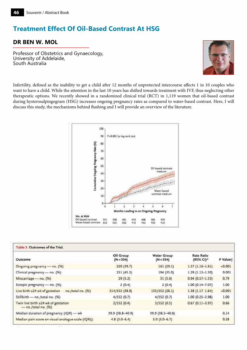
Souvenir / Abstract Book46
Dr Ben W. mol
professor of obstetrics and gynaecology,University of addelaide,South australia
Treatment effect of oil-Based contrast at hsG
Infertility, defined as the inability to get a child after 12 months of unprotected intercourse affects 1 in 10 couples who want to have a child. While the attention in the last 10 years has shifted towards treatment with IVF, thus neglecting other therapeutic options. We recently showed in a randomized clinical trial (RCT) in 1,119 women that oil-based contrast during hysterosalpingogram (HSG) increases ongoing pregnancy rates as compared to water-based contrast. Here, I will discuss this study, the mechanisms behind flushing and I will provide an overview of the literature.
47Souvenir / Abstract Book
interventions for endometriosis related infertility: a systematic review and network meta-analysis
What is known already
Most recognised therapies have not been directly compared in randomised controlled trials, therefore there is no direct evidence to inform clinical decision-making, hence selection of the most effective treatment is difficult. NMA compares multiple treatments in one statistical model.
MethodsA systematic review and network meta-analysis of relevant randomised control trials (RCTs) was performed. We searched electronic databases including, MEDLINE, CENTRAL, as well as reference lists to identify eligible studies. We included RCTs comparing any medical or surgical interventions to each other or placebo / no treatment in couples with endometriosis associated subfertility. The primary effectiveness outcome is a composite of clinical pregnancy.
Results4,252 titles/abstracts were identified through the literature search of which we included 27 trials reporting on 2,195 women with endometriosis associated subfertility. Network meta-analysis showed that compared to placebo, Lipiodol (OR 7.56, 95% CI 1.95-29.37) and surgical laparoscopy plus pentoxifylline (OR 3.44, 95 CI 1.08-10.93) resulted in more clinical pregnancies; GnRH-a (OR 1.54, 95% CI 0.93-2.56) and surgical laparoscopy OR 1.43, 95% CI 0.93-2.56) were likely to result in more clinical pregnancies. Dydrogesterone (OR 3.00, 95%CI 0.69-13.30), pentoxifylline ( OR 1.98, 95%CI 0.55-7.17) and laparoscopy plus danazol ( OR 1.72, 95%CI 0.34, 8.78) showed imprecise effect sizes.
FindingsThe most important conclusion is that more RCTs are needed to clarify the relative effectiveness of treatments for endometriosis-related infertility, in particular RCTs comparing IVF or IUI to other treatments including surgical laparoscopy and lipiodol to other treatments.
Souvenir / Abstract Book48
carmen morales, pHD
pgD labs Kuwait, Jabriya, Kuwait
preimplantation Genetic Tests -current consensus
Preimplantation Genetic Testing (PGT) has rapidly evolved in the recent years thanks to two main factors: the development of new technologies in the field of genetics and the great improvement of IVF laboratory conditions. We have moved from cytogenetic techniques (that allowed us to test a limited number of chromosomes on a single embryonic cell with fresh embryo transfer) to molecular diagnostic technologies (that allow us to massively analyze the entire genome targeting thousands of chromosomal regions from a pool of embryonic cells with frozen single embryo transfer). This last approach is currently known as “PGT 2.0”. However, these advances have generated great amount of new information and have raised new concerns and controversies.
Although the use PGT to prevent monogenic diseases or specific chromosomal abnormalities in known carriers has a clear acceptance as something beneficial, the use of PGT for aneuploidy screening (PGT-A) does not show the same wide consensus. The application of PGT-A is based on the fact that chromosomal abnormalities have a great negative impact in human reproduction and are responsible of large number of implantation failures and miscarriages (Macklon et al, 2002; Gardner et al, 2012). Thus, it seems logical to try to improve the success on IVF cycles by selecting and transferring the euploid embryos: we would expect higher options to achieve an ongoing pregnancy and the time to pregnancy would be reduced. But … are we succeeding?
Opinions against PGT-A have been classically based on no-beneficial (or damaging) effect. With the implementation of PGT 2.0, these opinions basically remained the same and one its main argument is the lack of reliable RTCs that support the beneficial effects for the patients (Orvieto, 2016) and the difficulty to move from research to clinical application. Some authors support the freezing-all transfer-all policy, although it can lead to ethical issues (based on our knowledge of the aneuploidy impact on IVF embryos and reproduction) (Geraets and Sermon, 2016).
Professionals supporting PGT-A (possibly the broadest group) believe on its beneficial effect by increasing implantation rate, reducing miscarriage rate (and its psychological burden), reducing time to pregnancy and reducing the risk of baby/fetus with aneuploidy. However, some discrepancies within this group have also arisen (Griffin and Ogur, 2018), mainly due to two reasons: 1) the effect of mosaicism on the diagnosis and decision taking and 2) the group of patients that can be benefited. The development of technologies such as NGS (Next Generation Sequencing) has made it possible to detect with greater precision (than array CGH) the percentage of mosaicism in the sample. We saw that mosaics (even in blastocyst stage) are not infrequent and that these embryos can lead to live born children (Munne et al, 2017). This lead to a third classification of embryos additional to euploid and aneuploid: mosaic embryos. Consequently, this has raised questions about whether these embryos are transferable (in case of not having euploid embryos), what kind of mosaicism (type of chromosome involved or percentage of mosaicism) is acceptable for transfer (Grati et al, 2018) or even if in fact can all or almost all embryos be mosaic.
The groups of patients that can preferably (or exclusively) benefit from PGT-A is also a controversial issue. Classical referrals for PGT-A (groups of patients with known increased risk of aneuploidies) are advance maternal age, recurrent IFV failure, recurrent miscarriage or genetic male factor. However, aneuploidy can be detected also in patients not belonging to any of these groups (i.e, egg donors) so based on that, it could be justified to offer the PGT-A to all patients undergoing IVF. Some recommendations has been published regarding mosaicism and patient selection, however it still remains a decision of each center to establish their policies regarding both issues.
In summary, despite PGT-A has been controversial since its introduction, there is a worldwide tendency to offer it at least to a certain subgroup of patients. The transfer of mosaic embryos remains a critical and challenging point, and although many clinics still choose not to transfer them, the trend is changing. Despite there is a general belief in the advantages of PGT-A, more evidence is needed especially regarding the mosaicism and its biological implications. However, publishing accurate blind RCT in IVF (and assisted reproduction in general) is not an easy task (and maybe not achievable considering
49Souvenir / Abstract Book
the characteristics of this specific medical field) (Griffin and Ogur, 2018). As it has been evidenced, not all IVF clinics neither all genetic labs are reporting the same rates of aneuploidy or mosaicism (nor pregnancy rate). There is a great number of internal factors that can influence the observed variability (from stimulation protocols to the type of technology used for PGT analysis, etc.) and results are frequently not comparable. Each clinic have to assess its own results, do the proper amendment if required, and base its decision regarding PGT-A according its own data.
References1. Macklon NS, Geraedts JP, Fauser BC. Conception to ongoing pregnancy: the ‘black box’ of early pregnancy loss. Hum Reprod
Update. 2002 Jul-Aug;8(4):333-43.2. Gardner R.J.M, Sutherland G.R and Shaffer L.G. Chromosome abnormalities and genetic counseling: Oxford monographs on
medical genetics(4th ed) New York: Oxford Press 2012.3. Raoul Orvieto. Preimplantation genetic screening- the required RCT that has not yet been carried out. Reprod Biol Endocrinol.
2016; 14: 35.4. Geraedts J and Sermon K. Preimplantation genetic screening 2.0: the theory. Mol Hum Reprod. 2016 Aug;22(8):839-44.5. Griffin DK and Ogur C. Chromosomal analysis in IVF: just how useful is it?. Reproduction. 2018 Jul;156(1):F29-F50.6. Munné S, Blazek J, Large M, Martinez-Ortiz PA, Nisson H, Liu E, Tarozzi N, Borini A, Becker A, Zhang J, Maxwell S, Grifo J,
Babariya D, Wells D, Fragouli E. Detailed investigation into the cytogenetic constitution and pregnancy outcome of replacing mosaic blastocysts detected with the use of high-resolution next-generation sequencing. Fertil Steril. 2017 Jul;108(1):62-71.
7. Grati FR, Gallazzi G, Branca L, Maggi F, Simoni G, Yaron Y. An evidence-based scoring system for prioritizing mosaic aneuploid embryos following preimplantation genetic screening. Reprod Biomed Online. 2018 Apr;36(4):442-449.
Souvenir / Abstract Book50
samuel Dos sanTos riBeiro, m.D. pH.D.
Scientific coordinatorIVIrma Lisbonportugal
understanding the poseiDon classification
final oocyte maturation: role of hcG/Gnrha/kisspeptin
Ovarian stimulation (OS) is historically regarded as a milestone for the treatment of infertility, as it allowed physicians to bypass the natural phenomena of follicular dominance and atresia by artificially overcoming the FSH threshold and inducing multi-follicular (instead of mono-follicular) development. The generalized use of exogenous gonadotropins during assisted reproductive technology (ART) has led to a substantial increase in pregnancy rates, from the typical 3-10% range (using no or minimal stimulation) to 20-50%.
Women with impaired ovarian reserve or poor ovarian response (POR) to exogenous gonadotropin stimulation present a challenge for reproductive specialists. Moreover, the current standards that define POR vary widely as several factors either isolated or in combination are used for identification of such patients. Not surprisingly, the reported prevalence of POR fluctuates markedly between 5.6 and 35.1%. Regardless of the chosen definition, it is clear that the POR population accounts for a substantial subset of women treated in IVF clinics nowadays. Driven by socioeconomic and other issues, many women are currently postponing motherhood which results in a higher number of patients seeking ART treatments in their late thirties and early forties.
Several standards have been developed for the definition of POR. Parameters related to patient demographics, ovarian reserve tests, and outcomes of previous IVF cycles — alone or combined — are used to define the POR population. Amongst these is the Patient Oriented Strategies Encompassing IndividualizeD Oocyte Number (POSEIDON) classification, which following its publication in 2016, has sparked interest among infertility practitioners. The new Poseidon criteria to identify and stratify infertility patients with “expected” or “unexpected” impaired ovarian response to exogenous gonadotropins undergoing ART. Four distinct groups of low prognosis patients can be established based on quantitative and qualitative parameters, namely:1. The age of the patient and the expected embryo aneuploidy rate;2. Ovarian biomarkers [antral follicle count [AFC] and/or anti-Müllerian hormone [AMH]]3. The ovarian response of the patient in terms of oocyte quantity provided a previous cycle of stimulation was carried
out.The new POSEIDON marker of successful outcome is the ability to retrieve the number of oocytes necessary to achieve at least one euploid embryo for transfer in each patient, which may represent a more pragmatic endpoint for clinicians providing care to infertility patients. Furthermore, it opens the possibility of developing prediction models to help clinicians counsel and set patient expectations and establish a working plan to reduce the time-to-pregnancy (TTP). This is essential to avoid any misunderstanding regarding the POSEIDON concept, as the intention of the concept is to help guide clinicians through the medical management. During this lecture, an overview of the potential benefits of the application of the POSEIDON criteria for research and clinical practice will be presented.
Prior to oocyte retrieval, it is necessary to artificially stimulate the final stages of oocyte maturation and ovulation. Traditionally, this has been performed by administering exogenous hCG, a molecule historically obtained in large quantities by purification of the urine of pregnant women while serving the same purpose of LH via its common site to the LH/hCG receptor.
While the administration of hCG is essential for the final stages of oocyte competence and to trigger luteinization, some investigators have alluded to the possible relationship between this drug and changes in endometrial receptivity. Specifically, a recent immunohistochemistry and flow cytometry analyses performed on endometrial biopsies deriving from 34 women with recurrent implantation failure postulated that hCG may deregulate T-cell activity.
51Souvenir / Abstract Book
The role of the endometrium in ivfART have developed vastly since the first live birth following IVF in 1978. This multidirectional improvement led to the optimisation of ovarian stimulation and to a better assessment of embryo quality, ultimately causing a steady increase of live birth delivery rates until the late 1990s. Despite that, livebirth rates have remained relatively low and, since the year 2000, rather stagnant.
Many authors have postulated whether the supraphysiologic milieu of hormones produced during ovarian stimulation (OS) may affect endometrial receptivity (ER) and hinder both pregnancy rates and neonatal outcomes. More specifically, the abnormal production of progesterone (P) during the later stages of the follicular phase (late-follicular elevated progesterone, LFEP) has been associated with both an abnormal endometrial gene expression and an atypical epigenetic profile in the luteal phase. This has led many fertility centers to change their clinical practice and to measure serum P levels on the day of hCG administration, adopting a freeze-all strategy whenever LFEP occurs. Nonetheless, the everyday use of a universal LFEP threshold as an ART outcome predictor has proven to be harder than originally expected, owing mostly to the fact that a) excessive P production is frequently encountered in good-prognosis women with an otherwise healthy
Another major concern around the use of hCG for ovulation triggering is its association with the most notable iatrogenic event in ART: ovarian hyperstimulation syndrome (OHSS), which is almost exclusively an ovarian-stimulation-related iatrogenic event that occurs in women who are frequently otherwise healthy. While its general incidence is approximately 2% to 3% per cycle, OHSS can occur in up to a third of all cases of high-risk patients, namely those with a previous history of OHSS or polycystic ovaries. In its most severe forms, this syndrome has the potential to cause serious morbidity or mortality, mainly due to the increased occurrence of ovarian torsion and thromboembolism.
OHSS is an exaggerated response to ovarian stimulation characterized by cystic enlargement of the ovaries, abdominal distention and pain, fluid shift from the intravascular space to the third space, which can result in ascites, pericardial and pleural effusions, and even generalized edema. Although this syndrome has been extensively studied, its pathophysiology remains rather elusive. Until now, there is sufficient evidence to believe that OHSS results from an excessive secretion of vasoactive substances during ovarian stimulation, namely VEGF and factors that derive from the renin–angiotensin system. VEGF is produced by the granulosa cells after stimulation with gonadotropins and increases sharply after the administration of hCG due to a hypersensitivity to this latter hormone. This vasoactive substance over-secretion seems to be almost entirely dependent on the activity of LH, which is present only for a short period in the natural cycle. During IVF, however, final oocyte maturation and ovulation is frequently triggered with hCG, which, in comparison to LH, has a substantially longer half-life. For this reason, many authors have proposed that women with a high-risk of developing OHSS should perform ovarian stimulation under GnRH antagonist suppression and then use a GnRH agonist for triggering instead. When administered, the GnRH agonist will cause the displacement of the GnRH antagonist from the pituitary receptors, resulting in the induction of a LH flare-up/”surge” that lasts only for approximately 24-36 hours in total. Although this approach has effectively reduced the incidence of OHSS, it has not eliminated the risk completely. GnRH agonist triggering has been increasingly acknowledged as a worthy strategy to minimize the risk of OHSS and is currently applied more broadly, namely in cycles with abnormal late-follicular P levels or in oocyte donation programs. However, while seeming equally efficient in terms of oocyte competence, the generalized use of GnRH agonist triggering has remained thus far limited by the fact that this approach seems to cause an artificially shortened luteal phase with abrupt luteolysis which significantly reduces IVF pregnancy outcomes. In light of the reduced CPR associated with GnRH agonist triggering, several possible strategies have been proposed. The first strategy is intensified luteal phase support with either an alternative supplementation of P/E2 or a low-dose administration of hCG immediately after oocyte retrieval. This approach seems to significantly increase pregnancy rates after GnRH triggering but may come potentially at the cost of once more increasing the risk of OHSS. The second strategy (i.e. freeze-all strategy) is to electively cryopreserve all embryos and then replace them in a subsequent artificially-supported cycle, which, until now, has been shown to be the method most effective at reducing the occurrence of severe OHSS. Although both approaches seem reasonable, there is little consensus on which is the most adequate.
Finally, another ovulation triggering drug – kisspeptin – is also currently under investigation as a potential alternative to both hCG and GnRH agonist triggering. When administered, kisspeptin induces a dose-dependent release of LH and FSH, thus mimicking the effect obtained by the administration of a GnRH agonist. However, concerns on the post-trigger endocrine profile of this relatively unstudied drug deem further studies necessary to confirm whether its routine use does not decrease ART pregnancy outcomes.
Souvenir / Abstract Book52
multi-follicular response and b) it seems to be non-linearly related with pregnancy outcomes. Furthermore, the universal use of the threshold of >1.50 ng/mL for LFEP on the day of hCG administration (which is the most frequently used cut-off to define LFEP in daily clinical practice) has been challenged by previous studies which have proposed that the detrimental effect of circulating P may be already set into motion below this arbitrary cut-off. Many investigators have attempted to enhance the predictive capacity of this single P measurement by using ratios such as P-to-follicle, P-to-oocyte and P-to-estradiol. However, such indexes have failed to perform superiorly, thus limiting their routine clinical use, an observation that may have led some to propose clinicians to disregard endocrine monitoring of P during OS altogether.
The knowledge that OS hinders ER has led to multiple efforts to adequately assess the endometrium prior to embryo transfer. These research groups stem from many scientific fields, including immunology, histology, endocrinology, microbiology, proteomics and genomics. Amongst these, the most notable was the development of customized microarrays that analyse the transcriptomic signature of freshly biopsied secretory endometria have recently been developed. By analysing the endometrium’s expression profile, these microarrays can accurately discriminate between receptive and non-receptive uteri. Although this innovative approach has an enormous potential, its use as a decision-making tool during ART has been hampered thus far by two factors: a) criticism stating that the increased pregnancy rates following the diagnostic biopsy may be due to the effect of endometrial scratching, and b) a biopsy during the secretory phase induces endometrial injury which, although temporary, effectively precludes the transfer of an embryo during that same window of implantation.During this lecture we will review the increasing evidence around the role of the endometrium during IVF and evaluate the existing evidence around the current tests available to diagnose ER abnormalities.
Ovarian stimulation (OS) is historically regarded as a milestone for the treatment of infertility, as it allowed physicians to bypass the natural phenomena of follicular dominance and atresia by artificially overcoming the FSH threshold and inducing multi-follicular (instead of mono-follicular) development. The generalized use of exogenous gonadotropins during assisted reproductive technology (ART) has led to a substantial increase in pregnancy rates, from the typical 3-10% range (using no or minimal stimulation) to 20-50%.
Over the last years, a fervid discussion regarding OS dosing and the “sweet spot” which may maximize ART pregnancy outcome without affecting the safety of the procedure has been ongoing. Most of the discussion has circulated around whether an increasing number of oocytes retrieved is always associated with better pregnancy outcomes or not. The two first large registry studies which analysed the outcome of only the first embryo transfer both concluded that live birth rates plateau as soon as at least 10-15 oocytes are collected. However, these studies did not account for the potential benefit of the transfer of supernumerary frozen embryos in subsequent cycles – i.e. the cumulative live birth rate after one oocyte retrieval cycle – which is considered by most to be the most accurate benchmark for ART success. When assessing the relationship between oocyte retrieval rates and cumulative ART outcomes, the results seem conflicting, with some studies proposing that there is a plateau as soon as approximately 15-20 oocytes are retrieved, while others conclude that cumulative live birth rates continue to increase with the number of oocytes retrieved. These contrasting results have led to a frequent debate among physicians who question what the best approach for their patients may be, namely to either (a) perform a tailored stimulation and thrive to obtain between 15 to 20 oocytes per oocyte retrieval in order to minimize potential complications as much as possible or (b) to allow for the development of more retrievable oocytes and apply secondary preventive measures to ensure patient safety when an excessive response occurs.
The homogeneity of the “normal” responding group has been recently debated, given that patients with 4–9 retrieved oocytes may have substantial different clinical prognosis in comparison to women with higher oocyte yields. This implies that several patients, who may even represent around 40% of the infertile cohort, may respond “sub-optimally” following ovarian stimulation, despite being predicted as normal responders based on their ovarian reserve markers. Although several explanations may be given for the nature of suboptimal response, the main dilemma is which treatment modality should be implemented in order to increase the number of oocytes in a subsequent IVF cycle. In this context, the adjustment of the gonadotropins’ dose in a following cycle represents one of the most common treatment measures used in clinical practice. However, in order to be able to evaluate this approach, the naturally existing individual variability in ovarian response between consecutive cycles should be taken into consideration and for such an assessment, repetitive cycles should be evaluated, which would ideally be performed under the same conditions.
During this lecture we will assess potential causes of unexpected inadequate response and evaluate the potential causes and rescue strategies.
unexpected poor response in Young patients - possible causes and rescue
53Souvenir / Abstract Book
sTuarT BenJamin John lonG (pcQI, mIbmS, mSc, pgcerT, bSc)
andrology Service Lead/Deputy Quality Lead of cellular pathologycellular pathology, andrology Departmentgood Hope Hospitalrectory road, Sutton coldfield, UK
role of Testicular sperms in non azoospermic patients
interpreting & reporting semen analysis - Today’s scenario:
There are two main methods of extracting sperm from the testes. Testicular sperm extraction (TESE) is a method of removing tissue directly from the testicle in order to try to locate and subsequently utilise sperm as required, whether this be with cryopreservation or treatment. Testicular sperm aspiration (TESA) is the process where negative pressure is used to aspirate sperm from the testis. Sperm retrieved from these methods are not fully matured and therefore intracytoplasmic sperm injection (ICSI) is required for use in treatment plans. Traditionally, the process of retrieving spermatozoa from the testes is considered a tool reserved for the azoospermic patient. There may however, be instances where the use of testicular sperm is used as a tool in bypassing other anomalies or in cases where there may be unexplained infertility. An example here can be drawn from circumstances where
Semen analysis is often the first test to be undertaken for the man within the fertility pathway. Currently, the most recognised and widespread method of analysis follows the World Health Organisation’s (WHO) 2010 guideline: Examination and processing of human semen, 5th addition. This test should aid the clinician manage the male patient and subsequently the couple, dependant on the female’s status. The results overall outcome may indicate to the clinician the necessity to undertake further diagnostic tests such as blood analysis, ultrasound scan (USS) or genetic analysis (CF, Karyotyping or AZF microdeletion testing). This may be highly dependent on local and national protocols and the knowledge of the clinician overseeing the patient’s treatment and management. The question to ask is: how relevant is semen analysis, the interpretation of the results and subsequent reporting? This talk aims to look at how useful the tool may be in modern healthcare, considering the continual improvements in ART, the use of ICSI and the varying methods by which semen analysis is undertaken which can adversely impact diagnosis. We will discuss the various outcomes of semen analysis and aim to review the potential impact this has on future management in today’s climate.
Souvenir / Abstract Book54
aleX c varGhese, phD
Scientific DirectoraSTra Fertility group4303, Village centre courtmississauaga, oN L4Z 1S2, canada
Temperature and ph maintenance from opu to eT
The implanting blastocyst derived in vivo from the human oocyte and sperm traverses the fallopian tube and uterine endometrium during its pre-implantation development. A perfect homeostasis is provided by the in vivo micro environment during the course of bastocyst development. However the scenario is of stark difference when the in vitro derived embryo develop in the confinement of a culture droplet in a petridish. Several physical parameters need to be controlled meticulously to obtain a healthy embryo in vitro.
Maintaining the oocytes and embryos at a constant temperature is of vital importance for the successful outcome and health of implanting blastocysts.There exist several different temperature measuring devices on the market today, from electronic thermometers with thermocouples to data-loggers, temp-tracers and continuous temperature monitoring probes that can be connected to a clinic’s IT network and the internet for instant access and easy monitoring and adjustment.
Recently, preliminary data have suggested that even slight rises in pHe during brief manipulations outside the laboratory incubator can significantly impact mouse blastocyst development and hatching, as well as significantly alter gene expression profiles.xxiv One manner in which pHe may be influencing the embryos is likely due, in part, to its impact on intracellular pH (pHi) and cellular resources expended in maintaining intracellular homeostasis. Various commercial media companies recommend differing pHe ranges, most within the range of 7.2–7.4. Historically, CO2 set-points in incubators are adjusted to attempt to create the ideal media pH environment for any development stage of embryos. CO2 adjustments are generally done by spot checking the pH of equilibrated media once a month or once a week.There are many gadgets for pH measurement for IVF culture systems. A bench top pH meter can be used for the routine pH measurement with proper calibration buffers. However, it is essential to keep the certificate of analysis of the media being used handy to check the pH of the given media lot each time. A new technology, SAFE Sens TrakStation, enables real time non-invasive pH monitoring within an incubator environment. Use of this technology can reveal incubator conditions which are not ideal for maintaining ideal pH levels.
55Souvenir / Abstract Book
Dr eThiraJ BalaJi prasaTh
Head, chief embryologistThomson Fertility centreSingapore
Disaster management in ivf lab
embryo morphokinetics -The Way ahead or Time for obituary
Every ART centre is committed to the safety of the staff and patients involved in the Centre during the infertility treatment. The safety also applies to gametes and embryos handled or stored in the Centre. Disaster may strike any time resulting in disruption of business continuity, affecting health of the patients and staff, losing or damaging gametes and embryos or lives at extreme condition.
Disaster may strike from many sources from Power loss to any Natural Calamity such as Earthquake or flood. Power loss, for instance, may disrupt access to Centre or lab thereby the workflow is broken. Power loss also can cause equipment fail to function at critical times such as during a procedure. A back up power supply is recommended so that such disruptions could be avoided. Access to Lab may be solved by manual override of the doors when electronic access fails due to power loss. Connecting all critical equipment to Uninterrupted Power Supply (UPS) is mandatory. Cryopreservation storage tanks must be closely monitored by personnel as well as electronically. Alerts should be set up in such a way that personnel allocated are alarmed immediately. A roster of personnel to handle such calls should also be in place.
Disaster due to protocol failure may lead to very severe problems such as mix up of samples. Also, it may result in loss of gametes and embryos or damage caused to them. Risk analysis of such events and managing them should be documented and drills may be required to see the effectiveness of the plans of managing them.Administrative errors may result in breach of confidentiality of the patient information and financial loss to the Centre. The former damage is a more dangerous one and is generally handled very seriously.
Natural disasters such as storm, earthquake or flood is beyond anybody’s control. However, plan to function at offsite, migration of gametes and embryos in hand, whether in the incubator or frozen, to the offsite should be effectively worked out with the aid of the chosen offsite. A comprehensive list of personnel in charge and properties or items to be moved to the offsite also should be thoroughly planned. Fire safety has become mandatory nowadays in almost all countries.
All safety information should be passed on to all staff. Emergency exit routes should be clearly indicated on the exits. Periodical audits of the effectiveness of safety plans must be in place and findings or shortcomings should be addressed and resolved immediately. Counseling and consent signing of the patient on safety issues also helps in minimising damages to the business.
Identifying a viable embryo to achieve a live birth is the targeted task of a Clinical Embryologist. With only a light microscope in hand to accomplish this, morphological evaluation has been the mainstream tool to choose a viable embryo. The demand for a stringent selection of embryos led to inclusion of developmental kinetics such as syngamy and early cleavage for fresh embryos or cleavage in culture for frozen-thawed embryos.
Time-lapse studies, meanwhile, were adding more information on embryo development in relation to time of insemination with reference to morphology. This information, collectively called morphokinetics, helps in selecting or deselecting embryos based on chronologically related stage of development.
Time taken to reach first and second mitotic cell division, duration of cell cycle to next cell division, time to reach 5 cell stage, etc are identified as important events to predict blastulation, implantation rate and live birth rate. These markers are mostly effective when they are combined rather than used individually. However, these markers cannot predict aneuploidy. Currently, using these markers early cleavage transfers such as transferring D3 embryos may be revisited to avoid embryo wastage in extended culture.
Souvenir / Abstract Book56
Although more information is being published in favour of using morphokinetics as a tool to select a viable embryo, evidences are available to show that morphology alone can predict a viable embryo. The literature available now is not enough to convince to keep morphokinetics as an effective tool in selection of viable embryo. It may be the way ahead but too early to for obituary as larger studies are required to validate morphokinetics as a tool of selecting embryo to achieve a live birth.
Quality is defined as a set of inherent characteristics fulfilling requirements to achieve preferred results. Management of coordinated activities to control and assure quality is Quality Management System (QMS). ART involves a wide range of variables from patient characteristics to inter and intra staff differences in performance. Assessment of quality requires identifying key performance indices (KPI), measuring them and adjusting them. Rates of oocyte retrieval, oocyte maturation, fertilization, cleavage, embryo utilization, blastulation, embryo transfer and cryopreservation are important KPI for fresh cycle while rates of survival and cleavage in culture are for frozen embryo transfers. Clinical and on-going pregnancy rates are critical KPI of the ART Centre. Live birth rate is the ultimate targeted outcome. OHSS rate and Multiple Pregnancy rate show the clinical management effectively. ESHRE has recommended a set of KPI for reference. KPIs must be monitored and reviewed periodically.
Maintenance of ambience of the lab includes air quality, temperature and circulation of air changes. References are prescribed by ISO, EU Tissue Directives, GMP and local governing bodies. The temperature controlling devices and equipment must be calibrated and maintained periodically. All critical equipment must be tested properly before use and maintained periodically. Electrical equipment must be connected to uninterrupted power supply to have continuous power. Consumables must be tested before use. Plastics should be aired well to get rid of VOCs. Media should be tested for pH. Cryopreservation equipment must be maintained well. Storage tanks must be monitored manually and electronically. Inter staff and intra staff variation must be managed. Performance all staff involved in clinical work must be monitored and remedial actions to be taken.
Documentation is critical in QMS. Creation, review and destruction of documents must be controlled with appropriate approver levels and adhering to requirements by local governance. All sections of the ART Centre must have written SOP for every procedure and protocol with periodical review. Documentation must include identity and traceability for every single case of the ART centre. Risk Assessment and management for the staff as well as patients must be identified and documented.
Quality control and its Documentation in ivf lab
57Souvenir / Abstract Book
Dr kulDeep Jain
Director, KJIVF and Laparoscopy centre,Delhi, Indiapast president, IFS
Disaster management in ivf lab
Endometriosis affects over 176 million women worldwide and 6-10% of women of reproductive age are affected . An incidence of 2.37-2.49/1000 has also been reported with a relative prevalence of 6-8% in the general population . There is a much higher incidence infertile women. 25% of patients undergoing ART are affected and 20-40% of these patients show ovarian endometriosis.
It is estimated that 25-35% of infertile women have endometriosis while 30-50% of women with endometriosis have infertility . 8% of women in ART programs have the primary diagnosis of endometriosis made. In a review of 61 women with endometriosis 48 of which underwent controlled ovarian hyper stimulation showed the need for higher total dose or ampoules of gonadotropins to produce an average of 3 metaphase II eggs, 50% fertilization rate but pregnancy rates were lower in those with moderate to severe disease . Fecundability is 0.20 in unaffected women but 0.02-0.20 in women with endometriosis . Another report indicates a fecundity of 0.15-0.20 per month in normal couples but 0.02-0.10 in untreated women with endometriosis and infertility. Therefore in women with stage III-IV monthly probability to conceive is 2-10% vs. 15-25% in healthy couples.
IVF is the appropriate treatment especially in the presence of tubal function compromise, male factor or failure of other treatments. IVF is an effective treatment in women with infertility and endometriosis. IVF is appropriate treatment especially if there are multiple causes of infertility and/or other treatments have failed . Surgery may have some role in mild endometriosis and cases with large endometriomas as surgical treatment may improve outcomes. IVF is an useful and effective procedure to treat infertile women with endometriosis although its success rate could be impaired by the disease itself. If there is a negative effect , the oocyte quality more than the endometrium seems to be affected ,this may explain why outcomes are better in women with endometriosis who have recipient cycles. Some large data bases did not show adverse effect on pregnancy rates (SART and HFEA) in women with endometriosis.
IVF protocols in women with Endometriosis: The debate about the best protocol is ongoing. The following are some proposed protocols for assisted reproduction in women with endometriosis.• Prolonged GnRH analog treatment( ultra long protocol) before IVF.• Long protocol.• Gonadotrophins plus GnRH antagonist.• prolonged Dinogest or danazol before IVF.• Short protocol. • Gonadotropins alone. • Clomid plus gonadotrophins. • Aromatase inhibitors plus gonadotrophins. • Natural cycle IVF.
COH is equally effective with GnRH-agonist and GnRH antagonist protocols in terms of fertilization rates and clinical pregnancy rates. However, GnRHa is preferred because of more MII oocytes and embryos . The use of GnRH analog pituitary suppression with the so-called “long protocol” is the gold standard among the COH protocols. It should be used as the Ist line of treatment in women with good ovarian reserve. However, it is less effective in women with low ovarian reserve. Short protocol with GnRH analogue, should be avoided in these women because it results in lower number of oocytes yield when compared with the long protocol. Another possible way to perform COH in women with endometriosis undergoing IVF is the use of GnRH antagonists (cetrorelix & Ganirelix), which help to avoid pituitary suppression. A recent publication on this issue made a comparison between GnRH antagonists versus long protocol with GnRH analogs, showing no difference at all .However, antagonist protocol may be useful in patients where the suppression with GnRH agonists does not allow follicular recruitment. Natural cycle IVF is another way assisted reproduction can be attempted in women with endometriosis. It involves stimulation of single naturally selected oocyte without COH. Possible problems
Souvenir / Abstract Book58
avoided without COH include disease progression, poor responsiveness to hyper stimulation with poor IVF outcomes. However it is not a favoured protocol because of low fecundity and high cancellation rates.
Should endometrioma be removed before IVF ?There is increasing debate on this issue .Laparoscopic ovarian cystectomy is indicated for ovarian endometrioma 4 cm or more in diameter. This helps to confirm diagnosis, reduce risk of infection , improve access to follicles and possibly improve ovarian response. Cystectomy improves fertility compared to drainage and coagulation and reduces risk of recurrence because of non-excision of pseudo capsule in the latter. It is worthy to note that surgery may reduce ovarian reserve or lead to loss of ovary without improving cycle outcome therefore the need for careful excision of capsule . Ovarian responsiveness in operated ovaries is reduced by about 50% . Premature ovarian failure (POF) after surgery for bilateral endometriomas has been reported . In 93 women who had IVF after resection for mono lateral endometrioma, absence of follicular growth was seen in 12 cases (13%) . Repeated surgeries should therefore be discouraged in women with endometriosis. Benaglia et al. found no significant diffrence in the ovarian responsiveness to COH or the number of follicles retrieved between the affected and non affected ovary in women with unilateral endometriomas .It may therefore be agreed that the presence of ovarian endometriomas does not affect IVF outcomes.
Conclusion Endometriosis may impact negatively on fertility and IVF outcomes through various mechanisms. However, IVF remains an appropriate means of management of women with endometriosis. The exact benefits of pre IVF treatment either medically or surgically must be carefully weighed. Protocols need to be individualized. IVF does not appear to adversely affect progression or recurrence of disease or risk of cancer. More research is needed to unmask the many remaining mysteries of endometriosis .
59Souvenir / Abstract Book
Dr kD naYar
Sr Vice president, IFSDirector, akanksha IVF, Delhi
luteal support in arT
The luteal phase is still an enigma in the field of in vitro fertilization (IVF). After ovulation, the formation of the corpus luteum occurs under the influence of luteinizing hormone (LH). Intriguingly, the luteal phase of almost all stimulated assisted reproductive technology (ART) cycles is defective and requires correction. The main etiology of the luteal phase defect observed in stimulated IVF cycles is the supraphysiologic levels of steroids secreted by a high number of corpora lutea during the early luteal phase, which directly inhibits LH release via negative feedback actions at the level of the hypothalamic-pituitary axis. Also there is a difference in LH activity between endogenous LH surge and exogenous LH surge produced by human chorionic gonadotropin (hCG) injection or GnRH agonist as ovulation triggering agents. This has sparked the concept of ‘‘luteal gap’’ between the stimulatory effects of exogenous hCG-used for triggering ovulation— and endogenous hCG originating from the conceptus. It is during this luteal gap that the endogenous progesterone (P) production may drop, thereby causing harm to the potentially developing embryo. Practically, GnRH-a causes lasting suppression of LH pulsatility during the luteal phase, thereby further impeding P production by corpus luteum. Therefore, Controlled ovarian hyperstimulation per se is an indication for luteal phase support.1 The potential effects of premature luteolysis can be compensated directly through addition of progesterone with or without estrogens during luteal phase or indirectly by increasing LH activity by adding hCG or GnRH agonists.
ProgesteronesProgesterone has different pharmacokinetics and pharmacodynamic properties when used in different routes of preparation. Progesterone is usually used through vaginal route (micronized), intramuscular route (progesterone in oil), subcutaneous (aqueous progesterone) or rectal route. Cochrane review in 2015 did not find any conclusive results regarding the different routes of administration of progesterone.2
DydrogesteronesMicronised progesterone usually has lower bioavailability through oral route, hence recently there is literature supporting oral dydrogesterone as a new standard for luteal phase support in ART cycles. LOTUS I was an international Phase III randomized controlled trial, performed across 38 sites, from August 2013 to March 2016. The concluded that pregnancy rates at 12 weeks of gestation were similar in the oral dydrogesterone and MVP treatment groups, respectively (37.6% and 33.1%, difference 4.7%; 95% CI: -1.2-10.6%).3
Human Chorionic GonadotropinInstead of providing exogenous P for LPS, an enhanced endogenous production of P by the CL can be achieved through exogenous administered hCG. Human chorionic gonadotropin (hCG) is similar to LH in its mode of action and physiological effects. Molecular structure is also similar. However hCG differs from LH in that elevated sialic acid residues are responsible for the longer serum half-life and potency. Cochrane review in 2015 concluded that there was no evidence of a difference between progesterone and hCG regimens in live birth or ongoing pregnancy rates (OR 0.95, 95% CI 0.65 to 1.38.) Progesterone was associated with lower OHSS rates than hCG regimens (OR 0.46, 95% CI 0.30 to 0.71).2 Daily microdoses of hCG of only 100–150 IU throughout the luteal phase without using any exogenous P preparation or hCG 1500 IU every 2-3 days can be used.
GnRH Agonist As Luteal SupportIf the GnRH-a is administered in the mid-luteal phase, an initial flare-up with increased levels of LH takes 3–4 days before receptor down-regulation kicks in. The increased LH results in increased support for the CL, leading to higher output of P and providing stronger LPS. According to Cochrane 2015, Live birth or ongoing pregnancy rates were lower in the progesterone-only group than the progesterone plus GnRH agonist group (OR 0.62, 95% CI 0.48 to 0.81, nine RCTs, 2861 women, I2 = 55%, random effects, low-quality evidence) 2.
Souvenir / Abstract Book60
Estrogens+ ProgesteronesThe CL produces P and E2. This has been the grounds on which coadministration of E2 has been proposed in OS. The literature data on this topic is markedly divergent. According to Cochrane 2015, there was no evidence of a difference between the groups in rates of live birth or ongoing pregnancy (OR 1.12, 95% CI 0.91 to 1.38, nine RCTs, 1651 women, I2 = 0%, low-quality evidence) or OHSS (OR 0.56, 95% CI 0.2 to 1.63, two RCTs, 461 women, I2 = 0%, low-quality evidence).2 A study published in Clinical Endocrinology in 2018 found out that In cycles with oestradiol (E2) levels less than 5000 pmol/L on the day of hCG trigger, E supplementation resulted in a significantly higher live birth rate.4
Luteal Phase Support In GnRH Agonist Triggered CyclesGnRH trigger induces a rapid and reversible luteolysis (therefore decreasing OHSS risk). However, this is concomitant with severe luteal phase defect, resulting from a short period of the induced LH and FSH peak. Evidence from Cochrane review 2016 suggests that GnRH agonist as a final oocyte maturation trigger in fresh autologous cycles is associated with a lower live birth rate( 31% vs. 12-24%), a lower ongoing pregnancy rate (pregnancy beyond 12 weeks) and a higher rate of early miscarriage (less than 12 weeks) though there was a reduction in OHSS in GnRH agonist triggered cycles(0-2%) vs. 5% in HCG triggered cycles.5 The use of intensive luteal phase support in the form of intramuscular progesterone combined with estradiol has been shown to overcome the luteal phase defect in agonist triggered cycles. The approach of using 1500 units of hcg at oocyte retrieval has also been shown to correct the luteal phase and pregnancy rates have improved.
Luteal Support In Frozen Thawed/Oocyte Donor CyclesIn these cases, the support of the luteal phase is an essential prerequisite due to absence of corpus luteum, and it needs to be continued till luteo placental shift.
Duration Of Luteal Phase SupportOnset of Progesterone SupplementationThere is no argument regarding the need for LPS in OS, but doubts still remain as to when it should be initiated. The defect to be corrected— deficient P production as a result of lack of LH support to the CL—only starts several days into the luteal phase, during the so-called luteal gap. Therefore it could be considered that LPS might be initiated only a few days after oocyte retrieval. In fact, some authors believe that early P administration could harm by advancing the closure of the window of receptivity. Conversely, arguments have been put forth for initiating P supplementation on the day of or the day after oocyte retrieval for suppressing uterine contractions (UC) at the time of ET.
When Can LPS Be Stopped?It has been common practice to pursue LPS until approximately the 10th week of pregnancy, when the luteoplacental shift is ascertained. Beyond that time, CL function is not necessary to the development of pregnancy. But it is now believed that luteal support can be stopped at the time of positive pregnancy test because it is the LH support of CL that is disrupted in OS. Therefore, interrupting LPS on the day of the positive pregnancy test concurs with this, because afterward the CL is supported by hCG of embryonic origin. This does not hold true for frozen-embryo transfers because there is no corpus luteum, hence luteal support needs to be continued till luteo-placental shift.
ConclusionLuteal function is compromised in ART cycles both in GnRH agonist and antagonist protocol. Both-hCG or progesterone given during the luteal phase are associated with higher rates of live birth or ongoing pregnancy than placebo or no treatment. The addition of GnRHa to progesterone appears to improve outcomes. Neither the addition of oestrogen nor the route of progesterone administration appears to be associated with an improvement in outcomes. Vaginal progesterone is currently the most commonly used formulation. The evidence does not support continuing Luteal Phase Support beyond 8 weeks gestation. Intensive luteal support is warranted in cycles triggered with GnRh agonist.
References1. de Ziegler D, Pirtea P, Andersen CY, Ayoubi JM. Role of gonadotropin releasing hormone agonists, human chorionic gonadotropin(hCG),
progesterone, and estrogen in luteal phase support after hCG triggering, and when in pregnancy hormonal support can be stopped. Fertil Steril. 2018 May;109(5):749-755
2. Van der Linden M, Buckingham K, Farquhar C, Kremer JA, Metwally M. Luteal phase support for assisted reproduction cycles. Cochrane Database Syst Rev 2015:CD009154.
3. Tournaye H. A Phase III randomized controlled trial comparing the efficacy, safety and tolerability of oral dydrogesterone versus micronized vaginal progesterone for luteal support in in vitro fertilization. Hum Reprod. 2017 May 1;32(5):1019-1027
4. Zhao W, Liu Y, Xu P, Wu Y, Chen K, Guo X, Zhang F, Huang Y, Zhu L, Zhang R, Zhang D. Effects of oestradiol for luteal phase support in fresh embryo transfer cycles: A retrospective cohort study.Clin Endocrinol (Oxf). 2018 May
5. Al-Inany HG, Youssef MA, Ayeleke RO, Brown J, Lam WS, Broekmans FJ. Gonadotrophin-releasing hormone antagonists for assisted reproductive technology.Cochrane Database Syst Rev. 2016 Apr 29;4:CD001750.
61Souvenir / Abstract Book
prof praTap kumar
Dept of of obstetrics and gynecologyKasturba medical collegemanipal academy of Higher educationmanipal, Karnataka state, India
stimulating patients with hypogonadotropic hypogonadism
Hypogonadotropic hypogonadism (HH) or secondary hypogonadism is defined as a clinical syndrome that results from gonadal failure due to abnormal pituitary gonadotropin levels. HH may result from either absent or inadequate hypothalamic GnRH secretion or failure of pituitary gonadotropin secretion1. Ovulation induction in patients with hypogonadotropic hypogonadism (HH) is a challenge to the treating physician. The threshold for ovarian response in HH may differ substantially from that of normal patients. To reach that threshold levels of follicle stimulating hormone, in a step-up protocol longer duration of stimulation is required in some cases so as to prevent multiple pregnancy and to eliminate the risk of ovarian hyperstimulation syndrome2. Synthetic gonadotropin releasing hormone (GnRH) pumps were very popular earlier and considered deal for OI in type I anovulation that is hypogonadotropic hypogonadism of hypothalamic origin. Synthetic GnRH is given in a pulsatile manner by a portable, programmable mini-pump. The pump delivers a small volume of fluid every 60 to 90 minutes through a needle placed beneath the skin (usually in the abdomen) or into a blood vessel mimicking the endogenous GnRH secretion. Seventy five ng/kg GnRH IV via portable mini infusion pump with 2.5 - 5 mcg every 60- 90 min is administered till ovulation Post ovulation, GnRH is administer at slower pulse frequency every 120/240 min.
With the introduction of and easy availability of gonadotropins these became the favored drug to stimulate ovaries for HH. GnRH pump could be used only if the pituitaries were functional whereas gonadotropins could be used to treat pituitary dysfunction also. These patients require administration of both FSH and LH, while HCG is injected for follicle rupture. Such patients need high dosage and longer duration of stimulation than other patients3.
LH is considered to be absolutely necessary in patients with HH as there is a role of luteinizing hormone in human follicular maturation and function. For this reason ovulation induction with human menopausal gonadotropin (hMG) (containing FSH and LH in equal proportion) has been the traditional practice. In recent years, there have been several studies to increase the success rate with FSH and LH. As a result of these studies, it seems that a combined stimulation with FSH and LH is a promising option. However, it was concluded that the FSH/LH ratio should be 2/1 in the first half of the stimulation cycle, and 1/2 in the second half. Retrospective studies showed that the results of ART in HH patients treated with HMG were comparable to those in women with tubal factor infertility and unexplained infertility4. HCG trigger is required to achieve ovulation as they are incapable of endogenous LH surge. The oocyte yield and ART outcomes in these women are comparable to those in male factor infertility undergoing ART.These patients often have less than normal size uterus, thin endometrial lining and amenorrhea hence pretreatment with sequential estrogen and progesterone help in achieving adequate uterine size as well as regenerating the endometrium.
References:1. Letícia Ferreira Gontijo Silveira, Ana Claudia Latronico; Approach to the Patient With Hypogonadotropic Hypogonadism, The
Journal of Clinical Endocrinology & Metabolism, 1 May 2013; 98 (5):1781-1788, https://doi.org/10.1210/jc.2012-3550.2. Pandurangi, M., Tamizharasi, M., & Reddy, N. S. Pregnancy outcome of assisted reproductive technology cycle in patients with
hypogonadotropic hypogonadism. Journal of human reproductive sciences, 2015;8(3):146-50.3. Rai S, Narayana N, Sharma P, Talwar P. IUI in hypogonadotropic hypogonadism: Do not give up. Ferti Sci Res 2014;1:112-3.4. Burgues, S. and Spanish Collaborative Group on Female Hypogonadotropic Hypogonadism. The effectiveness and safety of
recombinant human LH to support follicular development induced by recombinant human FSH in WHO group I anovulation: evidence from a multicentre study in Spain. Hum Reprod, 2001;16:2525–2532.
Souvenir / Abstract Book62
Dr. nalini mahaJan mD, m.meD ScI (arT), FIcog
Director, mother and child Hospital. Delhipresident, Fertility preservation Society of Indiapast president, IFS
Breast cancer - fertility preservation and pregnancy
Breast cancer (BC) treatment leads to a reduction in reproductive lifespan due to the use of gondotoxic agents and prolonged hormonal treatment. With increasing incidence of BC in the young and better survival rates fertility issues have come into focus. Pregnancy does not appear to have a detrimental effect and may even improve survival rates. Fertility counselling and offering fertility preservation therefore is standard of care. Amongst the fertility preservation procedures oocyte freezing is preferred as it allows reproductive autonomy. Ovarian stimulation required for oocyte recruitment, does not worsen BC prognosis. For hormone receptor positive patients Letrozole is used in the OS regime to keep the estradiol levels low. Antagonist protocol with an agonist trigger is preferred to save time and prevent OHSS. If there is paucity of time a random start protocol can be used. Reproductive outcome using cryopreserved gametes give live birth rates similar to non-oncological patients though studies are limited on this aspect. Ovarian Tissue Cryopreservation can be safely offered if neo-adjuvant therapy is required. Fertility issues are extremely relevant in BRCA mutation carriers and fertility preservation should be offered to them. Co-administration of GnRH agonist with chemotherapy is recommended in patients wishing to preserve ovarian function.
63Souvenir / Abstract Book
Dr umesh n JinDal
Jindal IVF and Sant memorial Nursing Home, chandigarh Indiaexecutive advisor, IFS
Progesterone (P) is a major compond in the steroid biosynthesis pathway both in ovaries and adrenals. P is mainly screted in the luteal phase from corpus luteum and is responsible for preimplantatation morphotype of endometrium. There is a small but significant rise of P in preovulatory phase. This small rise has important contribution in the Luteinising Hormone ( LH ) surge, a smaller but concommitent Follicular Stimulating hormone (FSH) surge. Intrafollicular P rise is responsible for many molecular changes responsible for follicular rupture.
There is evidence in literature that P rise byond a threshold may have deleterious effect on IVF outcome. There is wide range of threshold described (.3 to 3 ng/ml) but a rise of more than 1.5 ng/ml on the day of human chorionic gonadotrophin (hCG) is taken as evidence of premature P elevation (PE). The incidence again is highly variable but taken as 13-17%.
The rise of P has been correlated to premature rise of LH, LH activity from urinary gonadotrophins, high FSH dose, increased sensitivity of granulosa cells to FSH, the protocol used ( antagonist or agonist), patient profile, laboratory factors and ovarian response. The most important factor appears to be the supraphysiological stimulation of granulose cells and increased granulosa cell mass and longer duration of stimulation. This appears to be independent of type of gonadotropins used (1)
There are many studies in literature proving or refuting the association of PE to adverse in-vitro-fertilization outcome (IVF). Finally the meta-analysis by Venetis et al (1) which included 26 studies and 55,000 patients proved that PE does have an adverse effect with an odds ratio of 0.64; CI 0.54-0.76.
The mechanism of this deleterious effect is not very well understood. The PE is likely to affect endometrial receptivity due to early opening of window of implantation rather than having an impact on oocyte or embryo quality. Many studies conducted on oocyte donor and recipient cycles have supported this hypothesis. Important preventive and treatment strategies include no LH activity, no step up or step down, P estimation on day of hCG, early trigger if PE and freeze all policy. Unfortunately there is little evidence in support of any of these strategies.
Finally Venetis calculated the net impact of PE rise on a program. With an OR of .64, PE rate of 17%, and program success rate of 40% there is a decline of 1.5% in the overall pregnancy rate if no intervention is done. This also means that for every 10 patients with PE, three instead of four pregnancies will occur.
Overall impact is low on a program but may be very relevant in an individual case. Higher P levels are observed in individuals with higher response and the detrimental effect of elevated P offset by increased number of embryos available. The future research needs to address to identify subset of patients who are affected by high P levels Finally given the variability in P assays it is imperative for any program to decide own cutoffs.
References:1. Sandro CE, Khastgir G, Shah J, Murdia K, Mittal S, Durga G R, Dash S, Ingale K, Patil M, Kunji Moideen, Thakor P and Dewda
P .Association Between Progesterone Elevation on the Day of HumanChronic Gonadotropin Trigger and Pregnancy Outcomes After Fresh Embryo Transfer in In Vitro Fertilization/Intracytoplasmic Sperm Injection CyclesFront. Endocrinol. 9:201.2018
2. Venetis CA, Kolibianakis EM, Bosdou JK, Lainas GT, Sfontouris IA, Tarlatzis BC, et al. Estimating the net effect of progesterone elevation on the day of hCG on live birth rates after IVF: a cohort analysis of 3296 IVF cycles. Hum Reprod (2015) 30(3):684–91..
progesterone assay concentrationsTo Guide Whether To Transfer or freeze embryos
Souvenir / Abstract Book64
Dr surveen Ghumman sinDhu mD
Director & HeadDept of IVF & reproductive medicinemaX multispeciality Hospitals, New Delhieditor, IFS
The precise impact of fibroids, on reproductive function and infertility is unknown. Drawing clear guidelines for management of fibroids is difficult as randomized controlled studies are difficult. The need to treat submucosal fibroids is widely accepted, but fibroids in other locations and sizes continue to present a clinical conundrum.
Rationale of the association between Fibroids and ReproductionImpact on PregnancyIt is known that fibroids cause many pregnancy complications depending on their size, number and location Abortion: There is an increased risk of spontaneous abortion because of 1. Alterations in blood supply to the endometrium2. Uterine irritability.3. Rapid growth and degeneration of leiomyomata during pregnancy4. Difficulty in enlargement of uterine cavity to accommodate for growth of the fetus5. Interference with proper implantation and placental growth by poorly developed endometrium or by leiomyomata.
Other complications include premature delivery, still birth, abruption placentae, abnormal presentation, obstruction and postpartum hemorrhage. Besides this there may be red degeneration, torsion, infection with infarction of the fibroid
Impact on FertilityA critical and still unsolved question in this field is the relationship between fibroids and infertility. Causes of infertility in fibroid uterus. 1. Ovarian compression2. Anovulation3. Prostaglandin induced uterine contractions.4. Bilateral cornual obstruction due to manual compression5. Fibroids distort the endometrium and interfere with endometrial blood flow in a manner which inhibits appropriate
implantation6. Interference with sperm transport by distortion and increase in surface area within the uterine cavity, impingement of
leiomyomata on endocervical canal or interstitial portion of fallopian tube, interference with prostaglandin induced uterine contraction
7. Cervical fibroids alter position of the cervix and affect fertility.
As incidence of fibroids with infertility is going to rise because age of marriage is increasing, it is essential to clarify whether these tumours affect fertility and, if so, which kind of lesions deserves treatment.
The Evidence……Fibroids are traditionally classified according to their anatomical location and are divided in submucous, intramural or subserosal locations. Available evidence suggests that submucosal, intramural and subserosal fibroids interfere with fertility in decreasing order of importance. Uterine myoma which interfere neither with tubal structure nor with endometrium and its underlying myometrial architecture are unlikely to result in infertility and are best left alone. The localization of fibroid becomes imperative.
Although more limited, some evidence also supports an impact of the number and the dimension of the lesions. Accurate fibroid mapping, description of size, location and nature of fibroids, using ultrasound scan is a critical step for assessment. A Cochrane (2012)1 review of three RCT’s concludedthat there is insufficient evidence to recommend a myomectomy for the purpose of improving fertility outcomes in the case of intramural or subserosal fibroids. Removing submucous fibroidsmay cause an improvement in both conception and live birth rates With regard to intramural fibroids, both the evidence and
fibroids and reproduction: a critical analysis of evidence
65Souvenir / Abstract Book
consensus for myomectomy, purely for infertility, is weak. Given the risk of significant morbidity of surgery including that of postoperative adhesion formation further research is needed and cases have to be managed on an individual basis.
Practice committee ASRM (2017) stated that regarding removal of myomas in asymptomatic patients to improve fertility and/or reduce miscarriage rate
• There is insufficient evidence to conclude that the presence of myomas reduces the likelihood of achieving pregnancy. • Fair evidence that myomectomy (open or laparoscopic) for cavity-distorting myomas (intramural or intramural with a
submucosal component) improves pregnancy rates and reduces the risk of early pregnancy loss. • There is fair evidence that hysteroscopic myomectomy for cavity-distorting myomas improves clinical pregnancy rates
but insufficient evidence regarding the impact of this procedure on the likelihood of live birth or early pregnancy loss. • In women with asymptomatic cavity-distorting myomas, myomectomy may be considered to optimize pregnancy
outcomes.2
A recent Cochrane review (2015) stated that benefit with the hysteroscopic removal of submucous fibroids for improving the chance of clinical pregnancy in women with otherwise unexplained subfertility and in women prior to IUI cannot be excluded. However, more randomised studies are needed to substantiate this.3
Non-medical alternative options have been developed over the recent past. They include fibroid embolization, laparoscopic myolysis and RMI-guided focused ultrasound.
Data regarding pregnancy outcome tends to support a detrimental effect of fibroid embolization on fertility.4 Specifically, an increased risk of miscarriage, preterm delivery, IUGR, abnormal placentation, malpresentation and post-partum hemorrhage has been reportedNo randomized controlled trials have been conducted for any non surgical treatment.
ConclusionSurgical treatment should be decided after complete evaluation of other potential factors of infertility. It is imperative to adopt a comprehensive and personalized approach in the decision-making process to identify the best option for the woman keeping in mind(i) Age of the woman (ii) Location, dimension and number of the fibroids (iii) Concomitant presence of fibroids-related symptoms such as menorrhagia or hypermenorrhea (iv) Presence of other causes of infertility and whether or not there is an indication to IVF.
Many women may fall in a ‘grey’ zone where decision making for surgery is tough. It is important to adopt a individualized attitude, explaining the risks and to the patient, including risks associated to fibroids during pregnancy on one hand and those associated with surgery on the other hand. The decision must be well counseled and an informed consent for treatment decided should be taken. Problems with studies is that there are very a few randomized controlled trials as surgical treatment is involved. In most studies many variable factors are there like number of IVF cycles done, size of fibroid, method of assessing submucus component variable and no constant control in pooled studies. These problems cannot be addressed by metaanalyses and cochrane reviews and cases need to be individualized till we have better studies to guide our practice.
References1. Metwally M, Cheong YC, Horne AW Surgical treatment of fibroids for subfertility. Cochrane Database Syst Rev. 2012
Nov 14;11:CD003857. doi: 10.1002/14651858.CD003857.pub3.2. Practice Committee of the American Society for Reproductive Medicine Removal of myomas in asymptomatic patients to improve fertility and/or reduce miscarriage rate: a guideline Fertil Steril. 2017 Sep;108(3):416-4253. Bosteels J1, Kasius J, Weyers S, Broekmans FJ, Mol BW, D’Hooghe TM.Hysteroscopy for treating subfertility associated
with suspected major uterine cavity abnormalities. Cochrane Database Syst Rev.2015 Feb 21;(2):CD009461. doi: 10.1002/14651858.CD009461.pub3.
4. Gupta JK, Sinha A, Lumsden MA, Hickey M. Uterine artery embolization for symptomatic uterine fibroids. Cochrane Database Syst Rev. 2014 Dec 26;(12):CD005073. doi: 10.1002/14651858.CD005073.pub4.
Souvenir / Abstract Book66
Dr. GuruprasaD kalThur
professorDepartment of clinical embryologyKasturba medical college, manipal academy of Higher educationmanipal, Karnataka, INDIa
In the past two decades, tremendous advancements in science and technology have improved the success of Assisted reproductive technologies (ART) to a great extent. However, selecting the best embryos for transfer (ET) and improving the implantation process of embryos still remains a challenge. Embryo implantation is a complex multistep process which involves cross talk between embryo and uterine endometrium. While there are several reasons for the implantation failure, it is believed that failure to develop a sticky matrix between blastocyst and endometrium could be one of the main reasons. Various modifications have been made to ET techniques and ET medium to improve implantation and pregnancy rates. An example for commercially available ET medium is Embryo glue composed of albumin and hyaluronic acid has gained popularity over the last few years. EmbryoGlue is a medium, which is composed of Hyaluronic acid and human albumin. The presentation will highlight the scientific rationale of using EmbryoGlue in embryo transfer and the experience of ART practitioners based on the published reports available in the literature.
embryo Glue: current consensus
67Souvenir / Abstract Book
Dr s krishnakumar m.D., Dgo, FIcog, ecreS
DirectorJK Women’s Hospital and Nirmiti Fertility and IVF centre, mumbaiSecretory general, ISar
The current infertility work-up includes both diagnostic and therapeutic steps: in the first place, it includes the assessment of contributors to infertility and is based on specific investigations including tests of ovulation and tubal patency, as well as semen analysis. Subsequently, when indicated, pregnancy-oriented intercourse, induction of ovulation and IUI are attempted.The evaluation of the uterine capacity for reproduction is an important step during infertility work-up, either during initial assessment or when any ART procedure is scheduled. It’s a known fact, intrauterine lesions are more common in infertile women, compromising spontaneous fertility as well as reducing pregnancy rates in assisted reproductionIn routine clinical practice, the first-line investigation tools for uterine factor are indirect imaging techniques such as TVS, HSG and SIS/GIS. In contrast, although hysteroscopy is considered to be the gold standard for the evaluation of the uterine cavity worldwide, as well as enabling the treatment of any detected intrauterine anomaly, it continues to be considered a second-line procedure for the uterine factor in infertile women ( NICE, 2013 ); this being mainly related to its invasiveness and cost ( The Practice Committee of the American Society for Reproductive Medicine, 2012 ).
However, recent evidence suggests that the role of hysteroscopy should be re-evaluated, since its execution at specific steps of the clinical work-up may improve the reproductive outcome of infertile couples. Moreover, with improvements and innovations in instrumentation, “ SEE& TREAT” hysteroscopy is now an established protocol with office based hysteroscopy procedures. On the other hand, the current evidence is giving an increasing attention to the ‘time to pregnancy’, already defined as ‘an essential concept in human reproduction’. The prolonged time to pregnancy is becoming a crucial issue in the infertility work-up due to the dramatic increase in the mean age of women who attempt spontaneous conception and ART treatments. This social phenomenon has to be given consideration in light of the relevant acceleration of ovarian aging as well as the increase in the aneuploidy rates above 35 years of age.
Hence it is imperative that role of hysteroscopy in fertility promoting procedures be re-evaluated with robust data. Current data on this interesting topic remain limited by their paucity and fragmentation. In particular, it is not clear whether specific infertile populations may be more appropriate candidates for hysteroscopy and whether the timing of hysteroscopy could affect their reproductive prognosis. In other words, it is unclear at which specific step of the infertility work-up (e.g. at initial assessment, when an intrauterine abnormality is suspected by non-invasive methods, prior to timed intercourse/IUI, prior to first IVF/ICSI or after one or more failed IVF/ICSI, etc.) hysteroscopy should be performed in order to maximize its beneficial effects on reproductive outcomes.
In a systematic review and meta-analysis conducted in 2016 by Aghahosseini M which included 9 studies and 2976 patients, it was found that: there is no evidence about the role of hysteroscopy as a basic infertility evaluation tool; it is unclear whether hysteroscopy, performed before IVF, regardless of intrauterine abnormalities, improves LBR because of the very low-quality evidence; there is moderate quality evidence that hysteroscopy increases pregnancy rate if performed before IVF, regardless of intrauterine abnormalities; Also there was low-quality evidence that hysteroscopy may increase pregnancy rate when removing submucosal fibroids or endometrial polyps. No studies were found that looked at LBR when hysteroscopy was performed to remove submucosal fibroids or endometrial polyps.
Hence we conclude that, robust and high-quality RCTs are still needed before hysteroscopy can be regarded as a first-line procedure in all infertile women, especially during the initial clinical assessment of a couple where it could reduce the time-to-pregnancy and the need for ART.
hysteroscopy in infertility
Souvenir / Abstract Book68
Dr shWeTa miTTal GupTa
Senior consultantcentre of IVF & Human reprodcutionSir ganga ram Hosptial
Asherman syndrome is characterized by the presence of permanent intrauterine adhesions associated with clinical symptoms. Women generally present with amenorrhea, infertility, or recurrent pregnancy loss . Intrauterine adhesions are most commonly caused by a dilation and curettage (D&C) procedure for spontaneous or elective abortion. The risk of intrauterine adhesions after one or two D&C’s is about 14-16%, which increases to 32% after the third D&C. Repeated D&C increases the severity of adhesions.
Given there is no universal grading system for the severity of intrauterine adhesions, the severity was graded using the Valle and Sciarra’s 1988 classification. Mild disease indicates there were filmy adhesions composed of basal endometrium producing partial or complete uterine cavity occlusion. Moderate disease involves fibromuscular adhesions that are characteristically thick, covered by endometrium that may bleed, and partial or total occlusion of the uterine cavity. Severe disease is composed of connective tissue with no endometrial lining and is partially or totally occluding the endometrial cavity
A frequent and often frustrating outcome of patients who have hysteroscopic surgery is recurrence of uterine adhesions.It has been postulated that the basalis layer of the endometrium harbors endometrial stem/progenitor cells, which are responsible for the regenerative capacity . It is also possible that inflammation may prevent the endometrium from regenerating due to the deposition of fibrotic tissue
Gretchen et al reported in 2016 case series in the newer techniques attempts have been made to regenerate endometrium by inserting allogenic amnion graft post hysteroscopic adhesiolysis by wrapping it over cooks balloon catheter and leaving it intrauterine for two weeks with promising resulta in terms of endometrial regeneration.
Recently, the use of newer adhesion barriers in the prevention of intrauterine adhesions (IUA) has been explored. Tsapanos et al. performed a randomized, controlled study of postoperative, intrauterine insertion of Seprafilm, a bioresorbable carboxymethylcellulose membrane, to determine whether it would prevent or decrease IUA in women who had undergone surgical treatment for incomplete, missed, or recurrent abortion. The Seprafilm group demonstrated increased pregnancy rates and decreased incidence of IUA formation.
Guida et al. investigated the use of autocrosslinked hyaluronic acid gel after hysteroscopy in another prospective, randomized, controlled trial and found decreased incidence and severity of de novo IUA formation in patients treated with the gel. Additionally, Amer et al. have shown some success in reduction of recurrent IUA with the use of human amnion as a biologic barrier placed after hysteroscopic adhesiolysis. Although these results are promising, confirmatory studies are lacking to support the widespread use of any of these products.Recent human studies have documented successful pregnancy outcomes for bone marrow-derived stem cell (BMDSC) treatments following intermittent hysteroscopy are reported.
As per the AAGL report 2017, hysteroscopic adhesiolysis by direct vision is the standard recommended approach for symptomatic IUA.Adjunctive interventions to aid adhesiolysis include ultrasound, fluoroscopy, and laparoscopy.The application of an adhesion barrier following surgery that may lead to endometrial damage significantly reduces the development of IUAs in the short term, although limited fertility data are available following this interventionThe use of an IUD, stent or catheter appears to reduce the rate postoperative adhesion reformation. There are limited data regarding subsequent fertility outcomes when these barriers are used.Intrauterine devices that contain progestin or copper should not be used after surgical division of IUAsSemi-solid barriers such as hyaluronic acid and auto-cross-linked hyaluronic acid gel reduce adhesion reformation. At this time, their effect on post-treatment pregnancy rates is unknown. Following hysteroscopic directed adhesiolysis, postoperative hormone treatment using estrogen, with or without progestin, may reduce recurrence of IUAs.
Stem cell treatment may ultimately provide an effective adjuvant approach to the treatment of Asherman syndrome, however, evidence is very limited and this treatment should not be offered outside of rigorous research protocols
asherman’s syndrome: What is new ?
69Souvenir / Abstract Book
Dr poonam naYar
consultant clinical psychologyakanksha IVF centreDelhi
The ESHRE 2018 guidelines propose an approach to psychosocial care that differentiates between three complementary levels of psychosocial care:1. Routine Psychosocial Care2. Infertility Counseling (e.g. crisis intervention, grieving support, implications counseling);3. Psychotherapy (for patients with diagnosed mental health disorders).
These latter two are considered specialized psychosocial care. It is now consensual that most patients identify a relatively common set of challenges to the treatment process, but only around 20% of them develop clinically significant problems that lead them to seek or warrant referral to specialized psychosocial care (infertility counselling or psychotherapy) (Boivin, et al., 2010; Verhaak, et al., 2007).
By providing routine psychosocial care, clinics can address the common needs that most patients have. However, to be effective and impactful, this has to be provided in combination with medical care during routine practice in a way that makes it easily accessible for all patients. This implies that routine psychosocial care should be the responsibility of all staff members that have contact with patients (depending on how clinics organize their services, these may or may not include mental health professionals). This approach is in line with the biopsychosocial and patient-centred models of care and has been advocated and implemented across different health conditions ( Gameiro, et al., 2013).
The evidence shows that providing routine psychosocial care can reduce stress , and concerns about medical procedures (Gameiro, et al., 2013) .It can improve lifestyle outcomes ,knowledge and enhance patient well-being.This in turn improve compliance with the treatment and satisfaction with care.Indeed, in retrospect patients refer to specific needs not being met as important reasons for having discontinued treatment prematurely (Gameiro, et al., 2012) and discontinuation results in lower success rates for patients and clinics. If full compliance with treatment can be achieved, a 15% increase in pregnancy rates at fertility clinics can be expected (Gameiro, et al., 2013c).
Recent evidence suggests that fertility staff lack precise knowledge about how to address their patients’ concerns, needs, and preferences , find it hard to assess their performance in doing so, and believe they need detailed and clear guidance to improve their practice .The current presentation provides guidance to all fertility clinic staff (doctors, nurses, counsellors, social workers, embryologists, and administrative personnel) that have contact with patients and can deliver routine psychosocial care and/or make referrals to specialist psychosocial care services (i.e., infertility counselling or psychotherapy).
References:1. Boivin J, Lancastle D. Medical waiting periods: imminence, emotions and coping. Womens Health 2010;6: 59-69.2. ESHRE Psychology and Counselling Guideline Development Group March 2015 : Routine psychosocial care in infertility and
medically assisted reproduction – A guide for fertility staff.3. Gameiro S, Verhaak CM, Kremer JA, Boivin J. Why we should talk about compliance with assisted reproductive technologies
(ART): a systematic review and meta-analysis of ART compliance rates. Hum Reprod Update 2013;19: 124-135.4. Gameiro S, Boivin J, Peronace L, Verhaak CM. Why do patients discontinue fertility treatment? A systematic review of reasons and
predictors of discontinuation in fertility treatment. Hum Reprod Update 2012;18: 652-6695. Verhaak CM, Smeenk JM, van Minnen A, Kremer JA, Kraaimaat FW. A longitudinal, prospective study on emotional adjustment
before, during and after consecutive fertility treatment cycles. Hum Reprod 2005;20: 2253-2260.6. Verhaak CM, Smeenk JM, Evers AW, Kremer JA, Kraaimaat FW, Braat DD. Women’s emotional adjustment to IVF: a systematic
review of 25 years of research. Hum Reprod Update 2007;13: 27-36.
psychosocial care During assisted reproduction
Souvenir / Abstract Book70
Dr. p.m. GopinaTh
Director & HoD of obgY-IVF DepartmentSrm Institute for medical sciences (SImS)
The use of GnRH antagonists was traditionally restricted to selected patients, as “poor responders” and women at high-risk of developing OHSS such as Polycystic Ovary Syndrome (PCOS) and patients who had previously experienced OHSS. Recent study findings prompt a trend to change from the standard agonist protocol to the antagonist protocol in all categories of patients. By suppressing hypophyseal activity, it is possible to prevent untimely LH surge and allow the appropriate development of the leading follicle. Two kinds of drugs are currently available for this purpose: GnRH agonists (GnRH-a) and GnRH antagonists (GnRH-ant). Although these drugs share common clinical indications, their mechanisms of action are completely different. Contrary to GnRH-a, GnRH antagonists are competitive inhibitors of GnRH binding to its receptors. Therefore they induce a direct, dose-dependent and quickly reversible block of GnRH-receptors.
The Advantages are shorter duration of the treatment, the lower amount of gonadotropin required, the shorter hormonal and ultrasound monitoring of patients, milder physical and emotional stress (patient friendly), and a lower risk of Ovarian Hyperstimulation Syndrome (40% reduction in moderate to severe OHSS as compared to GnRH agonists).
The disadvantages are i)GnRH antagonist co-treatment represents a novel approach in ovarian stimulation for IVF and knowledge accumulation is vital for its optimization ii)GnRH antagonists offer less flexibility regarding cycle programming as compared with the long, but not with the short, GnRH agonist protocol iii)many comparative studies report a minor reduction in pregnancy rates per cycle with GnRH antagonists as compared with GnRH agonists.
GnRH-Antagonists in IVF-ICSI are administered in the late follicular phase according to three possible protocols: single-dose fixed protocol, multiple-dose fixed protocol, multiple-dose flexible protocol . In the daily dose antagonist protocol , antagonist is administered on day 6 of stimulation at a dose of 0.25mg, while in the single dose protocol, 3 mg of GnRH antagonist are administered on day 7 of stimulation, provided that E2 is >400 pg/ml.The multiple dose protocol and the single-dose protocol are equally effective in the prevention of premature LH surge over a wide range of body weights. The multiple-dose flexible protocol seems as effective as the multiple-dose fixed protocol, but it is associated with higher E2 levels and reduces the total amount of GnRH-ant used. With GnRH antagonist protocols, sufficient flexibility regarding the starting dates and the ability to achieve a daily volume control is still present, although this can be improved by using the oral contraceptive pill (OCP).However, the oocytes retrieved were significantly fewer and a trend towards lower pregnancy rate (PR) could be noticed in most of the RCTs. But, the greater safety of GnRH-ant over GnRH-a has been definitively demonstrated in the last version of the Cochrane review, in which a further clinical advantage has been detected, i.e. the reduction in the number of cycles cancelled due to OHSS risk. Despite the undeniable advantages provided by GnRH-ant, their efficacy is still debated. Several of the modifications of the GnRH antagonist protocol evaluated may not appear to enhance pregnancy rates; however, they may still be useful in optimizing the antagonist protocol.
GnRH Antagonists in Normal Responder Patients have demonstrated an efficacy overall comparable to GnRH-Yet the PR provided by GnRH-ant is slightly lower. In fact, every effort should be made to introduce the GnRH-ant in clinical practice because this protocol is more patient-friendly and almost as effective as the traditional long protocol. The main factors reducing PR in GnRH ant protocol are the quantitative and qualitative characteristics of the oocytes retrieved. But, higher PR can be achieved through the cycle delay or cancellation in patients with initially elevated E2 and P concentrations; Early administration of hCG limiting luteal phase duration(administration of hCG as soon as ≥ 3 follicles of ≥ 17mm were present at ultrasound; Prolongation of luteal phase support; Active oocyte selection.
GnRH Antagonists in Poor Responder patients is still controversial, though novel protocols including these drugs seem promising. GnRH-ant administration is associated with a lower PR and with a reduction in the oocyte yield. A possible solution is represented by a novel protocol combining high dose r-FSH, clomiphene citrate (CC) and GnRH-ant. In such regimen CC administration in the early follicular phase stimulates gonadotropin release by the pituitary gland and it promotes aromatase activity in the granulosa cells, increasing E2 levels. Furthermore, the GnRH-ant is started when follicular development is almost complete (diameter of the leading follicle = 16 mm) thus it should not produce detrimental effects on follicular development.
intricacies of antagonist protocol in arT
71Souvenir / Abstract Book
GnRH Antagonists in Patients at High Risk of OHSS (High Responders) could be both safer and more effective in high responders. GnRH-ant protocol using a GnRHa for ovulation triggering and including an aggressive luteal support shows interesting PR and minimizes or avoids altogether the risk of both early and late OHSS.
GnRH Antagonists in PCOS Patients is further improved by a pre-treatment with metformin. GnRH-ant regimen shows a similar efficacy and a greater safety than the traditional protocol in PCOS patients and hence it might be considered the protocol of choice in this category. Even though these studies demonstrated the excellent response to GnRH ant and the additional benefit of a pre-treatment with metformin in PCOS patients, more data are needed to confirm these findings.
GnRH antagonists in obese patients can be considered as efficient and acceptable treatment for both normal and high BMI patients. In fact, a GnRH-ant regimen is as effective as the traditional stimulation protocol and shows further advantages, such as a reduced amount of rFSH needed, a shorter duration of the stimulation period and a lower risk of OHSS. Even though these results sound convincing, the impact of GnRH-ant on obese women must be further investigated.
GnRH-Antagonists in Intrauterine Insemination (IUI) is to prevent premature LH surge and subsequent luteinisation has been widely demonstrated. The role of GnRH antagonists in ovarian stimulation for IUI as well as their application in mild stimulation protocols for IVF appears to be promising.
Hence concluded that Gonadotropin-releasing hormone (GnRH) agonists were introduced in ovarian stimulation for in-vitro fertilization to suppress the premature surge of luteinizing hormone (LH). But, subsequently became well accepted in clinical practice for its series of advantages. However, the introduction of GnRH antagonists in assisted reproductive technologies (ART) has simplified ovarian stimulation. It provides clinicians with flexibility in terms of administration, and offers patients a friendlier method of ovarian stimulation. Soon, use of antagonists for COS in IVF may probably replace GnRH agonists widely.
Souvenir / Abstract Book72
Dr. maDhuri paTil
mD, Dgo, FcpS, DFp,FIcogDr. patil’s Fertility & endoscopy clinicbangalore
Premature ovarian insufficiency is a clinical syndrome defined by loss of ovarian activity before the age of 40. The primary defect lies within the ovary and it is characterised by menstrual disturbance - amenorrhoea or oligomenorrhea for at least 4 months, raised gonadotropins –Elevated FSH level >25 IU/L on two occasions >4 weeks apartaccompanied by low oestradiol (<100pmol/l).No role of USG, Laparoscopy with ovarian biopsy or AMH in diagnosis of POI.
Apart of Amenorrhea and oligomenorrhoea women with POI• sufferfromvasomotorsymptomsandsymptomsassociatedwithE2deficiency• symptomsmayintermittentlydisappearduetofluctuatingovarianfunction• experienceinfertilityandpsychologicalproblems• havesignificantimpactontheirqualityoflifeAge cut-off level of 40 years as fertility drops significantly after the age of 40 and from a statistical point of view, the age limit of 40 is approximately two standard deviations (SD) below the average age at natural menopause (50 ± 4 years). It is important for us to differentiate women with early/normal menopause from POI. Timing of POI is likely to be regulated by follicle reserve at birth , rate of follicle depletion and local factors which control recruitment and survival.
Causes of PPOI
1. X Chromosomal abnormalities – 10-12%• X structural abnormalities or X aneuploidy (Turners syndrome), • Presence of Y chromosome , • Fragile-X syndrome mutation in FMR1 gene – 0.8 - 7.5% without other family members with POI and 13%in women
with a positive family history of POI • Small X chromosome defects • Perrault syndrome2. Other genetic causesAutosomal gene mutations
1. Galactosaemia 2.BPES syndrome (Blepharophimosis, ptosis and epicanthus inversus)3.Genes involved in a. folliculogenesis NR5A1, NOBOX, FIGLA, and FOXL2 b. Folliculogenesis growth factors BMP15, GDF9, inhibin c. ovarian steroidogenesis FSH, FSHR, LH, LHR3. Genes identified in syndromes BLM, WRN, RTS
3. AutoimmuneAutoimmune Addison’s disease (8-20%0 and polyendocrine syndrome (APS) (40-60%) predispose to POI
Others - thyroid diseases, thyroid autoimmunity (14–27%), hypoparathyroidism, hypophysitis, type 1 diabetes mellitus, and non-endocrine autoimmune diseases, including Systemic lupus erythematosus (SLE), Sjogren’s syndrome, rheumatoid arthritis, immune thrombocytopenic purpura, autoimmune haemolytic anaemia, pernicious anaemia, vitiligo, alopecia areata, coeliac disease, inflammatory bowel diseases, primary biliary cirrhosis, glomerulonephritis, multiple sclerosis, and myasthenia gravis
4. InfectiousMumps oophoritis - 3-7%Only case reports on HIV, herpes zoster, cytomegalovirus, tuberculosis, malaria, varicella, and shigella resulting in POI
emerging evidence and new approaches to management – premature ovarian insufficiency
73Souvenir / Abstract Book
5. Iatrogenic Chemotherapy – drug and dose dependent, maximum with Alkylating agents Radiotherapy - dependent on the radiation therapy field - abdominal pelvic radiation; total body irradiation, dose and age Surgery - ovarian surgery for endometrioma and endometriosis, Early Menopause in women undergone tubal sterilization6. Environmental Smoking, alcohol, nutrition, and exposure to endocrine disruptors are implicated as influencing the age of menopause, but are not readily diagnosable causes of POI
7. Enzyme deficienciesCholesterol desmolase deficiency17-alpha-hydrolase deficiency17-20-desmolase deficiency
8. Idiopathic Unknown causes – 90%
ManagementManaging POIis extremely difficult to establish criteria to define the population that should be treated and/or how to treat them. All types of empirical interventions are being tried, some with a hypothesis behind them, which might be biologically plausible, others with less plausibility
Current Strategy• Start stimulation when baseline US shows sufficient number of follicles • Synchronize the follicle pool with premenstrual estrogen treatment or long protocol • Use a combination of FSH and LH • Dual stimulation or pooling when appropriate • No adjuvants (Androgens, DHEA or GH) • Embryo selection-Transfer at the blastocyst stage, PGS, time lapse ?? • If there are no visible follicles success is unlikely as one cannot stimulate what is not there and oocyte donation is the
only option
New interventions include accumulation of oocytes or embryos and In Vitro Activation. Future Potential solutions of POI may be Mitochondrial Transfer for Gamete Repair, gametes from Stem Cells and pharmacogenomics
Before we implement these new technologies important to
• Determinecosteffectiveness• Evidenceforprovenbenefit• Beawareofpatientdemographics• Arewecausinganypotentialharm?• Longtermfollowupthechildren
Souvenir / Abstract Book74
Dr. leena WaDhWa
professor Department of obstetrics & gynecology,eSI-post graduate Institute of medical Sciences & research,basaidarapur, New Delhi
simplifying luteal support in ivf
In spontaneous cycles, there are no direct data on the efficiency of luteal phase support for improving fertility. In Controlled Ovarian Stimulation with gonadotropin, supraphysiologic estrogen levels in the follicular phase and rapid changes in the estrogen and progesterone levels in the luteal phase after follicle aspiration disrupt the luteal phase. Luteal function is compromised in ART cycles both in GnRH agonist and antagonist protocol and the IVF outcomes are optimised by luteal phase support. A systematic review demonstrated that the use of progestogens in IVF was associated with an improvement in the live birth rate (van der Linden et al., 2015).
Both-intermittent administration of hCG or progesterone given daily during the luteal phase are associated with higher rates of live birth or ongoing pregnancy than placebo or no treatment. But use of hCG for luteal phase support has been found to increase the likelihood of ovarian hyperstimulation syndrome (OHSS).
Progesterone can be administered orally, vaginally, rectally, subcutaneously or intramuscularly. Vaginal progesterone is currently the most commonly used formulation. However, synthetic oral progesterone (Dydrogesterone) has been found to be associated with a higher clinical pregnancy rate than micronized vaginal progesterone in few studies.A Phase III randomized controlled trial comparing the efficacy, safety and tolerability of oral dydrogesterone versus micronized vaginal progesterone for luteal support in in vitro fertilization demonstratednon-inferiority of oral dydrogesterone with pregnancy rates at 12 weeks of gestation of 37.6% and 33.1% in the oral dydrogesterone and micronised vaginal progesterone treatment groups, respectively (difference 4.7%; 95% CI: -1.2-10.6%).
A recent systematic review of the clinical efficacy of vaginal progesterone preparations Crinone, Cyclogest, Lutigest and Utrogestan Vaginal, found all equally safe and effective in luteal phase support in ART Cycles (Tim Child et al 2018). There is no consensus on optimum dosage of vaginal micronized progesterone (100-600 mg daily).
New aqueous subcutaneous progesterone preparation has been developed as a therapeutic alternative to I/M injections and vaginal route administration. This has been possible by encapsulation of Progesterone molecules in a starch residue, cyclodextrin. Dosage is 25 mg subcutaneous daily which corresponds to the actual amount of progesterone produced by the corpus luteum in mid luteal phase.
As per Cochrane Systemic Review 2015, there is no evidence of a difference between the groups in rates of live birth or ongoing pregnancy in women receiving Progesterone vs progesterone with oestrogen administration. (OR 1.12, 95% CI 0.91 to 1.38, nine RCTs, 1651 women, I2 = 0%, low-quality evidence) or OHSS (OR 0.56, 95% CI 0.2 to 1.63, two RCTs, 461 women, I2 = 0%, low-quality evidence).
The addition of GnRHa to progesterone appears to improve outcomes. Live birth or ongoing pregnancy rates were lower in the progesterone-only group than the progesterone plus GnRH agonist group (OR 0.62, 95% CI 0.48 to 0.81, nine RCTs, 2861 women, I2 = 55%, random effects, low-quality evidence). (Cochrane Syst Rev 2015). Luteal-phase GnRH agonist administration increases luteal phase serum hCG, estradiol and progesterone concentrations in both antagonist and agonist regimens.
Luteal Phase Support in GnRH-Agonist–Triggered CyclesModified luteal phase support protocols in form of one to two small doses of hCG (1,000–1,500 IU) has been used in GnRh agonist triggered cycles by Humaidan (2014) between the time of trigger and mid-luteal phase in addition to the standard progesterone supplementation with decrease incidence of OHSS and without compromising pregnancy rates. GnRH-antagonist cycles that are triggered with GnRH agonists and that involve fresh embryo transfers should have a modified luteal phase support regimen with small doses of hCG or LH preparations in addition to standard progesterone support for optimal outcomes.
75Souvenir / Abstract Book
Luteal support in Donor Oocyte and/ or Frozen embryo transfer (FET) cyclesLuteal phase support is an essential prerequisite in these cases as there is no endogenous progesterone production, and therefore instead of luteal phase supplementation, there is a need for luteal phase “creation” or replacement. (Yanushpolsky EH 2015)A Cochrane review analyzed the most effective endometrial preparation for women undergoing embryo transfer with frozen embryos and with embryos derived from donated oocytes. There was insufficient evidence to recommend any one particular protocol for endometrial preparation over another with regard to pregnancy rates after embryo transfers. However, starting progesterone on the day of oocyte aspiration (OR 1.92, 95 % CI 1.08 to 3.42) or on the day after (OR 1.81, 95 % CI 1.01 to 3.24) led to higher pregnancy rate than when progesterone was started the day before oocyte retrieval (Glujobsky D et al 2010).
Frozen embryo transfer: a review on the optimal endometrial preparation and timing done by Mackens et al (2017) have proposed to start progesterone intake on the theoretical day of oocyte retrieval in HRT and to perform blastocyst transfer at hCG + 7 or LH + 6 in modified or true Natural cycle, respectively.
Devine et al(2018) conducted Three-arm randomized controlled study to assess the noninferiority of vaginal P (Endometrin) compared with daily intramuscular P for replacement in programmed vitrified-warmed blastocyst transfer cycles and to assess the noninferiority of vaginal P in combination with intramuscular progesterone every third day compared with daily intramuscular P. Vaginal-only Progesterone(P) replacement for vitrified-warmed blastocyst transfer resulted in decreased ongoing pregnancy, due to increased miscarriage relative to regimens inclusive of intramuscular P, and so should be avoided. Randomization to the vaginal-only arm was terminated with these findings. This trial is ongoing to assess the noninferiority of the vaginal plus every 3rd day intramuscular P arm compared with daily intramuscular P in terms of live birth.
References1. Van der Linden M, Buckingham K, Farquhar C, Kremer JAM, Metwally M. Luteal phase support for assisted reproduction,
Cochrane Database Syst Rev.review 2015.2. Tournaye H, Sukhikh GT, Kahler E, Griesinger G. A Phase III randomized controlled trial comparing the efficacy, safety and
tolerability of oral dydrogesterone versus micronized vaginal progesterone for luteal support in in vitro fertilization.4Hum Reprod. 2017;32(5):1019-1027..
3. Child T, Evans JS, Lass A. Systematic review of the clinical efficacy of vaginal progesterone for luteal phase support in assisted reproductive technology cycles. Reprod Biomed Online.2018 ;36(6):630-45.
4. Yanushpolsky EH.Luteal phase support in in vitro fertilizationSemin Reprod Med. 2015 ;33(2):118-275. Griesinger G, Blockeel C, Tournaye H.Oral dydrogesterone for luteal phase support in fresh in vitro fertilization cycles: a new
standard? j.fertnstert 2018;(109), 756–762.6. Humaidan P, Alsbjerg B. GnRHa trigger for final oocyte maturation: is HCG trigger history? Reprod Biomed Online 2014; 29 (3)
274-2807. Devine K, Richter KS, Widra EA, McKeeby JL.Vitrified blastocyst transfer cycles with the use of only vaginal progesterone
replacement with Endometrin have inferior ongoing pregnancy rates: results from the planned interim analysis of a three-arm randomized controlled noninferiority trial.Fertil Steril. 2018 Feb;109(2):266-275.
8. Glujovsky D1, Pesce R, Fiszbajn G, Sueldo C, Hart RJ, Ciapponi A.Endometrial preparation for women undergoing embryo transfer with frozen embryos or embryos derived from donor oocytes. Cochrane Database Syst Rev. 2010 Jan 20;(1):CD006359.
9. Mackens S, Santos-Ribeiro S, van de Vijver A, Racca A, Van Landuyt L, Tournaye H, Blockeel C.Frozen embryo transfer: a review on the optimal endometrial preparation and timing. Hum Reprod. 2017 Nov 1;32(11):2234-2242.
Souvenir / Abstract Book76
Dr saumYa prasaDSenior resident,IVF and reproductive biology centre maulana azad medical college
fertility enhancing laparoscopic surgery-what’s the future?
The development of Assisted Reproductive Technologies has dramatically changed the surgical approach to the infertile patient. With more and more use of laparoscopy, the surgical approach has become less invasive and more acceptable. Fertility enhancing surgeries involve management of unexplained infertility, endometriosis, myomas, hydrosalpinges, proximal tubal occlusion, adhesions, ectopic pregnancy, tubal reversal, and laparoscopic ovarian drilling for polycystic ovarian disease.
Endometriosis for long is known for their notorious behavior affecting fertility. Ovarian endometriomas can be found in 6-10% in women of reproductive age.1 Theories for endometriosis‐related infertility include chronic inflammation, tuboperitoneal anatomic distortion and reduced endometrial receptivity, leading to compromised oocyte and embryo quality, and ovarian reserve. While surgical intervention may restore the pelvic anatomy, but there is still debate whether the inflammatory and biochemical damage caused would be restorable. Hence, consideration of age, duration of infertility, and ovarian reserve should be kept in mind. In endometriomas >3cm, surgical intervention is not advisable since it does not improves the pregnancy rates.2 Surgery should be envisaged only in specific circumstances like difficult access to growing follicles but not offered to every single patient with endometrioma-associated infertility.
Leiomyomas of the uterus are most common solid pelvic tumor. They occur in >25% of patients of more than 30 years.3 The sub mucous fibroids interfere with implantation by directly infringing upon the endometrial lining and by indirectly causing vascular compression. Myomectomy is recommended as the practice of choice for those ranked in FIGO stages between 3 and 6, are easily accessible and are 4 cm or more in diameter.4
Tubal disease accounts for 25%–35% of female factor infertility, with more than half of the cases due to salpingitis.5 Though there are no adequate trials comparing pregnancy rates with tubal surgery vs. IVF. IVF has a higher per-cycle pregnancy rate. Tubal anastomosis for reversal of tubal sterilization has a significantly higher cumulative pregnancy rate than IVF, and it is more cost efficient, even in women 40 years of age or older. A good prognosis is associated with patients who have no more than limited filmy adnexal adhesions, mildly dilated tubes (<3 cm) with thin and pliable walls, and a lush endosalpinx with preservation of the mucosal folds. But the risk for ectopic pregnancy increases with tubal surgery.6 In patients with a regret of prior tubal ligation the decision regarding whether to undergo tubal anastomosis or IVF should take into consideration the pros and cons of each treatment option for that individual couple.
Major procedures, like extensive adhesiolysis, tubal reconstruction, become rare due to individualized approach to the patient undergoing IVF. During laparoscopy, steps should be taken to prevent future adhesion formation. Liberal washing of tissues with physiological salt solution and other steps to minimize postoperative adhesion formation is necessary. 7Extensive adhesiolysis or complete removal of very large ovarian cyst wall which may lead to vascular compromise and thereby loss of function of pelvic organs, should be avoided. Restoration of anatomical competence of reproductive organs at the cost of functional competence is an useless effort, hence the pros-cons of laparoscopic surgeries should be weighed before treating a patient with infertility.
1. Review Pathogenic mechanisms in endometriosis-associated infertility. Gupta S, Goldberg JM, Aziz N, Goldberg E, Krajcir N, Agarwal A Fertil Steril. 2008 Aug; 90(2):247-57.
2. Jayaprakasan K, Becker C, Mittal M. The Effect of Surgery for Endometriomas on Fertility: Scientific Impact Paper No. 55. BJOG-An International Journal Of Obstetrics And Gynaecology. 2018 May 1;125(6):E19-28.
3. Wallach EE. Myomectomy. In: Thompson JD,Rock JA, editors. TeLinde’s operative gynecology. 7th ed. Philadelphia: Lippincott, 1992:647-62.
4. Zepiridis LI, Grimbizis GF, Tarlatzis BC, “Infertility and Uterine Fibroids”, Best Practice & Research Clinical Obstetrics & Gynaecology (2016), doi: 10.1016/j.bpobgyn.2015.12.001
5. Practice Committee of the American Society for Reproductive Medicine. Role of tubal surgery in the era of assisted reproductive technology: a committee opinion. Fertility and sterility. 2015 Jun 1;103(6):e37-43.
6. Practice Committee of the American Society for Reproductive Medicine. Role of tubal surgery in the era of assisted reproductive technology: a committee opinion. Fertility and sterility. 2015 Jun 1;103(6):e37-43.
7. Chatterjee S, Chatterjee A, Chowdhury RG, Dey S, Ganguly D. Fertility Promoting Laparoscopic Surgery: Our Experience. Journal of South Asian Federation of Obstetrics and Gynaecology. 2012 Jan;4(1):12-6.
77Souvenir / Abstract Book
Dr monica sinGh
mD, DNb, FmaS, FIcogbhopal
Routinely, a bolus of 5.000-10.000 IU human chorionic gonadotropin (hCG) is used for the final follicular maturation and ovulation as a standard method. HCG has the same effect of luteinizing hormone (LH) with long half-life. It has the long lutheotrophic effect which increases the risk of ovarian hyper stimulation syndrome (OHSS). Recently, gonadotropin-releasing hormone agonist (GnRH-a) trigger has been used for the induction of final follicular maturation and ovulation with the aim of reducing the OHSS risk. Several studies have shown that the releases of endogenous follicular stimulating hormone (FSH) and LH after administration of GnRH agonist in in vitro fertilization (IVF) cycles are able to precede the final follicular maturation leading to removal of fertile oocyte with normal development of the embryo and ultimately pregnancy. But based on the results of some studies, using GnRH-a trigger leads to defect luteal-phase resulting to reduce the implantation and clinical pregnancy rates and also increase abortion in fresh embryo transfer cycles compared to routine IVF cycle with hCG triggering . Also, in recent years, studies have continued to modify the luteal phase support, so that the fresh embryo transfer is possible too. In this review, we examined the benefits, problems, and also ways to reform GnRH agonist triggering complications.
Ovarian hyperstimulation syndrome (OHSS) is an iatrogenic complication of controlled ovarian stimulation. The use of gonadotropin releasing hormone (GnRH) agonist for the trigger of oocyte maturation is effective in the prevention of OHSS although it may result in a lower pregnancy rate. The use of adjuvant low dose human chorionic gonadotropin(hCG) at the time of trigger or at the time of oocyte retrieval may improve pregnancy rates.
Review article have shown that the outcome of fertility in GnRH-a trigger is similar to those after the hCG trigger.1 So, it has been proposed that GnRH-a trigger is a convenient way for patients at risk of OHSS and also oocyte donors. Despite the advantages of using hCG for LPS, currently, articles have shown that luteal phase support with low dose of hCG cannot completely eliminate the risk of OHSS. Since early OHSS may occur even after GnRH-a trigger and as well prescription of hCG 1500 IU (hCG rescue) and the risk of delayed OHSS will be remaining during pregnancy, especially in patients at risk OHSS. Also intensive LPS method could not be effective in all patients with luteal phase deficiency, in despite of acceptable outcomes. At present the most appropriate method for LPS after GnRH-a triggering is unknown and further studies are needed.
The goal of this dual trigger study is to evaluate the safety and efficacy of the use of low dose hCG administered at the time of GnRH agonist trigger or 35 h later as well as the potential impact on pregnancy rates. 2The population will consist of 82 women undergoing IVF treatment who are at risk of developing OHSS. This study will be a single center prospective randomized double-blind placebo controlled trial. The randomization schedule will be administered by the Investigational Drug Services of the University. After controlled ovarian stimulation, induction of oocyte maturation will be achieved using a GnRH agonist and patients will be randomized to receive either low dose hCG 1000 IU at the time of trigger and placebo at oocyte retrieval (Study group) or placebo at the time of trigger and hCG 1500 IU at the time of oocyte retrieval (Control group). The main outcomes will be live birth rates and incidence of OHSS. Two ancillary studies will include a quality of life survey and serum assessment of independent corpus luteum function.
ovulation Trigger - What is The right recipe?

Souvenir / Abstract Book78
The dual trigger study is a clinical trial designed to compare the effectiveness of adjuvant low dose hCG at the time of GnRHa trigger or at the time of oocyte retrieval. It will be the first study to directly compare the two protocols in high responders undergoing IVF treatment. There are several unique qualities of this trial. These include the double-blind nature of the trial thereby limiting any bias from study personnel, the fact that the randomization schedule will be performed and administered by the IDS who are independent of the study as well as the inclusion of different secondary hypotheses.
Final oocyte maturation triggering with GnRH agonist instead of HCG in fresh autologous GnRH antagonist IVF/ICSI treatment cycles prevents OHSS to the detriment of the live birth rate. In donor‐recipient cycles, use of GnRH agonists instead of HCG resulted in a lower incidence of OHSS, with no evidence of a difference in live birth rate.
Evidence suggests that GnRH agonist as a final oocyte maturation trigger in fresh autologous cycles is associated with a lower live birth rate, a lower ongoing pregnancy rate (pregnancy beyond 12 weeks) and a higher rate of early miscarriage (less than 12 weeks). GnRH agonist as an oocyte maturation trigger could be useful for women who choose to avoid fresh transfers (for whatever reason), women who donate oocytes to recipients or women who wish to freeze their eggs for later use in the context of fertility preservation.3
References1. Ashraf Alyasin, Shayesteh Mehdinejadiani, M. Ghasemi, Gnrh Agonist Trigger Versus HCG Trigger In Gnrh Antagonist
In Ivf/Icsi Cycles: A Review Article: 2016, 2. Danielgriffin ,Claudio Benadiva, Carolina Sueldo, Tara BudinetzThe Dual Trigger Study: Rationale And Study Design
Of A Prospective Double-Blind Randomized Clinical Trial Comparing Pregnancy Rates After Co-Administration Of Low Dose HCG At The Time Of Gnrh Agonist Trigger Or 35 H Later For The Prevention Of OHSS: 2017,
3. M A Youssef, Cochrane Database of Systematic Reviews:2014, Gonadotropin-Releasing Hormone Agonist Versus HCG For Oocyte Triggering In Antagonist-Assisted Reproductive Technology Cycles:
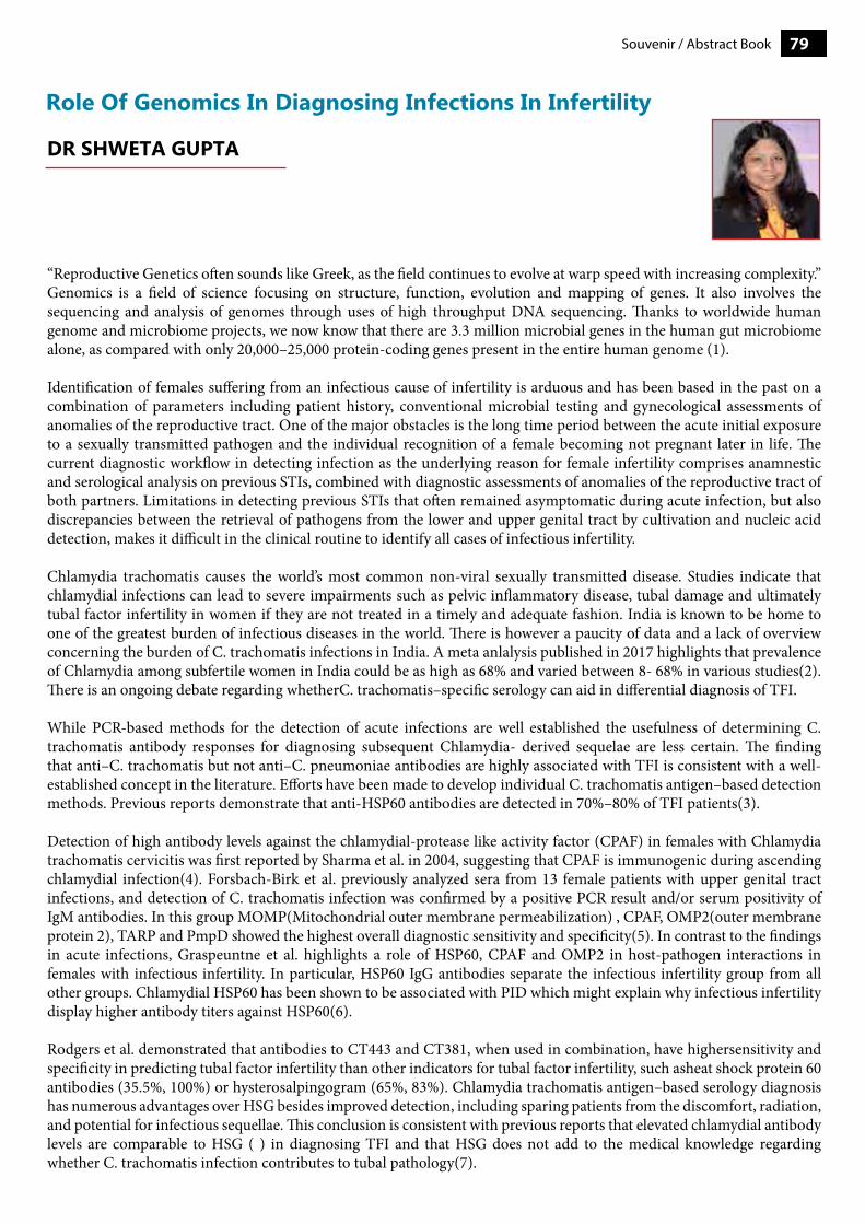
79Souvenir / Abstract Book
Dr shWeTa GupTa
‘‘Reproductive Genetics often sounds like Greek, as the field continues to evolve at warp speed with increasing complexity.’’ Genomics is a field of science focusing on structure, function, evolution and mapping of genes. It also involves the sequencing and analysis of genomes through uses of high throughput DNA sequencing. Thanks to worldwide human genome and microbiome projects, we now know that there are 3.3 million microbial genes in the human gut microbiome alone, as compared with only 20,000–25,000 protein-coding genes present in the entire human genome (1).
Identification of females suffering from an infectious cause of infertility is arduous and has been based in the past on a combination of parameters including patient history, conventional microbial testing and gynecological assessments of anomalies of the reproductive tract. One of the major obstacles is the long time period between the acute initial exposure to a sexually transmitted pathogen and the individual recognition of a female becoming not pregnant later in life. The current diagnostic workflow in detecting infection as the underlying reason for female infertility comprises anamnestic and serological analysis on previous STIs, combined with diagnostic assessments of anomalies of the reproductive tract of both partners. Limitations in detecting previous STIs that often remained asymptomatic during acute infection, but also discrepancies between the retrieval of pathogens from the lower and upper genital tract by cultivation and nucleic acid detection, makes it difficult in the clinical routine to identify all cases of infectious infertility.
Chlamydia trachomatis causes the world’s most common non-viral sexually transmitted disease. Studies indicate that chlamydial infections can lead to severe impairments such as pelvic inflammatory disease, tubal damage and ultimately tubal factor infertility in women if they are not treated in a timely and adequate fashion. India is known to be home to one of the greatest burden of infectious diseases in the world. There is however a paucity of data and a lack of overview concerning the burden of C. trachomatis infections in India. A meta anlalysis published in 2017 highlights that prevalence of Chlamydia among subfertile women in India could be as high as 68% and varied between 8- 68% in various studies(2). There is an ongoing debate regarding whetherC. trachomatis–specific serology can aid in differential diagnosis of TFI.
While PCR-based methods for the detection of acute infections are well established the usefulness of determining C. trachomatis antibody responses for diagnosing subsequent Chlamydia- derived sequelae are less certain. The finding that anti–C. trachomatis but not anti–C. pneumoniae antibodies are highly associated with TFI is consistent with a well-established concept in the literature. Efforts have been made to develop individual C. trachomatis antigen–based detection methods. Previous reports demonstrate that anti-HSP60 antibodies are detected in 70%–80% of TFI patients(3).
Detection of high antibody levels against the chlamydial-protease like activity factor (CPAF) in females with Chlamydia trachomatis cervicitis was first reported by Sharma et al. in 2004, suggesting that CPAF is immunogenic during ascending chlamydial infection(4). Forsbach-Birk et al. previously analyzed sera from 13 female patients with upper genital tract infections, and detection of C. trachomatis infection was confirmed by a positive PCR result and/or serum positivity of IgM antibodies. In this group MOMP(Mitochondrial outer membrane permeabilization) , CPAF, OMP2(outer membrane protein 2), TARP and PmpD showed the highest overall diagnostic sensitivity and specificity(5). In contrast to the findings in acute infections, Graspeuntne et al. highlights a role of HSP60, CPAF and OMP2 in host-pathogen interactions in females with infectious infertility. In particular, HSP60 IgG antibodies separate the infectious infertility group from all other groups. Chlamydial HSP60 has been shown to be associated with PID which might explain why infectious infertility display higher antibody titers against HSP60(6).
Rodgers et al. demonstrated that antibodies to CT443 and CT381, when used in combination, have highersensitivity and specificity in predicting tubal factor infertility than other indicators for tubal factor infertility, such asheat shock protein 60 antibodies (35.5%, 100%) or hysterosalpingogram (65%, 83%). Chlamydia trachomatis antigen–based serology diagnosis has numerous advantages over HSG besides improved detection, including sparing patients from the discomfort, radiation, and potential for infectious sequellae. This conclusion is consistent with previous reports that elevated chlamydial antibody levels are comparable to HSG ( ) in diagnosing TFI and that HSG does not add to the medical knowledge regarding whether C. trachomatis infection contributes to tubal pathology(7).
role of Genomics in Diagnosing infections in infertility

Souvenir / Abstract Book80
The use of novel genetic, proteomic, and metabolomic techniques may hold the key to more accurately diagnosing and treating infectious female infertility. It is within these categories that the search for potential biomarkers can begin. A biomarker is a distinctive biological or biologically derived indicator of a process, event, or condition that can be objectively measured, evaluated, and compared. The ideal biomarker should identify disease at an early stage, be easily detectable, cost-effective, and accurate, as well as having minimal side effects. The discovery of such noninvasive, highly sensitive and specific biomarkers would be helpful in eliminating the need for invasive testing.
References1. Mor A, Driggers PH, Segars JH. Molecular characterization of the human microbiome from a reproductive perspective.
Fertil Steril. 2015 Dec;104(6):1344-502. Thomas P, Spaargaren J, Kant R, Lawrence R, Dayal A, Lal JA, Morré SA. Burden of Chlamydia trachomatis in India: a
systematic literature review.Pathog Dis. 2017 Jul 31;75(5).3. Dadamessi, I., Eb, F., and Betsou, F. Combined detection of Chlamydia trachomatis-specific antibodies against the
10 and 60-kDa heat shock proteins as a diagnostic tool for tubal factor infertility: results from a case-control study in Cameroon. FEMS Immunol Med Microbiol. 2005; 45: 31–35
4. Sharma J, Bosnic AM, Piper JM, Zhong G. Human antibody responses to a Chlamydia-secreted protease factor. Infect Immun. 2004; 72(12):7164±71.
5. Forsbach-Birk V, Simnacher U, Pfrepper KI, Soutschek E, Kiselev AO, Lampe MF, et al. Identification and evaluation of a combination of chlamydial antigens to support the diagnosis of severe and invasive Chlamydia trachomatis infections. Clin Microbiol Infect. 2010; 16(8):1237±44.
6. Graspeuntner S, Bohlmann MK, Gillmann K, Speer R, Kuenzel S, Mark H, Hoellen F, Lettau R, Griesinger G, König IR, Baines JF, Rupp J.. Microbiota-based analysis reveals specific bacterial traits and a novel strategy for the diagnosis of infectious infertility. PLoS One. 2018 Jan 9;13(1):e0191047.
7. Rodgers AK, Budrys NM, Gong S, Wang J, Holden A, Schenken RS, Zhong G. Genome-wide identification of Chlamydia trachomatis antigens associated with tubal factor infertility. Fertil Steril. 2011 Sep;96(3):715-21.
81Souvenir / Abstract Book
Dr ranDhir sinGh mD, LLb
certifiedembryologist (eSHre )bhopal
Human ejaculate consists of a heterogeneous population of sperm in terms of morphology, maturity, motility, and functional quality. During natural conception, the sperm from these subpopulations compete in the female reproductive tract, while the best sperm is able to successfully fertilize an oocyte and contribute to the next generation. To mimic the natural selection process, sperm selection has become an integral part of assisted reproduction technique (ART) procedure, while the selection of physiologically fit sperm has been proven to improve fertilization and success following ARTs. Here, we discuss in detail some of the novel technologies developed to select sperm as they may have the ability to revolutionize ART by improving the success rate, even in patients with severely compromised sperm.
Selection For Icsi Using The Hyaluronic Acid Binding Assay: Sperm selection is an important part of the ICSI procedure. Routinely, sperm selection for ICSI is based solely on sperm morphology and motility. These latter parameters may not be sufficient to select sperm with intact chromatin. Therefore, sperm selected based on sperm functional characteristics may result in the most appropriate sperm for the ICSI procedure. The methodology describes the selection of sperm based on the ability of sperm to bind solid-state hyaluronic acid as its receptor, present on mature sperm with intact chromatin.
Electrophoretic Separation Of Spermatozoa: An Analysis Of Genotype, Surface Carbohydrate Composition And Potential For CapacitationStudies examines the properties of an electrophoretic device designed to effect the rapid isolation of spermatozoa for assisted conception purposes. In light of previous reports suggesting that X- and Y-bearing spermatozoa can be separated in an electric field, the first characteristic examined was the sex chromosome status of electrophoretically.
Fragmentation In Morphologically Normal Spermatozoa: How Much Should We Be Concerned In The Icsi Era?Intracytoplasmic sperm injection (ICSI) has revolutionized the treatment of male infertility. However, there are still unanswered questions about the safety of this technique. During ICSI, only morphologically normal and motile spermatozoa are typically used to fertilize an oocyte.
Relationship Between Hyaluronic Acid Binding Assay And Outcome In Art: A Pilot StudyThe sperm-hyaluronan binding assay (HBA) is a diagnostic kit for assessing sperm maturity, function and fertility. The aim of this prospective cohort pilot study was to evaluate the relationship between HBA and WHO sperm parameters (motility, concentration and detailed morphology) and possible influence of sperm processing on hyaluronic acid.
Relation Of Zeta And Ha-Binding Methods For Selection Of Spermatozoa With Normal Morphology, Protamine Content And Dna IntegritySperm selection parameters based on morphology and motility for ICSI might not be relevant to chromatin integrity. Thus sperm selection based on sperm characteristics has been suggested.
Spectroscopy Of Dna Packaging In Individual Human Sperm Cells Distinguishes Normal From Abnormal CellsHealthy human males produce sperm cells of which about 25-40% have abnormal head shapes. Increases in the percentage of sperm exhibiting aberrant sperm head morphologies have been correlated with male infertility, and biochemical studies of pooled sperm have suggested that sperm with abnormal shape may contain abnormal DNA.
Outcome Of Magnetic Activated Cell Sorting Of Non-Apoptotic Spermatozoa Before Density Gradient Centrifugation For Assisted ReproductionMagnetic activated cell sorting (MACS) eliminates apoptotic spermatozoa based on the presence of externalized phosphatidylserine residues. We evaluated the outcome of male fertility treatment when intracytoplasmic sperm injection (ICSI) into human oocytes was performed with non-apoptotic MACS-selected spermatozoa.
sperm selection Techniques - how close are We?

Souvenir / Abstract Book82
Incomplete Development Of Human Spermatozoa Is Associated With Increased Creatine Phosphokinase Concentrations And Abnormal Head MorphologyCreatine phosphokinase (CK) activity studies in human sperm revealed differences among men and among sperm populations within the same specimen. Samples with low sperm concentrations, high incidence of abnormal sperm morphology, and diminished fertility had higher per sperm CK activity.
Development Of A Microchamber Which Spontaneously Selects High-Quality Sperm For Use In In Vitro Fertilization Or MicromanipulationA microchamber has been developed which allows motile sperm to swim from a central loading site to peripheral sidewells. The sidewells are designed such that oocytes may be placed within them for in vitro fertilization (IVF) or sperm may be harvested from them for use in standard IVF or micromanipulation. Because only motile sperm can fertilize
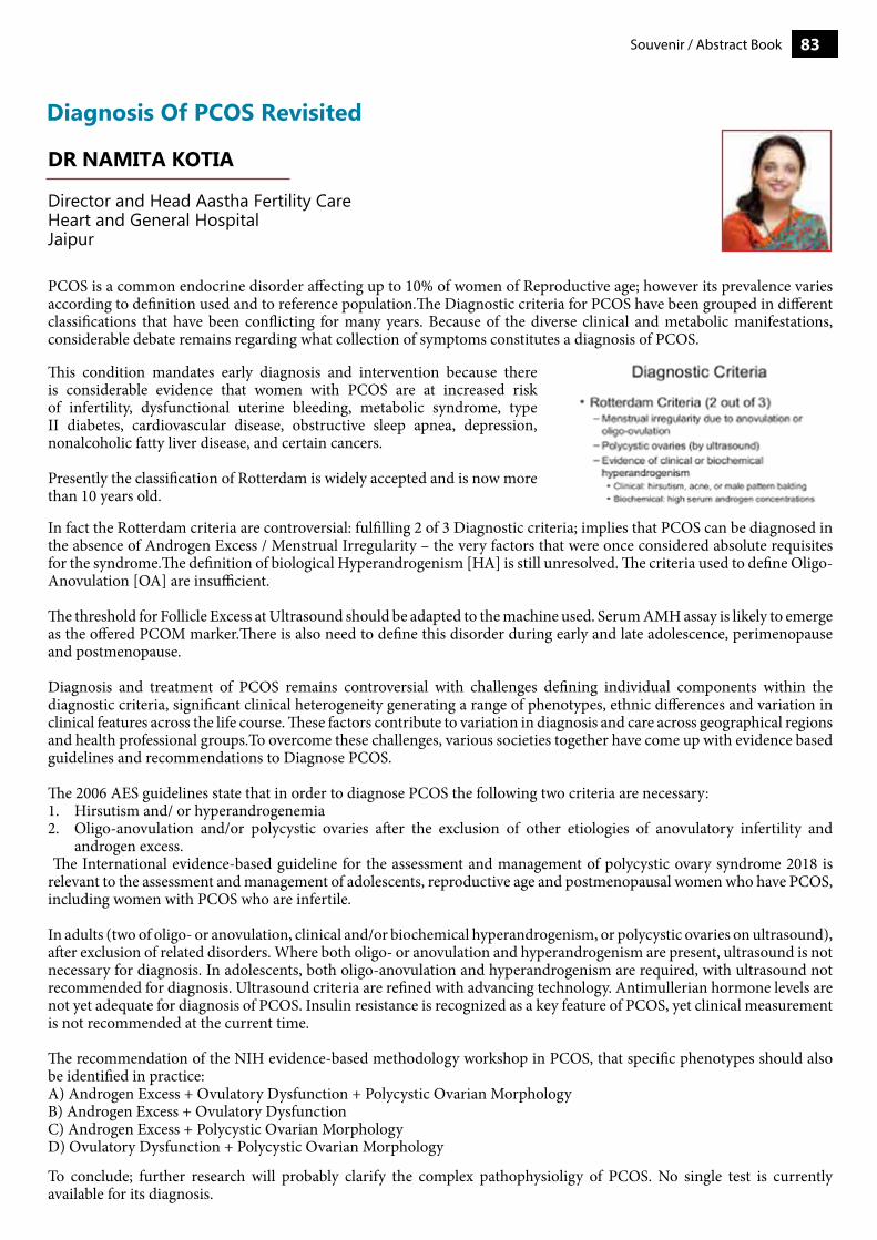
83Souvenir / Abstract Book
Dr namiTa koTia
Director and Head aastha Fertility careHeart and general HospitalJaipur
PCOS is a common endocrine disorder affecting up to 10% of women of Reproductive age; however its prevalence varies according to definition used and to reference population.The Diagnostic criteria for PCOS have been grouped in different classifications that have been conflicting for many years. Because of the diverse clinical and metabolic manifestations, considerable debate remains regarding what collection of symptoms constitutes a diagnosis of PCOS.
This condition mandates early diagnosis and intervention because there is considerable evidence that women with PCOS are at increased risk of infertility, dysfunctional uterine bleeding, metabolic syndrome, type II diabetes, cardiovascular disease, obstructive sleep apnea, depression, nonalcoholic fatty liver disease, and certain cancers.
Presently the classification of Rotterdam is widely accepted and is now more than 10 years old.
In fact the Rotterdam criteria are controversial: fulfilling 2 of 3 Diagnostic criteria; implies that PCOS can be diagnosed in the absence of Androgen Excess / Menstrual Irregularity – the very factors that were once considered absolute requisites for the syndrome.The definition of biological Hyperandrogenism [HA] is still unresolved. The criteria used to define Oligo-Anovulation [OA] are insufficient.
The threshold for Follicle Excess at Ultrasound should be adapted to the machine used. Serum AMH assay is likely to emerge as the offered PCOM marker.There is also need to define this disorder during early and late adolescence, perimenopause and postmenopause.
Diagnosis and treatment of PCOS remains controversial with challenges defining individual components within the diagnostic criteria, significant clinical heterogeneity generating a range of phenotypes, ethnic differences and variation in clinical features across the life course. These factors contribute to variation in diagnosis and care across geographical regions and health professional groups.To overcome these challenges, various societies together have come up with evidence based guidelines and recommendations to Diagnose PCOS.
The 2006 AES guidelines state that in order to diagnose PCOS the following two criteria are necessary:1. Hirsutism and/ or hyperandrogenemia2. Oligo-anovulation and/or polycystic ovaries after the exclusion of other etiologies of anovulatory infertility and
androgen excess. The International evidence-based guideline for the assessment and management of polycystic ovary syndrome 2018 is relevant to the assessment and management of adolescents, reproductive age and postmenopausal women who have PCOS, including women with PCOS who are infertile.
In adults (two of oligo- or anovulation, clinical and/or biochemical hyperandrogenism, or polycystic ovaries on ultrasound), after exclusion of related disorders. Where both oligo- or anovulation and hyperandrogenism are present, ultrasound is not necessary for diagnosis. In adolescents, both oligo-anovulation and hyperandrogenism are required, with ultrasound not recommended for diagnosis. Ultrasound criteria are refined with advancing technology. Antimullerian hormone levels are not yet adequate for diagnosis of PCOS. Insulin resistance is recognized as a key feature of PCOS, yet clinical measurement is not recommended at the current time.
The recommendation of the NIH evidence-based methodology workshop in PCOS, that specific phenotypes should also be identified in practice: A) Androgen Excess + Ovulatory Dysfunction + Polycystic Ovarian Morphology B) Androgen Excess + Ovulatory Dysfunction C) Androgen Excess + Polycystic Ovarian Morphology D) Ovulatory Dysfunction + Polycystic Ovarian Morphology
To conclude; further research will probably clarify the complex pathophysioligy of PCOS. No single test is currently available for its diagnosis.
Diagnosis of pcos revisited
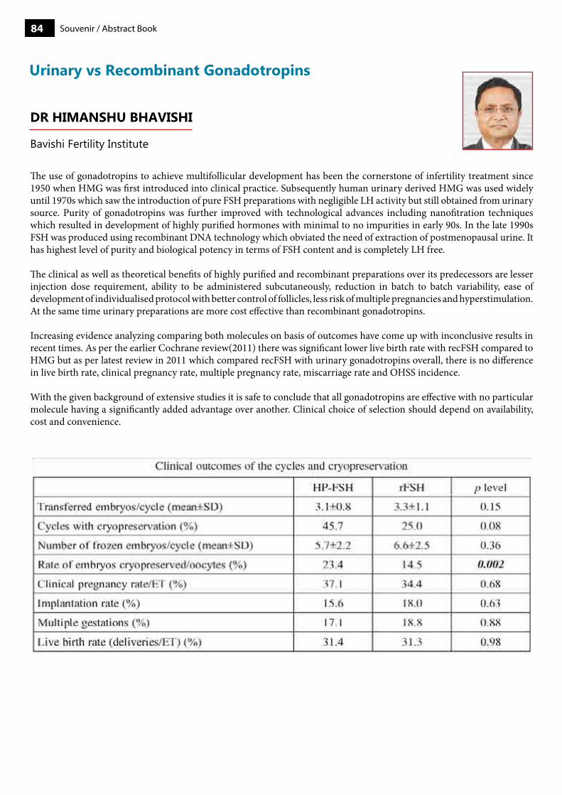
Souvenir / Abstract Book84
Dr himanshu Bhavishi
bavishi Fertility Institute
urinary vs recombinant Gonadotropins
The use of gonadotropins to achieve multifollicular development has been the cornerstone of infertility treatment since 1950 when HMG was first introduced into clinical practice. Subsequently human urinary derived HMG was used widely until 1970s which saw the introduction of pure FSH preparations with negligible LH activity but still obtained from urinary source. Purity of gonadotropins was further improved with technological advances including nanofitration techniques which resulted in development of highly purified hormones with minimal to no impurities in early 90s. In the late 1990s FSH was produced using recombinant DNA technology which obviated the need of extraction of postmenopausal urine. It has highest level of purity and biological potency in terms of FSH content and is completely LH free.
The clinical as well as theoretical benefits of highly purified and recombinant preparations over its predecessors are lesser injection dose requirement, ability to be administered subcutaneously, reduction in batch to batch variability, ease of development of individualised protocol with better control of follicles, less risk of multiple pregnancies and hyperstimulation. At the same time urinary preparations are more cost effective than recombinant gonadotropins.
Increasing evidence analyzing comparing both molecules on basis of outcomes have come up with inconclusive results in recent times. As per the earlier Cochrane review(2011) there was significant lower live birth rate with recFSH compared to HMG but as per latest review in 2011 which compared recFSH with urinary gonadotropins overall, there is no difference in live birth rate, clinical pregnancy rate, multiple pregnancy rate, miscarriage rate and OHSS incidence.
With the given background of extensive studies it is safe to conclude that all gonadotropins are effective with no particular molecule having a significantly added advantage over another. Clinical choice of selection should depend on availability, cost and convenience.
85Souvenir / Abstract Book
Dr nYmphea Walecha
Senior consultant Fortis ridge IVF,Shalimar bagh, Delhi
Technique of mitochondrial donation focus at eliminating the risk of mitochondrial diseases altogether from the oocyte or the fertilized egg. It is possible due to replacement of all the mitochondrial content of diseases mother’s oocyte with a healthy female’s oocyte. This method is called Mitochondrial Replacement Therapy. This implies is that the embryo is being produced with the DNA of both parents, as well as some DNA (only mitochondrial) from a healthy donor of mitochondrial contents.
Due to the uncharted nature of producing a child with three sources of DNA, this subject is currently quite contentious in the field of bioethics, as is the case with many other gene therapies. Currently, mitochondrial donation techniques are legal in the United Kingdom.[23] In February 2016, a report was issued by the US Food and Drug Administration declaring that further research into mitochondrial donation is ethically permissible.
The two most common techniques in the mitochondrial donation 1. Pronuclear transfer 2. Maternal spindle transfer.
Controversies1. The technique itself does not guarantee the complete removal of “Faulty Mitochondria” as the complete extraction of
nucleus without some mitochondria remaining attached to it is tricky even for experts. 2. The extent of mutation and frequency of division of faulty mitochondria depends on the kind of disease. If these faulty
mitochondria find their way into the embryo, it will tip‐off the balance between healthy and faulty mitochondria and the disease may still continue in the subsequent generations.
3. There may also be a risk of mismatch between the mtDNA haplotype of the surrogate and the donor mother.
Apart from these, the ethical, legal, and social issues that are raised with respect to these reproductive techniques are debatable and are mainly the reason why these techniques have not been legalized globally.Ethical Issues• Safety of the technique not yet established• The transfer of pronuclei results in destruction of the extra embryo which could develop into a healthy individual• DNA from three parents• Religious group objections of human’s meddling with the natural processes.
Legal IssuesGenetic content from three different parents thats not in agreement with the Human Fertilization and Embryology Authority Act 1990 as they involve the definite alteration of mitochondrial genome.
Social Issues• Expensivetechniquewithaccesstoonlyaffluentsocialgroups• Becauseoftriparentalaspect,childrenformedfromthesetechniquesmaysufferfrommentalagony• Thesemutationsmayreoccurlaterinlifeandmaybeinfuturegenerations.
Arguments in Favor1. Prevention of Disease: This technique uniquely provides women with mitochondrial mutations the ability to have
genetically related children free from disease.2. Physician Duties: Allowing patients to fulfill desires for genetically related children and reducing the incidence of
significant disease are consistent with a physician’s duty to act to benefit their patients.
Even though the research in this field still remains in infancy, mitochondrial replacement therapy may serve as a boon to a huge fraction of the population that is affected with mitochondrial diseases, for which no cure is available. This approach may eliminate these diseases from the subsequent generations as well making this a remarkable solution. As far as the ethical issues regarding the “Three‐Parent Baby” are concerned, the mitochondrial DNA plays no role in determining the physical appearance of the baby; only its overall metabolic fitness.Moreover it can also be utilized in poor reserve or poor quality oocyte patients who are not comfortable in taking a donor as they want their own genes in their children. Further research in analyzing the long‐term effects of this technique, however, is still due and subjected to considerations.
mitochondrial replacement Therapy:is it here to stay?
Souvenir / Abstract Book86
Dr. sanGiTa sharma mD(obgY), DNb(obgY), FNb(reproD. meDIcINe), mNamS, DgeS}Unit Head, Dept. of reproductive medicine,manipal Fertility, Jaipur
Dna fragmentation assay in clinical practice
Among the infertile couples, about 30-40% do have male subfertility. For male factor evaluation, conventional semen analysis had been thought enough, until the last couple of decades, after which sperm DNA fragmentation assays have been discussed and acknowledged as a complementary method for male subfertility evaluation.
The integrity of DNA in the chromosomes of the spermatozoon is a prerequisite to normal fertilization and transmission of paternal genetic information. Sperm DNA fragmentation (SDF) has been found to have a negative impact on spontaneous conception (1,2), Intrauterine Insemination (IUI) (3) & Assisted Reproductive Technology (ART) pregnancy outcome (4-7). High SDF has been found to be associated with high miscarriage rates, both in natural and ART conceptions (8). There is also a concern of transmission of genetic diseases and increased risk of cancer in the offspring, but adequate evidence is lacking. Although DNA fragmentation assays are being increasingly available, but there is still a debate on its clinical utility and exact clinical indications.
Guidelines and best practice statements published by the American Urological Association (AUA), European Association of Urology (EAU) and the American Society of Reproductive Medicine (ASRM) all acknowledge the potential contribution of sperm DNA fragmentation to male factor infertility, but do not recommend the routine use of sperm DNA integrity tests in the evaluation and treatment of infertile couples (9-11). Studies defining specific indications for DNA testing are now emerging. Practice recommendations based on different clinical scenarioshave also been published recently(12,13), but have been critically scrutinized by other authors (14 ).
Sperm DNA is specially susceptible to damage , as the chromatin has to undergo multiple steps for the final condensation and compaction, to be contained in such a small and highly motile cell. The DNA repair mechanisms are also suboptimal in the spermatozoa, making them more prone to damage.
Causes of Sperm DNA Damage:
Intrinsic Factors:• ProtaminedeficiencywhichcanleadtothehighersusceptibilityofDNAtodenaturation/instability• Oxidativestress(highROSfromleucocytesorimmaturespermatogeniccells,lowseminalantioxidants)• UnrepairedDNAbreaksduringchromatinremodeling• Abortiveapoptosisduringspermatogenesis
Extrinsic Factors:• Age• Obesity• Smoking,ExcessAlcohol• Varicocele• GenitalTractInflammation• Hyperthermia,Febrileillness• Spinalcordinjury• Testicularcancer• Environmentaltoxins• Drugs,chemotherapyandradiation
DNA Fragmentation Assays:There are different DNA fragmentation assays which have been propagated and used in different studies. The drawbacks are inter-observer variations, lack of standardization, cost involved and lack of any specific cut off values for clinical implications.
87Souvenir / Abstract Book
Below is the list of available different DNA Fragmentation Assays:
1. Acridine Orange Test (AO test)2. Aniline Blue staining (AB test)3. Toluidine Blue Staining (TB test)4. Chromomycin A3 staining (CMA3 staining)5. Single cell gel electrophoresis assay (Comet)6. Sperm Chromatin Dispersion (SCD) (Halo Test)7. Terminal deoxynucleotidyl transferase dUTP nick end labeling (TUNEL)8. Sperm Chromatin Structure Assay (SCSA)
While the AO, AB and TB tests are easy to perform, rapid, simple and relatively inexpensive, the disadvantages are lack of reproducibility and inter- observer variation. CMA3 staining is strongly correlated with other assays, but also has inter- observer variability. The comet assay uses a gel electrophoresis to compare the ratio of the smaller DNA fragments (tail of the comet) from the intact chromatin (head of the comet). While this test is sensitive, it is also operator-dependent.The SCD uses fluoroscopy to dye intact DNA and quantifies the normal DNA rather than the DNA fragmentation. It comes in an inexpensive, accessible kit, but clinically significant correlations between this method and treatment outcomes are limited. While standardization of SCSA has made this technique highly reliable, it is cost prohibitive for many medical centers as it requires the flow cytometer. In spite of the lack of standardization of TUNEL protocol, this assay is the preferred method of testing due to its established clinical value. Although different studies have mentioned different cutoff values for the various SDF assays, values above 25-30% are usually worrisome.
Evidence of SDF Testing:On reviewing the literature, there are systematic reviews and metaanalysis on SDF and its impact on various reproductive outcomes, both in natural and ART conceptions (5,6,8, 15,16 ), but the quality of the studies is compromised regarding the level of evidence. There are no well designed randomized controlled trials, which are required to study the predictive value of any diagnostic test. A diagnostic test is considered to be clinically useful when the results are reproducible with standard methodology, there are clear cut-off points, good sensitivity and specificity, clear clinical indications with ability to translate to a clinically meaningful difference and should change the further management of the patient with reasonably effective management options available. Sperm DNA fragmentation assays, unfortunately do not fulfill these criteria in absolute terms.
There is scarcity of high quality, prospective studies recruiting consecutive patients validating previously established cut off points with gold standard fertility outcomes. The systematic reviews and studies which are available have the following shortcomings: • Heterogenous studies (mostly cohort, case –control, retrospective)• Small sample size • Variable patient population• Lack of control for female factors and other confounding factors affecting the outcome• Weak statistical methodology in calculating threshold values and predictive ability of tests• Use of different assays for testing Sperm DNA damage
Although good quality evidence is still lacking, based on the available evidence, Agarwal et al &Cho et al. recently published a summary of the literature describing the clinical utility of sperm DNA fragmentation assays, in the context of commonly encountered clinical scenarios (12,13). The authors recommended that sperm DNA fragmentation testing should be offered to couples in each of these clinical scenarios, the results of which may facilitate decision making in further management and may also motivate the patients for lifestyle changes.
Clinical indications for SDF testing ( 12,13) :• Unexplained Infertility / Repeated IUI Failures:High levels of SDF may be associated with unexplained infertility and
multiple IUI failures in past. Lower IUI pregnancy rates have been found in patients with high SDF. It is reasonable to offer SDF testing to such couples as they may be better served by ART (more precisely ICSI) (Grade C recommendation).
• Infertility with Varicocoele : Here SDF testing can help in selecting the candidates for varicocoelectomy. SDF is recommended in patients with grade 2/3 varicocele with normal conventional semen parameters and in patients with grade 1 varicocele with borderline/abnormal conventional semen parameters ( grade C recommendation).
• Recurrent Pregnancy loss (RPL) workup: High SDFhas been found to be associated with RPL. Higher miscarriage rates are seen in both IVF and ICSI cycles. Lifestyle changes, addition of antioxidants or a decision for ICSI with
Souvenir / Abstract Book88
testicular sperm might be helpful.• Recurrent IVF/ICSI failures : High SDF has been found to be associated with lower fertilization rates, lesser number of
good quality embryos, lower implantation rates, pregnancy and Live birth rates. DNA fragmentation testing in patients with recurrent ART failure is indicated as it can provide useful prognostic information on subsequent ART cycles . The negative impact on LBR was not found in ICSI cycles, whereas it was there after conventional IVF. Thus, in couple with recurrent IVF failures and high SDF, ICSI is a better choice for subsequent cycles. Several studies have shown some benefit in using testicular sperm rather than ejaculated sperm in men with oligozoospermia, high SDF and recurrent IVF failure (Grade B–C recommendation).
• Infertility with SDF risk factors (viz smoking, high fat diet, alcohol intake, radiation exposure): Infertile men with evidence of exposure to pollutants or those found to have a modifiable lifestyle risk factor during evaluation should be offered SDF testing. The sperm DNA test can help reinforce the importance of lifestyle modification (e.g., cessation of cigarette smoking, antioxidant therapy(17)), predict fertility and monitor the patient’s response to intervention (Grade C recommendation).
Conclusions:Sperm DNA integrity is essential for human reproduction. Over the last couple of decades, research has shown significant correlation between SDF and the chances of conception (both naturally and by ART). SDF testing provides complementary information to semen analysis and both tests should be used in combination for a comprehensive assessment of infertile men. More studies are required to clarify the role of SDF testing in clinical practice, but there is emerging evidence supporting the use of SDF testing in specific clinical scenarios. Lifestyle modification, antioxidant supplementation and ART (ICSI with or without testicular sperms) can be considered for infertile couples with high SDF. The cost of the assays, lack of clinical value in the general population, and lack of standardization are the main disadvantages to SDF analysis .
References1. Agarwal A, Majzoub A, Esteves SC, Ko E, Ramasamy R, Zini A. Clinical utility of sperm DNA fragmentation
testing: practice recommendations based on clinical scenarios. Transl Androl Urol 2016;5(6):935-950. doi: 10.21037/ tau.2016.10.03
2. Cho CL, Agarwal A, Majzoub A, Esteves SC. Clinical utility of sperm DNA fragmentation testing: concise practice recommendations. Transl Androl Urol 2017;6(Suppl 4):S366-S373. doi:10.21037/tau.2017.07.28
3. Johnson D, Sandlow J. Sperm DNA fragmentation testing: proceed with care. Transl Androl Urol 2017;6(Suppl 4):S425-S427. doi: 10.21037/tau.2017.03.75
4. Cissen M, Wely Mv, Scholten I, Mansell S, Bruin JPd, Mol BW, et al. (2016) MeasuringSperm DNA Fragmentation and Clinical Outcomes of Medically Assisted Reproduction: A Systematic Review and Meta-Analysis. PLoS ONE 11(11): e0165125. doi:10.1371/journal.pone.0165125
5. 16. Jhao et al. Whether sperm deoxyribonucleic acid fragmentation has an effect on pregnancy and miscarriage after in vitro fertilization/intracytoplasmic sperm injection: a systematic reviewand meta-analysis Fertil Steril_ 2014;102: 998–1005. (American Society for Reproductive Medicine.)
6. 17. Showell MG, Brown J, Yazdani A, Stankiewicz MT, Hart RJ. Antioxidants formale subfertility. Cochrane Database Syst Rev 2011.
89Souvenir / Abstract Book
Dr. sonal panchal. mD
Dr. Nagori’s Institute for Infertility and IVF, ahmedabad, India
The peritoneum is a thin, translucent serosal membrane of mesodermal origin that covers the surface of theperitoneal cavity and its mesenteries. The peritoneum partially or completely covers the visceral organscontained within the peritoneal cavity. In females, the peritoneum is discontinuous at the ostia of the oviducts, allowingcommunication between the peritoneal cavity and extraperitoneal pelvis. Peritoneal fluid acts as a bacterial defense. Two layers of peritoneum that invest blood vessels, lymphatics, nerves,adipose tissue, and connective tissue form the peritoneal ligaments and mesenteries. The peritonealligaments subdivide the peritoneum into interconnected compartments that dictate the locationand routes of spread of primary and secondary malignancies and infection within the peritoneal cavity. In the pelvis, the dependent peritoneal recesses are the rectovaginal pouch in the female, and the lateral paravesical recesses inboth sexes.
The pathology of the peritoneum that is most commonly seen is the peritoneal inclusion cyst. Though less common lesions may also be seen like peritoneal lieomyomatosis, peritoneal carcinomatosis, multiloculated mesotheliomas, Though these have typical appearance, but still confident histopathological diagnosis based on ultrasound is not possible.
Fallopian tubes are portals for gamete transfer. These are divided into four parts. Tubes if normal cannot be appreciated on routine scan due to lack of fluid interface. Therefore when there is hyodrosalpinx or free fluid arounf tubes, that is when these can be appreciated on ultrasound. Hydrosalpinx on ultrasound can be described by certain typical description as an extraovarian cystic lesion, that changes shape on rotation of probe, has incomplete septa, is sausage shape and may show a cog-wheel appearance.
When due to acute inflammation, it has thick walls and ascites and when it is due to chronic inflammation, it is more likely to appear thin walled, rigid and may show adhesions. Tubal obstructions may be due to infections, endometriosis or due to previous surgery.Amongst other pathologies of the tubes are neoplasm, para tubal/ fimbrial cysts, etc. Tubal patency assessment can be done by x- ray HSG and laparoscopy wirth hysteroscopy is the gold standard. But ultrasound has been now widely used for tubal patency assessment, both by negative and positive contrast, using both B mode and 3D with contrast mode. Ultrasound has maintained its essential role in the assessment of the pelvic peritoneoum and the tubes.
peritoneal and tubal morphology and pathology by ultrasound
ultrasound monitoring of ovarian stimulation: from basic to advancedTransvaginal ultrasound is the tool of choice for follicular and endometrial monitoring. Ultrasound parameters like ovarian size , antral follicle count and ovarian stromal flows are used for assessing ovarian reserve and response and to decide the stimulation protocol. Scoring system has been devised using these parameters with age and BMI of the patient. This helps in individualizing the stimulation protocols and significantly reduces the incidence of OHSS and cancellation of cycle due to poor response, both.
Follicular monitoring is about assessing the maturity of the follicle and receptivity of the endometrium to decide the time of ovulation trigger. This can be done by hormonal assessments and/ or ultrasound. The follicular maturity can be assessed by oestradiol levels, but frequent assessment of blood oestradiol level is a cumbersome. When using ultrasound follicular diameter of 16mm for gonadotrophin stimulation and 18-20mm for clomiphene stimulation cycles has been considered to correlate with follicular maturity. At that stage of follicular growth, Doppler assessment of the follicle and endometrium should start. Doppler with transvaginal ultrasound this has been the most preferred method for assessment of follicle and endometrium. The vascular changes are reflection of the biochemical changes and can be studied by colour Doppler. 3D ultrasound gives a better assessment of the follicular and endometrial size, that is the anatomical maturity, than 2D ultrasound and 3D power Doppler gives not only qualitative but also quantitative idea of global vascularity, that is the reflection of functional/ physiological maturity.
FSH dependent dominant follicle and grows at a rate of 2-3mm per day has no internal echogenecity and has thin (pencil line like) walls. A mature follicle is 16 – 18 mm has thin walls, regular round shape. It shows a thin hypoechoic rim
Souvenir / Abstract Book90
surrounding the follicle. When functionally mature, on colour Doppler, the follicle shows blood vessels covering at least 3/4th of the follicular circumference with RI of 0.4 – 0.48 and PSV of > 10 cms/ sec. Though close to rupture the PSV of the follicle rises and therefore in follicles with high PSV, early IUI may be recommended. 3D US and 3D power Doppler have been widely used for the assessment of these follicles. It is used to asses the follicular volume and its global vascularity. adding 3D power Doppler parameters to the Doppler criteria for the assessment of the follicular vascularity, improves the conception rates. Follicles containing oocytes capable to produce a pregnancy have a perifollicular vascular network more uniform and distinctive.
On TVS an endometrial thickness of minimum 6 mm is required on the day of hCG , but 8-10 mm is optimum. Breach or irregularity of endometrio-myometrial junction is an indication of unhealthy endometrium and therefore poor receptivity. Endometrial morphology of grade A or B is preferred. On colour Doppler the endometrium that is mature shows vascularity in zone 3 and 4. The pulse Doppler of these vessels should have an RI of <0.6 and his vascularity should cover at least 5mm2 area of the endometrium. When the endometrial thickness is <8 mm, and if there are non-triple endometrial line and non-favorable blood flow zone on day of hCG in IVF/ICSI cycles, pregnancy is unlikely.Moreover the uterine artery PI should be < 3.2. Pregnancy and implantation rates were significantly lower when endometrial volume < 2ml. Better endometrial vascularity can be correlated with better pregnancy rates. 3D power Doppler may help to assess the global vascularity of the endometrium.
Ultrasound is a tool of choice for selection of stimulation protocol and dose requirement of gonadotrophins and also for assessing follicular maturity and endometrial receptivity and decides the time of trigger.
91Souvenir / Abstract Book
Dr.roYa rozaTi m.D, F.r.c.og
Dr. Nagori’s Institute for Infertility and IVF, ahmedabad, India
Endometriosis is an enigmatic and debilitating gynaecological disease of adolescents and reproductive-aged women. It affects 6-10% of women and is characterized by growth of endometrial tissue outside of the uterus, most often in the peritoneal cavity and is associated with infertility and pain.The etiology of endometriosis remains unclear but it is evident that endometriosis occurs due to the dissemination of endometrium to ectopic sites and the subsequent establishment of deposits of ectopic endometrium. Endometriosis causes dysmenorrhea, dysparunia, heavy menstrual bleeding, non-menstrual pelvic pain, pain at ovulation, dysphasia, dysuria, as well as chronic fatigue, and infertility. The goals for endometriosis treatment may be the relief of pain and to achieve a successful pregnancy in the infertile patients. Treatment must be individualized with a multidisciplinary approach.
Management of endometriosis has been changing over period of time, shifting focus from radical surgery to more conservative approach.
TreatmentThe treatment of women with endometriosis is a challenge. Assessing the success of Treatment of endometriosis is difficult but it is evaluated based on the individual symptoms, age and desire for fertility. The most common approaches consist of medical treatment, laparoscopic surgery and major surgical management.
Medical TreatmentAssessing the success of medical treatment for endometriosis is difficult. But is based on individual medical options by suppression of ovarian steroids and induction of a hypoestrogenic state that causes atrophy of ectopic endometrium. The medical treatment has limited value in patients with infertility because it inhibits ovulation. Standard medical therapies include analgesics (non steroidal anti-inflammatory drugs - NSAIDs) oral contraceptive pills (OCPS), androgenic agents (danazol), progesterone, gonadotropins releasing hormone analogues (GnRH analogue) and antiprogestogens (gestrinone) all have been used for the treatment of endometriosis
Nonsteroidal Anti-Inflammatory Drugs(NSAID): Endometriosis is a chronic inflammatory disease. empirical treatment, nonsteroidal anti-inflammatory drugs have been reported to be effecti ve in reducing endometriosis associated pain. First-line medical treatment for pain due to endometriosis is a nonsteroidal anti-inflammatory drug, either by prescription or over-the-counter. Although these antiprostaglandin agents have been shown to be effective for the treatment of primary dysmenorrhea.A Cochrane analysis found insufficient data to show that they significantly reduce endometriosis pain .
Hormonal Medical TreatmentEndometriosis is an estrogen dependent disease hence, hormonal study has been designed to suppress estrogen synthesis, thereby inducing atrophy of ectopic endometrial implants or interrupting the cycle of stimulation and bleeding. Hormonal therapy is not curative, hence it requires to be administered for years or until woman desires a pregnancy. It has been observed that the various hormonal therapies studies have similar efficacy.
Combined oral contraceptives: They should be considered as the first line option, both as an alternative to surgery and as a postoperative adjuvant measure. Combined hormonal contraceptives have been used in both a cyclic and a continuous fashion in the treatment of symptoms associated with endometriosis contain more androgenic progestogencombined OCs containing the new generation progestogen, desogestrel, also have proven effective Gonadotrophin releasing hormone analogues, danazol and gestrinone should be used when there is failure of progestins and oral contraceptives or are not tolerated or are contraindicated.
Progestins: They aremost commonly used for the treatment of endometriosis. progestogen-induced suppression of matrix metalloproteinases, a class of enzymes important in the growth and implantation of ectopic endometrium Progestins cause antiendometriotic effect by initial decidualization of endometrial tissues followed by atrophy. They are as effective as danazol
medical management of endometriosis-new concepts
Souvenir / Abstract Book92
or GnRH analogues and have lower cost and a lower incidence of side effects compared to these agents. After 3 to 6 months of evaluation Medroxy progesterone acetate was found to do effective in relieving pain when started at a dose of 30 mg/day and increased the dose based on the clinical response and bleeding patterns(7). Medroxy progesterone acetate 150 mg given intramuscularly every 3 months is also effective for relief of pain associated with endometriosis. Orally norethidrone 5 mg / day increased maximum to 15 mg / day, Megestrol acetate 40mg/day, Lynestrenol 10 mg/day and dydrogesterone 20 to 30 mg / day either continuous or cyclical are also effective in r e l i e v ing pa in a ssoc i a t ed with endometriosis. If effective, these agents can be used safely for longer periods of time. The levonorgestrel releasing intrauterine device has been reported very effective at relieving pain associated with endometriosis especially relieving dysmenorrhea and pain due to rectovaginal endometriosis(8). Progestins are associated with more adverse effects than OCPs. Side effects include nausea, weight gain, fluid retention, breakthrough bleeding and delayed return of fertility after the cessation of treatment. Danazol: Itis a synthetic androgen that suppresses gonadotrophin secretion resulting into direct inhibition of the LH surge and steroidogenesis.. The multiple effects of danazol cause a high androgen and low estrogen levels, which does not support the growth of endometriosis. Androgenic side effects include acne, edema, and hirsutism, deepening of voice and weight gain. . These immunological effects of danazol also help in suppression of endometriosis. . Typically this medication is administered orally; however, vaginal administration as well as vaginal and intrauterine delivery systems have been reported.The medication is administered orally; however, vaginal administration as well as vaginal and intrauterine Danazol provided comparable pain relief to GnRH-a but was not as well tolerated .
Gestrinone: It is an antiprogestational steroid. used for the treatment of endometriosis.The mechanism which includes a progestational withdrawal effect at the endometrial cellular level and inhibition of ovarian steroidogenesis, cellular in activation and degeneration of endometriotic implants .The drug is administered orally daily to weekly with doses ranging from 2.5–10 mg. Side effects relate to both androgenic and antiestrogenic effects. Gestrinone was shown to be as effective as danazol and GnRH analogues . Side effects are dose dependent and are similar to danazol but less intense.
Gonadotropin-releasing hormone agonist: It has been studied more extensively than other medical treatment regimens. GnRH Agonists bind to pituitary GnRH receptors and stimulate LH & FSH synthesis and release.It is administered by a calibrated nasal spray twice daily (nafarelin acetate), by injection of either a short-acting formulation daily, or by injection of a depot formulation (LA, goserelin acetate) every 1–3 months. . Various GnRH agonists available for treatment of endometriosis are leuprolide, buserelin, nafarelin, histrelin, goserelin, deslorelin and triptorelin. Side effects relate primarily to the induced hypoestrogenic state and include hot flushes, vaginal dryness, decreased libido, mood swings, headache, and bone mineral depletion. About 90 percent of patient experience pain relief. Prolonged pretreatment with GnRH analogue before IVF has been reported to improve clinical pregnancy rates in infertile women with endometriosis. Side effects relate primarily to the induced hypoestrogenic state and include hot flushes, vaginal dryness, osteoporosis,decreased libido, mood swings, headache, and bone mineral depletion.
Other Medical Treatments: Under Investigation for endometriosis include RU486 (mifepristone), selective PR modulators, selective ER modulators, GnRH antagonists, pentoxifylline, and agents that inhibit the effect of tumor necrosis factor (TNF)-a, matrix metalloproteinases, and angiogenesis
ConclusionEndometriosis is a chronic disease that requires a lifelong management plan with the goal of maximizing the use of medical treatment and avoiding repeated surgical procedures. Definitive treatment of endometriosis with hysterectomy and BSO should be reserved for women with debilitating symptoms that can reasonably be attributed to the disease. These women should have completed childbearing and have failed to respond to alternative treatments. Further studies designed to compare medical and surgical treatments are clearly warranted. Current medical treatment of endometriosis depends on drugs that suppress ovarian steroids. Recent research shows a very promising role for new hormonal medication in the management of endometriosis which may prevent or eradicate endometriosis and also allow the conception during treatment rather than merely relieving the symptoms.

93Souvenir / Abstract Book
Dr laXmi shrikhanDe
mD, FIcog, FIcmU consultant Fertility Specialist
Women can now choose between surgical and an array of new medical management options for fibroids. Surgical option is no more the best option for fibroids. The fibroid management has to be customised as per the patient requirements and medical management should be tried first whenever it is feasible. The various medical management options are-• Hormonemedications.• Progestin-releasingintrauterinedevice(IUD)• GnRHagonist• SPRM-selectiveprogesteronereceptormodulator
Ulipristal Acetate is a SPRM which has revolutionized the medical treatment of fibroids. FDA has approved the drug for fibroid treatment. It acts directly on the progesterone receptors in 3 target tissues - the endometrium, uterine fibroids, and the pituitary gland.
The safety and efficacy of UA has been evaluated in VENUS I and VENUS II, 2 phase 3 studies of more than 500 adult women. Additionally, the efficacy of UA has been demonstrated in a series of 4 phase 3 European trials involving more than 1,000 women with uterine fibroids. Treatment should be started within one week of menstruation. One tablet of 5 mg is to be taken daily with or without food for 3 months. Wait for the periods. Second course if needed should be started only after menstruation.4 such intermittent courses of UA can be given.
Ulipristal acetate should only be prescribed after careful diagnosis, only where it is indicated and after excluding the contra indications. It is contraindicated during breastfeeding Pregnancy should be excluded before starting treatment. Concomitant use of progestagen-only pills, a progestagen-releasing intrauterine device or combined oral contraceptive pills is not recommended . Although a majority of women taking a therapeutic dose of ulipristal acetate have anovulation, a non-hormonal contraceptive method is recommended during treatment.
Liver function tests must be performed before starting treatment. During treatment, liver function tests must be performed monthly during the first 2 treatment courses. For further treatment courses, liver function must be tested once before each new treatment course and when clinically indicated.Co-administration of moderate (e.g. erythromycin, grapefruit juice, verapamil) or potent (e.g. ketoconazole, ritonavir, nefazodone, itraconazole, telithromycin, clarithromycin) CYP3A4 inhibitors and ulipristal acetate is not recommended.
To summarize UA definitely has benefit in properly selected cases of fibroid uterus. It should be judiciously prescribed after excluding the contra indications. Woman should be properly monitored by USG and blood tests while taking the drug.
new frontiers in medical management of fibroids
Souvenir / Abstract Book94
Dr kokila Desai m.D gYNae
Fellow in reproductive medicine germanycFS - morpheus shivam IVF centre Surat
Oocyte Retrieval is basic procedure required in all the IVF cases .Before starting the Procedure one should be adequately trained (20 follicle aspiration under direct supervision ) to prevent problems. The most common problem is unsuccessful Oocyte retrieval is either due to inadequate HCG trigger or may be genuine. In case of inadequate HCG trigger one can try recombinant HCG, increase time interval between trigger and OPU , increase dose of HCG, readministeration of HCG, or agonist trigger in antagonist cycle. Genuine unsuccessful Oocyte retrieval can happen due to
• Loweraspirationpressure• Untrainedperson• Earlyruptureofoocyteor• Earlyoocyteatresia.
Other problems are due to surgical complications which stem from two basic facts about the surgery:
1) Needle must be pushed through the vagina into the ovary,• DifficultyinvisualizingOvaryandfollicleonbiopsylineonscreen• Puncturingthefollicle• NonvisualizationofneedletiponUSGscreen• Incompleteaspiration
2) Damage to number of other organs and sensitive tissues which lie nearby. • Vaginalbleeding• Intra-abdominalhaemorrhage• Retroperitonealhaemorrhage
Another complication that appeared on occasion was ovarian torsion, which occurs when the ovary twists around on itself, cutting off its blood supply.Rare complications like ruptured endometriotic/dermoid cyst ,injury to ureter , ureterovaginal fistulae , Rectus sheath hematoma, vertebral osteomyelitis can occur. women undergoing oocyte retrieval also face certain potential risks from the anesthesia used to handle their pain during the surgery ,which includes asphyxia caused by airway obstruction, apnea, hypotension and pulmonary aspiration of stomach contents.
problems in oocyte aspiration
95Souvenir / Abstract Book
Dr aarTi DeenaDaYal Tolani
Among uterine structural abnormalities, fibroids and adenomyosis represent two distinct, though frequently coexisting entities, with a remarkable prevalence in women of reproductive age. Adenomyosis and fibroids are benign conditions that are often responsible for uterine enlargement, menorrhagia, anemia, and infertility.
Uterine myomas, also called leiomyomata, fibroids, fibromyomas, leiomyofibromas, and fibroleiomyomas, are the most common benign uterine tumors. “The uterus is an organ that should bear something, if not a child then a fibroid”- is an age old saying. Fibroids also called liomymoas are benign and the most common tumours of the uterus. They are benign neoplasm’s of smooth muscle origin with various degrees of fibrous connective tissue. Fibroids mostly remain asymptomatic but sometimes are the cause of menorrhagia, pelvic pain, infertility, dysmenorrhoea and pressure symptoms. Submucous fibroids lower the pregnancy rates by 70% and surgical removal appeared to improve pregnancy rates. The surgical removal of intramural fibroids should be a well calculated decision weighing all the pros and cons and after considering if the fibroid is really the cause of the symptoms or not . Women with no fibroids and sub serous fibroids appear to have similar pregnancy outcomes.
Adenomyosis is a common gynecological disorder. Proliferation of endometrial glands and stroma within myometrium along with hypertrophy and hyperplasia of adjoining myometrium is adenomyosis. Reason for adenomyosis is not known, however, it may be associated with pelvis endometriosis.
Ultrasound Diagnosis of Fibroids and Adenomyosis:The gold standard diagnostic modality for uterine fibroids and adenomyosis is ultrasonography. Ultrasound has been proven to be a cost effective and adequate means to identify, map and evaluate fibroids and adenomyomas. Accurate findings not only work as a diagnostic tool but also can help guide the doctor towards the appropriate management (medical therapy, surgery, uterine artery embolization or conservative management).
Before beginning an ultrasound examination especially for fibroids and adenomyomas (where our main aim is to understand the position in the uterus and its relationship to the endometrium and serosa), the probe, optimum pre-set of the equipment, orientation of image, depth, frequency and gain should be adjusted to get an optimum image. For best images use lots of gel, press hard and use a darkened room. Especially in case of fibroids myometrial contractions can be mistaken and reported as fibroids, hence Sapir mode assesses fibroids better.
The major steps involved in the ultrasound assessment of fibroids and adenomyomas are
1. Vaginal entry: Must evaluate the vaginal wall and the bladder mucosa and its relationship to the uterus. Observe the cervix for any abnormality and document it.
2. Evaluating the Uterus: Observe the Uterus while rotating the probe to achieve true axial, coronal and sagittal views. Use the depth setting to fill at least three forth of the screen with the uterus to avoid missing any pathologies. Increase or decrease the gain to have a clear and sharp image.
3. Location, position and measurements: First identify the center of uterus by delineating the endometrial cavity. Find out the cervical canal and trace it upwards to the endometrial cavity to locate fibroids and adenomyomas. Trace the position which can be Anterior, posterior, lateral wall. Three orthogonal dimensions are suitable for the measurements. Probe should be twisted in different planes till a clear border can be observed for measurement. Power Doppler should be further used to differentiate pathologies. Volume of each fibroid or adenomyomas should be calculated and measured.
Other important parameters are determination of echogenicity, vascularity and the relationship of fibroid or adenomyomas with endometrium. Reporting of the results is crucial. It must include all the details right from location, number, distance from the endometrium or serosa, measurements, vascularity and diagrammatic representation.
role of imaging in Diagnosis and Treatment of fibroids and adenomyosis
Souvenir / Abstract Book96
Some of the salient ultrasound features and differences between fibroid and adenomyosis are, fibroids are regular, lobulated, well defined and smooth margins, whereas adenomyomas are regular, poorly defined and irregular margins. The fibroid show internal linear shadows and edge shadows but adenomyomas appear without edge shadow. The blood flow in fibroids is circumferential but in adenomyosis is intralesional flow. Another important feature to differentiate fibroid and adenomyosis is appearance of junctional zone (JZ). In fibroids JZ is well defined but may be stretched around the fibroid, whereas in ademonyomas the JZ shows a typical adenomyotic features such as ill defined, irregular, thickened, tiny cystic spaces and echogenic buds or lines.
Ultrasound has been shown to be an adequate and cost effective means of evaluating size, number and location of fibroids and adenomyomas. Both trans abdominal scan (TAS) and a trans vaginal scan (TVS) should ideally be performed for precise detection and 3D ultrasound is very good for volume measurement vascularity assessment and spatial orientation.
Ultrasound in management of Fibroids: The management of uterine fibroids can be approached medically, surgically, and even by minimal access techniques. However, it is highly dependent on the presentation and patient wishes. Besides, it also depends on the number, location and size of the fibroids. Available surgical and non-surgical treatment modalities include myomectomy by hysteroscopy, myomectomy by laparotomy or laparoscopy, uterine artery embolization and interventions performed under radiologic or ultrasound guidance to induce thermal ablation of the uterine fibroids.
There is growing evidence on the management of submucous fibroid by Lasmars STEP-W. STEP-W classification is to analyse specific parameters that make surgery more difficult as that the hysteroscopic myomectomies can be planned based on the expected degree of technical difficulty in carrying out the procedure and to recommend the most suitable course of action in each situation. STEP-W score between 0-4 is under group-I which has low complexity hysteroscopic myomectomy. The score between 5-6 is group-II with high complexity hysteroscopic myomectomy. Consider either GnRH use or two step hysteroscopic myomectomy. Score between 7-9 is group-III where alternative to hysteroscopic technique should be considered. A score is given to the submucous myoma based on the size, topography, extension of the base penetration in to the myometrium and presence of the myoma in the lateral wall. Based on the total score complexity of the surgery was defined and therapeutic options suggested.
97Souvenir / Abstract Book
Dr charulaTa chaTTerJee
Sr.consultant embryologistYashoda Fertility and research InstituteHyderabad
Modern IVF lab requires wealth of expertise and experience from embryologist to achieve the best In terms of fertilization, cleavage, implantation, pregnancy & take home baby rate.Implementation of quality management system helps to achieve this.Human gametes and embryos cultured in vitro are extremely sensitive to oscillations in temperature, humidity,pH, light exposure, contaminants and physical trauma.
A common goal for all the embryologist is to minimize detrimental environmental stress, which is often achieved through setting thresholds or ranges for various environmental/lab parameters and measuring these variables on a regular basis. One such example of an environmental variable that should be measured within the IVF lab is the pH of the culture medium (pHe). Gametes and embryos are very sensitive to perturbations in pHe, which can lead to alteration in internal pH (pHi) and ultimately impact function and development.
Homeostasis of the intra cellular ionic environment is necessary to maintain cell function and viability.Improper pHi can impede sperm function, impairs embryo metabolism, alters organelle localization and even retards-resulting fetal growth.Hence to maintain Intra cellular pH (pHi) regulation of external pH [pHe] is must. pHe is the result of a balance between concentrations of CO2 in the cell culture incubator and the amount of bicarbonate in the media.
Cell culture media contain carbonate-based buffers which work with elevated gaseous carbon dioxide levels in the incubator to stabilize cell culture pH.This bicarbonate concentration is regulated by commercial media company. Thus,set point CO2 gas, diffusion of CO2 into the media , volume of media, surface area, use of oil overlay and even the type of lid/dishware can influence this gas exchange and equilibration timing and can alter pHe.
Change in external pH of culture media (pHe) influences sperm binding and motility, oocyte maturation and embryo development. Denuded mature oocyte or cryo preserved/thawed embryos , which lack robust pHi regulatory mechanisms are susceptible to deviations in pHe. Therefore, it is readily apparent that buffers used to stabilize pHe are extremely important factors to consider in optimizing embryo culture systems .Knowledge about basic buffer system of IVF media will help to set CO2 concentration required to get optimum pH.
Media pH should be measured initially during setting up equipment and installing a culture system. It should then likely be monitored following any major maintenance or media change, perhaps even with new media lots. During routine incubator maintenance or gas cylinder change it is also advisable to check pH e of media. Varieties of pH meter available commercially but careful selection of pH meter as per lab need is mandatory.
In summary, proper setting, monitoring and stabilizing of pHe during IVF laboratory proceduresis a crucial component of a rigorous quality control programme. Here, importance of both pHi and pHe in respect to gamete and embryo quality are important. Furthermore, embryologist should be aware of factors influencing selection of pHe, as well as emerging methods to stabilize pHe in the IVF laboratory.
ph monitoring - The way forward!
Souvenir / Abstract Book98
Dr vanDana narula
consultant gynaecologist, obstetrician and infertility specialistgurgaon
Infertility is so rampant in today’s scenario that it takes a toll on the couples’s lives.
Visits to the infertility specialist requires so much of time and man hours.
There are few alternative therapies the couples can try before starting treatment and also in conjunction with the treatment to help them relax and can also aid conception.
The various techniques are:
YOGA is a form of calming exercise which help couples to relax, increase flexibility, and improve blood flow to the pelvis.MUSIC THERAPY to relax parts of brain which have a role in release of hormones involved in treatment and helps cope up with the trauma involved.MEDITATION helps calm the mind and emotions.MINDFULNESS helps to focus awareness on the present moment and also accept your thoughts and feelings.HYPNOTHERAPY is used to help couples relax and reduce anxiety .Couples feel less stressed and more in control of their lives.ACUPUNCTURE helps to regulate the natural flow of energy through the body by stimulating special points.CRYSTAL THERAPY stimulates electromagnetic charges that encourage body processes to work better. FENG SHUI aims to rebalance the energy of home.and energy called as CHI starts flowing.REFLEXOLOGY relaxes body and mind and body becomes more receptive to conceiving a baby. MASSAGE helps to de stress sending blood and oxygen to massaged parts of the body.DIET BRAZIL NUTS are full of selenium that improves the quality of the sperms.HERBS AND TEAS have antioxidants which boost fertility.
role of alternate therapies in infertility
99Souvenir / Abstract Book
harsha BhaDarka
Director –embryology Departmentakanksha Hospital and research Institute
A high incidence of chromosomal anomalies has been observed in embryos cultured in IVF techniques. Preimplantation genetic testing is done for testing such anomalies. It includes preimplantation genetic screening and preimplantation genetic diagnosis.
The aim of PGS/PGD is to detect whether embryo is chromosomally affected or not, thereby preventing transfer of a chromosomally abnormal embryo. With development of prenatal diagnostic techniques like amniocentesis and chorionic villi sampling (CVS), NIPT (Noninvasive prenatal testing), it has been possible for couples at risk of genetic diseases to give birth to healthy babies, however the experience of these diagnosis followed by termination of pregnancy in case of abnormal findings if detected can be a unwelcome memory. PGD is essentially an alternative to prenatal diagnosis, which helps in negative selection of affected embryo prior to implantation.
Indications for pre-implantation genetic testing :1. Patients with family history of X-linked disorders carriers 25% risk of having an affected embryo.2. Implantation failure, recurrent pregnancy loss, or mental or physical problems in offspring occurs due to Chromosome
translocations.3. Carriers of autosomal recessive diseases, carriers 25% risk of having an affected embryo.4. Carriers of autosomal dominant diseases, carriers 50% risk of having an affected embryo.5. Higher age of female patient6. History of recurrent pregnancy loss7. Repeated IVF failure8. Male factor infertility.
Sources of genetic material for PGDThere are potentially three types of cells suitable for PGD analysis including polar bodies (PBs) from the oocyte/zygote stage, blastomeres from cleavage stage embryos, or trophectoderm cells from blastocysts.
Preparation of biopsy procedureAs per ESHRE PGD Consortium/Embryology Special Interest Group the following ‘recommendations’ are made in lab for preparations prior to any biopsy procedure on human oocytes or embryos.
1. Confirm that all equipment is working correctly, calibrated and maintained as per requirements of procedures. Biopsies must be performed on a warmed stage. Laminar air flow should have sterile and cell free environment.
2. Ensure that appropriate media, chemicals and tools are available in proper maintained conditions.3. Ensure that biopsy is performed by a well-qualified person.4. During procedure, use of powder free gloves, disposable gown, cap and mask is mandatory to eliminate DNA
contamination.5. Biopsy dishes should be prepared before the procedure, and clearly labeled with the patient name and oocyte or
embryo numbers.6. Biopsy dishes should contain a drop of biopsy medium of sufficient size to maintain pH, osmolarity and temperature
during the procedure.7. It is necessary to haveproper labeling system to identify the cell number and the oocyte/embryo from which it was
biopsied and it is important that all stages have appropriate and recorded witnessing. This must include documented matching of the cell and oocyte/embryo after biopsy, of the cell and slide/tube during preparation and finally of the embryos recommended for transfer on the PGD/PGS report prior to embryo transfer.
8. Arrangement for transportation of cell should be done in advance. ICSI is preferable procedure for fertilization and all cumulus cells are removed before biopsy as these cells can contaminate and lead to misdiagnosis.
embryo Biopsy Techniques
Souvenir / Abstract Book100
Steps during embryo biopsyIt involves two main steps:
1) Drilling a hole in Zona Pellucida(ZP) 2) Removal of the Cell/cells 3) Transportation to the Genetic lab.
1) Drilling a hole in Zona Pellucida(ZP)There are three methods for zona drilling:
1. Mechanical drilling: It involves partial zona dissection making a cut using sharp and closed micro needle.This method is Simple,Safe and no chemicals, laser are used which may affect embryo development whereas it is Time-consuming and technically difficult and requires skill to perform.
2. Chemical drilling: It is done by using acidic tyrode’s solution which creates larger and rounder hole in zona.It may affects embryonic development due to effect of chemical.
3. Laser assisted hatching: It is most advanced technology in which laser beam is used to drill zona.It is fast, easy and safe method. But laser shot may slightly increases surrounding temperature in media droplet and the equipment is costly.
2) Removal of the Cell/cellsThree types of cells are taken for the PGS/PGD analysis.Polar body, Blastomere, Blastocyst cells depending on types of biopsy.
Types of biopsy:
1) Polar body biopsy 2) Blastomere biosy 3) Blastocyst biopsy
1. Polar body biopsyIts a kind of preconception diagnosis where inherited disease in oocyte are recognized before fertilization. It works on the principle that genetic makeup of polar body is complementary to genetic makeup of its oocyte.As this technique does not require cleavage of embryo, it is mainly done in a country where there are legel restrictions on embryo research.It is also applied for couples having religious, ethical and moral constrains towards discarding surplus embryo.
Method:First and/or second polar bodies are biopsied simultaneously or sequentially. First polar body is biopsied immediately after egg retrieval when oocyte is at metaphase II stage followed by second polar body after 3-4 hours of post fertilization.
Limitations: Biopsy of only first polar body has limitations as • It provides information about only maternal genetic defects.• Less quantity of material available for testing.
Hence, biopsy of first and second polar body is must to improve diagnostic effectiveness and so two times manipulation require for single embryo testing.
2. Blastomere biopsyIt is performed on day 3 post fertilization, when embryo is 6-8 cell stage. It works on the principle that at 6-8 cell stage embryo is totipotent and each cell is mirror image of other cells present in embryo. So, removal of one cell during biopsy will not affect developmental potentiality of embryo.
Method: A hole is made in zona pellucida and one/two blastomeres having nucleus are aspirated gently. One of the major problem is cells undergoes compaction leading to tight junction. Hence, it requires time, which , increases possibility of blastomere damage.Ca++ and Mg++ free biopsy medium is used to loosen membrane adhesions between blastomere.
Limitations:• Only 1-2 blastomeres are available for genetic diagnosis but higher percentage of cytoplasm removed as compare to
blastocyst biopsy.
101Souvenir / Abstract Book
• Higher chances of mosaicism at this stage.Increases the cost as all embryo may not reach up to the blastocyst stage.
3.Blastocyst biopsyIt works on the principle that at blastocyst embryo have two types of cells:Trophectoderm cells which forms placenta and inner cell mass which forms fetus.The genetic makeup of trophectoderm is similar to inner cell mass. So, removal of few trophectoderm cells do not affect development of fetus.
Method: There are several methods for blastocyst biopsy which includes breaching of zona on day 3 and removal of 5-7 cells of trophectoderm on day 5 or zona breaching and remove 5-7 trophectoderm cells on day 5.
Limitations:Require skilled embryologist.Mostly it requires embryo freezing.
3)Transportation to Genetic lab.:
After removal of cells embryo should be washed thoroughly in culture media and incubated in proper condition.Cells are washed and kept in prepared small PCR tube at the bottom and transported to genetic lab.
For genetic analysis there are three main techniques:
1) FISH: It is older technique and is used for the determination of X-linked diseases, chromosomal abnormalities and aneuploidy screening. Chromosomes that can be analyzed with FISH probes include X, Y, 1, 13, 16, 18, and 21.2) Microarray Comparative Genomic Hybridization (ACGH): It is newer advanced technique .This is an accelerated CGH protocol providing results in 24 hours for all chromosomes.3) Next generation sequencing NGS is the latest and most promising technique, which is followed by most of the genetic labs for PGD/PGS nowadays.
Souvenir / Abstract Book102
Dr Bela BhaTT
consultant rainbow Women’s Hospital, mumbai. mD (o&g)FmF (UK) certified Sonologist for Nuchal ScanFmF (UK) certified Sonologist for anomaly Detection
Evaluation of fallopian tubes forms an essential part of evaluation of fallopian tubes as tubal pathology is a cause of infertility in 30- 35% of infertile patients.
Treatment of tubal infertility is largely dependent on the site and extent of the disease. Accurate evaluation of the fallopian tubes is crucial because an incorrect diagnosis may result in unnecessary tubal reconstructive surgery or in vitro fertilization.
Tubal Assessment Fallopian tubes can be assessed by: 1. Hysterosalpingography (HSG)2. Sonosalpingography (SSG)3. Hysterosalpingo-contrast-sonography (HyCoSy)4. Laparoscopy & Chromotubation ( Lap-and-Dye) Let us discuss the merits & pitfalls of each of them.
1) Hysterosalpingography (HSG)
HSG is the traditional method of assessing tubal patency.Benefits:- It can be performed in the outpatient setting. - Safe, not much expensive, easily available- The site of tubal block can be identified. - It also enables concurrent assessment of the uterine cavity.Limitations:- Its use is limited in assessing pelvic pathology- It is unable to detect abnormalities in the ovaries and myometrium, such as endometriotic cysts and adenomyosis, which can lead to infertility. - It also involves exposure to ionizing radiation and is associated with a small risk of iodine allergy.
2) Sono-Hystero Salpingo-Graphy (SSG)
It is a procedure for testing tubal patency similar to HSG. SSG is done under transvaginal ultrasound guidance using sterile saline. It can also be used to investigate endometrial and the endometrial cavity.Benefits: - Office procedure; Low technology / Low cost; Better resolution; Better tolerability; No radiation exposure.- Provides an accurate and more complete assessment of uterine anatomy. Limitations:- Site of the tubal block not identified. Intra tubal mucosal pathology not identified .- May not differentiate between unilateral & bilateral patency.- Mobility of tube not assessed .- There is risk of infection and pain.
3) Hysterosalpingo Contrast Sonography (HyCOSy)
Procedure is similar to SSG but with modern contrast medium.Benefits: - Accuracy of HyCoSy for tubal patency has been shown to be comparable to that with hysterosalpingography (HSG) - Office procedure in the most minimally invasive way.- Site of the tubal block can be identified
Tubal patency in 2018
103Souvenir / Abstract Book
- Overcomes such major drawbacks as hospitalization, anesthesia (as in Lap-and-Dye) and radiation exposure & use of iodinated contrast media (as in HSG)- Sensitivity ranges from 75% to 96% and specificity from 67% to 100%. Limitations:- Needs higher end ultrasonography machine & appropriate expertise.
4) Lap-And-Dye
Lap-and-dye is considered the gold standard investigation for tubal assessment. Benefits:- It allows for direct inspection of the fimbrial ends and assessment of pelvic pathology such as endometriosis and adhesions.- It also enables hysteroscopy and therapeutic procedures such as tubal surgery, hysterosopic tubal cannulation, ablation of endometriosis, ovarian cystectomy, and adhesiolysis to be undertaken concurrently.Limitations: - The assessment of the internal architecture of the tube is not permitted and it is not always possible to identify the site of tubal occlusion.- Lap-and-dye is also more invasive and costly than other forms of tubal assessment.- Needs anaesthesia & carries the risk of surgical morbidity and mortality.
Recommendations by the American Society of Reproductive Medicine (ASRM) state that all available methods for evaluation of tubal factors have technical limitations that must be considered when any one technique yields abnormal results. Further evaluation with a second, complementary method is prudent whenever the specific diagnosis or best treatment strategy is uncertain.
Conclusion
1) Tubal evaluation is essential in subfertile patients.2) HSG has long been the standard & traditional method of assessing tubal patency, can be performed in the outpatient setting & is available almost everywhere. So, it is still a good tool in low resource settings.3) SSG/ HyCoSy can be considered as first line investigative tool in low risk subfertile women who are not known to have any reproductive co-morbidities provided higher end ultrasound machine & appropriate expertise is available.4) Women who are thought to have reproductive comorbidities should be offered laparoscopy and dye so that tubal and other pelvic pathology can be assessed & dealt with at the same time.
Suggested Readings
1. Hemashree Rajesh,Serene Liqing Lim, and Su Ling YuHysterosalpingo-foam sonography: patient selection and perspectivesInt J Womens Health. 2017; 9: 23–32. Published online 2016 Dec 28
2. Sonal Panchal and Chaitanya NagoriImaging techniques for assessment of tubal statusJ Hum Reprod Sci.2014 Jan-Mar; 7(1): 2–12.
3. Dr Sharda Jain, Dr Jyoti Agarwal ,Sonosalpingography Published on June 6, 2016Diagnostic evaluation of the infertile female: a committee Opinion - ASRM Apr 30, 2015
Souvenir / Abstract Book104
Dr BhavaTeJ enGanTi
asian Institute of Nephrology & Urology,Hyderabad, India
Life style modifications such as weight reduction, smoking cessation and alcohol moderation have sufficient literature to support its improvement in seminal parameters. But, quality of evidence for the above lifestyle changes in Idiopathic Male infertility is sparse. The reported literature suggested that antioxidants (such as carnitine and coenzyme Q10 only) had improved semen parameters and pregnancy rates. The recent Cochrane review meta-data determined that men taking antioxidants had a statistically significant increase in both live birth rates and pregnancy rates by 4 to 5 fold respectively.However, additional randomized controlled trials are required to confirm safety and efficacy of antioxidant supplementation in the treatment of Idiopathic male infertility.
life style modifications and antioxidants in idiopathic male infertility- evidence Based recommendations
105Souvenir / Abstract Book
Dr chanDan
Senior and chief embryologistmotherhood fertilitybangalore
In 1990, the medical procedure called assisted hatching was used to assist the implantation process during IVF. The technique is performed three to 5 days post-fertilization, after the embryo has had a few days to develop.During assisted hatching, the zona pellucida, which coats the embryo, is thinned or ruptured. Sometimes, the outer layer of the zona pellucida is dissolved with an acidic mixture. Alternately, a laser or fine needle may be used to break open the outer layer of the zona pellucida.
Assisted hatching is done with a micromanipulation technique requiring the use of microscopic tools, robotic assistance, and a microscope to view the minuscule embryo. If you use IVF with assisted hatching, the embryo will be transferred into your uterus a day after hatching. To reduce the risk of complications, steroids and antibiotics may be administered, which can sometimes cause side effects.
Problems with recurrent implantation failure may be caused by the embryo’s inability to “abandon” its protective shell or by the changes in the protective layer. In such cases, we perform the assisted hatching. In this technique we disrupt the hard shell of the embryo by laser or by a special needle, just before the transfer. Thus we make it easier for it to attach to the uterine wall.
For what cases is assisted hatching appropriate• forolderwomenwhohaveeggswithahardershell• forrepeatedimplantationfailure• forfrozenembryosIt is important for assisted hatching to be carried out by an experienced professional.
Assisted Hatching MethodsYou may assume that assisted hatching always involves making a small “break” or tear in the zona pellucida. But that’s not actually so. There are a few methods available, and every embryo lab approaches this differently. There are pros and cons to every way and the skill of the technician matters
• Mechanical hatching: With this technique, the embryologist keeps the embryo steady with the help of a pipette, while using a micro-needle to puncture through the zona pellucida, go just underneath the shell for a bit, and then come out the other end. (Imagine drawing a very thin line just alongside the embryo.) Then, the area between the two punctures is gently rubbed until a small tear occurs. It’s difficult to control the size of the opening with this method.
• Mechanical expansion of the shell: With this technique, the zona pellucida is not broken open. Instead, hydrostatic pressure is introduced just under the shell, to cause it to expand. The idea for this method comes from the natural expansion of the outer shell during the hatching process.
• Chemical hatching: This technique involves using a chemical known as Tyrode’s acid. Tiny amounts of acid are applied to the zona pellucida until the shell is breached. Then, the embryo is quickly cleaned to avoid unnecessary acid exposure.
• Drilling: With drilling, vibratory movements are used to create a conical opening. This technique uses something known as Piezo technology.
• Laser-assisted hatching: Using a specialized laser to breach the zona pellucida is another possibility. Laser-assisted hatching allows much more control of the size of the hole created, more so than mechanical hatching with a needle (as described above).
Of all the methods, laser-assisted hatching may be the safest and most effective. However, not every embryology lab is equipped to perform this specific technology. Chemical hatching is more commonly used. With all of these methods, the skill and experience level of the embryologist can make a big difference.Pros and cons of LAHPros: 1. Needing to transfer fewer embryos 2. Allows blastocyst culture to occur 3. Increased implantation success ratesCons: 1. Assisted hatching can sometimes damage the embryo or its internal blastomeres, leading to poor IVF outcomes. 2. Hatching can increase the chance of a twin pregnancy, which carries a higher risk to mother and babies.
The assisted hatching procedure
Souvenir / Abstract Book106
Dr. krishna chaiTanYa
Scientific Head & consultant embryologistoasis- centre for reproductive medicine,banjara Hills road No 2, Hyderabad
The ART process is as complex as any clinical procedure. Like the aircraft industry, even IVF unit has zero tolerance for any mishaps. Multiple personnel trained in multiple disciplines are involved in a concentrated, coordinated effort to produce integrated care requiring precise timing. Accurate and timely communication between the team is mandatory. It is therefore important to implement strategies to reduce the likelihood of patient safety incidents. Establishing the detail of a process and its context through process mapping is an important prerequisite for understanding its risk. Furthermore, established methodologies exist for the reactive and the proactive assessment of risk. Validated standard operating procedures (SOPs), practically feasible risk management strategies, informed consent, and other procedures are intrinsic to offering the best product in the socially charged process of assisted human reproduction.
“Quality is never an accident; it is always the result of high intention, sincere effort, intelligent direction and skilful execution; it represents the wise choice of many alternatives”William A. Foster
One of the most relevant aspects in assisted reproduction technology is the possibility of characterizing and identifying the most viable oocytes or embryos. In most cases, embryologists select them by visual examination and their evaluation is totally subjective. Recently, due to the rapid growth in the capacity to extract texture descriptors from a given image, a growing interest has been shown in the use of artificial intelligence methods for embryo or oocyte scoring/selection in IVF programs. This presentation concentrates the efforts on the possible prediction of the quality of embryos and oocytes in order to improve the performance of assisted reproductive techniques.
Traditionally selecting the highest quality embryos to transfer is still based on morphological analysis. Many morphological embryo-scoring systems have been proposed and reviewed for selecting embryos to transfer. The choice of the most suitable embryo to transfer can be achieved by extended culture of human embryos to the blastocyst stage. Application of time lapse imaging system to optimize embryo selection criteria has extensively experimented in the last decade. It has been a breakthrough technology in automizing the lab process, but still a long way to go.
On similar lines for optimizing gamete selection, polscope for oocyte selection and microfluidic sperm sorting for sperms have been a significant improvement for automizing the lab process.
identification, communication & Documentation
artificial intelligence and automation in ivf lab: Will it change The Way We practice?
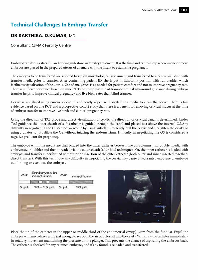
107Souvenir / Abstract Book
Dr karThika. D.kumar, mD
consultant, cImar Fertility centre
Embryo transfer is a stressful and exiting milestone in fertility treatment. It is the final and critical step wherein one or more embryos are placed in the prepared uterus of a female with the intent to establish a pregnancy.
The embryos to be transferred are selected based on morphological assessment and transferred to a centre well dish with transfer media prior to transfer. After confirming patient ID, she is put in lithotomy position with full bladder which facilitates visualisation of the uterus. Use of analgesics is as needed for patient comfort and not to improve pregnancy rate. There is sufficient evidence based on nine RCT’s to show that use of transabdominal ultrasound guidance during embryo transfer helps to improve clinical pregnancy and live birth rates than blind transfer.
Cervix is visualised using cuscus speculum and gently wiped with swab using media to clean the cervix. There is fair evidence based on one RCT and a prospective cohort study that there is a benefit to removing cervical mucus at the time of embryo transfer to improve live birth and clinical pregnancy rate.
Using the direction of TAS probe and direct visualisation of cervix, the direction of cervical canal is determined. Under TAS guidance the outer sheath of soft catheter is guided through the canal and placed just above the internal OS.Any difficulty in negotiating the OS can be overcome by using vulsellum to gently pull the cervix and straighten the cavity or using a dilator to just dilate the OS without injuring the endometrium. Difficulty in negotiating the OS is considered a negative predictor for pregnancy.
The embryos with little media are then loaded into the inner catheter between two air columns ( air bubble, media with embryo(s),air bubble) and then threaded via the outer sheath (after-load technique) . Or, the inner catheter is loaded with embryos and transfer is performed without prior insertion of the outer catheter (both outer and inner inserted together-direct transfer). With this technique any difficulty in negotiating the cervix may cause unwarranted exposure of embryos out for long or even lose the embryos.
Place the tip of the catheter in the upper or middle third of the endometrial cavity(1-2cm from the fundus). Expel the embryos with microlitre syring just enough to see both the air bubbles fall into the cavity. Withdraw the catheter immediately in rotatory movement maintaining the pressure on the plunger. This prevents the chance of aspirating the embryos back. The catheter is checked for any retained embryos, and if any found is reloaded and transferred.
Technical challenges in embryo Transfer
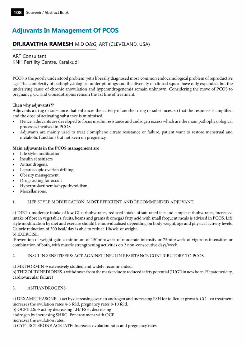
Souvenir / Abstract Book108
Dr.kaviTha ramesh m.D o&g, arT (cLeVeLaND, USa)
arT consultantKNH Fertility centre, Karaikudi
PCOS is the poorly understood problem, yet a liberally diagnosed most common endocrinological problem of reproductive age. The complexity of pathophysiological under pinnings and the diversity of clinical squeal have only expanded, but the underlying cause of chronic anovulation and hyperandrogenemia remain unknown. Considering the move of PCOS to pregnancy, CC and Gonadotropins remain the 1st line of treatment.
Then why adjuvants??Adjuvants a drug or substance that enhances the activity of another drug or substances, so that the response is amplified and the dose of activating substance is minimised.• Hence, adjuvants are developed to focus insulin resistance and androgen excess which are the main pathophysiological
processes involved in PCOS.• Adjuvants are mainly used to treat clomiphene citrate resistance or failure, patient want to restore menstrual and
metabolic functions but not keen on pregnancy.
Main adjuvants in the PCOS management are• Life style modification• Insulin sensitizers• Antiandrogens.• Laparoscopic ovarian drilling• Obesity management.• Drugs acting for occult• Hyperprolactinemia/hypothyroidism.• Miscellaneous. 1. LIFE STYLE MODIFICATION: MOST EFFICIENT AND RECOMMENDED ADJUVANT.
a) DIET→ moderate intake of low GI carbohydrates, reduced intake of saturated fats and simple carbohydrates, increased intake of fibre in vegetables, fruits, beans and grams & omega3 fatty acid with small frequent meals is advised in PCOS. Life style modification by diet and exercise should be individualised depending on body weight, age and physical activity levels. Calorie reduction of 500 kcal/ day is able to reduce 1lb/wk. of weight.b) EXERCISE: Prevention of weight gain a minimum of 150min/week of moderate intensity or 75min/week of vigorous intensities or combination of both, with muscle strengthening activities on 2 non-consecutive days/week.
2. INSULIN SENSITISERS: ACT AGAINST INSULIN RESISTANCE CONTRIBUTORY TO PCOS.
a) METFORMIN → extensively studied and widely recommended.b) THIZOLIDINEDIONES → withdrawn from the market due to reduced safety potential (IUGR in new born, Hepatotoxicity, cardiovascular failure)
3. ANTIANDROGENS
a) DEXAMETHASONE: → act by decreasing ovarian androgen and increasing FSH for follicular growth. CC – co treatment increases the ovulation rates 4-5 fold, pregnancy rates 8-10 fold.b) OCPILLS: → act by decreasing LH/ FSH, decreasingandrogen by increasing SHBG. Pre-treatment with OCPincreases the ovulation rates.c) CYPTROTERONE ACETATE: Increases ovulation rates and pregnancy rates.
adjuvants in management of pcos
109Souvenir / Abstract Book
4. LAPAROSCOPIC OVARIAN DRILLING
A monopolar needle approach, 20-30w, 5-10 punctures depending on size of ovaries. Benefitted are the 20% CC – resistant patients who are prone for multiple pregnancy and OHSS with the use of gonadotropins. Acts by reducing ovarian androgen production by granulosa and theca cells, by destructing them, thereby decreasing LH secretion. Also by reduction of inhibinproduction by granulosa cells, thereby FSH is increased and follicular growth is maximised.
5. OBESITY MANAGEMENT
a) ANTIOBESITY DRUGS – statins emerge as promising therapeutic agents in reducing androgen excess, decrease ovarian size and improve menstrual cyclicity by reducing proliferation and increasing apoptosis of ovarian theca cells and decreased androgen production by theca cells by decreased expression of CYPI7A, gene. Despite the promise, routine use in reproductive age is not yet recommended. Women of reproductive age, considering statins for clinical indications, should have concomitant contraception to reduce teratogenicity. b) BARIATRIC SURGERCY.Restores ovulation in morbidly obese women, who fail with conventional diet and exercise, and those with co- morbid metabolic abnormalities, also there is a reduction in pregnancy related complications, such as PIH, LBW, GDM, Preterm, JUGR and, Macrosomia. Patient should avoid pregnancy up to 1year of surgery. Weight loss benefits future pregnancies also. But because of its invasiveness, need of expertise, risk of mortality, large studies are needed before countering as a routine management.
6. DRUGS ACTING FOR OCCULT HYPERPROLACTINEMIA AND OCCULTHYPOTHYROIDISM REMAIN EXPERIMENTAL.
a) Bromocriptine / Cabergoline.b) Levothyroxine.7. MISCELLANCEOUS:a) Tamoxifenb) Vit D3c) Estradiol valarated) N-acetyl cysteine.e) Antioxidant decreasing Melatoninf) Galactomannang) Alternative medicine.i. Phytoestrogen.ii. Acupuncture.iii. Fenugreek seeds.
Decisions are made not only on RCTS but also on other basic scientific and clinical evidences supporting their use. Numerous adjuvants are known to be effective in PCOS – ovulation stimulation but most of them are approved for other uses but not specifically for as ovulation stimulation adjuvants. Because the risk benefit ratio is favourable, Physician should strongly recommenced in incorporating into ovulation stimulation protocols. Finally a valid therapeutic protocol for PCOS includes , ovulation stimulation with adjuvants like diet, exercise, insulin Sensitizing agents such as metformin and inositol, antiandrogen agents such as dexamethasone and oral contraception pills pre- treatment, with calcium VIT D3 supplementation and low sodium and laparoscopic ovarian drilling in needed cases remain satisfactory.
Souvenir / Abstract Book110
Dr nimmi nirmal
With the increased efficacy of medications and better quality of life among patients with chronic viral diseases more and more couples are coming forward to utilize facilities in fertility centres to help them conceive. Recruitment of persons affected with chronic viral diseases into an Assisted reproduction program still raises doubts into the minds of clinicians and embryologists due to the potential danger of cross- contamination thus putting into risk hospital personnel, patients and embryos. Many trials have recorded a decrease in the risk of transmission of viral infection to the partner in the case of a serodiscordant couple and also vertical transmission to the offspring.
The most common chronic viral infections seen in our country include HIV, HBV and HCV infections. Ideally when a couple with one or both infected partners are undergoing a procedure for assisted reproduction, that particular case is best separated from the rest of the batch in ‘time’ and ‘space’. By separation in space we mean that it be done in a separate “ infected “ laboratory, which is kept separate from the general embryology laboratory. However, in the current scenario the number of such cases may not warrant the construction of a separate facility and thus what we most probably do is to separate these cases ‘in time’, that is program them so that the oocyte retrieval comes just before the batch ends and no other cases are scheduled.
Special care has to be taken during the cryopreservation of the gametes and embryos as well. There have been case reports of HBV cross contamination across bone marrow samples which were stored together. Separate cryocans for the storage of infectious materials have to be arranged.
Universal safety precautions coupled with more efficient methods of handling the gametes combined with the use of antiviral medications holds great promise in reducing the potential risk of transmission to an uninfected partner, baby, staff members and disease-free gametes and embryos in the same laboratory.
management of seropositive cases in arT lab
111Souvenir / Abstract Book
Dr chiTra ThYaGaraJu
Chronic endometritis is a persistent inflammation of uterine endometrium. Histologically, the diagnosis of chronic endometritis is based on the presence of plasma cells in the endometrial stroma. In most of cases, the diagnosis of CE is an incidental finding based on histologic specimens obtained for various gynecologic indications (such as abnormal uterine bleeding or infertility). Chronic endometritis is often asymptomatic or accompanied by mild symptoms, which include pelvic pain, dysfunctional uterine bleeding, dyspareunia, and leukorrhea, Chronic endometritis may hamper endometrial receptivity and may cause infertility because the endometrium is characterized by an abnormal pattern of lymphocyte subsets and, consequently, an aberrant endometrial microenvironment. CE is diagnosed conventional HPE, Office hysteroscopy or by Culture. CE was identified in 30.3% of patients with repeated implantation failure at IVF and women diagnosed with CE had lower implantation rates (11.5%) after an IVF cycle. Indeed, untreated CE has been suggested to diminish the success rates of both spontaneous conception and IVF cycles, as well as to contribute to adverse obstetrical outcomes. Bacterial Vaginosis and Mycoplasma are the most frequent etiological agents for CE in women with RIF. in IVF cycles the presence of bacterial vaginosis may decrease conception rates, increase early pregnancy losses and may also increase the risk of preterm birth
chronic endometritis and reproductive outcome
Souvenir / Abstract Book112
Dr neeru Thakral
advanced Training reproductive endocrinology & arT germany.Diploma in advance gynae endoscopy cIce FranceDirector morpheus Thakral International IVF centerSecretary IFS Haryana chapter, Vice president Haryana endoscopy
In normally ovulating patients Ovarian stimulation is used to increase the number of follicles (Controlled ovarian Hyper stimulation) when planning procedures such as IUI, AID, IVF etc. or in cases of unexplained infertility.Ovarian stimulation may improve the results of IUI by Increasing the number of eggs available for fertilization, Overcoming subtle defects in ovulatory function and luteal phase, Controlling timing of ovulation and Prevention of premature LH surge.
Number of follicles pregnancies per cycle (%) details-One (5.7%), two (13.6%), Three (16.3%) & four or more (13.9%). COH has independent positive effect on pregnancy rate when combined with IUI Choice of ovulation stimulation protocol is individualized. Various drugs are used for this purpose. (A) CLOMIPHENE CITRATE (CC): Most widely usedMechanism of action: SERM- ANTI ESTROGEN CC is administered per day for 5 days starting from second to fifth day of cycle. The ant estrogenic effects of CC may be in the from of poor cervical mucous & poor endometrial thickness. So it better to administered gonadotropins along with low dose clomiphene rather than increasing the dose of clomiphene beyond 150mg per day. Clomiphene Resistance: No follicular development or improper follicle development leading to failure of ovulation.
(B) LETROZOLE:Dose: 2.5-mg/ day start cycle day 3-7, max 7.5 mg/day.Comparison with CC: High rate of monfolliculer, No direct antiestrogenic adverse effect on endometrium, shorter half-life & Lower serum E2. In a recent study conducted by Badawy et al, extended letrozole therapy (2.5mg daily from day 1 of menses for 10 days) was used for CC resistant PCOS women. Higher number of patients ovulated. Pregnancy rates were significantly greater.
(C) GONADOTROPINS:INDICATIONS: CC resistance / failure, Hypo gonadotropic hypogonadism, Pituitary dysfunction, COH and IUI in unexplained infertility & COH in IVF / ICSIADVANTAGE: High efficacy- Ovulation rate: >95 % per cycle & Conception rate : 20-30% per ovulatory cycle.COMPLICATIONS: multiple pregnancies & ovarian hyper stimulation syndrome. Require intensive monitoring.Step Up & Constant Dose was the type of regime we used up to now where dose is increased every 4-5 days according to ovarian response & serial E2 estimation. Once the desired response starts dose can be kept constant. Chronic Low Dose Step Up: Low initial daily FSH of 37.5-75IU and dose is increased by small amount usually 37.5IU/ day. The first increase in dose is done only after 14 daysStep Down Here FSH threshold can be reached relatively fast by rapid increase in dose & once ovarian response is achieved, gonadotropin dose should be reduced . Step Down Regimen Considered more physiological but higher incidence of multiple pregnancy and OHSS.Sequential Regimen: Combines an initial step up followed by a step down regimen after follicular selection (Leading follicle > 14mm).Commonest protocol; sequential CC/plus HMG regimen is as effective as Hmg regimen for ovulation induction, produces satisfactory pregnancy results and reduces the treatment cost.Cancellation (IUI): >4 follicles > 15mm irrespective of E2 level, 8 follicles > 10mm irrespective of E2 level & Estradiol >1500 pg/ ml.
Timing of Ovulation Trigger: 18-22 mm follicle size. If early or late trigger is given .it can fail to cause ovulation takes place, it will be premature or post mature egg hence embryo is not good quality
ovarian stimulation – Do’s and Don’ts
113Souvenir / Abstract Book
(D) GnRH ANALOGUES & GONADOTROPHINS:(1) Long Protocol: GnRH-a is given for 10-14 days. After two weeks when down regulation of the pituitary gland is achieved as confirmed by very low level of serum E2.The GnRHa is usually started in the mid luteal phase or early in the early follicular phase. Gonadotropins are started after down regulation(2) Short Protocol: GnRHa is administered on the first or second day of the cycle alone for 3 days is then continued in half the dose along with HMG. HMG is started on the 3rd or 4th day of the cycle. Here the stimulatory action of GnRHa is taken advantage of.This is used only in cases who have favourable LH levels. The flare effect increases the number of recruited follicles so is useful in poor responders & in patients of hypo gonadotrophic hypogonadism. (3) Ultra Short: Here GnRHa is given for only 3 days & stopped. Further supplementation is by HMG alone. Premature LH surge cannot be prevented in this regime.Indications of GnRHa : (a) PCOD cases where androgen or LH level are very high or when there is premature luteinization by gonadotropins repeatedly.(b) In IVF to prevent premature LH surge, to avoid cancellation of cycles as well as to time our aspirations.(E) GnRH ANTAGONISTS:
(1) Mechanism of Action: They act by competitive inhibition of GnRH receptors, which results in rapid decline in FSH / LH levels thus preventing premature LH surge. The drug can be given in a single dose or daily dose regimen.
Protocol: The 2 protocols for administering are –(a) Lubeck protocol: Gonadotropins are started as usual and antagonist is started when the follicle reaches a size of 14 mm, or from 6th day of stimulation onwards in a dose of 0.25mg / day till the day of HCG injection.(b) French protocol: Gonadotropins are started as usual and a single dose (3 mg) of antagonist is given when serum E2 level is about 150-200 pg / ml and follicular size is 14 mm.
ADVANTAGES: When compared to agonist it is relatively simple & inexpensive. There is no suppression of oestrogen and the effects are easily reversible and associated with lower rates of OHSS.
PROTOCOLS FOR ART:Choosing Stimulation Protocol in ART is an: ROPE WALK.On one side Recruite & grow enough number of oocytesOn the other side: Prevent OHSS .Aim of stimulation protocol:Develop 8 to 12 oocytes. approximately 70% are metaphase II 70% of metaphase II oocytes develop to become embryos(Thus 3 to 5 healthy embryos will be available for ET)Important aspects in ART protocol: Starting dose of gonadotropins, monitoring the ovarian response, controlling the LH surge, Dose and time of hCG Luteal support.
(A) Estimate of starting dose of FSH:
FSH dose calculation: Age & BMI 20 to 25yrs II ampoules (150 I.U)26 to 30yrs III ampoules (225 I.U)31 to 35yrs IV ampoules (300 I.U)36 to 40yrs V ampoules (375 I.U)(This table is for a lady with BMI 22-25)Add one ampoule for BMI>28Less one ampoule for BMI<22Add an extra ampoule for patients with H/O endometriosis, surgery done for fibroids, pelvic adhesions with discretion.
(B) Monitoring ovarian response: TVS: No. & size of follicles & Endometrial thickness and pattern.8 Estradiol (E2) blood level.( d4) E2: 150-200pg/ml continue same dose E2: 200-500pg/ml reduces one ampoule
Souvenir / Abstract Book114
E2>500pg/ml reduce two ampoules Caution: Caution is indicated when any of the following indicators for increasing risk of OHSS are present: Rapidly rising serum E2 levels,E2 concentration in excess of 2,500 pg/mL The emergence of a large number of intermediate sized follicles (10–14 mm).
Risk Factors: Young age, Low body weight, polycystic ovary syndrome (PCOS),Higher doses of exogenous gonadotropins, High absolute or rapidly rising serum E2 levelsPrevious episodes of OHSS.
COASTING: Withholding further gonadotropin stimulation and delaying hCG administration until E2 levels plateau or decrease significantly can reduce risks of OHSS. Available evidence suggests that such ‘‘coasting’’ does not adversely affect out- come in IVF cycles unless it is prolonged (>2 days).
(C) DOSE OF hCG: Given evidences suggest that hCG may play a pivotal role in the development of OHSS. A lower dose of hCG (e.g., 5,000 IU vs. the standard 10,000 IU dosage) may be prudent for patients judged to be at high risk for OHSS.• AllDownregulationprotocols10,000I.UhCGgivenwhenat-leasttwoleadingfolliclesreach18mmsize.• With(E2)Levelsupto2500pg/ml:7500I.UhCGgiven• When(E2)levelsbetween2500-3000pg/ml:5000I.UhCGgiven• When(E2)levels3000-3500pg/mlAvoidhCG&cancelcycle• IfOHSSisanticipated:AGnRHagonist(e.g.,leuprolide1.0–2.0mgSC)ratherthanhCGmightbeusedtostimulatean endogenous LH surge to promote final oocyte maturation and induce ovulation. This approach would be useful only in Antagonist cycleAnticipating OHSS:• ChoosinganappropriateprotocolandstrictregularmonitoringusingE2andUSG• Coasting• LessdoseofhCG• NottogivehCG/InsteadgiveGnRhagonist• Oralcabergoline,0.5mgdaily,wasgivenasaninterventionfromdayofOPU.• Cryopreserveembryos&transfernextcycle• UseofPandnothCGaslutealsupport• Cancelcycle.ULTRA LONG PROTOCOL: GnRH could be given for three months before the start of Hmg. May be used in patients with severe endometriosis before the start of the treatment. MILD STIMULATION PROTOCOL:Definition – FSH or HMG is administered at a lower dose (100-150) and oral compounds (CC or Letrozole) are used either alone or in combination with gonadotropins, aim of collecting fewer oocytes, Antagonist are used for pituitary suppression.
(a) Advantages – lower doses of gonadotropins and association with better embryo quality; lower per-cycle dropout rates.(b) Disadvantages – Higher per cycle cancellation rate; may required multiple stimulated cycles to achieve a pregnancy; few embryos available for cryopreservation; increased cumulative costs associated with multiple fresh cycles.
115Souvenir / Abstract Book
Dr m suJaTha suresh mSc., pH.D
Scientific head/Senior embryologistVamsam Fertility Research CentreCoimbatore
Vitrification as a cryopreservation technique involves rapid freezing at ultra high cooling rates so that a glassy state is achieved without ice formation. With the discovery and usage of cryoprotectants along with liquid nitrogen and different cell carrier devices, successful vitrification techniques have been applied to different species of embryos and a few other cell lines. Vitrification of human embryos is now done routinely in all ART clinics.
Vitrification has 2 major advantages over slow freezing. They are 1. The techniques and methods are quick and easy to learn do not require the use of programmed freezers. This makes
vitrification cost effective.2. The use of non-permeating cryoprotectants in vitrification protects the cell from the toxicity of permeating
cryoprotectants such as osmotic shock, lipid membrane damage and DNA damage.
Human sperm vitrification was first reported by Nawroth and Isachenko et al Vitrification has been explored by several groups as an alternative method for freezing human spermatozoa . Vitrification of oocytes and embryos and other tissues requires rapid cooling rates and high CPA concentrations. Spermatozoa have low tolerance levels for high concentration of cryoprotectants (CPA) with these agents causing possible lethal osmotic effects and chemical alterations in spermatozoa. Vitrification without the use of conventional CPA has therefore been described for human spermatozoa.
Different non permeating sugars have been successfully used in the vitrification protocol. This is based on the principle that human sperm cell contain an intracellular milieu that is high in proteins/sugars and low water, thus allowing vitrification to occur at lower temperatures in the absence of permeating cryoprotectants. The use of sucrose as a non permeating cryoprotectant in sperm vitrification was associated with good rates of sperm motility, vitality and mitochondrial membrane potential. The combination of sucrose and albumin in the cryoprotectant solution have equivalent results in terms of sperm capacitation, acrosome reaction and mitochondrial membrane potential when compared with conventional slow freezing of sperm.
In terms of methodology of vitrification a simple micro capillary technique of sperm vitrification has been reported. Different techniques that involve use of carrier devices like cryo loop ,cryotubes,grid, and cell sleeper have also been recently reported. Isachenkos group suggested directly placing 30ul droplet of semen mixed cryoprotectant into liquid nitrogen. Although this technique is feasible and no reported case of viral cross contamination has been reported, the development and use of open pulled straws(OPS) and cut stranded straws have further refined the techniques with aseptic vitrification
Moskovtsev group also reported excellent recovery in terms of sperm kinetic parameters and DNA fragmentation in a study on infertile men, where they compared slow freezing vs vitrification of sperm. An interesting finding in this study though, was that although sperm kinetic parameters were found to be better, sperm DNA fragmentation index did not differ between the two groups. Of clinical importance the report of live birth of healthy twins after ICSI with vitrified sperm .Sanchez etal also reported the live birth of after IUI with sperm vitrified without cryoprotectant in a patient with oligoasthenospermia, thereby making vitrification feasible for day to day clinical practice
Vitrification of spermatozoa provides simpler, faster, more cost effective alternative to conventional cryopreservation methods.
novel sperm vitrification Techniques
Souvenir / Abstract Book116
free comunicaTionsoraL/VIDeo preSeNTaTIoNS
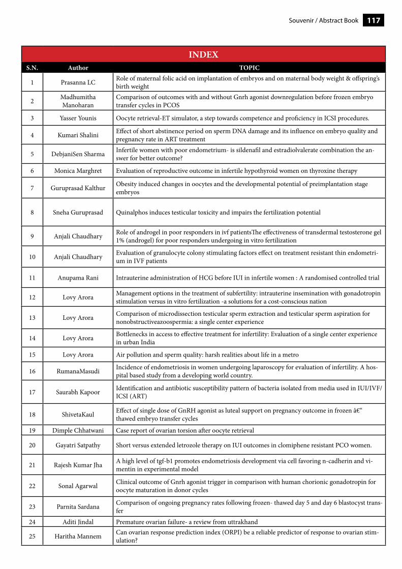
117Souvenir / Abstract Book
INDEXS.N. Author TOPIC
1 Prasanna LC Role of maternal folic acid on implantation of embryos and on maternal body weight & offspring’s birth weight
2 MadhumithaManoharan
Comparison of outcomes with and without Gnrh agonist downregulation before frozen embryo transfer cycles in PCOS
3 Yasser Younis Oocyte retrieval-ET simulator, a step towards competence and proficiency in ICSI procedures.
4 Kumari Shalini Effect of short abstinence period on sperm DNA damage and its influence on embryo quality and pregnancy rate in ART treatment
5 DebjaniSen Sharma Infertile women with poor endometrium- is sildenafil and estradiolvalerate combination the an-swer for better outcome?
6 Monica Marghret Evaluation of reproductive outcome in infertile hypothyroid women on thyroxine therapy
7 Guruprasad Kalthur Obesity induced changes in oocytes and the developmental potential of preimplantation stage embryos
8 Sneha Guruprasad Quinalphos induces testicular toxicity and impairs the fertilization potential
9 Anjali Chaudhary Role of androgel in poor responders in ivf patientsThe effectiveness of transdermal testosterone gel 1% (androgel) for poor responders undergoing in vitro fertilization
10 Anjali Chaudhary Evaluation of granulocyte colony stimulating factors effect on treatment resistant thin endometri-um in IVF patients
11 Anupama Rani Intrauterine administration of HCG before IUI in infertile women : A randomised controlled trial
12 Lovy Arora Management options in the treatment of subfertility: intrauterine insemination with gonadotropin stimulation versus in vitro fertilization -a solutions for a cost-conscious nation
13 Lovy Arora Comparison of microdissection testicular sperm extraction and testicular sperm aspiration for nonobstructiveazoospermia: a single center experience
14 Lovy Arora Bottlenecks in access to effective treatment for infertility: Evaluation of a single center experience in urban India
15 Lovy Arora Air pollution and sperm quality: harsh realities about life in a metro
16 RumanaMasudi Incidence of endometriosis in women undergoing laparoscopy for evaluation of infertility. A hos-pital based study from a developing world country.
17 Saurabh Kapoor Identification and antibiotic susceptibility pattern of bacteria isolated from media used in IUI/IVF/ICSI (ART)
18 ShivetaKaul Effect of single dose of GnRH agonist as luteal support on pregnancy outcome in frozen – thawed embryo transfer cycles
19 Dimple Chhatwani Case report of ovarian torsion after oocyte retrieval
20 Gayatri Satpathy Short versus extended letrozole therapy on IUI outcomes in clomiphene resistant PCO women.
21 Rajesh Kumar Jha A high level of tgf-b1 promotes endometriosis development via cell favoring n-cadherin and vi-mentin in experimental model
22 Sonal Agarwal Clinical outcome of Gnrh agonist trigger in comparison with human chorionic gonadotropin for oocyte maturation in donor cycles
23 Parnita Sardana Comparison of ongoing pregnancy rates following frozen- thawed day 5 and day 6 blastocyst trans-fer
24 Aditi Jindal Premature ovarian failure- a review from uttrakhand
25 Haritha Mannem Can ovarian response prediction index (ORPI) be a reliable predictor of response to ovarian stim-ulation?
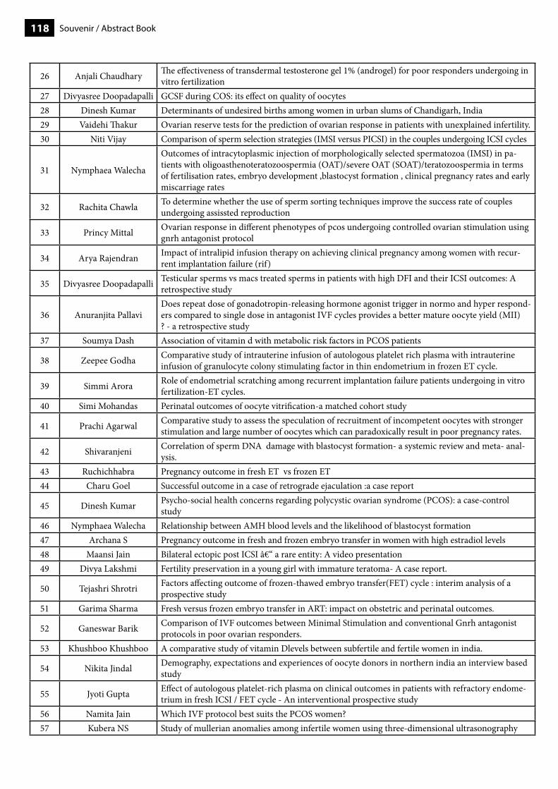
Souvenir / Abstract Book118
26 Anjali Chaudhary The effectiveness of transdermal testosterone gel 1% (androgel) for poor responders undergoing in vitro fertilization
27 Divyasree Doopadapalli GCSF during COS: its effect on quality of oocytes28 Dinesh Kumar Determinants of undesired births among women in urban slums of Chandigarh, India29 Vaidehi Thakur Ovarian reserve tests for the prediction of ovarian response in patients with unexplained infertility.30 Niti Vijay Comparison of sperm selection strategies (IMSI versus PICSI) in the couples undergoing ICSI cycles
31 Nymphaea Walecha
Outcomes of intracytoplasmic injection of morphologically selected spermatozoa (IMSI) in pa-tients with oligoasthenoteratozoospermia (OAT)/severe OAT (SOAT)/teratozoospermia in terms of fertilisation rates, embryo development ,blastocyst formation , clinical pregnancy rates and early miscarriage rates
32 Rachita Chawla To determine whether the use of sperm sorting techniques improve the success rate of couples undergoing assissted reproduction
33 Princy Mittal Ovarian response in different phenotypes of pcos undergoing controlled ovarian stimulation using gnrh antagonist protocol
34 Arya Rajendran Impact of intralipid infusion therapy on achieving clinical pregnancy among women with recur-rent implantation failure (rif)
35 Divyasree Doopadapalli Testicular sperms vs macs treated sperms in patients with high DFI and their ICSI outcomes: A retrospective study
36 Anuranjita PallaviDoes repeat dose of gonadotropin-releasing hormone agonist trigger in normo and hyper respond-ers compared to single dose in antagonist IVF cycles provides a better mature oocyte yield (MII) ? - a retrospective study
37 Soumya Dash Association of vitamin d with metabolic risk factors in PCOS patients
38 Zeepee Godha Comparative study of intrauterine infusion of autologous platelet rich plasma with intrauterine infusion of granulocyte colony stimulating factor in thin endometrium in frozen ET cycle.
39 Simmi Arora Role of endometrial scratching among recurrent implantation failure patients undergoing in vitro fertilization-ET cycles.
40 Simi Mohandas Perinatal outcomes of oocyte vitrification-a matched cohort study
41 Prachi Agarwal Comparative study to assess the speculation of recruitment of incompetent oocytes with stronger stimulation and large number of oocytes which can paradoxically result in poor pregnancy rates.
42 Shivaranjeni Correlation of sperm DNA damage with blastocyst formation- a systemic review and meta- anal-ysis.
43 Ruchichhabra Pregnancy outcome in fresh ET vs frozen ET44 Charu Goel Successful outcome in a case of retrograde ejaculation :a case report
45 Dinesh Kumar Psycho-social health concerns regarding polycystic ovarian syndrome (PCOS): a case-control study
46 Nymphaea Walecha Relationship between AMH blood levels and the likelihood of blastocyst formation47 Archana S Pregnancy outcome in fresh and frozen embryo transfer in women with high estradiol levels48 Maansi Jain Bilateral ectopic post ICSI – a rare entity: A video presentation49 Divya Lakshmi Fertility preservation in a young girl with immature teratoma- A case report.
50 Tejashri Shrotri Factors affecting outcome of frozen-thawed embryo transfer(FET) cycle : interim analysis of a prospective study
51 Garima Sharma Fresh versus frozen embryo transfer in ART: impact on obstetric and perinatal outcomes.
52 Ganeswar Barik Comparison of IVF outcomes between Minimal Stimulation and conventional Gnrh antagonist protocols in poor ovarian responders.
53 Khushboo Khushboo A comparative study of vitamin Dlevels between subfertile and fertile women in india.
54 Nikita Jindal Demography, expectations and experiences of oocyte donors in northern india an interview based study
55 Jyoti Gupta Effect of autologous platelet-rich plasma on clinical outcomes in patients with refractory endome-trium in fresh ICSI / FET cycle - An interventional prospective study
56 Namita Jain Which IVF protocol best suits the PCOS women?57 Kubera NS Study of mullerian anomalies among infertile women using three-dimensional ultrasonography
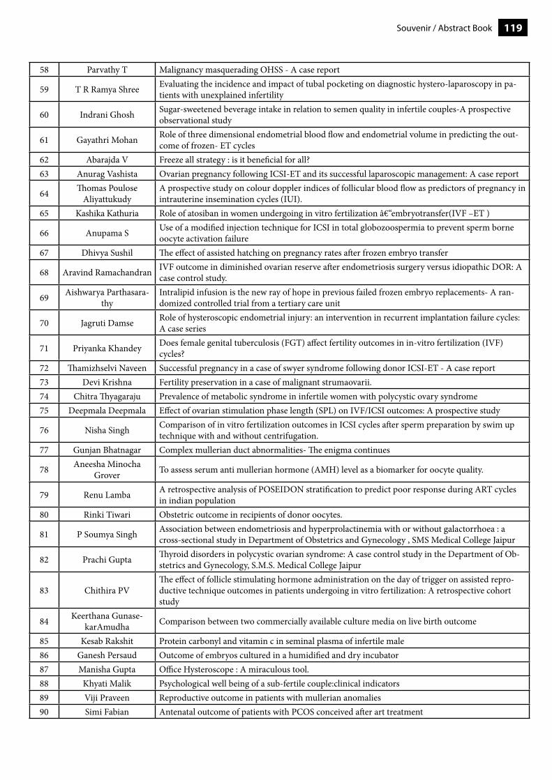
119Souvenir / Abstract Book
58 Parvathy T Malignancy masquerading OHSS - A case report
59 T R Ramya Shree Evaluating the incidence and impact of tubal pocketing on diagnostic hystero-laparoscopy in pa-tients with unexplained infertility
60 Indrani Ghosh Sugar-sweetened beverage intake in relation to semen quality in infertile couples-A prospective observational study
61 Gayathri Mohan Role of three dimensional endometrial blood flow and endometrial volume in predicting the out-come of frozen- ET cycles
62 Abarajda V Freeze all strategy : is it beneficial for all?63 Anurag Vashista Ovarian pregnancy following ICSI-ET and its successful laparoscopic management: A case report
64 Thomas PouloseAliyattukudy
A prospective study on colour doppler indices of follicular blood flow as predictors of pregnancy in intrauterine insemination cycles (IUI).
65 Kashika Kathuria Role of atosiban in women undergoing in vitro fertilization –embryotransfer(IVF –ET )
66 Anupama S Use of a modified injection technique for ICSI in total globozoospermia to prevent sperm borne oocyte activation failure
67 Dhivya Sushil The effect of assisted hatching on pregnancy rates after frozen embryo transfer
68 Aravind Ramachandran IVF outcome in diminished ovarian reserve after endometriosis surgery versus idiopathic DOR: A case control study.
69 Aishwarya Parthasara-thy
Intralipid infusion is the new ray of hope in previous failed frozen embryo replacements- A ran-domized controlled trial from a tertiary care unit
70 Jagruti Damse Role of hysteroscopic endometrial injury: an intervention in recurrent implantation failure cycles: A case series
71 Priyanka Khandey Does female genital tuberculosis (FGT) affect fertility outcomes in in-vitro fertilization (IVF) cycles?
72 Thamizhselvi Naveen Successful pregnancy in a case of swyer syndrome following donor ICSI-ET - A case report73 Devi Krishna Fertility preservation in a case of malignant strumaovarii.74 Chitra Thyagaraju Prevalence of metabolic syndrome in infertile women with polycystic ovary syndrome75 Deepmala Deepmala Effect of ovarian stimulation phase length (SPL) on IVF/ICSI outcomes: A prospective study
76 Nisha Singh Comparison of in vitro fertilization outcomes in ICSI cycles after sperm preparation by swim up technique with and without centrifugation.
77 Gunjan Bhatnagar Complex mullerian duct abnormalities- The enigma continues
78 Aneesha Minocha Grover To assess serum anti mullerian hormone (AMH) level as a biomarker for oocyte quality.
79 Renu Lamba A retrospective analysis of POSEIDON stratification to predict poor response during ART cycles in indian population
80 Rinki Tiwari Obstetric outcome in recipients of donor oocytes.
81 P Soumya Singh Association between endometriosis and hyperprolactinemia with or without galactorrhoea : a cross-sectional study in Department of Obstetrics and Gynecology , SMS Medical College Jaipur
82 Prachi Gupta Thyroid disorders in polycystic ovarian syndrome: A case control study in the Department of Ob-stetrics and Gynecology, S.M.S. Medical College Jaipur
83 Chithira PVThe effect of follicle stimulating hormone administration on the day of trigger on assisted repro-ductive technique outcomes in patients undergoing in vitro fertilization: A retrospective cohort study
84 Keerthana Gunase-karAmudha Comparison between two commercially available culture media on live birth outcome
85 Kesab Rakshit Protein carbonyl and vitamin c in seminal plasma of infertile male86 Ganesh Persaud Outcome of embryos cultured in a humidified and dry incubator87 Manisha Gupta Office Hysteroscope : A miraculous tool.88 Khyati Malik Psychological well being of a sub-fertile couple:clinical indicators89 Viji Praveen Reproductive outcome in patients with mullerian anomalies90 Simi Fabian Antenatal outcome of patients with PCOS conceived after art treatment
Souvenir / Abstract Book120
1. Role of maternal folic acid on implantation of embryos and on maternal body weight & offspring’s birth weightPrasanna LCKasturba Medical College, Manipal
Introduction: Maternal deficiency of FA or excess of FA would likely to affect the developing fetus at critical stages of development to cause long-lasting health consequences in their offspring. The present study aimed to know the effect of folic acid supplementation and folic acid absence diet during pregnancy on the number of implantation, endometrial changes at the implantation site and on maternal body weight & offspring’s birth weight.
Material & Methods: Thirty-Six Wistar strained female rats were randomly divided into two groups with 18 rats in each group. Further, each group has been divided into three sets with 6 rats in each. The control set was fed a normal diet, the second set was fed a diet containing no FA. 1% Succinyl sulfathiazole was added to the diet with no FA content to reduce/ inhibit the gut flora responsible for the synthesis of FA in their body. The third set was given diet with an excess of FA (40mg/kg body weight/day). Rats were given respective diet for 4 weeks before mating, continued throughout their pregnancy and 5 weeks after the delivery of pups (weening period). All female rats were paired with males of their own strain overnight at room temperature (25°C). To check whether they mated or not, the vaginal smear was taken in the early morning and stained with methylene blue and confirmed the presence of sperm plug under microscopy. On confirmation of sperm plug, the gestational day was termed as day 1 (GD1). Rats have remained in their cage and diets until they were sacrificed during gestation from day GD6-GD9. Delivered pups were counted for each rat and gross malformations noted & weighed individually in the digital weigh machine.
Results: Average number of pups born to pregnant dams fed with FA supplementation, FA absent diet, and in normal diet was found to be 8,8, and 9 respectively. Regarding the birth weight of pups born to pregnant dams fed with FA supplementation the average weight was found to be 6.01-5.09 gm, with normal diet it was 5.51-5.08 gm, and least weight was observed in pups born with FA absent diet (4.74-3.31 gm). Pair-wise comparison showed significant difference when compared with pups born to FA supplementation diet to pups born to FA absent diet. Also, significant differences were noted when compared pups born to FA absent diet mother with pups born to FA supplementation and with pups born to normal diet.
Conclusion: Our study provides clear evidence that FA is essential in successful implantation by providing favorable receptive environment to receive the implantation-competent blastocyst for a successful pregnancy. Birth weight of pups born to pregnant dams fed with FA supplementation was found to be significantly increased when compared to those with the diet with normal amount of FA, and least weight was observed in pups born with FA absent diet. Further research from longitudinal studies is warranted to confirm these results before its recommendation to the public health.
2. Comparison of outcomes with and without GnRH agonist downregulation before frozen embryo transfer cycles in PCOSMadhumitha Manoharan, Arun Karthik PonnusamyChandra Nursing home, Namakkal
Introduction : PCOS is the most common cause of anovulation in the reproductive age group. Incidence amongreproductive age women ranges between 5-10% worldwide.Among the PCOS women undergoingIVF, frozen thawed embryo transfer is most often opted for in order to reduce the risk of OHSS.Because of hyperandrogenism the endometrial function is compromised in these women. Use ofGnRH agonist for pituitary downregulation may help to improve the endometrial function in suchcases.
Aim : To determine if GnRH agonist given before the frozen thawed embryo transfer cycles would improvethe clinical pregnancy rate, ongoing pregnancy rate and miscarriage rate in PCOS women.
Materials and Methods : Retrospective study conducted from December 2016 to November 2017.Group A-Women who were given GnRH agonist before HRT FET cycleGroup B-Women who were not given GnRH agonist before HRT FET cycle
Results: The two groups of women were comparable in terms of age, BMI, duration of infertility, ovarianreserve, basal FSH and LH and associated male factor.
The clinical pregnancy rate was 66.6% in the GnRH agonist downregulated group compared to 57.6%in the other group,
121Souvenir / Abstract Book
though not statistically significant. The ongoing pregnancy rates weresignificantly increased in the former group. There was a statistically significant decrease in themiscarriage rate in the group that were given GnRH agonist for downregulation as compared to thegroup that were not downregulated – 6% versus 14%.
Conclusion : The decrease in androgens in GnRH agonist downregulated FET cycles in PCOS women might helpdecrease the miscarriage rates and hence as a result improve the ongoing pregnancy rates.
3. Oocyte retrieval-ET simulator, a step towards competence and proficiency in ICSI procedures.yasser younisLotus Fertility Centre, Talkha
Oocyte retrieval (OR) and Embryo transfer (ET) are mainly operator-dependent and requires training to be performed successfully. Acquisition of oocytes is the first step towards successful outcomes in an ART program and is considered to be easily mastered. However, the number of procedures required for a trainee to learn the procedure and reach proficiency is not well defined. To our knowledge little data exist about minimum number of retrievals physicians should perform under direct supervision prior to independent practice.
ET is a critical step in the overall success of (ICSI). A successful ET should deliver the embryos atraumatically to the point in the endometrial lining where implantation is most likely to occur. Despite its apparent simplicity, it is an integral part of ICSI cycle that can be difficult to teach and perform well.Standard practice is currently to perform a recommended number of procedures under supervision till the trainee acquires proficiency,
Few ICSI procedures training protocols have been reported in scientific literature. Moreover, commonly used training schemes are not tailored to the trainee and do not allow for individualized assessment of proficiency. Moreover, the difficulties encountered by clinics and hospitals to teach ICSI procedures and to operate on real patients, opens ethical issues of great relevance both from the legal and practical point of view.
As a training tool, simulation engages learners and allows for deliberate practice and allows trainees to experience learning in an immersive environment. Simulation allows educators to control the environment and ensure desired learning objectives are met while permitting increased trainee autonomy and provides a safe environment to practice and make mistakes without jeopardizing patient care.Oocyte retrieval-ET simulator might be a crucial step towards the creation of true training schools in all ART procedures.
4. Effect of short abstinence period on sperm DNA damage and its influence on embryo quality and pregnancy rate in ART treatmentKumari Shalini , Dayanidhi Kumar , Rakesh Kumar, Ajit Kumar, Nitiz Murdia, Kshitiz Murdia, Ajay MurdiaIndira IVF,Patna
Background: Some studies have shown decrease in sperm DNA fragmentation in patients with shorter abstinence period. However, the effect of shorter abstinence period on embryo quality and implantation rate derived from the sibling oocytes in not known.
Aims & Objective: To find out the effect of short ejaculatory abstinence period on sperm DNA damage and its influence of embryo quality derived from sibling oocytes in human.
Methods & Materials: This is a prospective study. This study includes 50 subjects who underwent infertility treatment at Indira IVF. The semen samples were collected at 1 day and 60 minutes of sexual abstinence period respectively from male partner. The semen analysis and DNA fragmentation index (DFI) was performed in both the samples immediately after liquefaction. At the same time sibling oocytes were divided in to two groups and ICSI was performed by using these spermatozoa. The fertilization rate, cleavage rate and blastocyst rate and pregnancy rate was observed from the embryos derived from these spermatozoa. One way analysis of variance (ANOVA) was used to compare the group.
Results: We did not find any significant difference in the sperm characteristics in both the groups. However, DFI was significantly reduced in 60 minutes abstinence group compared to 1 day. In addition, fertilization rate and cleavage rate
Souvenir / Abstract Book122
was not affected in both the groups. In contrast, we observe higher blastocyst rate and number of expanded blastocyst in the 60 min abstinence group compared to 1 day. Interestingly, we have also observed higher implantation rate in the 60 min group. However, the take home baby rate is yet to analyze.Conclusion: This study clearly showed that sperm DNA fragmentation can be decreased in 60 min abstinence period. In addition, shorter abstinence period of 60 min also improves blastocyst and implantation rate in the infertile couple. This study provides first evidence on effects of shorter abstinence period on sperm DNA fragmentation, embryo quality and pregnancy rate in ART treatment.
5. Infertile women with poor endometrium- Is sildenafil and estradiol valerate combination the answer for better outcome?Debjani Sen SharmaShristi Health Care, Bhangapool
Introduction: Thin endometrium is a challenging entity in the treatment of infertility for the clinicians. Successful implantation requires good embryo quality, appropriate timed and arranged endometrial receptivity and efficient crosslink between embryo and receptive endometrium. Impairment of any of these factors results in implantation failure.
Aim: The objective of this study is to evaluate the effect of oral sildenafil + estradiol valerate combination on endometrial thickness , blood flow, implantation rate and pregnancy rate in comparison to estradiol valerate alone in poor endometrium in stimulated cycles in infertile women .
Material And Method: It is a prospective comparative study including 20 women with primary or secondary infertility who had antecedent poor endometrial response. Inclusion criteria:patient with ovulatory infertility,age 25- 40yrs, BMI < 30kg/m2. Exclusion criteria: women with congenital or acquired uterine anomaly, tubal and male factor infertility,contraindication for estrogen use.Patients were randomised into 2 groups. Gr A( n=10) received Sildenafil+Estradiol valerate combination twice daily from D2 till the day of HCG trigger. Gr B (n = 10) received Estradiol valerate 2mg 8hrly from D8 of mens till the day of HCG trigger. Ovulation induction is done with tab Letrozole(5mg) from D2 to D6 of mens or gonadotrophin depending on the age, hormonal status and previous cycle response. Patients were evaluated by TVS on D14 for endometrial thickness and pattern, number and size of dominant follicle, Pulsatility Index(PI) of endometrium.Mean endometrial thicknessat the time of HCG trigger was 9.8 ± 2.3mm in Gr A Vs 7.8 ± 1.6 mm in Gr B ( p < 0.05). 85% of patient with Sildenafil had vascularity upto zone 3 and decrease in pulsatility index compared to 55% with Estradiol valerate. Implantation rate was 26% in Gr A Vs 7% in Gr B. Clinical pregnancy rate was 32.1% in Gr A Vs 15.9% in Gr B ( P< 0.015).
Conclusion: A receptive endometrium plays a critical role in embryo implantation and adequate endometrial growth is essential to this process. Sildenafil when compared to Estradiol valerate has a better outcome in terms of uterine receptivity, endometrial vascularity, marginally increase pregnancy outcome.
6. Evaluation of reproductive outcome in infertile hypothyroid women on thyroxine therapyMonica Marghret, Leena WadhwaESI PGIMSR and Model Hospital , Basaidarapur, New Delhi
Introduction: Hypothyroidism in women of reproductive age group is associated with increased risk of infertility. According to American thyroid association 2017 evaluation of serum Tsh is recommended for women seeking treatment for infertility. It recommends thyroxine treatment for infertile women with clinical and subclinical hypothyroidism to achieve a Tsh concentration <2.5 Iu/ml.
Material and Methods: We examined 46 female infertile women with hypothyroidism and effects of thyroxine therapy on pregnancy rates and outcomes were observed for a period of 18 months.
Results : 24 women conceived successfully during treatment(group A) of which 3 had miscarriage afterward. The remaining 22 patients did not conceive (group B). The median Tsh in group A before thyroxine treatment was 6.82+/-2.83 and this reduced to 3.2 +/- 1.36 during the treatment.The estimated duration of infertility before thyroxine treatment 4.2 +/- 1.3 yrs and infertile period after thyroxine treatment was 1.8 +/- 0.6 yrs. Administered thyroxine dose was 44+/- 13.82ug .
123Souvenir / Abstract Book
Conclusion: Increase in pregnancy rates and shorter duration of infertility until pregnancy after thyroxine treatment suggests that thyroxine might enhance fertility in infertile hypothyroid women.
7. Obesity induced changes in oocytes and the developmental potential of preimplantation stage embryosGuruprasad Kalthur, Aparna Satheesh , Guruprasad Nayak, Pooja SP, Satish AdigaKasturba Medical College, Manipal Academy of Higher Education, Manipal
Introduction: Obesity is a major health issue globally, associated with various pathologies including cardiovascular, musculoskeletal disorders, hypertension, certain cancers and infertility. Even though adipose tissue is required for proper reproductive functioning, excessive adipose tissue accumulation can cause perturbation in regulation of hypothalamo-pituitary-gonadal axis. These changes in the endocrine levels due to obesity can have adverse effects on follicular microenvironment and oocyte development.
Aim : The present study is planned to assess the cytoplasmic changes in the oocytes and to correlate it with the developmental potential of embryos using murine model.
Material and Method: Swiss albino female mice of 3 weeks old were fed with normal diet (control) and high fat diet (HFD) for 8 weeks. After 8 weeks, these mice were assesed for estrous cycling by vaginal cytology, seerum endocrine profiling and biochemical analysis. The ovaries were collected and germinal vescicle oocytes were subjected to in vitro maturation (IVM). The intracellular ROS level (DCHFDA staining), lipid droplet accumulation (nile red staining), XBP1 expression for ER stress were assessed in oocytes. To determine the effect of high fat diet in embryos, in vitro fertilization was performed and embryos were cultured till blastocyst stage. The fertilization rate, blastocyst rate, DNA intergrity in blastocyst (TUNEL assay) were assessed.
Results : The HFD fed femlae mice showed significant increase in body weight (p<0.0001) and abdominal fat accumulation (p<0.01). The percentage of mice showing normal estrous cycling was significantly reduced in the HFD group (p<0.0001), along with significant decrease in ovarian weight (p<0.0001) as compared control. Multiple cystic follicles, increased number of atritic follicles and decresed number of follicle reserve was observed in ovaries of mice fed with HFD. In addition, lower in vitro maturation potential of oocytes (39.09% v/s 63.75% , p<0.01) and high lipid accumulation was was observed in HFD group (p<0.05). Increased intracellular ROS levels (p<0.01) and XBP1 expression indicated that oocytes from HFD group have high intracellular oxidative stress and ER stress. Further, a significant decrease in fertilization rate (Control: 80.64%; HFD: 57.45% , p<0.001) and blastocyst rate (Control: 70%; HFD; 29.16%, p <0.0001), low total cell number and high DNA damage was observed compared to control group.
Conclusion: The diet induced obesity resulted in elevated levels of lipid accumulation, oxidative and endoplasmic reticulum stress which might have contributed to decreased in vitro maturation potential, poor fertilization and embryonic developmental potential and compromised DNA integrity in blastocyst.
8. Quinalphos induces testicular toxicity and impairs the fertilization potentialSneha Guruprasad, Sandhya Kumari, Sahil Sanghvi, Satish Adiga , Guruprasad KalthurKasturba Medical College Manipal Academy of Higher Education, Manipal
Introduction: Quinalphos is one of the widely used insecticide in India which falls under organophosphate group of pesticides. It is commonly preferred pesticide in cultivation of paddy, mustard and wheat etc. It is highly neurotoxic which inhibits acetylcholinesterase and known to cause neuronal dysfunction. However, there is no much information on its reproductive toxicity.
Aim : Therefore, this study was undertaken to investigate the effect of quinalphos induced testicular toxicity.
Material and methods : For the study, 8 weeks old male Swiss albino mice were orally administered with 0.25, 0.5 and 1 mg /kg body weight of commercially available quinalphos (25%) for 10 days and were observed till 35 days. The mice were dissected to assess the testicular function and epididymal sperm parameters.
Results : At none of the doses used in the study, quinalphos induced any significant effect on the sperm count. However, the
Souvenir / Abstract Book124
motility was significantly lower (p<0.01) and sperm head abnormalities were significantly higher (p<0.01) in quinalphos treated mice indicating that the quality of spermatozoa produced are poor. In addition, an elevated level of DNA damage was observed in the testicular cells of these mice. The pharmacokinetic studies revealed that a high tissue distribution and slow clearance was observed in testicular tissue.
9. Role of androgel in poor responders in IVF patients
The effectiveness of transdermal testosterone gel 1% (androgel) for poor responders undergoing in vitro fertilizationAnjali Chaudhary, Amrit Swain , Umesh Varma Aarogya Hospital, Delhi.
Objective: To investigate the effectiveness of treatment with transdermal testosterone gel(TTG) 1%(androgel) before ovarian stimulation (COS) using GnRH antagonist in low responders undergoing IVF/intracytoplasmic sperm injection (ICCSI)s
Design: Prospective randomized controlled trial.
Setting : Aarogya hospital (IVF CLINIC) delhi/ vaishali.
Study : A total of 60 low responder, who were defined as patient who failed to produce <3 follicles with a mean diameter of < 16 mm with the result that <3 oocytes were retrieved despite the use of a high gonadotropin dose in a previous failed IVF/ICSI cycle from 1.1.17 to 31.3.18 (15 months).
Intervention(s): Patient were randomized into TTG pretreatment group and control group. Fot TTG pretreatment group, 12.5mg TTG were applied daily for 21 days in the cycle preceding COS for IVF.
Main outcome measure(s): COS result and IVF outcome.
Result: There were no differences in patients characteristics between the two group. Total dose of FSH used were significantly fewer in the TTG pretreatment group than in the control group. The number of oocytes retrieved , mature oocytes, fertilized oocytes, and good quality embryos were significantly higher in the TTG pretreatment group. Embryos implantation rate and clinical pregnancy rate per cycle also were significantly higher in the women pretreated with TTG. No patient reported adverse effects attributed to TTG use.
Conclusion(s): TTG pretreatment might be beneficial in improving both response to COS and IVF outcome in low responders undergoing IVF/ICSI.(fertil steril 2011;95:679-83. 2011 by American society for reproductive medicine).
Key words: Transdermal testosterone gel, controlled ovarian stimulation,IVF, low responders
10. Evaluationof granulocyte colony stimulating factors effect on treatment resistant thin endometrium in IVF patients.Anjali Chaudhary , Sandeep Goel Aarogya Hospital, Delhi
Objective: Resistant Endometrium affects pregnancy outcome. ET below 7 mm on USG is sub-optimal for transfer with reduced pregnancy chances. Decrease in ET leads to cycle cancellations or vitrification of embryos or having blastocyte transfer despite inadequate endometrium or need for gestational carrier. The study was done to show the result of GCSF on treatment resistant thin endometrium and its effects on pregnancy
Method: Most patients were recurrent IVF failures with age > 35 yrs & reduced AMH & ET < 7 mm. Antagonist cycle was used. ET was monitored with TVS. GCSF was used in patients who remain unresponsive even after using estrogen, low dose aspirin, sildenafil. On the day of tigger (If ET < 7mm) then 300ug (1ml) GCSF was used as intrauterine perfusion. ET was again assessed during OPU. If more response was needed then 2nd infusion was planned 24 hrs before ET with a gap of 72 hrs between 1st and 2nd infusion.
125Souvenir / Abstract Book
Results: The patient population were women of age (35.5 + 6.6 yrs) with prior IVF failure (2+2.1) and diminished ovarian reserves (AMH ↓) with ET < 7mm on day of tigger. GCSF treatment was followed. At the time of first infusion, ET was (6.4 + 0.1). By the time embryo transfer, it had increased to (9.3 + 2.1). With significant increase (2.9 +2.0mm) in patient having two infusions (72 hrs apart). However increase in endometrial thickness leading to pregnancy (P= 0.034) and those not resulting in pregnancy (0.001) remained the same.
Conclusion: Although ET in response to GCSF is documented by USG.But the success rate of pregnancy observed needs bigger study group to establish the relationship of GCSF and improved pregnancy out come.
11. Intrauterine administration of HCG before IUI in infertile women :A randomised controlled trialAnupama Rani, Leena WadhwaESIC Model hospital,Basaidarapur, New Delhi
Study Question: Does intrauterine hCG administration before IUI( intra uterine insemination ) improves clinical pregnancy rates in infertile women?
Aims and Objectives: To study the impact of intrauterine hCG administration before IUI on clinical pregnancy rate.
Introduction: Implantation is the rate limiting step both in IUI and IVF cycles. Various modalities have been applied to improve the implantation and thereby clinical pregnancy rate. One of them is intrauterine injection of hCG 500 IU in luteal phase which acts as an immunomodulator and improves implantation by decidualization of endometrial stromal cells, trophoblast invasion, proliferation of uterine natural killer cells, immunological modulation at the maternal foetal interface, stimulation of endometrial angiogenesis and maintenance of progesterone secretion by corpus luteum. Since intrauterine hCG has shown some promising results in improving implantation rate in IVF cycles. This study was planned to evaluate the impact of intrauterine hCG before IUI presuming that it will have similar beneficial effect as of intrauterine hCG administration in IVF cycles.
Materialand Methods: This study is a parallel, prospective, double blind RCT, in ESIC Model hospital. 96 women were selected after applying inclusion and exclusion criteria .After taking informed consent participants were randomly divided into study group ( Group A ) and control group ( Group B).Group A participants ( n=50) received intrauterine 500 IU hCG in 0.5 ml solution while Group B participants (n=46 ) received 0.5 ml normal saline via three-way cannula before IUI.
Result: Patient’s demographic and baseline characteristics were comparable. Clinical pregnancy rates were significantly more in Group A (9 in 50; 18%) than in Group B (2 in 46 ; 4.35 %) with a p value of 0.053.
12. Management options in the treatment of subfertility: intrauterine insemination with gonadotropin stimulation versus in vitro fertilization -A solutions for a cost-conscious nation.Lovy Arora ,Parul KatiyarNova IVI , New Delhi
Objective: To evaluate the best first line management option for the treatment of unexplained sub fertility-controlled ovarian hyper stimulation (COH) with gonadotropins and IUI or IVF.
Design: Retrospective Matched Pair Analysis
Setting: Single center study in a tertiary referral unit.
Patient(s): Couples with unexplained sub fertility.Intervention(s): A retrospective matched pair analysis was conducted on subfertile couples who received either three cycles of IUI plus Controlled Ovarian Hyperstimulation (COH) or one cycle of IVF.
Main Outcome Measure(s): Singleton pregnancy rate (PR) per couple.

Souvenir / Abstract Book126
Result(s): A total of 300 couples were assessed over an 18 month period starting from 2016 June who either received three cycles of IUI plus COH (n=141) or one cycle of IVF (n=159). There were 38 (26.95%) singleton live births for the IUI plus COH group and 44 (27.67%) for the IVF group (relative risk, 0.97; 95% confidence interval [CI] 0.71–1.57). The multiple pregnancies per live birth were 8 (5.6%) for the IUI plus COH group and 10(6.2%) for the IVF group (relative risk, 0.9; 95% CI 0.44–1.7). There were no cases of ovarian hyper stimulation syndrome (OHSS) in the IUI group and three cases of OHSS (1.88%) in the IVF group. The average cost of three cycles of IUI plus COH was only one third of a single IVF cycle at our centre.
Conclusion(s): The singleton live birth rate with one cycle of IVF was not significantly different than three cycles of IUI plus COH. In the present setting of punitive scrutiny of cost of care, it is recommended that subfertile couples can be counseled regarding the comparable results of IUI plus COH versus a single cycle of IVF in view of the significant cost differential.
13. Comparison of microdissection testicular sperm extraction and testicular sperm aspiration for nonobstructive azoospermia: a single center experienceLovy Arora, Parul KatiyarNova IVI , New Delhi
Objective: To investigate the relative differences in outcomes among microdissection testicular sperm extraction (micro-TESE) and testicular sperm aspiration (TESA) in men with nonobstructive azoospermia in a urban tertiary care centre in North India.
Design: Prospective Randomized Control Trial
Setting: A single urban tertiary care facility in North India
Patients(s): Men with nonobstructive azoospermia.
Intervention(s): Micro-TESE or TESA.
Main Outcome Measure(s): Sperm retrieval (SR).
Result(s): Patients with non-obstructive azoospermia were prospectively randomized into the TESA/mTESE arm in our center over a 17 month period. A total of 98 were studied. The weighted average age of the patients was 35.2 years, the follicular stimulating hormone level was 18.7 mIU/mL, the T was 353 ng/dL, and the testicular volume was 11.3 mL. In a direct comparison, performance of micro-TESE was 1.89 times more likely (95% confidence interval 1.6–2.1) to result in successful SR as compared with TESA. All patients undergoing the procedures were operated upon by a single surgeon and the choice for either of the procedures was generated by a random number generation counter. There were no differences in pregnancy outcomes in either of the procedures although it was outside the parameters of the study
Conclusion(s): Sperm retrieval was higher for micro-TESE compared with TESA. Given the higher success rate of the procedure-mTESE, it is prudent to offer the service to patients who have no cost constraint in the management of non obstructive azoospermia (NOA).
14. Bottlenecks in access to effective treatment for infertility: Evaluation of a single center experience in urban IndiaLovy Arora, Parul KatiyarNova IVI , New Delhi Objective: To evaluate the various factors contributing to disparity in access to effective treatment of infertility in the population distributed in and around Delhi-National Capital Region.
Design: Prospective data accrual based on history taking.
Setting: Single center study in a tertiary referral unit.
127Souvenir / Abstract Book
Patient(s): Couples seeking treatment for sub fertility/infertility.
Data Measure(s): Distribution of factors causing disparity in access to infertility treatment.
Result(s): A total of 1000 couples were assessed over a 24 month period, who visited our center for treatment of infertility/sub fertility. In India, economic, religious, geographic, and other disparities exist in access to fertility treatments and in treatment outcomes. Economic factors are the chief contributors to disparities in access to effective treatment in a vast majority of our patients (73%). The second most common barrier to access of specialized care is the average education of the couple. We found that patients who were graduates sought treatment earlier (within 2.3 years of attempted natural conception vs. 5 years in the case of patients who had not passed high school). Patients residing in urban areas sought treatment earlier as compared to rural population (89% vs. 43%). There existed geographical barriers to access of care as most North Indian states have poorly developed secondary care eco systems and referral to tertiary care facilities is poorly structured. A large majority of all patients (61%) reaching specialized infertility treatment centers have had an incomplete or unscientific clinical assessment and/or treatment by general obstetricians/MBBS doctors/registered medical practioners/ AYUSH practioners.
Conclusion(s): Further research is needed to understand disparities in access to specialized care and treatment success and to improve treatment methods to reduce those disparities. It is the responsibility of all assisted reproductive technology (ART) stakeholders, including physicians, policy makers and educators and media to address and lessen existing barriers to infertility care. Efforts should target increasing public awareness, reducing the economic and non-economic burdens of treatment, improving public and physician awareness of the existence and causes of treatment disparities, and reaching under-served populations and geographic areas.
15. Air pollution and sperm quality: harsh realities about life in a metroLovy Arora, Parul KatiyarNova IVI , New Delhi
Objective: To evaluate the influence of ambient air quality on sperm quality in patients seeking treatment for infertility in and around Delhi-National Capital Region.
Design: Retrospective Cross Sectional Study.
Setting: Single center study in a tertiary referral unit.
Patient(s): Couples seeking treatment for sub fertility/infertility.
Data Measure(s): Volume, motility, morphology concentration, DNA fragmentation and progression of sperm. An initial univariate analysis of all particulate pollutants was undertaken and statistically significant factors underwent subsequent multivariate analysis.
Result(s): A total of 97 age matched consecutive male patients were evaluated for male factor infertility. Of this 53(54.63%) patients were from Delhi-NCR and 44 (45.36%) were from rural communities more than 75 kilometers outside NCR borders. Air quality indices were accessed from national meteorological centre for Delhi NCR and various other neighboring states. 27(50.94%) of patients from the Delhi-NCR region were found to suffer from male factor infertility whereas 11(25%) patients from rural communities suffered from the same. Patients with high exposure to air pollution had a significantly higher percentage of sperm with fragmented DNA than those with low exposure (β-0.17, 95% confidence interval [CI] 0.03–0.39; and β-0.32, 95% CI 0.07 -0.58 respectively. We found a statistically significant decrease of 4.4% in sperm motility, associated with a significant increase in PM 2.5. We found a significant association between air pollution and sperm count (76.08%). The association was found to be stronger with PM 2.5 and O3 rather than PM 10. Significant association was found between sperm morphology and air quality with PM 10 having the strongest association (p<0.003).
Conclusion(s): Further research is needed to understand the linear association between air quality and spermatogenesis. Spermatogenesis is a 70-75 day process with dynamic variation in air quality during the said period. The biological impact
Souvenir / Abstract Book128
of the same needs to be further studied. However in the current setting we were able to demonstrate a strong statistical correlation between air pollution indices and sperm quality. There exists a wide diversity in assessing both air pollution and sperm quality and further research and standardization is required because worsening air quality is a clear and present danger with far reaching consequences for the young couples in urban India.
16. Incidence of endometriosis in women undergoing laparoscopy for evaluation of infertility. A hospital based study from a developing world country.Rumana Masudi, Anjum Mailk, Mehbooba BeighSKIMS Soura, Srinagar
One of the major contributing factors for infertilaty in females is the presence of endometriosis. The exact prevalence in females is difficult to determine in reproductive age population. However, if detected and managed in time, the condition is reserveable with chances of pregnancy increasing exponentially.It is a hospital based study from a developing world country which included 219 patients, aged between 19 to 41 years ,who were undergoing diagnostic workup for inertility from august,2013 to june ,2015. On radiological examination 31 patients were detected to have an ovarian endometrioma. On diagnostic laparosopy 47 patients were detected to have endometriosis. majority of patients had stage 1 disease.
17. Identification and antibiotic susceptibility pattern of bacteria isolated from media used in IUI/IVF/ICSI (ART)Saurabh Kapoor, Snjiv Kalia , Shrikant yatnale , Geeta Digra , Surjeet K Bhatia ISO 9001:2015 Inside Doaba Hospital ,Jalandhar
Aim : The present study was carried out to isolate, identify and to find out the Antibiotic Sensitivity Pattern of Microorganisms by which we can improve the fertilization rate as well as pregnancy rates.
Material and Methods: The study was carried out in the ART laboratory for a period of 4 months to identify various bacteria, grown in the samples and in media used in the Embryology Laboratory. Rarely, bacterial contamination occurs in the IUI/IVF/ICSI programme becauseIVF/ICSI laboratory and OT is maintained with filtered air by using air filtration unit. In case of disturbance or any other technical problem in the air filtration unit there are chances of bacterial infection. Bacterial growth in the media is occurs only due to improper storage/handling (whenever used during the specific type of procedure).The main source of contamination is not only the environment in which work is done, patient’s body fluids like follicular aspirates, semen, vaginal, cervical regions and collected oocytes are also the potential sources of bacterial contamination.Total 246 samples were observed/examined in 4 months.
Results : Out of 246 samples, total (40, 16.26%) samples were found as positive (bacterial growth) and (206, 83.73%) Negative or with no growth afer 24 hours of incubation in bacteriological incubator. Total 5 bacterial genus were identified (n = 02, 40%) gram positive and (n = 03, 60%) gram negative bacteria. In gram positive bacteria Lactobacillus spp. (09, 22.50%) and staph. aureus (08, 20.00%) and gram negative bacteria E. coli (09, 22.50%), Klebsiella spp. (08, 20.00%), Pseudomonas spp. (06, 15.00%) wereidentified.
Conclusion : The presence of bacterial contamination on catheter tips during embryo transfer is evidently limited and does not significantly affect the cycle outcomes. end point measures are not affected by commensal contamination due to presence of different types of antibiotics in medium.
Keywords: Pathogens, Microorganisms, media, contamination, Antibiotics, ART, Embryo transfer,Embryology laboratory, commensa.
18. Effect of single dose of GnRH agonist as luteal support on pregnancy outcome in frozen thawed embryo transfer cyclesShiveta Kaul, Rita Bakshi, Bavya JhaInternational Fertility Centre, New Delhi
Aim : To determine whether an additional GnRH agonist administered at the time of implantation for luteal phase support in frozen – thawed embryo transfer improves the embryo developmental potential.
129Souvenir / Abstract Book
Material and Method : This was a retrospective study conducted in International Fertility centre new delhi for a period of 6 months .In this study , 180 Fet cycles were included.Patients were randomized on the day of embryo transfer into group 1 (n = 90 ) to whom a single dose of GnRH agonist ( 0.1 mg triptorelin ) was administered 3 days after the transfer and group 2 ( n = 90 ),who did not received agonist.Bot the groups received vaginal progesterone and estradiol valerate 6 mg /day.Primary outcome was clinical pregnancy rate. Secondary outcome were implantation rate ,chemical ,ongoing pregnancy rate and abortion rate
Result : A total of 180 FET cycles were analysed.Demographic data and embryo quality were comparable between the two groups.there was a significant increase in implantation rate in the GnRH agonist group (group 1 ) compared to the other group (group 2 ). The pregnancy rate was also significantly higher in group 1 compared to group 2.
Conclusions: GnRH agonist administration during endometrial preparation for FET increases the implantation and pregnancy rates.
19. Case report of ovarian torsion after oocyte retrievalDimple Chhatwani, Mona Shroff, Anil Jasani, Sujata Lalit Kumar, Manish BankerNova IVI , Surat
Introduction: Adnexal torsion is a gynecological emergency. Timely diagnosis and intervention can be life saving and help in ovarian presevation. Incidence of ovarian torsion after ART is 0.2%.
Case Report : We report a case of 33 year old female with secondary infertility who had right ovarian torsion after 5 days of oocyte retrieval done for ICSI. Patient presented in emergency with severe abdominal pain. Patient was taken for laproscopy. In situ, three twists of right ovarian pedicle seen. Detorsion was done and vascularisation was restored as judged by changing colour of ovary. Early diagnosis and timely intervention is the key for ovarian preservation. Patient was laready planned for feezing all embryos. Two thawed embryos were transferred later resulting in pregnancy.
20. Short versus extended letrozole therapy on IUI outcomes in clomiphene resistant PCO women.Gayatri Satpathy, Kundavi ShankarInstitute Of Reproductive Medicine, Madras Medical Mission Hospital, Chennai
Introduction: Clomiphene citrate (CC) has been the first-line treatment for chronic anovulation that characterizes PCOS. It has a long half-life (2 weeks) and prolonged estrogen receptor depletion. This has a negative effect on the cervical mucus and endometrium, leading to discrepancy between ovulation and pregnancy rates.Nearly 25% of PCOS women are clomiphene resistant. Letrozole, an aromatase inhibitor does not possess the anti-estrogenic effects of clomiphene. It has been introduced for the treatment of women with CC- resistant anovulation but the optimum duration of letrozole administration is not well established.
Aims And Objectives: To compare the effect of short and extended letrozole therapy on IUI outcomes in CC-resistant PCO women.
Materials And Methods: Forty five CC-resistant PCOS women were randomized to treatment with either short letrozole therapy, 5 mg /day from cycle day 2 to 6 (n=28; 46 cycles) or long letrozole therapy, 5 mg/day from cycle day 2 to 11 (n=17; 27cycles). In both groups 75 IU HMG was administered on cycle days 5, 7 and 9. Insemination was performed 36 hours after hCG administration. The outcomes evaluated were number of follicles, days to reach mature follicle, endometrial thickness and pregnancy.
Results: The total number of follicles during stimulation was significantly greater in the short letrozole group (2.26 +/- 1.08 Vs 1.51 +/- 0.69). Both groups were comparable with regard to number of mature follicles (≥18 mm) and the day of HCG administration. The endometrial thickness (8.22 +/- 1.67 Vs 7.14 +/- 0.92 mm, p = 0.001) and cumulative pregnancy rate (37% vs 11%, p=0.04) were significantly greater in the short letrozole group.
Conclusion: The short letrozole regimen had a superior efficacy as compared with extended letrozole regimen in CC-resistant PCO patients undergoing superovulation and IUI.
Souvenir / Abstract Book130
21. A high level of tgf-b1 promotes endometriosis development via cell favoring n-cadherin and vimentin in experimental modelRajesh Kumar Jha, Upendra Kumar Soni, Sangappa Basanna Chadchan , Rituraj KonwarCSIR-Central Drug Research Institute ,Lucknow
Introduction: Endometriosis is a very prevalent gynecological disorder thateventually gives rise to painful invasive lesions and other morbidities. This can arise due to proliferation, migration, attachment, colonization, and invasion of floating endometrial tissue/ cells to ectopic/extra-uterine sites (such as the pelvic peritoneum, mesentery area, ovarian cortex, and rectovaginal septum).Thiseventually leads to severe pelvic pain due to the formation of endometriotic lesions and chronic inflammation [1]. Female infertility is also associated with endometriosis. The prevalence of endometriosis can range from 10-14% in women of reproductive age. However, the frequency of its occurrence is 35–50% among patients with pelvic pain and infertility [2]. The precise molecular pathways that allow the establishment and survival of the endometrial cells at ectopic sites remains obscure, leading endometriosis to belabeled an enigmatic disease.The disorder is estradiol dependent and involves various other cellular and molecular pathways. Increased levels of transforming growth factor-β1 (TGF-β1) havebeen reported in endometriosis; however, details of theeffects of high TGF-B1mediated downstream signalinginectopic endometrial tissue remainunknown.
Material and methods : We induced endometriotic lesions in miceby surgical auto-transpl¬¬antation of endometrial tissuesto the peritoneal regions.At the same time, we used the human endometriotic cells (ovarian cyst derived cells from endometriosis patient) for understanding the humanoid system responses. We thentreatedendometriotic (ectopic and eutopic endometrial tissues) and non-endometriotic (only eutopic endometrial tissues) animal groupswith either active TGF-B1 or PBS.
Results: Our results demonstrate that externally supplemented TGF-B1increases the growth of ectopically implanted endometrial tissues in mice involving SMAD2/3 activation and PTEN suppression. Adhesion molecules integrins (beta3 and beta8) and FAK were upregulated in the ectopic endometrial tissue when TGF-B1 was administered.Phosphorylated E-cadherin, N-cadherin, and vimentin were enhanced in the ectopic endometrial tissue in the presence of TGF-B1inthemouse model,and correlatedwith Epithelial-Mesenchymal Transition(EMT)in ovarian endometriotic cells of human origin.
Conclusion : We conclude that TGF-B1potentiates the adhesionof ectopic endometrial cells/tissues in the peritoneal region by enhancing the N-cadherin and Vimentin, epitheial to mesenchymal biomarkers, and suppressing the E-cadherin in the endometriotic cells and autologous endometriosis mouse model.
The study is supported by DBT, New Delhi BT/PR14480/MED/97/265/2015.
22. Clinical outcome of GnRH agonist trigger in comparison with human chorionic gonadotropin for oocyte maturation in donor cyclesSonal Agarwal, Kamini RaoMilann fertility center Objective: To compare effect of agonist versus hCG trigger for final oocyte maturation in oocyte donor undergoing in vitro fertilization cycles with GnRH antagonist protocol.
Design: It is a prospective study in which donors triggered with HCG and agonist were evaluated and enumerated as controls and cases respectively. Number of cases-224and controls-124 were recruited. Duration of study was from april 2017 to November 2017.
Setting: Milann fertility center.
Material And Methods: Inclusion criteria was age 21-35 years,one child,BMI 19.5kg/m2 to 25kg/m2,AMH>1.25ng/ml and AFC >=6.Exclusion criteria was age>35 years,AMH<1.25ng/ml,AFC<6 and BOH.Medical Records were reviewed after their consent and following variables were noted:age,AFC,BMI,estradiol levels,number and quality of oocytes,fertilisation rates and occurence of OHSS.
Statistical Analysis: Continuous variables were expressed in terms of mean ± SD.Categorical variables were analysed by
131Souvenir / Abstract Book
Chi square test.Outcome Measures:Primary outcome: OHSS(mild,moderate and severe)rates.Secondary outcomes: Maturity of oocytes, fertilization rate,estradiol levels,availability of top quality embryos on day 3.
Results: The incidence of OHSS in the hCG group and in GnRHa group was significantly different with p<0.001.The GnRHa group had significantly more mature oocytes retrieved ,more fertilized oocytes and higher number of top quality cleavage embryos on day3 than the hCG group.
Conclusions: Oocyte donors are more at risk for developing ovarian hyperstimulation syndrome (OHSS), given that donors are often young and lean, both of which are independent risk factors for the development of OHSS. The risk of severe OHSS appears to be lower with the agonist trigger.This alternative to hCG as trigger in oocyte donors will thus lead in reduction of the risk of OHSS, because hCG plays an integral role in the pathogenesis of OHSS without reducing oocyte number or embryo quality.
Key Words: GnRHa trigger ,hCG ,OHSS.
23. Comparison of ongoing pregnancy rates following frozen- thawed day 5 and day 6 blastocyst transferParnita Sardana ,Saneep Shah, Deven Patel, Aditi Kotdawala, Parul Arora, Manish BankerNova IVI Fertility, Ahmedabad
Introduction : Cryopreservation of gametes and embryos have become integral part of ART with the advent of vitrification. It is well known that pregnancy rates are significantly higher after transferring blastocysts expanded on day 5 than blastocysts expanded on day 6 in fresh cycles. This could be explained by endometrial- embryonic asynchrony and also may be due to slow developing potential of day 6 blastocysts. But the results are inconsistent in frozen embryo transfer cycles.
Aim: The aim of the present study was to evaluate ongoing pregnancy rates after frozen thawed transfer with blastocysts expanded on Day 5 and Day 6.
Materials and methods: The present study was carried out at Nova IVI Fertility Ahmedabad on 300 patients who underwent frozen embryo transfers in HRT cycle following ovum donation. 250 patients underwent frozen thawed transfers with blastocysts expanded on day 5, whereas 50 patients underwent frozen embryo transfers with blastocysts expanded on day 6. Exclusion criteria were patients who received GnRH agonist pre-treatment, cancelled embryo transfers and natural cycle embryo transfers.
Statistical analysis: The analysis was carried out using chi-square tests at 5% level of significance to determine whether there was any association between ongoing pregnancy rates in frozen thawed cycles with blastocysts expanded on day 5 and day 6.
Results: The ongoing pregnancy rates was 48.4% [n=121] in D5 group whereas it was 38% [n=19] in D6 group. The p value was 0.17 and hence, it was not found to be statistically significant.
Conclusion: No difference was observed in ongoing pregnancy rates following frozen embryo transfer with blastocysts expanded on day 5 and day 6.
24. Premature ovarian failure- a review from UttrakhandAditi Jindal, Anupama Bahadar, Latika Chawla, Rajlaxmi Mundhra, Jaya Chaturvedi, Rashmi Rajput, Kiran Kunwar, Mamta Kumawat, Payal Kumari, Neha VermaAarogaya Hospital, Delhi
Introduction: Premature ovarian insuuficiency(POI) is a depletion of normal ovarian function before the age of 40 years accompanied by cessation of menstruation. A couple must be counselled on the effect of POI on future fertility and once diagnosed a woman needs to be evaluated annually. She required hormone replacement therapy for bone support, cardiovascular and sexual health. In a state of Uttrakhand a rising trend in POI has been observed.
Souvenir / Abstract Book132
Material and method: A prospective study was conducted on department of obstetrics and gyecology at AIIMS Rishikesh from Feb- July 2016. We studied the sociodemographic profile, etiology, clinical presentation of 33 patients presenting with POI. Women at less than 40 years of age with ammenorrhoea lasting for six months with two FSH levels in menopausal range(>40 miu/ml) obtained atleast one month apart were included in the study.
Result: Total of 33 patients met the inclusion criterion . 56.67% of patient resided from urban area. The most common pressenting symptom in 83.33% was hot flushes. 90% of them had infertility.
Conclusion: When POI is diagnosed in the adolescent females, the patient and her family are often unprepared for such news and its implication for compromised fertility and impaired self image and the need for long term hormonal therapy. As clinician we should be mindful of the sensitive nature of this medical condition.
25. Can ovarian response prediction index (ORPI) be a reliable predictor of response to ovarian stimulation?Haritha MannemMilann Hospital , Bangalore
Introduction: ORPI is based on antral follicle count (AFC), antimullerian hormone(AMH) and age of the patient
Aim And Objectives: To evaluate ORPI as an index to predict the response to ovarian stimulation.
Material And Mehtods: A total 753 patients who underwent IVF treatment during the period of 2 years(Jan 2016 – Dec 2017) are taken in the study after applying inclusion and exclusion criteria. Medical records of these patients were used to calculate ORPI which is derived by multiplying AMH level(ng/ml) and AFC , divided by age of the patient. The primary outcomes measured are the total number of oocytes retrieved and number of MII among the total oocytes.
Results: Pearson correlation test showed positive correlations between ORPI and total number of oocytes retrieved and number of MII oocytes. Regarding the probability of collecting >4 oocytes under the ROC curve , the AUC for ORPI is 0.68(95%CI 0.65-0.72) with sensitivity of 78.4 and specificity of 51.4 and with a cut off of >0.44.In relation to the probability of collecting > 15 ooytes according to ORPI values the ROC curve had an AUC of 0.72 with sensitivity of 66.7 and specificity of 73.4 and cut off of >1.28.The ROC curve for the probability of collecting >4 MII oocytes resulted in an AUC of 0.67 with cut off of >0.77.
Conclusion: The results of our study concluded that in a patient undergoing IVF treatment, ORPI has a fair ability to predict retrieval of >15 oocytes. Compared to AMH, ORPI has better AUC to predict the retrieval of > 4 oocytes and >4 MII indicating that this index can be a cost effective option to guide the stimulation and also to predict the response.
26. Role of androgel in poor responders in IVF patientsThe effectiveness of transdermal testosterone gel 1% (androgel) for poor responders undergoing in vitro fertilizationAnjali Chaudhary , Amrit Swain , Umesh VarmaAarogya Hospital, Delhi
Aim : To investigate the effectiveness of transdermal androgel before using controlled ovarian stimulation on patient undergoing IVF
Objective: To investigate the effectiveness of treatment with transdermal testosterone gel(TTG) 1%(androgel) before ovarian stimulation (COS) using GnRH antagonist in low responders undergoing IVF/intracytoplasmic sperm injection (ICCSI)s
Design: prospective randomized controlled trial.
Setting: Aarogya hospital (IVF CLINIC) delhi/ vaishali.
Study: A total of 60 low responder, who were defined as patient who failed to produce <3 follicles with a mean diameter of < 16 mm with the result that <3 oocytes were retrieved despite the use of a high gonadotropin dose in a previous failed IVF/ICSI cycle from 1.1.17 to 31.3.18 (15 months).
133Souvenir / Abstract Book
Intervention(s): Patient were randomized into TTG pretreatment group and control group. Fot TTG pretreatment group, 12.5mg TTG were applied daily for 21 days in the cycle preceding COS for IVF.
Main outcome measure(s): COS result and IVF outcome.
Result: There were no differences in patients characteristics between the two group. Total dose of FSH used were significantly fewer in the TTG pretreatment group than in the control group. The number of oocytes retrieved , mature oocytes, fertilized oocytes, and good quality embryos were significantly higher in the TTG pretreatment group. Embryos implantation rate and clinical pregnancy rate per cycle also were significantly higher in the wome pretreated with TTG. No patient reported adverse effects attributed to TTG use.
Conclusion(s): TTG pretreatment might be beneficial in improving both response to COS and IVF outcome in low responders undergoing IVF/ICSI.(fertil steril 2011;95:679-83. 2011 by American society for reproductive medicine).
Key words: transdermal testosterone gel, controlled ovarian stimulation,IVF, low responders
27. GCSF during COS: its effect on quality of oocytes .Divyasree Doopadapalli, Chinmay Kulkarni Crafts Kodangallur
Back Ground: GCSF gradually boosts during the follicular phase and reaches its peak inovulation time. GCSF leads to leukocyte accumulation in the follicle, follicular wall and accelerates ovulation. GCSF concentration had been directly correlated to the quality of oocyte, and also it was related to the patient’s age. In all types of assisted reproductive technology (ART),the number of retrieved oocyte and quality of them is absolutely vital for the success of the process.
Objective: To evaluate the effect of granulocyte colony stimulating factor in cos on the quality of oocytes.
Study Design: Retrospective cohort study.
Material And Methods : Women undergoing ivf -icsi from july 2017 to july 2018 with previous h/o less mature oocytes retrieved with respect to number of optimum follicles on the day of trigger and those with less than 50% of the optimum oocytes retrieved and h/o EFS are included in the study.
Intervention : Inj Endokine(filgrastim) 300µg sc ones daily was started when lead follicle has reached 14mm and continued till the day of OCR.
Primary outcome: no of mature oocytes retrieved
Secondary outcome : fertilization rate and embryo quality.
Results : A total of 100 women were treated with GCSF and it was found that the no of mature oocytes retrieved and good quality embryos were more in gscf group and stastically significant when compared to historical controls.
28. Determinants of undesired births among women in urban slums of Chandigarh, IndiaDinesh Kumar ,Naveen Krishan Goel, Munesh Kumar SharmaGovernment Medical College Hospital (GMCH), Chandigarh
Background : Population explosions neutralize all the gains and developments achieved in developing countries. The couple protection rate (CPR) continues to be inadequate in India, resulting in undesired births resulting in improvident maternity. Present study aims at estimating prevalence of undesired last pregnancy among women and to investigate its correlates.
Materials and methods: Cross-sectional survey was conducted among 667women in reproductive age along with their spouses selected by two-stage systematic random sampling design. Study variablesincluded socio-cultural and
Souvenir / Abstract Book134
demographic characteristics, fertility behavior in terms of past/future fertility desires/preferences, contraceptive choices, gender preference, interpersonal communications.
Results: Last pregnancy was undesired by 40% couples and prevalence of improvident maternity rate was 43.6%. Acceptance of unplanned births among couples was 64.8%. Unmet need of contraception was found among 40.0%. Overall contraceptive prevalence rate was found to be 57.1% only in spite of high contraceptive awareness level of 81.7%. Undesired pregnancy was influenced by educational status, employment of women, type of family, contraceptive awareness and son preference.
Conclusions: High percentages of undesired births and improvidentmaternity indicate poor fertility decisions of women and theirspouses. There is an urgent need of developing strategies for reducing undesired births by improving fertility related spousal communications for better reproductivehealth outcomes .
Acknowledgements: Authors acknowledge the grant received from Indian Council of Medical Research (ICMR) for the study.
29. Ovarian reserve tests for the prediction of ovarian response in patients with unexplained infertility.Vaidehi Thakur, Sandeep KarunakaranINHS Kalyani, Vishakhapatnam Introduction: Ovarian reserve tests in unexplained infertility patients before management by ovulation induction is a worthy procedure as it saves unnecessary procedures, induction complications, cancelled cycles, wasted resources and emotional stress to the couple in case of low estimate and can as well help in adjusting the doses to obtain the most appropriate response.
Aim: To evaluate the relationship between various endocrinological markers and antral follicle count by ultrasound as a marker of ovarian reserve in infertile women undergoing controlled ovarian hyperstimulation in IVF cycles.Outcome: Early follicular phase serum levels of various markers, baseline antral follicle count and the number of mature oocytes retrieved.
Design: Prospective study
Material And Methods: Study population: 200 women undergoing first cycle of IVF received treatment in the form of long luteal or short luteal protocol.Patient intervention: Transvaginal ultrasound on day 2/3 of menstrual cycle to assess number of antral follicles and the total ovarian volume by ultrasound and endocrinological profile on the same day comprising of FSH, Inhibin B, AMH and serum estradiol.Primary outcome -The response was evaluated according to number of mature oocytes follicles retrieved. All analyses were analyzed according to SPSS Version 20.
Results: Total AFC and AMH correlate significantly with the ovarian response indicating that they are good predictors of ovarian reserve. The basal FSH and ovarian volume do not correlate with the ovarian response indicating poor predictors.
Conclusion: Total AFC is the single best predictor of ovarian response and pregnancy outcome. Total AFC and AMH taken together are best classifier for decision making.
30. Comparison of sperm selection strategies (IMSI versus PICSI) in the couples undergoing ICSI cyclesNiti Vijay, M Gouri Devi Ridge IVF centre Objectives: To compare the clinical outcome in IMSI vs PICSI cycles
Design: Prospective randomised control study
135Souvenir / Abstract Book
Setting : Ridge IVF centre , Malka Ganj , Delhi
Materials and Methods: After taking informed consent ,sperm preparation was done by density gradient method followed by swim up. Ovarian stimulation done with antagonist protocol and oocyte retrieval following HCG trigger. Randomisation of patients for PICSI/IMSI was done by lottery system.Biochemical pregnancy was tested by urine pregnancy test after 15 days of embryo transfer. Clinical pregnancy was confirmed by the presence of cardiac activity by ultrasound at 4 weeks post embryo transfer.Inclusion criteria:Moderate to severe male infertility, Unexplained infertility, Previous ICSI failures, Female 20 – 40 yrs Exclusion criteria:Cycles that used PESA TESA or microTESE procedures, Female Age > 40 yrs.Study Period: August, 2018 to January, 2019Sample size :25 (sample of convenience)
Results: In total, 38 patients, 19 in each group (PICSI and IMSI) have been recruited for the study till date (August, 2018 to October, 2018). Analysis of the data showed no difference in the two groups as far as the patient characteristics (female age, male partner’s semen parameters) were concerned. Comparison of the outcome in the two groups (PICSI versus IMSI) showed clinical pregnancy rates in PICSI cycles to be much higher compared to IMSI cycles (70% versus 55%; p-value: 0.04). There was no difference amongst the two techniques as far as the fertilization rate and the number of grade 1 embryos are concerned.
Conclusion: The analysis of data achieved so far suggests that PICSI does improve clinical pregnancy rates compared to IMSI in cases of males having normal sperm count and motility but poor morphology according to strict criteria.
31. Outcomes of intracytoplasmic injection of morphologically selected spermatozoa (IMSI) in patients with oligoasthenoteratozoospermia (OAT)/severe OAT (SOAT)/teratozoospermia in terms of fertilisation rates, embryo development ,blastocyst formation , clinical pregnancy rates and early miscarriage rates. Nymphaea Walecha, Gouri Devi ,Geeta GoswamiRidge IVF centre
Objective: Outcomes of Intracytoplasmic injection of morphologically selected spermatozoa (IMSI) in patients with oligoasthenoteratozoospermia (OAT)/severe OAT (SOAT)/teratozoospermia in terms of fertilisation rates, embryo development,blastocyst formation,clinical pregnancy rates and early miscarriages. Setting: Institution-based, in vitro fertilization center. Study Design: It was a prospective study of IMSI including 86 patients with a semen analysis report showing oligoasthenoteratozoospermia (OAT)/severe OAT (SOAT)/teratozoospermia. Materials and Methods: Real-time selection of sperms was done using IMSI as it allows the assessment of fine nuclear morphology and vacuoles in the sperm head at a high magnification (>6000×) with differential interference contrast optics. Therefore, IMSI was applied in couples having OAT, SOAT or teratozoospermia as male factor . Statistical analysis was carried out using GraphPad Prism. Results: Overall rates of fertilisation were 52%, D3 Gade A embryos 56.4%, implantation rates 53.7% , Clinical Pregnancy rates of 68.5%, miscarriage rate of 12.6%. Conclusion: IMSI can improve the normal fertilization rates in couples with male factor infertility (including obstructive azoospermia and teratozoospermia) , improve clinical pregnancy rates and reduce miscarriage rates in cases of oligoasthenoteratozoospermia (OAT)/severe OAT (SOAT)/teratozoospermia.It can be taken up as the treatment of choice in cases of severe male factor infertility.
32. To determine whether the use of sperm sorting techniques improve the success rate of couples undergoing assisted reproductionRachita ChawlaMukhi Hospital, Ambala
Aim: To determine whether the use of sperm selection techniques influence the outcome in ART
Souvenir / Abstract Book136
Methods: Retrospective case control study. the study group ( n =48)comprised of patients using sperm sorting techniques ( MACS/ MFSS/ IMSI) in addition to DGC whereas control group comprised of patients using DGC only. the fertilisation rate and clinical pregnancy rate were compared between 2 groups.
Result: Study comprised 96 patients . there was significant difference in fertilization rate (81.79 vs 71.56 ) p value 0.011. The clinical pregnancy rate was also significant ( 33% vs 14.6 %) p value 0.054
Conclusion: Sperm selection techniques appear to be a proficient tool to select functional good quality sperms with better results as compared to standard density gradient centrifugation.
33. Ovarian response in different phenotypes of PCOS undergoing controlled ovarian stimulation using GnRH antagonist protocolPrincy Mittal, KD Nayar , Minal Singh Akanksha IVF Centre
Introduction: Polycystic ovary syndrome (PCOS) is a common cause of femaleinfertility and affects 15–25% of women. Currently, the commonest criteria used for diagnosis of PCOS is the “Rotterdam criteria” which includes any two of the following features: Oligo/anovulation (OA), clinical and/or biochemical hyperandrogenemia (HA), polycystic ovaries on ultrasound (PCO), with exclusion of other known disorders of hyperandrogenemia. This generates four different phenotypes: (A) PCO + HA + OA (PCOS complete), (B) PCO + OA, (C) HA + OA, and (D) PCO + HA.
Aim: The aim of our study is to observe ovarian response in different phenotypes of PCOS undergoing controlled ovarian stimulation using GnRH antagonist protocol.
Material and Methods: PCOS infertile patients were enrolled in study and they underwent IVF using GnRH antagonist protocol and COH was performed with 152-225 IU of recombinant FSH and trigger was carried out using 250 microgram of recombinant hCG or GnRH analogous accordingly.
Results: It was observed that more dose of gonadotropins was used in phenotype B and more number of follicles were retrieved in phenotype A. AFC were also seen more in phenotype A as compared to other phenotypes. So it was concluded that different PCOS phenotypes reflect the variety of ovarian response to COH and OHSS risk.
34. Impact of intralipid infusion therapy on achieving clinical pregnancy among women with recurrent implantation failure (RIF) Dr. Arya Rajendran, Dr. Sheetal ,Dr. Sonal Aggarwal, Dr Deepika Krishna, Dr. Kamini A Rao.Milann-The Fertility Centre no7, Kumarapark East
Introduction: With advances in stimulation protocols, advent of better gonadotrophins and perfection of embryology laboratory technology, the one area of reproductive medicine that still remains enigmatic is Implantation.The possibility of a correlation between uterine NK cells recurrent implantation failure and recurrent miscarriage has been a matter of debate.Intralipids is a synthetic fat emulsion composed of 10% soyabean oil, 1.2 % egg yolk phospholipids, 2.25% glycerine and water. It is given as an intravenous infusion during the period of endometrial preparation with the benefit of immunomodulation at endometrial level.This study aims at analysing the relevance of uterine NK cell levels among women with RIF and the benefit of intralipid therapy on implantation. Aims & Objectives: 1. To assess uterine NK cell levels among women with RIF and correlate the pregnancy rates among women who received
Intralipid therapy with those who did not.2. To derive a cut-off level of uterine NK cells at which maximum benefit of intralipid therapy is obtained. Materials and Methods :Inclusion criteria : Woman who had Recurrent Implantation Failure were included in this study.Exclusion criteria : Women who had other predisposing factors for RIF such as known thrombophilias, uterine cavity
137Souvenir / Abstract Book
abnormalities, fibroid uterus, adenomyosis, endometriosis, previous genital TB were excluded from the study. Study Design :A retrospective analysis was performed among two patients with RIF who did / did not receive Intralipid therapy during their embryo transfer preparation. Intralipid infusions were given on day of starting progesterone therapy to all these patients as part of hospital standard operating practice. The pregnancy rates among both groups were compared.S
Results: The groups of patients who received and did not receive Intralipid therapy were comparable with respect to patient characteristics such as age, BMI, endocrine status and peak endometrial thickness.The clinical pregnancy rate was comparable among those who received intralipid intravenous infusion therapy, compared to those who didn’t receive the same (43.2% vs 42.5%) although statistical significance was not reached (p-0.937). This certainly signifies a trend towards no benefit in the therapy.Receiver operator curve analysis has shown that at an elevated uterine NK cell level of upto 38% has some benefit with Intralipid intravenous infusion therapy in getting a positive clinical pregnancy. The area under the curve is 53% and p value is 0.54. Beyond this level, there is no benefit at all in using intralipids towards clinical pregnancy rate.
Conclusions: There was no apparent benefit in use of intralipid therapy among patients with RIF, with regard to clinical pregnancy rate. Marginal benefit (positive predictive value of 53%) towards clinical pregnancy was noted among patients with uterine NK cell level upto 38%.
35. Testicular sperms vs MACs treated sperms in patients with high DFI and their ICSI outcomes: A retrospective studyDivyasree Doopadapalli , Akila ChalmallCraft Hospital And Research Centre, Bangalore
Background: A high DFI in sperms used in IVF/ICSI has been associated with poor pregnancy outcome. Several methods have been proposed to procure sperms with higher DNA integrity in ART procedures to ensure more favorable results. Sperms obtained by TESA have lower DFI, thus more promising pregnancy rates. More recently, there has been growing emphasis on non-invasive methods such as MACS, wherein apoptotic sperms are sorted prior to the procedure with the aim of reducing negative outcomes.
Design: retrospective cohort study from May 2017-May 2018 at Craft Hospital and Research Centre.Inclusion Criteria: Females- Aged between 22-35yrs; Normo-responders, PCOS, H/O of RIF and RPLMales: High DFI patients (>30%).Exclusion Criteria : Female: Fibroid uterus, adenomyosis, Decreased Ovarian Reserve, polyp, anatomical defects in uterus.Males: SOAT, Azoospermia
Methodology: A total of 100 couples undergoing ICSI and frozen embryo transfer were included. Male patients scheduled for ICSI were given 3 months of antioxidants and were screened for DFI testing. Patients with DFI between 30-50% underwent ICSI with MACS/TESA prepared sperms.
Results: Results of 100 patients were analyzed MACS prepared sperms had similar ICSI outcome compared to Testicular sperms.
Conclusion: MACS being a non invasive proceedure can be preferred over TESTI for patients with high DFI.
36. Does repeat dose of gonadotropin-releasing hormone agonist trigger in normo and hyper responders compared to single dose in antagonist IVF cycles provides a better mature oocyte yield (MII) ? - a retrospective studyAnuranjita Pallavi , Sankalp singhCraft hospital and Research Centre,Chandapura Introduction : Gonadotropin-releasing hormone agonist (GnRHa) trigger for final oocyte maturation in gonadotropin-releasing hormone antagonist (GnRHA) protocol has been a revolutionary tool in the armamentarium of assisted reproductive technology. The GnRHa trigger effectively results in the induction of final oocyte maturation and ovulation in comparison with the standard human chorionic gonadotropin.A single dose of GnRHa could possibly be insufficient to yield an optimal response.
Souvenir / Abstract Book138
Hypothesis : The hypothesis of this study was to establish if a second dose of GnRHa repeated 8 h following the initial dose optimizes the cycle outcome in terms of oocyte maturity in normo and hyper responders women.
Design: This is a retrospective cohort study
Setting: This study was carried out at Craft Hospital and Research Centre ,Kodungallur ,Kerala between December 2017-August 2018.
Material and Methods :
Inclusion Criteria:•Age18-37years•Normoresponders•Hyperresponders(accordingtoRotterdam’sCriteria)Exclusion Criteria:•Age>37years•PoorrespondersandDiminishedOvarianreserve(accordingtoBolognaCriteria).
Methodology : A total of 118 patients, Normo and Hyper responders undergoing IVF in antagonist protocol were divided into two groups.Group A: Single dose of GnRHa (Triporelin) 0.4 mg followed by oocyte retrieval after 37 hrs. Post trigger LH was estimated after 12 hrsGroup B: 0.2 mg GnRHa (Triptorelin) + repeat dose of 0.2 mg 8 h following the 1st dose. Oocyte retrieval was carried out 36 hrs after the first dose. Post-trigger, luteinizing hormone (LH) was estimated after 8 hrs after each dose.
Results: Our study showed that Single dose of Gnrh agonist was better when compared to repeat dose of Gnrh agonist in terms of oocyte maturity but it was not statistically significant.In group A- Out of 1082 oocytes aspirated, 898 were mature oocytes (MII) - 82.99 %In group B- Out of 1120 oocytes aspirated, 860 were mature oocytes(MII) – 76.78%P Value- 0.402 (Statistically not significant)
Conclusion: In conclusion, Single dose of Gnrh agonist was better when compared to repeat dose of Gnrh agonist in terms of oocyte maturity but it was not statistically significant.
37. Association of vitamin D with metabolic risk factors in PCOS patientsSoumya Dash106/D , K- Pocket Sheikh Sarai Phase 2, New Delhi
PCOS is one of the most common ovulatory disorder of woman in reproductive age group with a prevalence of 5-10%.In recent years it was discovered that this syndrome is frequently associated with hypertension,type II diabetes, insulin resistance ,lipid abnormalities and cardiovascular diease. Vitamin D plays an important role in ovarian physiology by altering AMH signaling, FSH sensitivity and progesterone production and release.Many observational studies show that there is an inverse relationship between serum Vitamin D level with obesity and metabolic parameters in women with PCOS such as fasting blood glucose,insulin resistance,deranged lipid profile , fertility and other clinical and biochemical parameters .Vitamin D deficiency is highly prevalent in general population specially women with PCOS but there are few studies that establish the causal relationship between Vitamin D and metabolic disturbances in PCOS patients and very scanty data in Indian patients. Hence we are conducting the study to determine the association of Vitamin D with metabolic Parameters in PCOS patients.
139Souvenir / Abstract Book
38. Comparative study of intrauterine infusion of autologous platelet rich plasma with intrauterine infusion of granulocyte colony stimulating factor in thin endometrium in frozen ET cycle.Zeepee Godha,K.D. Nayar, Minal Singh , Monica Gupta ,Shweta GuptaAkanksha IVF center
Aims and Objectives- Aims of our study is to evaluate the effect of intrauterine infusion of platelet rich plasma and to compare its effect with intrauterine infusion with granulocyte colony stimulating factor in patients with thin endometrium in frozen embryo transfer cycle.
Material and Methods - our study is prospective clinical intervention study, done at Akanksha IVF center, at Mata channan devi hospital, New delhi from period of August 2018 to Nov 2018 after approval of ethical committee. Patients with history of thin endometriun(less than 7 mm) , with normal hysteroscopic examination were enrolled in the study for frozen embryo transfer cycle. Patients with platelet count less than 1.50000/dl, uncorrected asherman syndrome, submucosal polyp, fibroid or congenital uterine anomaly and with history of systemic diseases were excluded from study. from day 2 of menses tab estradiol valerate was started in dose of 6-8mg/day it was increased up to 12 mg/ day gradually after reviewing endometrial thickness serially. Patients with thin endometriun were divided in two groups by computer generated random number system. Patients in group A relieved platelet rich plasma on day 11 and repeat dose after 48 hr if endometrial thickness was less than 7mm, while patients in group B received 300 micro gram recombinant G-CSF infusion on day 11 , repeat dose after 48 hr if endometrial thickness was less than 7mm. Frozen embryo transfer was done in patients who achieved endometrial thickness 7mm or more. After 15 days of embryo transfer urine pregnancy test was done to check for pregnancy. Patients are kept in follow up till fetal cardiac activity came.
Results- total 20 patients were enrolled in the study, 10 in group A and 10 in group B. endometrial thickness was increased (7mm or more )in 6 patients out of 10 in group A and in 5 patients in group B. Mean increase in endometrial thickness was in group A and this was in group B. Embryo transfer was done in 6 patients of group A , out of them 2 were turned in clinical pregnancy and embryo transfer was done in 5 patients of group B , out of which 1 was pregnant . still few more results are awaited so final statistical analysis will be presented in the presentation.
Conclusion –According to this study platelet rich plasma is equally effective and safe to G-CSF in patients of thin endometrium. But large clinical trial are needed.
39. Role of endometrial scratching among recurrent implantation failure patients undergoing in vitro fertilization-ET cycles.Simmi Arora , Sanjay MakwanaVasundhara Hospital And Fertility Research Centre, Jodhpur Title : Role of endometrial scratching among recurrent implantation failure patients undergoing in vitro fertilization-ET cycles. Aim & Objectives:1. To evaluate the influence of endometrial scratching on increasing implantation rate in patients with recurrent
implantation failure.2. To assess the effectiveness and safety of endometrial scratching performed before embryo transfer in women
undergoing IVF. Materials And Methods : Study design : Prospective interventional studyThe study was conducted at ART department, Vasundhara Hospital And Fertility Research Centre, Jodhpur, Rajasthan. In this randomised control trial study, 40 patients each with at least two implantation failures were randomly assigned into two groups. In the case group (20 patients), endometrial scratching was done with a disposable pipelle catheter (GYNETICS)in the mid luteal phase & embryo transfer (DAY 3) was done in the next cycle and implantation & clinical pregnancy rates were compared with patients in the control group (20 patients) after at least two cycles of IVF-ET.
Results : Endometrial scratching was associated with higher rates of implantation and clinical pregnancy rate and it was found to be effective and safe technique in women undergoing IVF. Conclusion : Endometrial scratching performed once before IVF-ET increased the chance of clinical pregnancy, but as the
Souvenir / Abstract Book140
study was performed on a smaller group, its reliability in clinical practice needs further research by randomized control trials on a larger study group.
40. Perinatal outcomes of oocyte vitrification-a matched cohort studySimi Mohandas , Sankalp SinghCraft Hospital And Research Centre Objective : To assess outcomes after oocyte vitrification on embryo transfer, obstetric and perinatal outcomes compared with those achieved with frozen embryo transfer
Design : Retrospective matched cohort studySetting : Craft hospital and research centre.
Material and Methods : ET outcomes of 100 patients who under went vitrified oocyte thaw ICSI-ET and 200 matched controls who under went frozen ET -matched for all potential confounders were assessed .
Main Outcome Measures : Implantation rate, obstetric , and neonatal outcomes.
Result : Oocyte vitrification did not adverse effects on obstetric and perinatal outcomes. No differences were found between the two groups in the rate of obstetric problems , gestational age at birth, birth weight, Apgar score,anomalies, admission to NICU, perinatal morbidity and mortality
Conclusion : Oocyte vitrification perse did not increase the obstetric or perinatal risks in this study
41. Comparative study to assess the speculation of recruitment of incompetent oocytes with stronger stimulation and large number of oocytes which can paradoxically result in poor pregnancy rates.Prachi Agarwal, Khushbu AnandH2/88, Sector D, LDA Colony, Kanpur Road
Purpose of study: To assess the outcome of increase number of follicles with stronger stimulation in donors and effect on blastocyst rate.
Methods: This is a retrospective study from January 2018 to September 2018.
Results:Average M2 and blastocyst increase with Follicle #. M2 shows stronger dependence (larger slope). Spread in M2 is also larger for larger follicle #. Blast rate and fert rate is almost constant with the first showing slightly decreasing trend and second showing increasing trend.Further subdivide into 3 groups of follicle no. (low, sheet5; medium, sheet 3; high sheet 4). Since blast rate is almost independent of follicle # we study properties as function of it.Fert rate is found to be independent of blast rate for all three samples. No. of blast increases with blast rate (as expected?). However the slope is higher for higher follicle no.
42. Correlation of sperm DNA damage with blastocyst formation- a systemic review and meta- analysis.Shivaranjeni , Srisailesh Vitthala Ovum Hospitals. Bangalore. India. Introduction: Male infertility impacts 50 % of infertile couples and Sperm DNA damage (expressed by DFI -DNA fragmentation index) is prevalent among infertile men.Hence, sperm with normal genetic material is essential to obtain a successful pregnancy in natural conception and Assisted reproductive techniques (ART).Paternal genome plays an important role in activation of embryonic genome between 4 & 8 cell stage of embryo. The sperm DNA damage is impairs the blastocyst formation from 8 cell stage of embryo. Henceforth, blastocyst formation assumes importance in the sperm DNA damage.Publications so far did not report on actual blastocyst formation in relation to sperm DNA damage.
Aim : The objective of this study is to find out correlation between sperm DNA damage and blastocyst formation in ART.
141Souvenir / Abstract Book
Methodology : We searched MEDLINE, EMBASE, Google Scholar and SCISEARCH for studies that reported on sperm DNA damage and blastocyst formation in ART.
The outcome measured was blastocyst formation from M II oocytes in regards to sperm DNA damage expressed as DFI in ART. The studies included were that reported on 1)DFI by SCSA, SCD, COMET and TUNEL assay 2)IVF, ICSI or mixed (IVF + ICSI) treatment method. 3)Number of blastocysts formed and the number of couples.
The following studies were excluded from our analysis that did not report
1. DFI, 2) On blastocysts formation. 3)Number of couples involved in the study.
A meta-analysis of the was performed using fixed effects model with the Stats Data software. Results (Figures 1 & 2) : The search revealed 272 publications reporting on the relation between sperm DNA damage and blastocysts formation after ART.Meta-analysis was performed on 5 studies which met the inclusion criteria. The outcomes analysed in two groups 1) DFI < 30 % (sperm DNA damage <30 %), 2) DFI > 30 % (sperm DNA damage >30 %)In DFI < 30 %, 9181.9 blastocysts yielded from 61880 MII oocytes from 910 couples in comparison to DFI > 30 % group, where 715.35 blastocysts yielded from 11492 MII oocytes from 169 couples.The pooled proportion rate of blastocyst formation in two groups is 0.15 (95% CI = 0.1 to 0.22); p= 0.989. The pooled odds ratio of blastocysts in two groups is 1.02 (95% CI = 0.39 to 2.65); p= 0.838.There is no statistical difference in the formation of blastocyst in DFI < 30 % and DFI > 30%. Conclusion : There was no correlation between sperm DNA damage and blastocyst formation. The larger studies reporting on sperm DNA damage and blastocyst formation are warranted before definitive conclusions can be drawn based on negative correlation between DFI and blastocyst formation. Key words: DFI, IVF, ICSI, Blastocysts. 43. Pregnancy outcome in fresh ET vs frozen ETRuchi chhabraA 4 Vandana Apartment, Ramdaspeth, Nagpur Objective: The aim of this study is to evaluate the pregnancy rate and outcome in Frozen embryo transfers compared to fresh cycles.
Design: single centre retrospective study. Materials and methods: In this study, 55 patients with fresh ET and 48 patients underwent FET were investigated regarding live birth rate as primary outcome and abortions , stillbirth, ectopic pregnancy, preterm birth as secondary outcome.
Results: Our study showed that there is no difference between FET and fresh ET regarding conception rate and live birth rate. Abortions and ectopic pregnancy were similar in both the groups.
Conclusion: This study highlights that fresh embryo transfers yield equivalent pregnancy outcomes as frozen thawed embryo transfers.
44. Successful outcome in a case of retrograde ejaculation :a case reportCharu Goel, Umesh Jindal ,Sanjeev Maheshwari, Simmi Maheshwari
We present a case where ICSI with sperm retrieved from urine. Here testicular extraction of sperms was not done as patient was on immune suppressants with multiple co morbidities. Case report: 37 years primary infertility 9 years with husband having retrograde ejaculation.. She was hypothyroid 8 years, BP – 145/90 mmHg and BMI 32. USG – 1- 2 AFC, Hb– 13 g/dl, GTT– 121/ 218/241 mg/dl , FSH 16.03 mIU/ ml, AMH – 0.09 ng/ml, TSH – 2.35 ng/ml, HBA1C–6%, endometrial biopsy TB PCR negative
Souvenir / Abstract Book142
Husband 35 years non smoker, non alcoholic, h/o Kidney transplant 2013 , on immunosuppressants – Tacrolimus and Mycophenolate mofetil BD. He was diagnosed c/o Type 1 DM 15 years and hypertension since 2010, diabetic retinopathy and cataract operated in 2010.HBA1C – 8.6, renal function test – normal, Post erection Urine for sperms - sperms seen, all immotile HOS not done.
IVF- OD with retrieval of sperms from urine with back-up of donor sperm was planned.Semen sample preparation: Patient given alkalizer one day prior of semen collection three times a day.On day of OPUUrine passed. After dry ejaculation, 25 ml urine collected with sperm culture media in multiple tubes and centrifuged at 300 g for 10 minutes. Then pellet removed and mixed with HEPES media to form 2 ml concentrated urine /semen sample which showed 10 million count, 50% motility and 50% normal morphology which was prepared by double density gradient to final volume of 0.5ml in CO2 based media for ICSI. ICSI of 8 M II oocytes done. day 3, all embryos 8 C grade I.ET with 2 grade I embryos done, 6 embryos vitrified. Bhcg day 14 – 333 mIU/ ml, USG after 5 weeks –SLIUG 6.1 weeks.
Discussion: RE accounts for 0.4%–2% clinical cases of male-factor infertility.. Medications used include alpha-agonists, tricyclic antidepressants up to 2 weeks. Although published data are insufficient to make firm conclusions, length of treatment varying between studies, anecdotal reports show better retrieval rates with 2 weeks than with 1 or 2 days of medical therapy. Though no major side effects have been reported, the incidence of minor ones might be significant. Because none of the studies of medical management have reported pregnancy outcomes, it is impossible to reach any conclusions about efficacy. (3)The successful recovery of viable spermatozoa from the urine is dependent upon careful regulation of pH and osmolarity of the urine at the time of ejaculation. Careful handling of the retrieved supermatozoa enables isolation of sperm cells with good quality for insemination of oocytes (2)Treatments must be tailored the individual and should involve consideration of ease of administration, degree of invasiveness and anticipated success.
45. Psycho-social health concerns regarding polycystic ovarian syndrome (PCOS): a case-control studyDinesh Kumar, Meenu Kalia, Naveen Krishan Goel, Bharti Goel, Navpreet SinghGovernment Medical College Hospital, Chandigarh
Background: Polycystic ovarian syndrome (PCOS) is one of the most common reproductive endocrinological disorders with a broad spectrum of clinical manifestations affecting about 6-8% of women of reproductive years.Changing life style and urbanization are the major risk factors for PCOS.
Objectives: 1)To assess the awareness of patients regarding PCOS. 2)To investigate the psycho-social factors related to PCOS.3) To assess clinical profile of PCOS cases.
Methods:A case-control study covering 159 cases and 233 controls aged above 18 years matched for confounding factors was conducted during 2017-18 at GMCH Chandigarh in a detailed DST sponsored project. Information regarding socio-demographic characteristics, psycho-social characteristics, life style related factors and clinical profile of cases was collected.
Results:About 52% cases and 70% controls did not have any prior knowledge about PCOS. Among 67.6% cases, source of awareness was doctors while friends came out to be the major source of awareness as reported by 31.9% women in controls group. According to 47.2% cases and 6% controls psychological problems occur due to PCOS and this difference was highly significant (P<0.001). Depression was considered to be the most common psychological problem reported by 23.3% cases and 11% of controls. Problem in married life was perceived to be the social problem among cases (6.9%) and controls (3%) only. Hormonal imbalance was considered as the major cause of PCOS by 47.8% cases and 10.7% controls and increasing physical activity was the most common preventive measure reported by 47.2% cases and 12.0% cases. Clinical profile of cases included higher proportions of women with oligmenorrhea, secondary amenorrhea, irregular cycle, acne, excessive hair growth, anxiety, depression, obesity, oily skin, dandruff and infertility. About 25% cases related PCOS with weight gain with while an equal percentage of controls related PCOS with irregular cycle. The relation between being overweight and PCOS was found to be statistically significant.
Conclusions: PCOS is emerging as a major public health problem Awareness regarding PCOS among women in Chandigarh is poor. Several psychological problems are associated with PCOS. Social problems due to PCOS are not perceived by women. Regular orientation program should be conducted among women for promotion of healthy life style and improving their treatment seeking behavior. Females should also be educated regarding the symptoms and management of the disease.
143Souvenir / Abstract Book
More detailed epidemiological studies should be conducted to find answers to many clinical and the theoretical aspects of the syndrome.
46. Relationship between AMH blood levels and the likelihood of blastocyst formation. Nymphaea Walecha, Gouri Devi, Geeta GoswamiRidge IVF Centre, New Delhi Objective: To investigate the relationship between AMH blood levels and the likelihood of blastocyst formation. Methods: 100 pts, 22-44 years of age, undergoing IVF at our Centre, were studied. Serum AMH and FSH levels were measured and laboratory data was obtained after ovulation induction with an antagonist protocol.Participants were sorted into two different groups paired by age. The first group involved women having no blastocyst formation; the second group was made up of those women who were considered eligible to undergo 5 days of embryo culture. Patients were divided according to the rate of blastocyst formation. The Statistical analysis was performed using SPSS version 20.0. We ran Student’s t-test for independent samples and Pearson’s correlation. A P < 0.05 was considered significant.
Results: AMH levels were statistically different (P=0.002) between the YES and NO blasto groups. Number of oocytes, MII oocytes and embryos were higher in Yes Blas to group. FSH levels were similar between the groups (P=0.149). Pearson correlation coefficient shows that the rate of blastocyst formation is inversely correlated to AMH levels.
Conclusions: We conclude that patients that were considered eligible to undergo blastocyst formation have higher levels of serum AMH.
47. Pregnancy outcome in fresh and frozen embryo transfer in women with high estradiol levelsArchana S, Gayatri UtturSri Ramachandra University, Porur, Chennai
Introduction : Exogenous elevation of serum estradiol shortly after the time of ovulation is known to reduce the endometrial receptivity in natural cycle. High serumestradiol may affect the synthesis and secretion of glycogen by endometrial epithelial cells. Studies on endometrial morphology, biochemistry and endometrial genomic pattern at the time of implantation showed that high estradiol may negatively affect the endometrial receptivity during infertility treatment.Highestradiol is one of the contributing factors for OHSS.Controlled Ovarian Hyperstimulation(COH) creates a supraphysiologic environment which affects endometrial receptivity . Frozen embryo transfer avoids supraphysiological environment which alters the endometrial receptivity .Endometrial development can be controlled precisely in frozen embryo transfer cycle than in COH with gonadotropins.High estradiol levels showed lower implantation and pregnancy rates (Forman et al., 1988; Simon et al., 1995) where as some other studies (Chenette et al., 1990) have showed no adverse effects.Elevation of estradiol levels during the First trimester may lead to impaired angiogenesis and results in abnormal placentation. Objective: To compare pregnancy outcomes in women with estradiol levelsgreater than 2500pg/ml and number of oocytes obtained ≥14. Methodology: Retrospective study conducted at Gunasheela IVF centre from 2011 to2013. 122 patients were meeting the study criteria. All the patients were ≤ 35years old.Total number of oocytes obtained per patient ≥14 and Estradiol levels on the day of HCG ≥ 2500pg/ml were the inclusion criteria . Oocyte Donor/recipient cases and Intracytoplasmic sperm injection with donor spermatozoa were excluded. Two groups were included in the study .Group I had 79 patients who had undergone fresh embryo transfer. GroupII had 43 patients who had undergone frozen embryo transfer(FET) with no fresh embryo transfer.Fresh embryo transfer was cancelled in group II patients due fluid collection >70cc showed in scan in 32 patients and 11 patients had lupride trigger.All the patients in group II had PCOS.Patients in groupII was diagnosedas PCOS based on ultrasound findings. Fresh embryo transfer was cancelled in group II due to risk of OHSS.Controlled ovarian stimulation using gonadotropins was done either along with agonist or antagonist protocol.HCG trigger was given when the leading follicle size was ≥18mm for all the patients in group I and 32patients in group II.11 patients in group II had lupride trigger(leuprolide acetate 1mg).Oocyte retrieval was done 34-36hours after HCG injection .Estradiol levels were checked on the day of HCG in all the patients.In Group II, patients had stimulation by straight HRT(hormone replacement Therapy) or Luteal phasedownregulation HRT(LPDR+HRT) in Frozen embryo transfer cycle.Ovral-L(levenorgestrol +
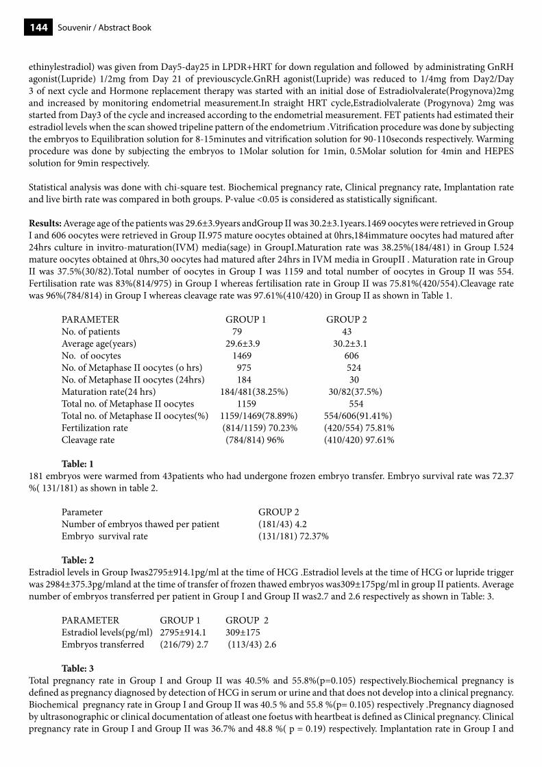
Souvenir / Abstract Book144
ethinylestradiol) was given from Day5-day25 in LPDR+HRT for down regulation and followed by administrating GnRH agonist(Lupride) 1/2mg from Day 21 of previouscycle.GnRH agonist(Lupride) was reduced to 1/4mg from Day2/Day 3 of next cycle and Hormone replacement therapy was started with an initial dose of Estradiolvalerate(Progynova)2mg and increased by monitoring endometrial measurement.In straight HRT cycle,Estradiolvalerate (Progynova) 2mg was started from Day3 of the cycle and increased according to the endometrial measurement. FET patients had estimated their estradiol levels when the scan showed tripeline pattern of the endometrium .Vitrification procedure was done by subjecting the embryos to Equilibration solution for 8-15minutes and vitrification solution for 90-110seconds respectively. Warming procedure was done by subjecting the embryos to 1Molar solution for 1min, 0.5Molar solution for 4min and HEPES solution for 9min respectively. Statistical analysis was done with chi-square test. Biochemical pregnancy rate, Clinical pregnancy rate, Implantation rate and live birth rate was compared in both groups. P-value <0.05 is considered as statistically significant. Results: Average age of the patients was 29.6±3.9years andGroup II was 30.2±3.1years.1469 oocytes were retrieved in Group I and 606 oocytes were retrieved in Group II.975 mature oocytes obtained at 0hrs,184immature oocytes had matured after 24hrs culture in invitro-maturation(IVM) media(sage) in GroupI.Maturation rate was 38.25%(184/481) in Group I.524 mature oocytes obtained at 0hrs,30 oocytes had matured after 24hrs in IVM media in GroupII . Maturation rate in Group II was 37.5%(30/82).Total number of oocytes in Group I was 1159 and total number of oocytes in Group II was 554.Fertilisation rate was 83%(814/975) in Group I whereas fertilisation rate in Group II was 75.81%(420/554).Cleavage rate was 96%(784/814) in Group I whereas cleavage rate was 97.61%(410/420) in Group II as shown in Table 1. PARAMETER GROUP 1 GROUP 2 No. of patients 79 43 Average age(years) 29.6±3.9 30.2±3.1 No. of oocytes 1469 606 No. of Metaphase II oocytes (o hrs) 975 524 No. of Metaphase II oocytes (24hrs) 184 30 Maturation rate(24 hrs) 184/481(38.25%) 30/82(37.5%) Total no. of Metaphase II oocytes 1159 554 Total no. of Metaphase II oocytes(%) 1159/1469(78.89%) 554/606(91.41%) Fertilization rate (814/1159) 70.23% (420/554) 75.81% Cleavage rate (784/814) 96% (410/420) 97.61% Table: 1181 embryos were warmed from 43patients who had undergone frozen embryo transfer. Embryo survival rate was 72.37 %( 131/181) as shown in table 2. Parameter GROUP 2 Number of embryos thawed per patient (181/43) 4.2 Embryo survival rate (131/181) 72.37% Table: 2Estradiol levels in Group Iwas2795±914.1pg/ml at the time of HCG .Estradiol levels at the time of HCG or lupride trigger was 2984±375.3pg/mland at the time of transfer of frozen thawed embryos was309±175pg/ml in group II patients. Average number of embryos transferred per patient in Group I and Group II was2.7 and 2.6 respectively as shown in Table: 3. PARAMETER GROUP 1 GROUP 2 Estradiol levels(pg/ml) 2795±914.1 309±175 Embryos transferred (216/79) 2.7 (113/43) 2.6 Table: 3Total pregnancy rate in Group I and Group II was 40.5% and 55.8%(p=0.105) respectively.Biochemical pregnancy is defined as pregnancy diagnosed by detection of HCG in serum or urine and that does not develop into a clinical pregnancy.Biochemical pregnancy rate in Group I and Group II was 40.5 % and 55.8 %(p= 0.105) respectively .Pregnancy diagnosed by ultrasonographic or clinical documentation of atleast one foetus with heartbeat is defined as Clinical pregnancy. Clinical pregnancy rate in Group I and Group II was 36.7% and 48.8 %( p = 0.19) respectively. Implantation rate in Group I and
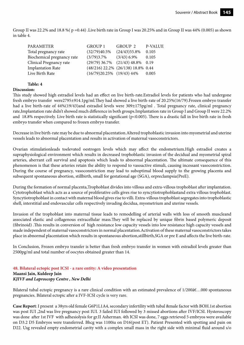
145Souvenir / Abstract Book
Group II was 22.2% and 18.8 %( p =0.44) .Live birth rate in Group I was 20.25% and in Group II was 44% (0.005) as shown in table 4. PARAMETER GROUP 1 GROUP 2 P-VALUE Total pregnancy rate (32/79)40.5% (24/43)55.8% 0.105 Biochemical pregnancy rate (3/79)3.7% (3/43) 6.9% 0.105 Clinical Pregnancy rate (29/79) 36.7% (21/43) 48.8% 0.19 Implantation Rate (48/216) 22.2% (26/138) 18.8% 0.44 Live Birth Rate (16/79)20.25% (19/43) 44% 0.005 Table: 4Discussion:This study showed high estradiol levels had an effect on live birth-rate.Estradiol levels for patients who had undergone fresh embryo transfer were2795±914.1pg/ml.They had showed a live birth-rate of 20.25%(16/79).Frozen embryo transfer had a live birth-rate of 44%(19/43)and estradiol levels were 309±175pg/ml . Total pregnancy rate, clinical pregnancy rate,Implantation rate didn’t showed much difference in both groups.Implantation rate in Group I and Group II were 22.2% and 18.8% respectively. Live birth rate is statistically significant (p<0.005). There is a drastic fall in live birth rate in fresh embryo transfer when compared to frozen embryo transfer. Decrease in live birth-rate may be due to abnormal placentation.Altered trophoblastic invasion into myometrial and uterine vessels leads to abnormal placentation and results in activation of maternal vasoconstrictors.
Ovarian stimulationleads toelevated oestrogen levels which may affect the endometrium.High estradiol creates a supraphysiological environment which results in decreased trophoblastic invasion of the decidual and myometrial spiral arteries, aberrant cell survival and apoptosis which leads to abnormal placentation. The ultimate consequence of this phenomenon is that these arteries retain the ability to respond to vasoactive stimuli, causing incessant vasoconstriction. During the course of pregnancy, vasoconstriction may lead to suboptimal blood supply to the growing placenta and subsequent spontaneous abortion, stillbirth, small for gestational age (SGA), orpreclampsia(PreE).
During the formation of normal placenta,Trophoblast divides into villous and extra-villous trophoblast after implantation.Cytotrophoblast which acts as a source of proliferative cells gives rise to syncytiotrophoblastand extra villous trophoblast. Syncytiotrophoblast in contact with maternal blood gives rise to villi. Extra-villous trophoblast segregates into trophoblastic shell, interstitial and endovascular cells respectively invading decidua, myometrium and uterine vessels.
Invasion of the trophoblast into maternal tissue leads to remodelling of arterial walls with loss of smooth muscleand associated elastic and collagenous extracellular mass.They will be replaced by unique fibrin based polymeric deposit (fibrinoid) .This results in conversion of high resistance low capacity vessels into low resistance high capacity vessels and made independent of maternal vasoconstrictors in normal placentation.Activation of these maternal vasoconstrictors takes place in abnormal placentation which results in spontaneous abortion,stillbirth,SGA or pre E and affects the live birth-rate.
In Conclusion, Frozen embryo transfer is better than fresh embryo transfer in women with estradiol levels greater than 2500pg/ml and total number of oocytes obtained greater than 14.
48. Bilateral ectopic post ICSI - a rare entity: A video presentationMaansi Jain, Kuldeep JainKJIVF and Laproscopy Centre , New Delhi
Bilateral tubal ectopic pregnancy is a rare clinical condition with an estimated prevalence of 1/200 000 spontaneous pregnancies. Bilateral ectopic after a IVF-ICSI cycle is very rare.
Case Report: I present a 38yrs old female G6P1L1A4, secondary infertility with tubal &male factor with BOH.1st abortion was post IUI ,2nd was live pregnancy post IUI. 3 failed IUI followed by 3 missed abortions after IVF/ICSI. Hysteroscopy was done after 1st IVF with adhesiolysis for gr.II Asherman. 4th ICSI was done, 7 eggs retrieved 5 embryos were available on D3.2 D5 Embryos were transferred. Bhcg was 1100iu on D16(post ET). Patient Presented with spotting and pain on D22. Usg revealed empty endometrial cavity with a complex small mass in the right side with minimal fluid around s/o
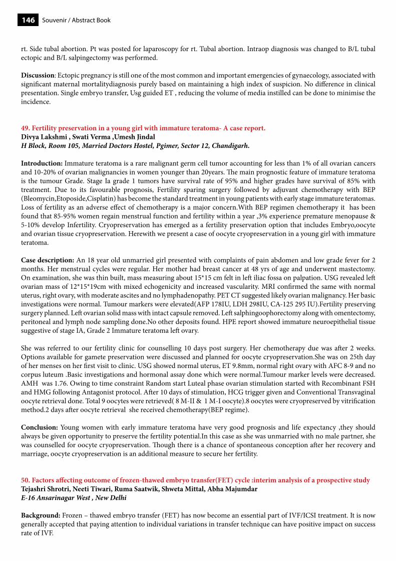
Souvenir / Abstract Book146
rt. Side tubal abortion. Pt was posted for laparoscopy for rt. Tubal abortion. Intraop diagnosis was changed to B/L tubal ectopic and B/L salpingectomy was performed.
Discussion: Ectopic pregnancy is still one of the most common and important emergencies of gynaecology, associated with significant maternal mortalitydiagnosis purely based on maintaining a high index of suspicion. No difference in clinical presentation. Single embryo transfer, Usg guided ET , reducing the volume of media instilled can be done to minimise the incidence.
49. Fertility preservation in a young girl with immature teratoma- A case report.Divya Lakshmi , Swati Verma ,Umesh JindalH Block, Room 105, Married Doctors Hostel, Pgimer, Sector 12, Chandigarh.
Introduction: Immature teratoma is a rare malignant germ cell tumor accounting for less than 1% of all ovarian cancers and 10-20% of ovarian malignancies in women younger than 20years. The main prognostic feature of immature teratoma is the tumour Grade. Stage Ia grade 1 tumors have survival rate of 95% and higher grades have survival of 85% with treatment. Due to its favourable prognosis, Fertility sparing surgery followed by adjuvant chemotherapy with BEP (Bleomycin,Etoposide,Cisplatin) has become the standard treatment in young patients with early stage immature teratomas.Loss of fertility as an adverse effect of chemotherapy is a major concern.With BEP regimen chemotherapy it has been found that 85-95% women regain menstrual function and fertility within a year ,3% experience premature menopause & 5-10% develop Infertility. Cryopreservation has emerged as a fertility preservation option that includes Embryo,oocyte and ovarian tissue cryopreservation. Herewith we present a case of oocyte cryopreservation in a young girl with immature teratoma.
Case description: An 18 year old unmarried girl presented with complaints of pain abdomen and low grade fever for 2 months. Her menstrual cycles were regular. Her mother had breast cancer at 48 yrs of age and underwent mastectomy. On examination, she was thin built, mass measuring about 15*15 cm felt in left iliac fossa on palpation. USG revealed left ovarian mass of 12*15*19cm with mixed echogenicity and increased vascularity. MRI confirmed the same with normal uterus, right ovary, with moderate ascites and no lymphadenopathy. PET CT suggested likely ovarian malignancy. Her basic investigations were normal. Tumour markers were elevated(AFP 178IU, LDH 298IU, CA-125 295 IU).Fertility preserving surgery planned. Left ovarian solid mass with intact capsule removed. Left salphingoophorectomy along with omentectomy, peritoneal and lymph node sampling done.No other deposits found. HPE report showed immature neuroepithelial tissue suggestive of stage IA, Grade 2 Immature teratoma left ovary.
She was referred to our fertility clinic for counselling 10 days post surgery. Her chemotherapy due was after 2 weeks. Options available for gamete preservation were discussed and planned for oocyte cryopreservation.She was on 25th day of her menses on her first visit to clinic. USG showed normal uterus, ET 9.8mm, normal right ovary with AFC 8-9 and no corpus luteum .Basic investigations and hormonal assay done which were normal.Tumour marker levels were decreased.AMH was 1.76. Owing to time constraint Random start Luteal phase ovarian stimulation started with Recombinant FSH and HMG following Antagonist protocol. After 10 days of stimulation, HCG trigger given and Conventional Transvaginal oocyte retrieval done. Total 9 oocytes were retrieved( 8 M-II & 1 M-I oocyte).8 oocytes were cryopreserved by vitrification method.2 days after oocyte retrieval she received chemotherapy(BEP regime).
Conclusion: Young women with early immature teratoma have very good prognosis and life expectancy ,they should always be given opportunity to preserve the fertility potential.In this case as she was unmarried with no male partner, she was counselled for oocyte cryopreservation. Though there is a chance of spontaneous conception after her recovery and marriage, oocyte cryopreservation is an additional measure to secure her fertility.
50. Factors affecting outcome of frozen-thawed embryo transfer(FET) cycle :interim analysis of a prospective studyTejashri Shrotri, Neeti Tiwari, Ruma Saatwik, Shweta Mittal, Abha MajumdarE-16 Ansarinagar West , New Delhi Background: Frozen – thawed embryo transfer (FET) has now become an essential part of IVF/ICSI treatment. It is now generally accepted that paying attention to individual variations in transfer technique can have positive impact on success rate of IVF.
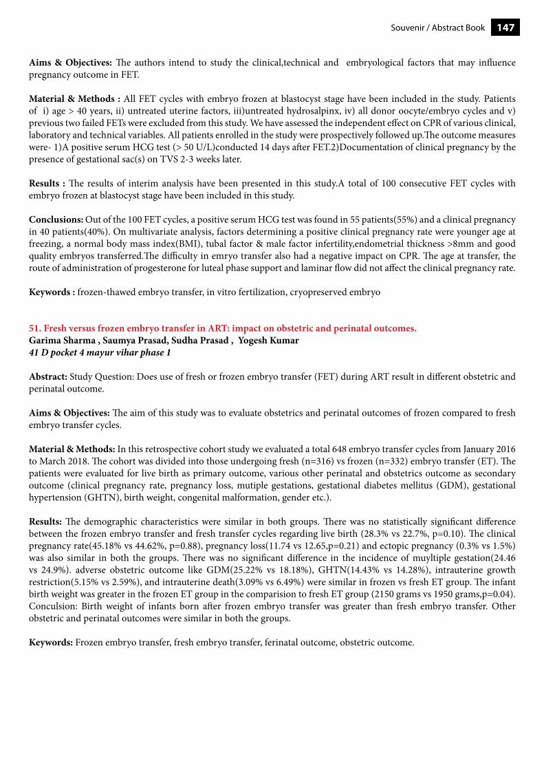
147Souvenir / Abstract Book
Aims & Objectives: The authors intend to study the clinical,technical and embryological factors that may influence pregnancy outcome in FET.
Material & Methods : All FET cycles with embryo frozen at blastocyst stage have been included in the study. Patients of i) age > 40 years, ii) untreated uterine factors, iii)untreated hydrosalpinx, iv) all donor oocyte/embryo cycles and v)previous two failed FETs were excluded from this study. We have assessed the independent effect on CPR of various clinical, laboratory and technical variables. All patients enrolled in the study were prospectively followed up.The outcome measures were- 1)A positive serum HCG test (> 50 U/L)conducted 14 days after FET.2)Documentation of clinical pregnancy by the presence of gestational sac(s) on TVS 2-3 weeks later. Results : The results of interim analysis have been presented in this study.A total of 100 consecutive FET cycles with embryo frozen at blastocyst stage have been included in this study.
Conclusions: Out of the 100 FET cycles, a positive serum HCG test was found in 55 patients(55%) and a clinical pregnancy in 40 patients(40%). On multivariate analysis, factors determining a positive clinical pregnancy rate were younger age at freezing, a normal body mass index(BMI), tubal factor & male factor infertility,endometrial thickness >8mm and good quality embryos transferred.The difficulty in emryo transfer also had a negative impact on CPR. The age at transfer, the route of administration of progesterone for luteal phase support and laminar flow did not affect the clinical pregnancy rate. Keywords : frozen-thawed embryo transfer, in vitro fertilization, cryopreserved embryo
51. Fresh versus frozen embryo transfer in ART: impact on obstetric and perinatal outcomes.Garima Sharma , Saumya Prasad, Sudha Prasad , yogesh Kumar41 D pocket 4 mayur vihar phase 1 Abstract: Study Question: Does use of fresh or frozen embryo transfer (FET) during ART result in different obstetric and perinatal outcome.
Aims & Objectives: The aim of this study was to evaluate obstetrics and perinatal outcomes of frozen compared to fresh embryo transfer cycles.
Material & Methods: In this retrospective cohort study we evaluated a total 648 embryo transfer cycles from January 2016 to March 2018. The cohort was divided into those undergoing fresh (n=316) vs frozen (n=332) embryo transfer (ET). The patients were evaluated for live birth as primary outcome, various other perinatal and obstetrics outcome as secondary outcome (clinical pregnancy rate, pregnancy loss, mutiple gestations, gestational diabetes mellitus (GDM), gestational hypertension (GHTN), birth weight, congenital malformation, gender etc.).
Results: The demographic characteristics were similar in both groups. There was no statistically significant difference between the frozen embryo transfer and fresh transfer cycles regarding live birth (28.3% vs 22.7%, p=0.10). The clinical pregnancy rate(45.18% vs 44.62%, p=0.88), pregnancy loss(11.74 vs 12.65,p=0.21) and ectopic pregnancy (0.3% vs 1.5%) was also similar in both the groups. There was no significant difference in the incidence of muyltiple gestation(24.46 vs 24.9%). adverse obstetric outcome like GDM(25.22% vs 18.18%), GHTN(14.43% vs 14.28%), intrauterine growth restriction(5.15% vs 2.59%), and intrauterine death(3.09% vs 6.49%) were similar in frozen vs fresh ET group. The infant birth weight was greater in the frozen ET group in the comparision to fresh ET group (2150 grams vs 1950 grams,p=0.04).Conculsion: Birth weight of infants born after frozen embryo transfer was greater than fresh embryo transfer. Other obstetric and perinatal outcomes were similar in both the groups.
Keywords: Frozen embryo transfer, fresh embryo transfer, ferinatal outcome, obstetric outcome.
Souvenir / Abstract Book148
52. Comparison of IVF outcomes between Minimal Stimulation and conventional GnRH antagonist protocols in poor ovarian responders.Ganeswar Barik , Vasundra Thaigarajan, A Aishwarya38 Th Street, Greata Opal, Nanganallur, Chennai, Tamilnadu
Background: Patients with poor ovarian responders (PORs) are both challenging to treat and represent a large proportion of patients presenting with infertility. Patients with POR, who are often of advanced maternal age, have a high cycle cancellation rate, higher miscarriage rate, and significantly reduced live birth rate per cycle. The management of POR is highly controversial as well. There is still no consensus regarding the “ideal” protocol and so far no one treatment protocol has proven to be superior for this group.
Objective: The object of this study was to compare IVF outcomes between minimal stimulation and conventional antagonist protocols in poor ovarian responders (PORs).Materials and methods: In this retrospective cohort study 40 PORs undergoing IVF were selected and divided into two groups. First group was the minimal stimulation group (n = 20) receiving 100 mg/day clomiphene citrate on day 2of the cycle for 5 day that was followed by 150IU/day human menopausal gonadotropin (hMG) on day 7 of the cycle. Second group was the conventional group (n = 20) receiving gonadotropin on day 2 of the cycle. Gonadotropin-releasing hormone (GnRH) antagonist protocol was applied for both groups according to flexible protocol. Number of retrieved oocytes and chemical pregnancy rate were the main outcomes.Results: There was no difference in number of retrieved oocyte and pregnancy rate (3.85 ± 2.03 vs. 4.10 ±1.71 and 20% vs. 25% between both groups. The gonadotropin dose used in the minimal stimulation group was lower than conventional group (1207 ± 124 vs. 2520 ± 406).
Conclusion: Minimal stimulation protocol with lower gonadotropin used is likely to be considered as a patient- friendly and cost-effective substitute for PORs.Keywords: Antagonist Protocol, Minimal Stimulation ProtocoL, Poor Ovarian Responders
53. A comparative study of Vitamin Dlevels between subfertile and fertile women in India.Khushboo Khushboo , Himanshu RoyNavrattan Hata, Purnea Introduction: The role of Vitamin D in human reproduction and the role of its deficiency in cases of subfertility has become a matter of much scientific scrutiny throughout the world. Vitam D deficiency has been found to be associated with PCOD, fibroid, endometriosis and poor ovarian reserve and also negatively affecting the outcome of Assisted Reproductive Techniques. There is serious dearth of data on prevalence of Vitamin D in subfertile women in our country. This missing piece of data could assist in studying the magnitude of problem and also the effect of Vitamin D deficiency and benefit if any of vitamin D supplements in these women.
Aim and Objective : To study the prevalence of Vitamin D deficiency in subfertile women in India and to compare it with levels of Vitamin D in fertile women.
Material and Method : In this study, 150 women were included in our study group who were undergoing treatment for infertility. Their age, AMH levels and vitamin D levels were noted. Our control group consisted of 141 fertile women.
Result : The mean level of Vitamin D in study group was 20.37 ng/ml (SEM - 0.66) and that in control group was 16.58 ng/ml (SEM - 0.46). Therefore, there was no significant difference in Vitamin D levels in both the groups.
Conclusion : There are many studies supporting the salutory effects of Vitamin D upon female reproduction. However, our study found no difference in Vitamin D levels between fertile and subfertile women.
149Souvenir / Abstract Book
54. Demography, expectations and experiences of oocyte donors in northern india an interview based studyNikita Jindal95/H34/Sector 3, Rohini
Study question - Oocyte donation is an increasingly popular treatment option for pregnancy and parenthood in women with ovarian dysfunction or advanced age. Indian literature still lacks a formal interview based study focusing expectations and experiences of oocyte donors.
Aim – To assess demography, expectations and experiences of oocyte donors in Northern India by an interview based questionnaire.
Material and Methods – This is a Prospective Cross sectional survey at tertiary care IVF centre (Sample size of convenience – 50 to 60) over a period of August 2018 to January 2019 (ongoing study). Interviews were taken using structured questionnaire at the time of registration and after oocyte retrieval.
Results – A total of 32 donors were evaluated till 31st October 2018 (ongoing study). Mean age of donors was 24.8 years. Most of the donors were housewife (65.6%) rest were in a private job. Only 34.3% of donors were educated above tenth standard. All the donors were commercial donors introduced by paid agents or friends. Almost all donors had very poor understanding of details of the oocyte retrieval procedure. Only 28.1% of donors felt that compensation given is adequate, rest expected a higher compensation. Mean compensation given to donors was INR 23000, but three donors were not aware of compensation amount. All donors were satisfied with the procedure and did not feel exploited or anxious or depressed but few complained of discomfort experienced in visits and minimal complications like pain. None of the donor developed signs/symptoms of OHSS. Average number of oocyte retrieved per donor was 17. Mean dose of Gonadotropins required per donor was 1387 IU. Conclusion – Our findings show that the oocyte donation has an overall good acceptance and satisfaction among the donors. However there is a need to impart more detailed information to donors in order to prevent their any possible exploitation.
55. Effect of autologous platelet-rich plasma on clinical outcomes in patients with refractory endometrium in fresh ICSI / FET cycle -An interventional prospective studyJyoti Gupta, Kuldeep Jain , Bharti JainKJIVF and Laproscopy center, New Delhi. Background: Endometrial receptivity is determined independently by endometrial thickness, vascularity and pattern. Refractory endometrium has been associated with poor clinical outcome. Conventionally, many therapies have been tried such as estradiol valerate, sildenafil, intrauterine Granulocyte-colony stimulating factor (G-CSF) instillation etc but they lack consistency in delivering results. Intrauterine infusion of platelet-rich plasma (PRP) is a new approach that has been suggested for the treatment of refractory endometrium. The benefits of PRP for these infertile females with refractory endometrium may be due to the four times higher platelets and significantly greater amounts of growth factors in PRP than peripheral blood however it’s efficacy in endometrial growth has not been fully elucidated. Aim & Objectives : To evaluate the efficacy of autologous PRP in improving the endometrial parameters (thickness, pattern and vascularity) in females with refractory endometrium in fresh Intra cytoplasmic sperm injection (ICSI)/frozen embryo transfer (FET) cycle. Material and Methods: This is an Interventional prospective study being conducted at a KJIVF and Laproscopy center, New Delhi. A sample size of 30 females was taken for the study period from August 2018 to December 2018. Patients undergoing IVF-ICSI//FET who failed implantation or had history of cancelled cycles with suboptimal endometrial parameters in previous cycle were included in the study. All patients with Ashermann’s syndrome documented and corrected by hysteroscopy were also included. Patients with poor embryo quality, bleeding dyscracias, platelet count < 105/µL, hemoglobin < 10 g/dl and other concomitant active infections were excluded. Ultrasound screening was performed as per protocol for regular assessment of size of follicle and endometrial parameters (thickness, morphology and vascularity). In females with compromised endometrial parameter defined as endometrial thickness <7mm, high resistance flow in uterine vessels or RI > 3.2, poor vascularity in zone 3 and 4 or regular pattern of endometrium intra-uterine PRP infusion was offered. Outcomes were assessed in the enrolled females by endometrial assessment on day of ET, after 15 days of ET, β-hCG estimation was done for the confirmation of the pregnancy. Clinical pregnancy was diagnosed one weeks after the β-hCG estimation by the presence of the fetal cardiac activity.
Souvenir / Abstract Book150
Results and Conclusion: As it is an outgoing study, data compilation is under process. Data was collected in a pre-tested data entry. Continuous data with normal distribution is being analysed by student t-test and non-normally distributed data by Mann-Whitney U test. Categorical data is being analysed by chi-square or Fischer exact test. A p value of <0.05 is considered significant. Performance of PRP in thin endometrium is being analysed by measuring sensitivity, specificity, positive predictive value, negative predictive value, receiver-operating characteristic curve and area under curve. 95% confidence interval will be calculated.
56. Which IVF protocol best suits the PCOS women?Namita Jain, Sonia MalikSouthend IVF Centre , New Delhi Study question: Which IVF protocol best suits the PCOS women?
Aims & Objective: The present study was undertaken to compare various IVF protocols in women with PCOS.
Material and methods: This was a retrospective observational study. 102 women under 40 years of age presenting with PCOS as one of the main cause for infertility who underwent IVF/ICSI at our centre over a period of one year (May 2016-Apr 2017), were enrolled for this study. Clinical data, investigations, treatment and the outcome of IVF/ICSI were recorded.Results: Three protocols were used for IVF stimulation which included Antagonist protocol, stop protocol and AACEP protocol depending upon the clinician discretion. Gonadotropin dose required for stimulation was not significantly different with different protocols used. Similarly no difference was noticed in terms of number of oocytes retrieved and good quality embryos formed. But a statistically significant difference was found in terms of clinical pregnancy rates disfavouring the AACEP protocol for PCOS women.
Conclusion: Clinical pregnancy rates were significantly higher with Stop protocol, which should be considered for IVF stimulation in PCOS women. However, randomized control trials are needed to further support it.
57. Study of Mullerian anomalies among infertile women using three-dimensional ultrasonographyKubera NS , Sunayana Kamala,Dilip Kumar MauryaKalpatharu, Plot No 120,121, 3 Cross,Dr Annie Besant Nagar, Navarkulam, Lawspet Introduction: Congenital Mullerian duct anomaly (MDA) is quite often encountered in infertility, preterm labor, first-trimester abortion, and fetal malpresentation. It accounts for 0.1% - 3.5% in the general population. MDA is 3% to 38% in patients with repeated spontaneous miscarriages or with infertility. Many noninvasive techniques such as pelvic ultrasound, which is widely used and Magnetic resonance imaging (MRI) being used in selected cases. Hysterosalpingography has been the primary diagnostic tool used to evaluate uterine anomalies in infertile women as an office procedure. Conventional 2D ultrasound and Hysterosalpingography has been used. Invasive methods like combined laparoscopy and hysteroscopy for uterine size, contour and morphology have been the method that is widely used. Three dimensional (3D) ultrasonography has higher accuracy for uterine anomalies classification and diagnosis compared to hysteroscopy or MRI. It assesses both external and internal uterine morphology using the coronal plane, it is a non-invasive and also reproducible procedure.
Aims and objectives:
Primary objective:To estimate the proportion of congenital Mullerian anomalies among infertile womenSecondary objective:To assess the accuracy of three-dimensional ultrasound in detecting Mullerian anomaliesMethods:The study has included 590 patients attending infertility OPD with infertility. After obtaining relevant history and examination, three-dimensional ultrasound was done using Volusion E8 machine. Measurements for uterus size like length breadth and width were taken. Intercornual distance, external and internal indentation measurements taken. Myometrial thickness at the level body of uterus measured. Anomalies are classified based on ASRM classification. Data was entered in MS Office Excel spreadsheet and was analyzed using SPSS Version 19.0. The outcome variables are expressed as frequencies.Results:Among the 590 infertility population under study, 493 (83.5%) were primary infertility and 97 (16.5%) were secondary infertility. Mean age of the study population is 28±3.9 years. In the present study 43 (7.3%) patients had the uterine anomaly, Among the 43 Mullerian anomalies noted, 24 (55.8%) were subseptate uterus, 9 (20.9%) were septate uterus, 8 (18.6%) were the bicornuate uterus, 2 (4.6%) were arcuate uterus.
151Souvenir / Abstract Book
Conclusion: The study has shown that the prevalence of Mullerian anomalies is 7.3% which is almost similar to western prevalence. The study has also confirmed that three-dimensional ultrasound is low cost, non-invasive, highly reproducible, reliable modality of screening for Mullerian anomalies. It showed high concordance with all standard investigations.
58. Malignancy masquerading OHSS - Acase reportParvathy T, Fessy Louis TAmrita Institute of Medial Science
Introduction : OHSS is a serious complication of ART. The incidence of OHSS varies between 0.6-6% among various studies in the setting of ART.
Case : 40 year G4A3 post IVF conception at 13 weeks referred as a case of OHSS not responding to routine management . Outside hospital tapping done twice each time about 2-2.5 litres . MRI done at our institution suggestive of gross ascites with grossly enlarged ovaries with mild pleural effusion on both sides favouring possibility of OHSS with viable fetus and normal interval growth. Initial ascitic fluid cytology report was normal. As ascites not responding to conservative management MRI with USG correlation showed solid areas and mural nodule in left ovary with provisional diagnosis of carcinoma ovary was made. CA 125 level came to be 5756U/ml. FNAC from the palpable left scalene lymph node revealed metastatic adenocarcinoma. The option of staging laparotomy given to the patient. As patient and husband were unwilling for surgery, they were given the option of chemotherapy. She underwent 6 cycles of carboplatin and paclitaxel combination. She had elective LSCS at term delivering a baby of 1.85kg. Along with LSCS gynaec oncology team proceeded with omentectomy and bilateral tumor debulking. Post surgery patient was stable. No chemotherapy was advised post operatively. CA 125 monitoring was advised serially. Histopathology report came to poorly differentiated serous adenocarcinoma. Discussion : On analyzing the case in detail – initially itself the clinical condition was not in favor of OHSS namely the age of the patient, gestational age of presentation, atypical clinical features and abnormal lab parameters . Malignancy suspicion started only when there was recurrent ascites with the imaging showed enhancing mural nodule in the ovary.Conclusion : Although OHSS is a known and accepted complication of ART , non-resolution of clinical symptoms with mismatch in lab parameters , keep the suspicion of malignancy
59. Evaluating the incidence and impact of tubal pocketing on diagnostic hystero-laparoscopy in patients with unexplained infertilityT R Ramya Shree, Parasuram GopinathBlock 1 5a, Ace Amrutha Retreat Appartments, Amrita Nagar
Objective: The aim of this study is to assess tubal pocketing observed during hysterolaparoscopy and its incidence and impact on the pregnancy outcome in couples with unexplained infertility.Pocketing is defined as inconsistent flow of dye through the tubes with irregular tubal distention seen during chromopertubation. Pocketing is of a)minimal b)severe.
Design: This is a retrospective cum prospective observational study.
Patients and Methods: Data of 105 unexplained infertility patients were collected from patient case records and they included factors such as age, duration and type of infertility, clinical examination findings, pelvic ultrasound, HSG/SSG records and semen analysis details. Previous treatment history included details of ovulation stimulation with or without intrauterine insemination (IUI). Intraoperative findings for the presence of tubal pocketing with no other pathology
Results: Hystero-laparoscopy revealed 73 patients with tubal pocketing out of which minimal and severe pocketing are 49 & 24 respectively. There is no significant impact of tubal pocketing on pregnancy outcome.
Conclusion: We have not found any significant correlation between tubal pocketing and its impact on pregnancy outcome, so the couple can be given a 6months duration for natural conception before directly going for ART treatment.
Keywords: Diagnostic hysterolaparoscopy, tubal pocketing , unexplained infertility
Souvenir / Abstract Book152
60. Sugar-sweetened beverage intake in relation to semen quality in infertile couples-A prospective observational studyIndrani Ghosh, Borbari, Pramod Sharma, Mujibur Rahman, Kabita Lahkar
Study Question- Is consumption of sugar-sweetened beverages ( SSB) associated with semen quality?
Aims And Objectives- To investigate the effect of SSB intake on semen quality in a cohort of infertile couples.The following outcomes were studied-
Primary objective: 1. Semen parameters- -Volume (in mL) -Viscosity -Fructose levels(qualitative) - Total sperm count -Sperm concentration(million/mL) -Sperm total motility (WHO A+B+C) and progressive motility (WHO A+B) -Sperm morphology(% normal)
The main independent variable was SSB intake.
The other demographic and clinical variables studied as potential confounders were: age, BMI, educational level, physical activity , abstinence time ( in hours), alcohol intake, caffeine intake, tobacco (smoking, chewing) and brief reproductive history. Materials And Methods- Study Population: Men attending infertility clinics, 18-50 years of age from August, 2018 to December, 2018.
Study Design: Prospective cross-sectional observational study
Sample Size: n (sample size) = Zα2 p (1-p) /e2 17 where Zα is Z-score at 95% confidence interval; p - proportion, e -precision, α=5%hence, Zα=1.96p (proportion of subfertile male= 50%), e=5% so, n= 385.Semen analysis was done according to WHO 2010 criteria.Intake of SSBs was assessed using a previously validated 15-item beverage intake questionnaire (food frequency questionnaire, FFQ).
Men were classified in quartiles of SSB intake. Linear regression models were used to estimate the adjusted difference and 95% confidence interval (CI) in semen quality parameters in increasing quartiles of SSB intake using men in the lowest quartile as reference, while adjusting for potential confounders.Continuous variables were expressed as mean±standard error and compared across groups using unpaired t test/ one way ANOVA if the data followed normal distribution and Mann- Whitney U test/ Kruskal Wallis test, if it did not. Categorical variables expressed as number of patients and percentage of patients and compared across the groups using Pearson’s Chi-square test for independence of attributes/ Fisher’s Exact test as appropriate.
Results And Conclusion- AWAITED Keywords-diet/ sugar-sweetened beverage/ semen quality/ food frequency questionnaire
61. Role of three dimensional endometrial blood flow and endometrial volume in predicting the outcome of frozen- ET cyclesGayathri Mohan, Asha Rao, Damodar Rao, Padmashri, Abarajda VCentre for assisted reproduction and endoscopy, Rao Hospital, Coimbatore, Tamil Nadu Aim & Objective: To analyze which is a better predictor- Power Doppler or endometrial volume for better pregnancy outcomes in frozen –ET cycles.
Design: A retrospective study.Setting: Rao hospital.
153Souvenir / Abstract Book
Materials and methods: 89 patients aged between 23 and 38 years who underwent Frozen ET (FET) cycles from january 2016 to january 2018, whose uteri were morphologically normal as confirmed by 3-D ultrasound were included in the study and those women with uterine pathology like fibroid, polyp etc., hydrosalpinx were excluded from the study.A 3D ultrasound on the day of progesterone supplementation in patients undergoing FET cycles were measured using voluson E8, GE Wipro medical system aided by VOCAL imaging software and volume histogram analysis to obtain Endometrial volume (EV), Vascular index (VI), Flow index (FI) and Vascular flow index (VFI) by a single person to avoid inter observer variations. Six contour planes were analyzed with a 30o rotation step for the endometrium to cover 180o.
Results: We analyzed EV, VI, FI and VFI in conception and non conception cycles. Mean endometrial volume in conception and non conception cycles by independent t test were (2.717, 3.446) with a significant p value of 0.011. Mean VI was (4.489, 4.118) with p value 0.864, mean FI was (23.866, 24.181) with p value 0.729 which were not significant and mean VFI was (2.948, 1.350) with a significant p value of 0.009.Hence in our study,(A) Vascular flow index (VFI) in predicting the pregnancy outcome of frozen ET cycles was superior to that using vascular index or endometrial volume.(B) VFI was significantly higher in pregnant women with live births than those with a miscarriage.
Conclusions: The detection of endometrial blood flow by 3-D power Doppler ultrasound may be a useful ultrasound parameter in the prediction of pregnancy outcome in Frozen ET cycles, especially endometrial VFI.Key words:Frozen ET (FET) cycles, Endometrial volume (EV), Vascular index (VI), Flow index (FI) and Vascular flow index (VFI)
62. Freeze all strategy : Is it beneficial for all?Abarajda V, Asha Rao, Damodar Rao, Padmashri GCentre For Assisted Reproduction And Endoscopy, Rao Hospital, Coimbatore, Tamil Nadu
Introduction: Freeze-for-all concept, based on the principle that controlled ovarian stimulation may cause a less physiological milieu for embryo to implant, is not uncommon these days. Yet, we must wonder when and for whom is this approach most appropriate.
Aim & Objectives: To assess the impact of ovarian response on clinical pregnancy rates (CPR) and live birth rates (LBR) in patients having their first fresh embryo transfer (ET) versus similar patients having their first frozen embryo transfer (FET).
Material & Methods: This is a retrospective cohort study conducted between January 2017 – September 2018 at Rao Hospital, Coimbatore. A total of 143 IVF cycles (Fresh group -67, Freeze –all group -76) were included. The cycles were subdivided into cohorts based on the number of oocytes retrieved: 1-5 (low), 6-10 (normal), >11 (high responders). Statistical analysis was done using SPSS 20.0 software.
Results: Frozen embryo transfer resulted in higher CPR and LBR than the fresh embryo transfer in high responders (57.14% vs 42.86%, 52.38% vs 28.57%) and in normal responders (50.00% vs 42.30%, 50.00% vs 38.46%). In low responders, CPR and LBR were higher after a fresh compared with FET (43.59% vs 35.00%, 35.90 % vs 25.00%). However miscarriage rate is more in fresh ET cycles in all the three sub groups.
Conclusion: The results of this study show that although the freeze-all strategy may be related to better IVF outcomes in high responders and normal responders, these advantages are not beneficial in low responders.Key words:Clinical pregnancy rates (CPR), Live birth rate (LBR), Embryo transfer (ET), Frozen embryo transfer (FET)
63. Ovarian pregnancy following ICSI-ET and its successful laparoscopic management: A case reportAnurag Vashista, K K Gopinathan,Soumya Nair, Sreehari ArunkumarEdappal, Malappuram Case: A 28 year old primary infertility case was evaluated and decided for ART for severe male factor infertility. She underwent ICSI-ET following long agonist protocol as per institutional policy. ICSI done and 14 embryos resulted. 2 grade I cleavage stage embryos were transferred by USG guidance about 1 cm away from the fundus, 8 embryos cryopreserved and 4 were donated with consent.Serum βhCG on day 15 following embryo transfer (ET) was 57.57 mIU/ml and doubled
Souvenir / Abstract Book154
after 48 hours (112.6 mIU/ml). Patient had intermittent abdominal pain managed by supportive measures. βhCG on day 24 showed rising trend to 1570 mIU/ml and on day 27 was 2481 mIU/ml.
Transvaginal USG showed good endometrial reaction with no intrauterine sac, single right ovarian unruptured ectopic pregnancy. Operative laparoscopy was decided and done.
Findings:1. Uterus was normal2. Left tube, left ovary and right tube were normal.3. Right ovary was seat of ectopic pregnancy of 2 x 2 cm that had ruptured.4. Hemoperitoneum of around 100 ml.5. Due to high vascularity and encountered bleeding, diluted vasopressin (1:20) was injected in uterus and right ovary.Hemostasis was secured. Gestational sac with products of conception enucleated from right ovary.
Discussion:Ovarian ectopic pregnancy is rare but possible entity after ART (IVF/ICSI). The Spiegelberg’s criteria for diagnosis are:1. Fallopian tubes with fimbria must be intact and separate from the ovary.2. The pregnancy must occupy the normal position of the ovary.3. The ovary must be attached to the uterus through utero ovarian ligament.4. There must be ovarian tissue attached to the pregnancy in the specimen.
The possibility of ovarian ectopic pregnancy should be kept in mind in background of normal appearing uterus, tubes and serum βhCG values suggestive of ectopic pregnancy. The exact aetiology of ovarian pregnancy is however unknown. Possibilities implicated are:1. Reverse migration of embryo after deep deposition in uterine cavity.2. Large volume of culture fluid.3. Presence of tubal pathology and pathology and pelvic inflammatory disease.
A high degree of suspicion is required for ovarian pregnancy. Differential diagnosis of hemorrhagic corpus luteum or ruptured lutein cyst can be easily missed. Meticulous preoperative scans and careful interpretation of βhCG values helps in early diagnosis and management decisions. We believe in laparoscopy as a gold standard for management of ovarian ectopic pregnancy. Use of methotrexate (MTX) though suggested is still controversial.
64. A prospective study on colour doppler indices of follicular blood flow as predictors of pregnancy in intrauterine insemination cycles (IUI).Thomas Poulose Aliyattukudy, Parasuram Gopinath, Karthika Bijoy, Betty John Ferns, Revathy,K K GopinathanCIMAR Cochin Background : The overall outcome of intrauterine insemination is still more often a failure (more than 80%) than success. Assessing perifollicular blood flow is one of the techniques to improve the success. Literature search has showed that Doppler indices of perifollicular blood flow as predictors of pregnancy in assisted reproductive techniques. The peak systolic velocity (PSV) of individual follicles has been correlated with oocyte recovery, fertilization rate and embryo quality. Aims & Objective : The aim of the study is to check the prognostic value of perifollicular blood flow in intrauterine insemination (IUI). The objective of the study is to assess the role of quantitative and qualitative indices of follicular vascularity in predicting pregnancy Methods : This is a prospective observational study. A total of one hundred patients were studied at CIMAR Cochin between May 2018 and November 2018
PSV was measured from the largest follicle on the day of human chorionic gonadotropin (HCG) administration. The quality of follicular flow was graded from 1 to 4 according to the amount of visible color flow around the follicle. Fifty patients underwent perifollicular Doppler blood flow in the dominant follicle before the trigger. Resistance Index (RI), Pulsatility Index (PI), Peak Systolic Velocity (PSV) were assessed and the patients had intrauterine insemination. This group was sub grouped into those having Doppler indices with peak systolic velocity >10cm/s and those with peak systolic velocity <10
155Souvenir / Abstract Book
cm/s. Pregnancy outcome was assessed between these subgroups. Remaining fifty patients did not had Doppler studies and had intrauterine insemination based on the follicular study. Results : The results are very hetrogenous.22% of experimental Doppler arm had successful pregnancies. Among conceived 94% of patients had grade 3 and grade 4 perifollicular blood flow. The pregnancy rate in the non-Doppler group was 18%. There was no significant correlation between the outcome of pregnancy in Doppler and non-Doppler group. Pregnancies were more in those with good peak systolic blood flow of more than 10cm/s Conclusion : Perifollicular blood flow seems to provide a favorable prediction of the pregnancy outcome as only 6% of patients with poor Doppler indices conceived. The knowledge gained from our study may have a use in future management of patients undergoing IVF treatment. Pregnancy rates would be expected to increase by selecting for transfer of embryos derived from follicles with PSV >10cm/s. Prospective larger clinical trials are necessary to test these hypothesis
65. Role of atosiban in women undergoing in vitro fertilization and embryo transfer(IVF –ET )Kashika Kathuria , Anjali Tempe Maulana Azad Medical College and Lok Nayak Hospital.
Objectives: To determine the role of oxytocin antagonist (atosiban) in improving pregnancy outcomes(implantation rate, pregnancy rate and live birth rate) in patients undergoing In vitro fertilization- embryo transfer( IVF-ET ). Methods: A prospective study was conducted in the IVF Centre at Maulana Azad Medical College and Lok Nayak Hospital. Patients undergoing IVF-ET with top quality embryos were offered atosiban at the time of embryo transfer. Atosiban was given as an intravenous bolus 6.75 mg(0.9 ml) 30 minutes prior to ET and continued as an infusion at 18 mg/hr for one hour, which was then reduced to 6 mg/ hour for another two hours. Outcomes were compared between the group of patients receiving atosiban(n=50) and those not receiving atosiban(n=53).
Results: The treatment (atosiban) group and the control group were comparable in the baseline characteristics. The causes of infertility were tubal factor, anovulation, unexplained infertility,endometriosis and male factor infertility. In the treatment group (n=50), the clinical pregnancy rate was 32 %(16 pregnancies in 50 patients) and implantation rate was 18.8%. In the control group (n=53), the clinical pregnancy rate was 39.6% (21 pregnancies) and the implantation rate was 21.8%. There was no statistically significant difference in the pregnancy rate and implantation rate between the two groups. There were two twin pregnancies in atosiban group compared to three twins in the control group. Five patients had a miscarriage in atosiban group as compared to 7 patients in the control group. Of the 16 clinical pregnancies in atosiban group, ten resulted in live births and there is one ongoing pregnancy; while in the group without atosiban intervention, there were nine live births and three ongoing pregnancies.
Conclusion: Use of atosiban at the time of embryo transfer is not associated with significantly higher pregnancy rates or implantation rates. Also there is no significant difference in the incidence of multiple pregnancy or miscarriage between the groups. However, these findings need to be confirmed in larger studies 66. Use of a modified injection technique for ICSI in total globozoospermia to prevent sperm borne oocyte activation failureAnupama S, K.K. Gopinath, Parasuram Gopinath, Karthika .D.KumarCIMAR Fertility Centre Cheranelloore Objective : To test whether oocyte activation failure due to Globozoospermia can be overcome by mechanical means that entail modifying the ICSI technique
Design : Case report Series
Setting : Private clinic
Patients : Three infertile patients with known Globozoospermia and previous fertilisation failure following ICSI
Intervention : Standard ICSI and Modified ICSI based on mechanical manipulation on sibling oocytes and thereby presumed changes in calcium oscillations
Souvenir / Abstract Book156
Method of Modified ICSI : Sibling oocytes of the same patients are allocated for standard ICSI and Modified ICSI procedure. Modified ICSI here means achieving oolemmal breakage by vigorous aspiration of ooplasm into the needle. This was done by placing the tip of the needle in central region of oocyte to reduce the risk of chromosomal damage.
Main Outcome: Fertilisation rate, Clinical pregnancy rate , Live birth rate, Follow up of children till one year of ageResult: In all three cases of Globozoospermia and previous fertilisation failure; Modified ICSI injection technique enabled normal fertilisation , clinical pregnancy and live birth. The children were followed up for one year post delivery and were found normal.
Conclusion: Complete oocyte activation failure due to Total Globozoospermia can be overcome by Modified ICSI injection technique.This modification obviates the need for using insufficiently tested and potentially harmful medications. This modified ICSI needs large RCTs to confirm its effectiveness and safety.
Keywords : Globozoospermia , Modified ICSI , oocyte activation failure
67. The effect of assisted hatching on pregnancy rates after frozen embryo transferDhivya Sushil , Parasuram Gopinath, CIMAR Fertility Centre Cochin
Objective: To compare clinical pregnancy rates, ongoing pregnancy rates and implantation rates after transfer of frozen thawed embryos prepared according to an assisted hatching protocol and nonassisted hatching protocol. The hypothesis was Laser assisted hatching improved implantation and clinical pregnancy rates.
Design: A Prospectiveobservational study done in CIMAR FERTILITY CENTRE, COCHIN from DEC 2016 to JUNE 2018.
Patients And Methods: Data of Thirty patients with matched controls with inclusion criteria of age, basal FSH , IVF protocol, quality of embryos and previous two IVF failures. Factors like duration of infertility, number of embryos transferred and number of FET attempts were taken in to consideration.
Outcome Measures: Implantation rate, clinical pregnancy rate, ongoing pregnancy rate
Results: The Ongoing pregnancy rate was 6 and 6 clinical pregnancies per transfer in non hatched group while Ongoing pregnancy rate was 4 and there were 9 clinical pregnancies per transfer in assisted hatching group. In non hatched group 33 embryos transfers done and 7 implanted and there were 6 pregnancies ( 1 twins, 1 triplet) . In hatched group, 30 embryo transfers done and 15 implanted and there were 4 pregnancies (1 triplet).
Conclusion: The clinical pregnancy rate and implantation rates were higher for group having assisted hatching protocol but the ongoing pregnancy rate was higher in the non hatched group. Key Words: assisted hatching, ongoing pregnancy rate, IVF failures.
68. IVF outcome in diminished ovarian reserve after endometriosis surgery versus idiopathic DOR: A case control study.Aravind Ramachandran ,Gopinathan KKCIMAR Fertility Centre Edappal, Kerala
Introduction : Endometriosis is a common cause of subfertility affecting 10-15% of women of reproductive age group. The ESHRE guidelines 2008 recommend laparoscopic surgical management for endometrioma > 4 cm so as to improve fertility outcome. In recent years, the safety of surgery for endometriosis in terms of damage to ovarian reserve has been debated. Several studies have reported a poor response to ovarian stimulation and significantly worse IVF outcome in women with Diminished ovarian reserve (DOR) following surgery for endometriosis. Hence our study is intended to compare the IVF outcome in Surgical DOR after endometrioma surgery versus Idiopathic DOR. Aims and Objectives : To Compare IVF outcome in DOR after endometriosis surgery versus Idiopathic DOR.
157Souvenir / Abstract Book
Materials and Methods : A retrospective case control study was conducted in women undergoing IVF in CIMAR Fertility centre Edappal,Kerala between June 2017 to June 2018. All women were under age 40, with duration of infertility > 1 year .AMH levels <2 ng/ml was used to define DOR.Group A consisted of patients with DOR after surgery for endometriosis and Group B had patients with Idiopathic DOR without evidence of endometriosis in pelvic ultrasound. Women with uterine factors, severe male partner pathology (sperm conc <1 million/ml, Recurrent pregnancy loss, age> 40 years were excluded from the study group.
30 cycles of IVF in group A was matched with 62 IVF cycles in group B and outcome compared. All patients underwent long protocol with HMG stimulation followed by fresh/frozen transfer. Primary end point was clinical pregnancy as evidenced by ultrasound. Secondary end points were total dose of gonadotropin required, No of M2 oocytes, fertilization rate, Implantation rate and cycle cancellation rate. Results : The demographic data in both groups were comparable. The mean age in group A (DOR after surgery for endometriosis) was 30.9 and Group B 32.9 years. Mean AMH in both groups were not different statistically ( Group A 1.31 ng/ml, Group B 1.32ng/ml). The HMG dose required in Group A was higher than Group B and was statistically significant (4313 IU vs 3765 IU). The M2 oocyte yield was not different statistically among both groups (Group A 7.7, Group B 6.09). The fertilization rate was comparable in both groups ( 83.2% vs 81.7%). However, the clinical pregnancy rate was significantly lower in Group A (23.3% vs 35.4%)
Conclusions : Surgery for endometriosis is not without harmful effects. It is important fertility specialists be aware of the significant harmful effect of endometriosis surgery on ovarian reserve. Meticulous patient selection for surgery is of paramount importance.
69. Intralipid infusion is the new ray of hope in previous failed frozen embryo replacements- A randomized controlled trial from a tertiary care unitAishwarya Parthasarathy , Indumathi M A, Kundavi Shankarno.15A/50A, 3rd street, Krishna nagar, Virugamabakkam Introduction: Immunotherapy remains a largely unexplored area in the field of assisted reproduction although pregnancy is a state of complex immunotolerance. The latest addition is the use of intralipid fat emulsion, that has been used in recurrent pregnancy loss.
Aims And Objectives: To study the effect of 20 % intralipid infusion in previously failed frozen embryo replacement cycles with good embryos
Materials And Methods : 100 women undergoing frozen embryo transfer cycles were randomized on the day of embryo transfer into Group A (n=50) to whom 100 ml of 20% Intralipid infusion and Group B (n=50), who did not receive any drug. Both groups received standard luteal support. Those patients who had positive clinical pregnancies, had one more dose (100 ml of 20% intralipid) after the confirmation in ultrasound.The primary outcomes analyzed were clinical pregnancy (CPR) and ongoing pregnancy rates (OPR). Secondary outcomes analysed were miscarriage rate(MR) and implantation rates(IR) .
Results: Both groups were comparable with respect to the demographics ,endometrial thickness and number of embryos tranasferred. Clinical pregnancy (28/50 vs 11/50; p<0.001) and Ongoing pregnancy rates (23/50% vs 11/50%;p=0.006) were significantly higher in the intervention group. Miscarriage rates (5/28 vs 2/11;p=0.09) were not different. Implantation rates were also not significantly different(34/160 vs 41/148);p=0.09) .
Conclusion: Administration of intralipids prior to frozen transfer is a promising intervention to increase the pregnancy rates in previous failed frozen transfers. We conclude that 20% intralipid infusion should be studied in larger population to be incorporated into practice
Souvenir / Abstract Book158
70. Role of hysteroscopic endometrial injury: an intervention in recurrent implantation failure cycles: A case seriesJagruti Damse , KK Gopinathan , Saumya Nair , Shreehari Arunkumar CIMAR, Edappal Hospital
Introduction: Implantation is the rate limiting step in the process of fertilization in IVF cycles.Sucessful implantation of embryo requires a receptive uterus.Poor endometrial receptivity is an important cause of implantation failure.Dysregulation in these factors leads to RIF .In controlled ovarian stimulation (COS) the implantation rate is decreased due to abnormally advanced endometrial maturation and disturbed endocrine milieu
Endometrial scratching by stimulating delay in endometrial maturation corrects asynchrony between endometrium and embryo and hence promotes wound healing by inducing a significant increase in the local secretion of pro inflammatory cytokines,which in turn promotes sucessful implantation.
Aims And Objectives:1. To evaluate the therapeutic efficacy of hysteroscopic endometrial scratching in repeated implantation failure.Pregnancy
outcome by endometrial scratching in follicular and luteal phase
Background :• It is a case series of 7 cases• Patients attending infertility CIMAR (Center for Infertility Management and Assisted Reproduction),Edappal Hospital
(period January 2018 to June 2018) and who have two or more repeated implantation failure cycles were included in the study.
• A total of 7 cases were randomly recruited and observations made Endometrial scratching was performed during follicular phase of same cycle (5 cases) or luteal phase of preceding cycles (2 cases)
• Endometrial scratching ( injury) was done following diagnostic hysteroscopy, making sure that there is no obvious other uterine/endometrial pathology present.
• Endometrial scratching was done using curette – only Anterior and Posterior walls of uterus.• In all the 7 cases, Donor Oocyte was used• All patients underwent endometrial preparation (using estradiol valerate 4-6 mg from D2 of cycle till the endometrial
thickness becomes 7-8mm, & 3 line ) according to standard protocol used in the center.• All cases had Frozen( Day 3) Embryo transfers.• B Hcg was performed on day 14 of cycle.• Clinical pregnancy was confirmed by USG evidence of intrauterine gestational sac. Case Series Observations :• 7 Patients were selected with H/O of 2 or more RIF cycles with high quality embryos in past ART cycles• Age group : average age 37 years• Average duration of infertility 10 years• Male indication : mostly Oligoasthenozospermia• All donor oocytes were used• Hysteroscopic endometrial scratching was done during: 1. Luteal phase of preceding cycle (2 cases). OR2. Follicular phase of same cycle (5 cases).• Average endometrial thickness on day of embryo transfer was 9 mm.• All had frozen( DAY 3) Embryo transfer• Average positive B hcg value (day 14) was 729 mIU/ml.
Conclusion : • Endometrial scratching during diagnostic hysteroscopy seems to enhance implantation as well as pregnancy rate
It is simple and inexpensive procedure with benefits ,as we combined diagnostic hysteroscopy with endometrial scratching with added advantage that other endometrial/uterine pathology were not missed for optimum ART results.
159Souvenir / Abstract Book
71. Does female genital tuberculosis (FGT) affect fertility outcomes in in-vitro fertilization (IVF) cycles?Priyanka Khandey,Anjali TempeMaulana Azad Medical College, New Delhi.
Objective: To study the effect of genital tuberculosis on fertility outcomes in women underwent in-vitro fertilization (IVF) cycles and comparison of results of IVF outcomes in women with or without genital tuberculosis.
Introduction: Tuberculosis caused by Mycobacterium tuberculosis is a major health problem in developing countries. Female genital tuberculosis (FGT) causes significant morbidity, subfertility and infertility by damaging fallopian tubes, endometrium and ovarian reserve.
Methodology: This is a retrospective cohort study conducted at IVF centre of Central Delhi. Total 177 infertile women were recruited who underwent IVF from January 2016 to December 2017 of whom 50 women had history of genital tuberculosis (Group A) and 127 had no history of tuberculosis or anti-tubercular treatment (ATT) (Group B). Female genital tuberculosis was diagnosed by histopathological examination of endometrial aspirate, acid fast bacilli staining of smear, positive culture or laparoscopy or hysteroscopic findings suggestive of genital tuberculosis. Frozen-thaw cycles and donor oocyte cycles were excluded from the study. IVF outcomes like number of oocytes retrieved, embryo quality, pregnancy outcomes and live birth rates were compared in both the groups.
Results: Both the groups had mean age of 29 years. Total numbers of oocytes retrieved and fertilization rate were also similar in both the groups. There were no statistical differences in the two groups in the overall pregnancy rate, 34% in group A vs 40% in group B (p value 0.68).
Conclusion: Infertile women with or without history of genital tuberculosis have similar fertility outcomes in IVF cycles
72. Successful pregnancy in a case of Swyer Syndrome following donor ICSI-ET - A case reportThamizhselvi Naveen, K. K. Gopinathan, Soumya Nair , Sreehari ArunkumarCIMAR, Edappal Hospital Swyer syndrome is a Disorder of Sex Development (DSD) characterised by the failure of development of sex glands.Typical characteristics of a Swyer Syndrome being female phenotype, bilateral gonadal dysgenesis, sexual infantilism with primary amenorrhoea
We report an interesting case of Swyer Syndrome with a successful pregnancy following ICSI with donor oocyte and embryo transfer.A 29 year old housewife who is married for 4 years presented to CIMAR with history of primary infertility. She had been investigated for primary amenorrhoea at 16 years of age and the USG of pelvis showed hypoplastic uterus with no visualisation of ovaries or testis. Her FSH in 2016 was 22.5 iu/ml, Lh being 14.2miu/ml and her karyotyping showed 46, XY. She had been counselled then about her status including the need for lifelong HRT for having regular menses and the need of Donor oocyte for conception in future.
The patient is on cyclical pills since 16years of age and is having cyclical bleeding since then. She presented with her husband for infertility treatment at CIMAR in April,2018. She had no other significant past medical or surgical history. She had a normal sexual life. On general examination, she had a tall stature with axillary hair, stage V pubic hair and tanner stage IV breasts. Her Cardiovascular, respiratory and CNS system examination was within normal limits. Her abdomen was soft. She had a normal external genitalia with a healthy cervix and vagina on speculum examination. On Vaginal examination she had an anteverted , normal sized, mobile uterus.The male partner history and examination were within normal limits. Routine blood investigation of the couple was within normal limits.TVS showed an anteverted , normal sized uterus with collection inside the cavity. Both ovaries were not visualised.
The patient was started on HRT with estradiol valerate 2mg 1OD and norethisterone 5mg 1HS. After 2 months of proper withdrawal bleeding, TVS on Day3 showed a very minimal collection inside the cavity. She was started on estradiol valerate 2mg 1tds from day 3 along with folic acid 5mg 1OD and aspirin 75mg 0-1-0. On day 12 she had a good 3 line endometrium measuring 12mm in thickness. And she was started on Inj. Progesterone 100mg for 4days and fresh ET was done with day3 embryos on 16thday of the same cycle.
Souvenir / Abstract Book160
ICSI with Donor oocyte and Husband sperm done. 5 oocytes injected, 5 fertilised and 5 cleaved.4 embryos were grade I and 1 was grade III .
All the four GradeI embryos were transferred. She became pregnancy positive in the same cycle with se Beta HCG being 1273miu/ml on day15 after ET. TVS on 6 weeks showed a TCTA gestation. She had undergone a selective fetal reduction at 11wks 6days to DCDA twins with consent and cervical encirclage following it. She was diagnosed to have GDM at 12weeks and started on Metformin followed by Inj. Insulin. Her 20weeks anomaly scan was normal and now she is in her 25thweek of pregnancy
73. Fertility preservation in a case of malignant struma ovarii. Devi Krishna, Shameema Anwar SadathEdapally Cochin
Struma ovarii is a rare tumor defined as a mature ovarian teratoma containing 50% or more thyroid tissue, and it accounts for approximately 5% of all ovarian teratomas that are mostly benign. Malignant transformation of Struma ovarii is reported to occur in less than 5% of all cases.
We present here the case of 29 year old, with history of abdominal pain, in the right iliac fossa. Ultrasound showed bilateral complex adnexal masses.She underwent open bilateral ovarian cystectomy-Intra operatively 1)Bilateral ovarian cyst noted- Right measuring 6*7cm with irregular surface and Left measuring 5*4cm also with irregular surface 2)multiple nodular peritoneal and omental deposits3)Uterus appeared normal 4)Bilateral ovarian cyst with part of omentum sent for frozen section.Frozen section? malignant Epithelial tumour ovary;Omentum showing similar lesionsPatient was planned for definitive treatment after detailed histopathology report.The Histopatholgy showed Struma ovarii with multiple peritoneal deposits; consistent with highly differentiated follicular carcinoma. Her case was discussed in the multidisciplinary tumour board, and a decision was taken to proceed with staging laparotomy and total thyroidectomy, followed by a radio iodine ablation. As she was nulliparous, and desired fertility preservation, she was referred to the fertility clinic and was counselled for embryo freezing.She had controlled ovarian stimulation with Antagonist protocol-Recombinant FSH 225units for 9 days followed by retrival of 3 M2 oocytes .ICSI done and 2 good quality embryos were cryopreserved on day 2.Patient underwent Staging Laparotomy with Total thyroidectomy (Bilateral salphingo-oopherectomy + omentectomy+ stripping of peritoneum over bladder, abdominal side walls,pelvic peritoneum+ appendectomy with preservation of uterus.).She is planned for Iodine ablation and for embryo transfer after 6 months of completion of iodine therapy.
Conclusion: There are only limited literature available on fertility preservation in malignant struma ovarii.Though ovarian cryo preservation is an option, there is always a chance of neoplastic contamination and poor success rates with this experimental method. We couldn’t find any case of controlled ovarian stimulation in malignant struma ovarii-this may be the first reported case of embryo cryo preservation in this scenario
74. Prevalence of metabolic syndrome in infertile women with polycystic ovary syndromeChitra Thyagaraju , Lalitha K, Nandeesh H, Sadish KJIPMERPuducherry The prevalence of Metabolic Syndrome (MS) in Polycystic Ovary Syndrome (PCOS) women varies between ethnic groups and countries due to differences in lifestyle, diet and genetic factors. The prevalence of MS in PCOS is not well studied in Indian non-urban population. Though many international studies exist, there is no study comparing the prevalence of MS in infertile PCOS women and prevalence of MS in infertile non-PCOS women. This is the first attempt to compare the prevalence of MS in infertile PCOS and non PCOS women. Objective: To compare the prevalence of MS among infertile PCOS and non PCOS women attending infertility clinic and to identify the socio-demographic and clinical factors associated with the MS in infertile women. Study Design: A cross-sectional study was conducted in the Department of Obstetrics and Gynaecology of Jawaharlal Institute of Postgraduate Medical Education and Research (JIPMER) Puducherry, India between Mar 2016 and Feb 2018. A total of 130 infertile women with PCOS (as per the Rotterdam consensus criteria) and 130 infertile non PCOS women were included in the study. All 260 women were considered for screening for metabolic syndrome as defined by the International
161Souvenir / Abstract Book
Diabetes Federation (IDF) criteria. Further, Cross-tabulation analysis and appropriate statistical significance tests such as Unpaired T-test (for continuous variables) and Chi-square test (for categorical variables) were carried out to find the prevalence of metabolic syndrome in PCOS and non-PCOS women and also to develop the correlation between the socio-demographic and clinical factors associated with the MS in infertile women. Results: The prevalence of metabolic syndrome (according to IDF criteria) was found as 42.3% and 19.3% in infertile PCOS and non PCOS women respectively. The prevalence for individual components of MS within PCOS group were high-density lipoprotein cholesterol level < 50mg/dL in 56.92% and triglycerides >150 mg/dl in 46.15%, waist circumference >80 cm in 71.5%, blood pressure>130/85 mm Hg in 31.5%%, and 37.69% were impaired fasting glucose, 46.5% were impaired glucose tolerant and 6.15% were diagnosed to have Type 2 DM. The prevalence of MS increased with body mass index: 11.34%, 52.87% and 56.25% for normal, overweight, and obese women, respectively and it is also found that prevalence of metabolic syndrome increases with age. PCOS women are more insulin resistant and have high HOMA IR values (2.35 +_ 0.21) compare to controls (1.41+- 0.39). Conclusion: In our study, age more than 30 years and presence of central obesity (i.e., waist circumference more than 80cm) and BMI more than 25 were identified as risk factors for MS.The findings can be used to formulate screening policy for MS particularly in the low resource setting.
75. Effect of ovarian stimulation phase length (SPL) on IVF/ICSI outcomes: A prospective studyDeepmala Deepmala, Kuldeep Jain, Bharti JainKJIVF and Laproscopy center, New Delhi.
SPL or stimulation phase length is defined as the time period from beginning follicle stimulating hormone (FSH) administration to the day of human chorionic gonadotropin (hCG) administration. Determination of an optimal SPL that results in competent oocytes and successful IVF outcomes would allow clinicians to tailor patient stimulation protocols to maximize the chance of a successful treatment. Aims And Objectives : This is a prospective observational study to determine whether SPL influences oocyte quality, fertilization rate, cleavage rate and clinical pregnancy rate in IVF/ICSI cycles.
Materials And Methods : All the patients enrolled for IVF/ICSI cycles during the study period of six months (August 2018 to January 2019) are included except those undergoing frozen embryo transfer.Demographic parameters: Age , Body mass index, Parity, Duration, Cause and type of infertility.
Method: Individualized controlled ovarian stimulation done as per patients profile, Anti Mullerian Hormone (AMH) levels, Day 2 Hormone profile and Antral Follicle Count (AFC). Serial follicular monitoring done and trigger given once 3 or more follicles reach a diameter of 18 mm and ovum pickup done after 36 hours of trigger. Embryo transfer done on day 3 of fertilization and vitrification done as per the quality and number of embryos. Beta HCG done 15 days after embryo transfer and if positive, ultrasonography done 7 days later and follow up USG if needed to detect viable pregnancy. Study parameters:Protocol used (agonist/antagonist),Starting dose of gonadotropin, Days of gonadotropin (SPL), Total dose of gonadotropin.
Outcome parameters: Number of follicles on the day of trigger, Total number of oocyes retrieved, Number of M2 oocytes and oocyte quality, Fertilization rate, Cleavage rate, Number of embryos transfered on Day 3. Clinical pregnancy rate.SPL is considered an independent variable and its association with outcomes will be analysed using SPSS software.
Results : Since this is an ongoing study the results upto 15th November 2018 will be analysed and presented.
76. Comparison of in vitro fertilization outcomes in ICSI cycles after sperm preparation by swim up technique with and without centrifugation.Nisha Singh,Nisha Singh , Hemanth Kumar,.Divyashree PS Milann Fertility Centre,Banglore Objective: The aim of this study was to evaluate the efficacy of a non-expensive, easy and fast technique (SWIM UP)for
Souvenir / Abstract Book162
sperm preparation in intracytoplasmic sperm injection (ICSI) treatments without the use of centrifuge.Methods:A retrospective study at Milann fertility centre,Banglore where 100 ICSI cycles performed in the year 2018 were included.Only cycles with Normozoospermic analysis were included in analysis.Oocytes obtained were divided into two groups according to semen preparation technique:Group A:Direct swim up without centrifugation(n=366 oocytes) and Group B:Swim up with centrifugation(n=288 oocytes).We analyzed differences in some key performance indicators.Results:Fertilization rates and cleavage rates were not statistically significant between two groups(99.4% vs 98.6%).But difference was noted in the quality of D3 embryos obtained.
Conclusion: The SU procedure has the advantage of reducing costs,time and mismatches,while ensuring comparable and in some cases better results than centrifugation procedures. The technique can be cost effective alternative method to other conventional treatments.
77. Complex Mullerian duct abnormalities- The enigma continues. Gunjan Bhatnagar, Sanjay Makwana , Renu MakwanaVasundhara Hospital And Fertility Research Centre, JodhpurP/18, Circular Road, Awas Vikas Colony, Mullerian abnormalities are often associated with other systemic abnormalities like renal, skeletal and cardiovascular. Making a proper diagnosis and management poses a great challenge for the gynaecologist and a multidisciplinary team may be required. Imaging modalities may be misleading at times.Herein we report a rare case of mullerian duct abnormality with bilateral pelvic wall uterine buds, absent cervix and a blind vaginal pouch in an amenorrheic female with past history of congenital cyanotic heart disease (VSD). This case report stresses on the importance of proper parental and patient counselling in such cases as reconstructive surgery is not possible many a times ,as in our case. Also, till now there is no classification which can describe all possible mullerian anomalies.For long the American Fertility Society (AFS) currently known as American Society of Reproductive Medicine system (ASRM) classification was the most accepted worldwide, being easy to interpret.But this also had some fallacies, mainly classification of mixed uterine anomalies difficult- Our case doesn’t fit in anywhere.In 2013, the European Society of Human Reproduction and Embryology (ESHRE) and the European Society for Gynaecological Endoscopy (ESGE) gave a new classification based primarily on anatomy of the female genital tract.Our case probably lies in the Class Vb - aplastic uterus with rudimentary horns with no cavity.
Key words: Mullerian, multi system, Congenital cyanotic heart disease
78. To assess serum anti mullerian hormone (AMH) level as a biomarker for oocyte quality. Aneesha Minocha Grover, Deepu Gupta, Vandana Bhatia, Ved Prakash, Sonia MalikSouthend Fertility and IVF, Vasant Vihar, New Delhi
Introduction: AMH is a dimeric glycoprotein secreted by the granulosa cells of the primary, secondary, pre-antral and early antral follicles. AMH levels reflect the primordial follicle pool and is a predictor of ovarian response to hormonal stimulation in ART cycles. It is also a marker of ovarian ageing and ovarian reserve. Relation of AMH with oocyte quality is subject to debate. Oocyte quality is assessed by examining them for various intra cytoplasmic and extra cytoplasmic abnormalities by standardised scores like Total oocyte score (TOS). Relationship of oocyte abnormalities like dark central granulation with low AMH and aggregations of sER with high AMH levels are well described. Current study tries to assess whether AMH levels can be used to predict oocyte quality.
Objective: The current study was undertaken as a prospective observational study to evaluate any correlation between AMH levels, oocyte quality and fertilization rate.
Methods: The study was conducted at Southend Fertility and IVF, Delhi. Women in age group of 20-45years, undergoing COS and not having any severe male factor or use of surgically retrieved sperms were included. AMH levels were measured on Day 2/3 of the cycle. The oocyte quality was assessed using the Total Oocyte Score(TOS) as proposed by Lazzaroni-Tealdi et al. The patients were divided into three groups according to the AMH levels, taking the 25th and the 75th percentile as the cutoff. Correlation between the AMH level in each group with total number of oocytes retrieved, oocyte quality(Total oocyte score/TOS), and fertilisation was sought.
163Souvenir / Abstract Book
Results: A total of 51 patients with mean age of 33.23 + 4.5 years and mean BMI of 24.82+3.96 undergoing IVF-ICSI were studied. The day 2 hormone profiles were FSH 6.22 + 2.67, LH=3.73 + 1.84, E2=37.59 + 16.42, P4=0.54 + 0.34 and AMH= 4.07 + 4.8.
A median of 9 (IQR: 5.5-12) oocytes per patient, with a total of 474 oocytes [401 M-II, 44 M-I and 23 GV] were studied. The mean Total Oocyte Score(TOS) was 1.84 + 1.4. Fertilisation as determined by presence of 2PN was seen in (91%) oocytes.AMH levels showed trends of decline with increasing age and BMI(not statistically significant). AMH levels did not correlate with total oocytes retrieved(p=0.27), with number of fertilized oocytes (p=0.096) or with total oocyte score (p=0.21).In the predefined subgroups of subjects with low AMH <0.97ngm/ml (<25th percentile of group), a significant correlation was seen with poor TOS(R2= 0.66, p=0.018). However, it did not correlate with number of oocytes retrieved or with fertilisation rates. In patients with normal AMH (25-75 percentile) or high AMH of >5.82ngm/ml( >75th percentile), no correlation was observed with TOS.
Conclusion: The current study indicates that decrease in AMH below the 25th percentile is associated with decrease in the oocyte quality as assessed by Total Oocyte Score. However, there was no significant impact on fertilisation rate. This may be a limitation of a smaller sample size and a larger study is required to draw any definite conclusion.
79. A retrospective analysis of POSEIDON stratification to predict poor response during ART cycles in Indian populationRenu Lamba, Sonia Malik, Ved PrakashSouthend Fertility & IVF , New Delhi Introduction: Poor ovarian response is one of the main challenges of modern reproductive medicine. The population of poor ovarian responders among patients undergoing IVF has been suggested to vary between 9 and 25%. It is rate limiting factor in success of ART. Early detection and active management is essential to minimise the need of egg donation. The main goal of this study was to evaluate the ART outcome among various subgroups of poor responders defined by the POSEIDON stratification.
Aims and objectives: To evaluate application of POSIEDON criteria for detection and to identify percentage of poor ovarian responders in Indian population during ART using POSIEDON stratification.
Methods: Data of 100 poor ovarian responders from jan 2017 to july 2018 with primary and secondary infertility was analysed retrospectively. All women with normal investigations specific to infertility having poor ovarian response to controlled ovarian hyperstimulation independently of aggressiveness of protocol were enrolled in this study. Based upon their response they were categorised according to POSEIDON stratification. Severe oligospermia, azoospermia, structural or numerical chromosomal errors necessitating pre-implantation genetic diagnosis or screening and frozen embryo transfer cycles were excluded from study. The selected PORs were categorized into four groups based on the POSEIDON classification. After oocyte retrieval the number of mature oocytes (M2) and quality of oocytes were judged followed by numbering and grading of embryos od day 2/day 3 of fertilisation were judged. The best embryos were transferred on day2/Day 3 under ultrasound guidance. Luteal support was given to all patients using dydrogesterone 10 mg twice daily and 8%progesterone vaginal gel once daily until menstruation or 10 weeks after embryo transfer in case of clinical pregnancy. Serum beta HCG was measured in 16 days after embryo transfer and a TVS was performed 3 weeks after positive beta HCG test for documentation of the presence of gestational sac and fetal heart activity.
Results: In this retrospective analysis of 100 poor ovarian responders, clinical pregnancy rate was 20%. And out of these 20 % patients, majority (80%) was from group 1&2 and rest from group 3 as per POSEIDON’s classification.Conclusion: Poor ovarian response is an indicator of reduced size of primordial follicle pool and the resulting eggs are likely to be of suboptimal quality as well. The best available evidence supports that the treatment of the expected poor ovarian response patient should be individualized in all steps of ART, including the choice of protocol, the gonadotropin type and dose, ovulation trigger, and the use of adjuvant therapies.
Souvenir / Abstract Book164
80. Obstetric outcome in recipients of donor oocytes.Rinki Tiwari , Divyashree PS Flat 1107,west block, axis aspira appartments Opp T R hospital J P nagar phase 7 Banglore
Pregnancy achieved from donor oocyte/embryo is unique,since it is immunological foreign to the mother.This is a retrospective observational study.A total 104 women were studied over the peroid of 3 years.All 104 women were infertile.Donor egg IVF cycles were done for various indications in all 104 women.All 104 women has crossed 20 weeks of gestation.In our study we found incidence of PIH was high in donor egg conception.Incidence of Gestational hypertension and pre-eclampsia.Incidence of threatened abortion was increased in these patients. In our study we found that pregnancy can be possible at advance maternal age with donor egg .Highest age in our study at which women got pregnant was 51.
81. Association between endometriosis and hyperprolactinemia with or without galactorrhoea : a cross-sectional study P Soumya Singh , Pushpa Nagar SMS Medical College Jaipur
Introduction: Endometriosis is a progressive estrogen-dependent disease affecting women during their reproductive years. It is a benign disease frequently associated in more than 50 % of cases infertility and chronic pelvic pain. It is characterized by presence of ectopic endometrial tissue outside endometrial cavity. Angiogenesis, fibrosis, adhesion formation pathophysiological process involved in development of endometriosis.VEGF a proangiogenic factor is known to increase in peritoneal fluid of patients with endometriosis.Interestingly ,prolactin ; an anterior pituatory hormone is known to stimulate expression of VEGF receptor. Hence prolactin has an important role in angiogenesis.Therefore prolactin might play an important role in establishment of endometriosis. There is positive correlation between VEGF and severity of the disease. Drugs decreasing serum prolactin levels can help in management of endometriosis.
Aim & Objectives: To detect association between endometriosis and hyperprolactinemia with or without galactorrhoea.
Materials & Methods: A Cross-sectional study was done at department of Obstetrics and gynecology at SMS Medical college between OCT 2016 to SEPT 2018 in which total of 30 women with endometriosis and 30 control without endometriosis were included . Study subjects were patients attending routine gynecology OPD with complain of infertility or chronic pelvic pain who were admitted for diagnostic laparoscopy. Laproscopically confirmed 30 endometriosis subjects were considered as cases while 30 subjects without endometriosis were controls . Serum prolactin levels were assessed and presence of galactorrhoea was recorded in both the groups. Results were analysed.
Results: Mean value of serum prolactin in endometriosis was 34.30 ± 13.81 vs 12.30±4.95 in control group. Association of hyperprolactinemia with endometriosis was statistically significant (p<0.001).30%(9 out of 30) of women in the endometriosis group had galactorrhoea as compared to none in the control group.Significant association between endometriosis and galactorrea in cases group compared to control group and this was statistically significant (p=0.0019).
82. Thyroid disorders in polycystic ovarian syndrome: A case control study Prachi Gupta, Divya MishraPushpa Nagar Introduction : Polycystic ovarian syndrome (PCOS) and thyroid disorders are the most common endocrine disorders in women of reproductive age. Current incidence of PCOS is increasing due to change in life style and stress. In recent years, a number of publications have reported close association between thyroid disorders and PCOS. The pathophysiological pathway connecting these two disorders has not been clearly delineated as of now. However, genetic polymorphisms, insulin resistence and leptin have been implicated to play a role. Increase in BMI is the most obvious connection between these two diseases. Obesity increases proinflammatory markers which in turn increases the insulin resistance. Hyperestrogenism (seen in PCOS) has also been proposed as a mechanism of increased autoimmune diseases in females, including thyroid autoimmunity. Thyroid disorders can thus be assumed as an integral part of this syndrome and may worsen the morbidities of PCOS.
Aims and Objective : To study the occurrence and association of thyroid disorders in Polycystic Ovarian Syndrome WomenMethodology : This was a case control study done in the Department of Obstetrics and Gynecology at S.M.S. Medical college and attached group of hospitals, Jaipur, Rajasthan. Women of age group 13-45 years, diagnosed as having PCOS
165Souvenir / Abstract Book
according to the Androgen Excess and PCOS Society (AE & PCOS) Criteria 2006 were labelled as cases. Women of the same age group with problems unrelated to PCOS or thyroid dysfunction and with normal menses became controls. 54 women were included in each group. Thyroid function tests and Anti TPO Ab level was done in all and data was analysed.
Results : Biochemically thyroid dysfunctions were found in 17 (31.4%) PCOS cases out of 54. Mean TSH level was significantly higher in PCOS group (5.08±6.62 µ IU/ml) as compared to the control group (2.97 ± 1.53 µ IU/ml), p value = 0.023. Anti-TPO Ab level was significantly very high in PCOS women(p value =0.015). No significant difference was found in mean free T3 level among the two groups. A significantly higher incidence of thyroid abnormalities, especially of subclinical hypothyroidism (p value = 0.01) and autoimmune thyroiditis (p value = 0.0023), was found in polycystic ovarian cases. The risk of thyroid disorder was 7 fold more in PCOS group than the control group. (OR = 7.2, 95% CI 2.5-21.1; p value=0.0003)
Conclusion : Polycystic ovarian syndrome and thyroid disorders are two of the most common endocrine disorders in the general population. In the present study, these disorders were found to be associated with each other. We recommend that all PCOS women should be screened for thyroid dysfunction and thyroid specific antibodies.
83. The effect of follicle stimulating hormone administration on the day of trigger on assisted reproductive technique outcomes in patients undergoing in vitro fertilization: A retrospective cohort studyChithira PV , Neeta Singh , Neena Malhotra , Reeta MaheyAll India Institute Of Medical Sciences, New Delhi, India
Study Objective: To evaluate the Assisted Reproductive Technique (ART) outcomes after FSH administration on the day of trigger in patients undergoing In Vitro Fertilization-Embryo Transfer (IVF-ET)
Design: Retrospective cohort study
Setting: All India Institute of Medical Sciences, New Delhi, India
Patients: A total of 290 patients who underwent IVF by agonist protocol were included in the study. One forty nine patients had received FSH on the day of trigger. One forty one patients did not receive FSH on the day of trigger.
Measurements and Main Results: The total number of oocytes retrieved (9.4 vs 8.5, p Ë‚ 0.001), the number of metaphase ii oocytes (7.3 vs 5.2, p Ë‚ 0.001), the total number of embryos (6.7 vs 4.5, p Ë‚ 0.001), the number of grade 1 embryos (3.7 vs 3.2, p Ë‚ 0.001) and the number of embryos available for cryopreservation (2.2 vs 1.3, p Ë‚ 0.001) were significantly higher in the FSH administered group. However, there was no significant difference between the two groups with respect to cleavage rate (0.96 vs 0.95, p= 0.19), percentage of cases that had blastocyst transfer (25.5 vs 24.1, p= 0.78), implantation rates (13.8% vs 12.4%, p= 0.62), clinical pregnancy rates (28.2% vs 24.8%, p= 0.52), miscarriage rates (2% vs 1.4%, p = 0.69) and live birth rates (26.2% vs 23.4%, p= 0.59).
Conclusions: FSH administration should be considered on the day of trigger in patients undergoing IVF-ET. Even though there was no significant difference between the two groups with regards to pregnancy rates, the significantly higher number of oocytes and embryos might have bearing on the cumulative pregnancy rates.
84. Comparison between two commercially available culture media on live birth outcomeKeerthana Gunasekar Amudha, Alex. C.Varghese , Sreesha viswamCraft Hospital & Research Centre, Kodungallur Background: Media that is used to culture pre implanted embryos are considered to have a significant influence on live birth outcomes. A wide variety of media exists for culturing the preimplanted embryos in IVF. Yet it is unknown which culture medium leads to highest live birth rates which is considered to be most relevant for evaluating IVF programmes.
Objective : The main objective of the study is to compare live birth outcome, miscarriage and biochemical pregnancy rate of embryos cultured in two different types of commercially available single step continous culture media.
Design: A Retrospective study
Souvenir / Abstract Book166
Methods: This study included 673 patients who underwent Intracytoplasmic sperm injection cycles during the period of January 2017 to August 2017 in a private IVF Centre, Kerala. Primary outcome of the study was Live birth outcome and secondary outcome of the study were biochemical pregnancy rate, Clinical pregnancy rate and miscarriage rate of embryos cultured in two different types of commercially available single step culture media viz Life Global and Vitromed Single step media.Patients were randomly distributed in two different groups . Group-1 included embryos cultured in Vitromed single step media and Group-2 included embryos cultured in life global media. The embryos used for culture were frozen embryos and day 4 or day 5 transfer were performed. Biochemical Pregnancy Rate, a Beta hcg Value >40mIU/ml post 12 to 14 days of embryo transfer and a clinical positive pregnancy is confirmed by Ultrasound confirmation of a gestational sac and heart beat ( fetal pole) by 6-8weeks.
Result: The percentage of live birth outcome were 34.63% and 35.06% (p=0.966) for LG and VM media respectively. The biochemical pregnancy rates for LG and VM media were 62.77% and 58.60%(p=0.776).The Clinical Pregnancy Rates were 55.41% and 50.90% (p=0.305) for LG and VM media respectively and miscarriage rates were 11.6% and 9.27% (p=0.494) for LG and VM media respectively.
Conclusion : Neither of the medium showed significant difference and had equal outcomes. Hence embryos cultured in both media contribute equally to live birth outcome, Biochemical pregnancy, Clinical Pregnancy Rate and miscarriage. Our study is limited by its retrospective design. More research is necessary to investigate the potential influence of confounding factors such as BMI, Age and stimulation protocols.
Support:NoneDisclosure: NoneKey Words: Single Step Embryo culture media, Live birth outcome, ICSI
85. Protein Carbonyl and Vitamin C in seminal plasma of infertile maleKesab Rakshit , Jayanta RoutA-2/6, Falguni Abasan, Block -Fb, Salt Lake
Objective: To assess seminal plasma PC and VC in male infertile (case) and fertile (control) subjects and their correlation with other seminal parameters.
Materials and Methods: Semen samples of 124 males (Group A; 68 infertile males) and (Group B; 56 fertile males) were tested. Seminal fluid analysis was done with Makler counting chamber. PC and VC measured by Levin’s method (1990, 1994) and Tietz’s photometric method (1995) respectively. The power of the study is 0.97.
Results: In group A, Sperm Count was positively correlated with Motility (r=0.428, p<0.001) & VC (r=0.348, p<0.01) but negatively correlated with PC (r=0.361, p<0.01).
In group B, Sperm Count positively correlated with Motility (r = 0.566, p< 0.001) & VC (r=0.504, p<0.001) but negatively correlated with PC (r=0.561, p<0.001).Moreover, PC was negatively correlated with VC in group A (r=0.375, p<0.01) and group B (r=0.601, p<0.001). In group A, significant negative correlations were found between PC, sperm count (r=-0.361) and motility (r=-0.243) and positive correlations between VC, sperm count (r=0.348) and motility (r=0.392). This means in infertile subjects the balance between PC and VC is disturbed. Mann-Whittney-U test significant (p<0.001)
Conclusion: Evaluation of oxidative status may aid the clinician in further management of idiopathic male infertility.
86. Outcome of embryos cultured in a humidified and dry incubatorGanesh Persaud , Dhannya Binoy, Alex Varghese , C Mohamed AshrafCraft Hospital Craft Hospital & Research Center Background: High humidity is recommended for embryo culture specifically when an oil overlay is not used; however, the literature has found no difference in the outcome of embryos cultured in humid or dry incubator when an oil overlay is used. This study was designed to determine whether culturing embryos using an oil overlay in a humidified and dry incubator would affect subsequent outcomes. Beta hCG and clinical pregnancy rates (CPR) were measured as the primary outcomes and the total percentage of D3 top quality embryos as the secondary outcome.
167Souvenir / Abstract Book
Design: Prospective studyMaterials and Methods: This study was conducted in a private fertility center to analyze the outcome of 113 patients’ embryos cultured between the period of June, 2017 to December, 2017. Patients were randomly assigned either to a humidified incubator with an open petri dish with ultrapure tissue culture water for creating humidity (ESCO MIRI) (Group I, N: 60); or to a dry incubator (ESCO MIRI) (Group II, N: 53).
Results: Beta hCG results from patients’ embryos that were cultured in the humidified MIRI were higher than the control group (Group I: 70% vs Group II: 63%). However, this difference was not statistically significant (p>0.05). There was a total of 14 patients in both groups who did not undergo embryo transfer due to poor D3 quality embryos and also patients who are yet to be prepared for transfer.With regards to CPR, there was a 6% difference between the two groups with favor being for those embryos cultured in the dry incubator (Group I: 83% vs Group II: 89%). Though the early miscarriages were slightly higher in Group I, there was no statistically significant difference between the two groups (p>0.05). The percentage of D3 top quality embryos were similar in both groups (Group I: 65% vs Group II: 64%; P>0.05), yet again there was no significant difference. Even though the overall results are in favor of the humidified MIRI it should be noted that there is no statistically significant difference in the Beta hCG, CPR and percentage of D3 top quality embryos cultured in humidified MIRI or dry MIRI.
Conclusions: The results suggest that outcome of embryos cultured in a humid or dry incubator are effectively the same provided that an oil overlay is used. However, since the results are still preliminary all subsequent transfers will have to be performed and analyzed before a final conclusion is made. The current results show no advantages of using a humid versus a dry incubator since using the later can also reduce the risks of possible contamination from humidified condensation. The author recommends that a multi-center study with a larger sample size be conducted along with long term follow up.Support: NoneDisclosure: None
87. Office Hysteroscope : A miraculous tool.Manisha Gupta, Sudershan Gupta 279 SEC 7A
Introduction: Office hysteroscopy is an essential and well tolerated procedure..it is rightly said to be stethoscope of a gynaecologist.Its uses,side effects and level of pelvic pain were evaluated in office settings.
Materials And Methods: 800 hysteroscopy were performed for different indications in minor ot.an oral painkiller was given 30 min before the procedure.with the patient in lithotomy position ,a bimanual pv examination was performed.using 2.9mm bettochi hysteroscope vaginoscopy was done. Then hysteroscope was introduced into uterus without using speculum , tenaculum or dilators.Indicators of hysteroscopy were AUB,infertility,misplaced iucd visualisation and removal.
Results: *7patients were diagnosed with submucosal fibroid.*4 had polyps.*3 patients with septate uterus.*2 had didelphys uterus.*26 had iucd with lost thread.out of them 12 patients were previous cesaerean.all iucd were removed in same sitting.*1 patient had embedded iucd in fundus which was referred for laprohysteroscopy.
Conclusion: Use of hysteroscope has brought a revolution in field of gynae.Blind procedures like d&c should not be done in AUB or infertility . IUCD should be seen and removed. See and treat should be the rule.
88. Psychological well being of a sub-fertile couple:clinical indicatorsKhyati Malik, Puneet Arora B-181,Raheja Atlantis
Aim: To assess if Patient psychological well being was dependent on duration of sub fertility and duration of treatment of subfertility?
Souvenir / Abstract Book168
Methodology: 75 couples who visited a tertiary hospital for seeking sub fertility treatment were assessed for their mood, societal pressure, coping skills and outcome of psychotherapy received. They were asked to fill in a questionnaire designed by dedicated psychologist. Patients were divided into subgroups of age and duration of sub fertility. This was compared with age of women seeking treatment. This is an ongoing study and a pilot is being presented.
Results: It was seen that duration of sub fertility of more than 4 years was associated with more marked changes in mood, societal pressure and coping with stigma of being sub fertile and extremely benefited from psychotherapy. The younger the patient the more were changes seen in mood, societal pressure and coping with stigma of being sub fertile and the need for aggressive psychotherapy. The more the treatment duration was for infertility, the more were the changes seen in mood, societal pressure and coping skills. Couples who had undergone more than 5 cycles of IUI demonstrated more difficulty in coping with stigma of sub fertility.no treatment was linked to minimal changes in psychological behavior of couples.
Conclusion and Discussion: Being diagnosed with infertility can be an extremely confronting and challenging experience. People often talk about the ‘roller coaster’, or ups and downs of emotions associated with infertility. It is normal that females and couples to feel stressed about what is often considered a life crisis. There should be a continuous psychological support in the journey of sub fertile couple till they are successful in their journey. This aspect should be included in training modules of psychology to give psychologist a holistic approach as there is a strong need for trained psychologist specializing in the field of infertility related counseling.
89. Reproductive outcome in patients with Mullerian anomaliesViji Praveen, Venugopal M, Praveen R, Simi Fabian Sukriti, Skvc Road,Chungam, Ayyanthole
Introduction: The prevalence of Mullerian or congenital uterine anomaly is 2% in the general population.It has a negative impact on the reproductive outcome and has a high incidence of pregnancy losses,infertlilty, preterm deliveries,malpresentations,abruptio placenta,IUGR,increased caesarian section rate and NICU admissions. Corrective surgeries or metroplasty is indicated in women with history of miscarriages or in whom ART is being contemplated.
Methods: A retrospective study was conducted.We evaluated the reproductive outcome of these patients from 2013 till date.These patients were followed up and Mullerian anomaly present,interventions required,overall pregnancy rate and live birth rate were studied.
Conclusion: The prevalence of Mullerian anomaly in women with recurrent pregnancy loss and infertility is more than that in the general population. Metroplasty is a safe and effective procedure resulting in higher pregnancy rate and live birth rate. 90. Antenatal outcome of patients with PCOS conceived after ART treatmentSimi Fabian, Venugopal M , Viji Praveen ARMC IVF Centre Ayyanthole P O Thrissur Introduction:- PCOS- Most common endocrine disorder in women of reproductive age.(4-12%).It is the leading cause of anovulatory infertility.
Aim :- To study the antenatal outcome of patients with PCOS conceived after ART treatment.
Materials And Methods-:- Retrospective observational study conducted in ARMC IVF-THRISSUR (2013-2015)56 singleton primigravidas with PCOS who had conceived after IVF/ICSI treatment,were considered for the study and followed up till term.Results:- Women with PCOS exhibit clinically significant increased risk of pregnancy complications compared with general population.5 patients had preterm labour 5/56(8.92%).The increased risk of PTB<37 weeks was eliminated after adjusting for development of hypertensive disorders of pregnancy, where as the increased risk of LGA remained significant after adjusting for GDM status. 5 patients had preterm birth between 34-37 weeks. 40 patients delivered after 37 weeks.
Conclusion:- Women with PCOS shows an increased risk of pregnancy complications.Heterogenous aetiological factors involved in PCO and associated co-morbidities may all be involved in compromised pregnancy and child outcome.

169Souvenir / Abstract Book
noTes


![Panasonic Th-42pz80u Th-46pz80u Th-50pz80u Th-42pz85u Th-46pz85u Th-50pz85u Th-42pz800u Th-46pz800u Th-50pz800u Th-46pz850u Th-50pz850u Training Guide [Tm]](https://img.pdfslide.us/doc/110x75/55cf9446550346f57ba0e070/panasonic-th-42pz80u-th-46pz80u-th-50pz80u-th-42pz85u-th-46pz85u-th-50pz85u.jpg)