Embed Size (px)
Citation preview
Sophocleous, F., Milano, E. G., Pontecorboli, G., Chivasso, P., Caputo, M.,Rajakaruna, C., ... Biglino, G. (2018). Enlightening the Association betweenBicuspid Aortic Valve and Aortopathy. Journal of CardiovascularDevelopment and Disease , 5(2), [21]. https://doi.org/10.3390/jcdd5020021
Publisher's PDF, also known as Version of record
License (if available):CC BY
Link to published version (if available):10.3390/jcdd5020021
Link to publication record in Explore Bristol ResearchPDF-document
University of Bristol - Explore Bristol ResearchGeneral rights
This document is made available in accordance with publisher policies. Please cite only the publishedversion using the reference above. Full terms of use are available:http://www.bristol.ac.uk/pure/about/ebr-terms
Journal of
Cardiovascular
Development and Disease
Review
Enlightening the Association between BicuspidAortic Valve and Aortopathy
Froso Sophocleous 1 ID , Elena Giulia Milano 1,2, Giulia Pontecorboli 3, Pierpaolo Chivasso 4,Massimo Caputo 1,4, Cha Rajakaruna 1,4, Chiara Bucciarelli-Ducci 1,4, Costanza Emanueli 1,4,5
and Giovanni Biglino 1,6,* ID
1 Bristol Heart Institute, Bristol Medical School, University of Bristol, Bristol BS2 89HW, UK;[email protected] (F.S.); [email protected] (E.G.M.); [email protected] (M.C.);[email protected] (C.R.); [email protected] (C.B.-D.);[email protected] (C.E.)
2 Department of Medicine, Division of Cardiology, University of Verona, 37100 Verona, Italy3 Structural Interventional Cardiology Division, Department of Experimental and Clinical Medicine,
University of Florence, 50100 Florence, Italy; [email protected] Cardiac Surgery, University Hospitals Bristol, NHS Foundation Trust, Bristol BS2 8HW, UK;
[email protected] National Heart and Lung Institute, Imperial College London, London SW7 2AZ, UK6 Cardiorespiratory Unit, Great Ormond Street Hospital for Children, NHS Foundation Trust,
London WC1N 3JH, UK* Correspondence: [email protected]; Tel.: +44-117-342-3287
Received: 22 March 2018; Accepted: 16 April 2018; Published: 19 April 2018�����������������
Abstract: Bicuspid aortic valve (BAV) patients have an increased incidence of developing aorticdilation. Despite its importance, the pathogenesis of aortopathy in BAV is still largely undetermined.Nowadays, intense focus falls both on BAV morphology and progression of valvular dysfunction andon the development of aortic dilation. However, less is known about the relationship between aorticvalve morphology and aortic dilation. A better understanding of the molecular pathways involved inthe homeostasis of the aortic wall, including the extracellular matrix, the plasticity of the vascularsmooth cells, TGFβ signaling, and epigenetic dysregulation, is key to enlighten the mechanismsunderpinning BAV-aortopathy development and progression. To date, there are two main theories onthis subject, i.e., the genetic and the hemodynamic theory, with an ongoing debate over the pathogenesisof BAV-aortopathy. Furthermore, the lack of early detection biomarkers leads to challenges in themanagement of patients affected by BAV-aortopathy. Here, we critically review the current knowledgeon the driving mechanisms of BAV-aortopathy together with the current clinical management andlack of available biomarkers allowing for early detection and better treatment optimization.
Keywords: bicuspid aortic valve; aortopathy; molecular pathways; hemodynamics; clinicalmanagement; microRNAs
1. Introduction
Bicuspid aortic valve (BAV) is a congenital and highly heterogeneous disorder characterised byaortic valve malformations associated with aortopathy, other congenital heart defects, and geneticsyndromes [1,2]. High intervention rates due to aortic valve and ascending aortic complications areobserved in more than 35% of individuals born with BAV [3,4]. Despite it being the most prevalentcongenital heart defect, affecting over 1% of the population [5], there are gaps in our current knowledgeof different aspects of this pathology, which indeed is significantly more complex than “just” a disorderof valvulogenesis [6]. This review focuses specifically on the association between BAV and the
J. Cardiovasc. Dev. Dis. 2018, 5, 21; doi:10.3390/jcdd5020021 www.mdpi.com/journal/jcdd
J. Cardiovasc. Dev. Dis. 2018, 5, 21 2 of 26
development of aortic dilation, which is the key feature of bicuspid aortopathy [7]. The dilation caninvolve the ascending aorta and/or the aortic root or aortic arch [8], and BAV patients are at risk ofdeveloping thoracic aortic aneurysm (TAA) at least 10–15 years earlier than patients with a tricuspidaortic valve (TAV) [9,10]. The aim of this review is to discuss genetic and hemodynamic factors leadingto or associated with BAV aortopathy and considering potential biomarkers in the light of currentBAV patients’ management. There is a need to predict disease onset and elucidate its progression,thus potentially guiding the choice of optimal treatment strategies and timing [4,11,12]. Such aneed is reflected by the importance of better stratifying patients, identifying peripheral biomarkersof aneurysm susceptibility, and better understanding the potential interplay between genetic andhemodynamic factors involved in the process of dilation.
Please refer to Table 1 for a list of abbreviations used throughout the article.
Table 1. List of abbreviations.
Abbreviation Full Name
AAo Ascending aortaACE Angiotensin-converting enzymeBAV Bicuspid aortic valveECM Extracellular matrixGWA Genome wide associationLLC Large latent complexLTBP Latent transforming growth factor beta binding protein
miRNA microRNAMMP Matrix metalloproteinaseTAA Thoracic aortic aneurysmTAV Tricuspid aortic valveTGF Transforming growth factor
TIMP Tissue inhibitor matrix metalloproteinaseSLC Small latent complexSMC Smooth muscle cellsWSS Wall shear stress
2. Tissue Biology
2.1. Distinct Genetic Aetiologies or Common Embryological Origin?
Linkage analyses of BAV pedigrees showed significant genetic associations, such as those locatedon chromosomes 5q15–21, 9q22.33 (TGFBR1), 3p22 (TGFBR2), 9q34–35 (NOTCH1), 10q23.3 (ACTA2),13q33–qter, 15q25–q26.1, 17q24 (KCNJ2), and 18q, but there is not a single-gene model to explain BAVinheritance yet [13]. According to several genetic and familial clustering studies, BAV is inherited in anautosomal dominant pattern, with increasing prevalence among first-degree relatives (9%) and almostthree times higher prevalence in families with more than one affected individual [4,14]. Non-valvularcomplications, such as TAA, are found in 10% of family members [1]. In the general population,regardless of valve morphology, 20% of the individuals with no genetic syndrome (e.g., Marfan, EhlersDanlos, Loeys-Dietz) have a family history of thoracic aortic disease [15]. Although ascending aorta(AAo) dilation is heritable with a higher prevalence between BAV family members, it is also heritablebetween TAV family members, suggesting that BAV-aortopathy and valve defect may have separategenetic aetiologies. In support of the latter, an echocardiography study of 209 families enriched forBAV showed significant heritability of increasing aortic diameters, with BAV acting as an independentpredictor [16]. This sets the question whether TAA is an associated, secondary, phenomenon of BAV,or whether BAV and its associated aortopathy have an oligogenic inheritance pattern, where at leasttwo co-segregated genetic mutations are required for the development of the disease [4].
J. Cardiovasc. Dev. Dis. 2018, 5, 21 3 of 26
Familial TAAs are clinically characterized as syndromic or non-syndromic, where abnormalitiesin syndromic TAA go beyond the cardiovascular system [7]. Whether syndromic or non-syndromic,the association between BAV and aneurysms could be based on cell embryologic patterning [17,18].Genetic mutations link to BAV (i.e., Nkx2.5, Alk2, eNOS, GATA5, NOTCH, Fgf8, Rock1,2, and Pax3),either in mice or human, can be subdivided in either second heart field- or neural crest cell-related.Both neural crest cells and second heart field cells contribute to semilunar valve formation [19].Also, neural crest cell- and second heart field cell-derived SMCs populate the media of the AAowith differential distribution [20]. This suggests a common embryological pattern between BAVand its associated aortopathy. In a try to map the anatomical boundaries of second heart field cells,experiments on Nkx2-5 lineage tracing showed that second heart field cell-derived SMCs meet neuralcrest cell-derived-SMCs at the base of the aorta [21], and this should be further examined to betterunderstand the associated pathologies.
Because of the evidence of a common origin between the aortic valve and the ascending aorta, itis reasonable, in fact, that genes responsible for BAV are also involved in BAV-aortopathy. Consideringthat the aortic root, AAo, and aortic arch derive from neural crest cells, whereas the descending aortaderives from paraxial mesoderm, a study tested the hypothesis that defective differentiation of neuralcrest cells-derived vascular smooth muscle cells (SMC) but not of paraxial mesoderm cells-derivedSMCs contribute to BAV-aortopathy. In this study, induced pluripotent stem cells were generated frompatients’ white blood cells and reprogrammed into neural or paraxial stem cells and then into SMCs.This study suggested that SMCs derived from neural crest cells in BAV-TAA subjects had impairedcontractile function, decreased transforming growth factor (TGF)-β signalling, and increased mTORsignalling, unlike those derived from paraxial mesoderm cells [22].
There are two important genes that have been discussed to cause BAV. Mutations of the NOTCH1gene have been shown to cause abnormal migration of neural crest cells, and several studies haveindicated its role in BAV disease [23–26]. Similar to NOTCH1, GATA5 is known to play a role in aorticvalve development and has been discussed to cause BAV [24]. Considering the shared embryologicalorigin of the aortic valve and AAo and in light of the fact that both these genes are associated withoutflow tract formation as well as valve development, it may be suggested that they are also, to someextent, implicated in the development of aortopathy in BAV patients [27]. In fact, non-synonymousvariants of NOTCH1 and GATA5 have been associated with the coexistence of BAV and BAV-aortopathyin sporadic cases. One study observed that GATA5-depleted mice with BAV had also a decrease inNOTCH1 signalling pathway [28]. However, the molecular mechanisms by which those variants resultin BAV and BAV-TAA are still unknown [29]. Further studies are needed to explore these mechanismsand to clarify whether BAV and its associated aortopathy arise from distinct genetic events or areinstead potentially linked.
Finally, the role of eNOS during cardiovascular development was investigated through aneNOS knockout mouse model, showing association with BAV [30]. Polymorphisms in eNOS,angiotensin-converting enzyme (ACE), and matrix metalloproteinase (MMP)-2 and -9 genes have alsobeen associated with increased risk of aneurysm development in BAV patients by examining tissuesamples of different segments of the aorta, suggesting the importance of considering the histologicalfeatures of the whole AAo and the genetic risk profile of the patient [31].
2.2. Histological Abnormalities
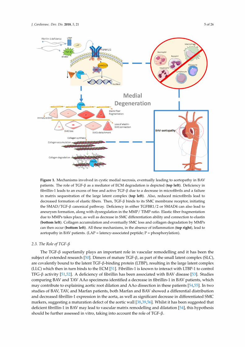
Many studies support the presence of medial degeneration in BAV disease, characterized by: SMCloss, in the absence of inflammation; altered collagen content; elastic fiber fragmentation; accumulationof mucopolysaccharide ground substance within cell-depleted areas of the AAo media [32–34]. Indeed,medial degeneration (Figure 1) is considered the underling abnormality in AAo aneurysm anddissection, regardless of aetiology [32]. Considering the process of aneurysm formation in general,aortic media remodelling is characterised by altered extracellular matrix (ECM) proteins (e.g., collagen,elastin, fibrillin) production and deposition. In the context of BAV, a study of cultured BAV-SMCs
J. Cardiovasc. Dev. Dis. 2018, 5, 21 4 of 26
obtained from aneurysmal tissue showed SMC loss in the absence of inflammation and intracellularaccumulation of fibrillin, fibronectin, and tenascin, suggesting that a defect in cellular transport affectstheir secretion [35]. Another study on patients with BAV-TAA confirmed protein accumulation andsmooth muscle cell loss and also observed elastic fiber fragmentation and decreased collagen I and IIImostly in the convexity of the aorta rather than in the concavity [36]. However, it should be noted thathistological research on BAV compared to TAV aortic vessel wall does not always support the increasein medial degeneration [34,37–39].
Ascending aortic aneurysms also exhibit elevated MMP expression. A significant increase in theMMP-2/tissue inhibitor matrix metalloproteinase (TIMP)-2 ratio has been associated with increasedlikelihood of BAV aneurysm formation [5,40]. Furthermore, in contrast to TAV, BAV aneurysms havebeen characterised by a lack of inflammation, preservation of elastin content, and increased MMP-2,implying that the pattern of MMP expression and the degree of inflammation differ between BAV andTAV scenarios, while the variations in the molecular mechanisms underlying different types of TAAneed further investigation [40].
With regard to histological differences in TAA between BAV and TAV, Philippi et al. discussedthat whilst TAV-TAA has a random, multidirectional collagen and elastin organisation, BAV-TAA has ahighly aligned unidirectional and parallel fiber architecture, indicating that the aorta in BAV patientsremodels following a unique mechanism. In addition, BAV-non-aneurysmal patients were identified toexhibit the same fiber orientation as in BAV-aortopathy patients, suggesting that such microarchitectureis a feature of BAV disease [9]. The highly aligned oriented fibers can also-at least in part-explainthe relationship between BAV and increased AAo stiffness [41,42]. The same study suggests thatprogressive aortic dilation in BAV but not TAV patients might be associated with decreased fiberalignment as a secondary remodelling mechanism, and this could be ascribed to local changes in wallshear stress (WSS) [43–45].
As mentioned, SMCs play an important role in aortic aneurysm development, considering theirinvolvement in inflammation and aortic wall homeostasis. Loss of SMC-TGFβR1 activates pathways,e.g., TGFβR2, ERK, and AngII/AT1R signals, which disrupt aortic wall homeostasis thus leading toaneurysm formation [46]. Furthermore, different molecular alterations in SMCs have been linkedwith aortic aneurysm formation, e.g., Smad4 or TGF-β receptor type II deficiency and subsequentincrease in Cathepsin S and MMP-12, which are proteases essential for elastin degradation (Figure 1).Given the role of Smad4 as a central mediator of the canonical TFG-β signalling pathway, it hasalso been discussed that Smad4-deficient SMCs directly initiate aortic wall inflammation throughthe production of chemokines to recruit macrophages [47]. Studies have tried to unravel the role ofSMCs specifically in BAV-aortopathy. Inability of differentiation in neural crest cells-derived SMCswas related to decreased expression of MYH11, caused by decreased TGF-β signalling based on thephosphorylation of SMAD2, thus indicating that decreased contractile function in these cells maycontribute to the development of AAo dilation in BAV [22]. Differences in the responsiveness ofSMCs to NOTCH and proosteogenic induction were identified between BAV and TAV with associatedaortopathies. Osteogenic induction caused elevated RUNX2 expression in BAV patients, enhancingcalcification in BAV-derived SMCs. In addition, NOTCH activation was identified to significantlyincrease ACTA2 expression specifically in BAV patients, leading to increased osteogenic differentiationin SMCs [48]. Finally, decreased expression of Bcl-2, a mediator of apoptosis, has been identified inBAV aortas and may be involved in SMC apoptosis [49].
In summary, despite current gaps in the knowledge of mechanisms of matrix degeneration inBAV-aortopathy, it has been suggested that SMCs in the BAV aorta exhibit an inherent defect that canlead to changes in vessel wall mechanical properties, thus contributing to aneurysm formation.
J. Cardiovasc. Dev. Dis. 2018, 5, 21 5 of 26J. Cardiovasc. Dev. Dis. 2017, 4, x FOR PEER REVIEW 5 of 26
Figure 1. Mechanisms involved in cystic medial necrosis, eventually leading to aortopathy in BAV
patients. The role of TGF-β as a mediator of ECM degradation is depicted (top left). Deficiency in
fibrillin-1 leads to an excess of free and active TGF-β due to a decrease in microfibrils and a failure in
matrix sequestration of the large latent complex (top left). Also, reduced microfibrils lead to
decreased formation of elastic fibers. Then, TGF-β binds to its SMC membrane receptor, initiating the
SMAD/TGF-β canonical pathway. Deficiency in either TGFBR1/2 or SMAD4 can also lead to
aneurysm formation, along with dysregulation in the MMP/ TIMP ratio. Elastic fiber fragmentation
due to MMPs takes place, as well as decrease in SMC differentiation ability and connection to elastin
(bottom left). Collagen accumulation and eventually SMC loss and collagen degradation by MMPs
can then occur (bottom left). All these mechanisms, in the absence of inflammation (top right), lead
to aortopathy in BAV patients. (LAP = latency-associated peptide; P = phosphorylation).
2.3. The Role of TGF-β
The TGF-β superfamily plays an important role in vascular remodelling and it has been the
subject of extended research [50]. Dimers of mature TGF-β, as part of the small latent complex (SLC),
are covalently bound to the latent TGF-β-binding protein (LTBP), resulting in the large latent complex
(LLC) which then in turn binds to the ECM [51]. Fibrillin-1 is known to interact with LTBP-1 to control
TFG-β activity [51,52]. A deficiency of fibrillin has been associated with BAV disease [53]. Studies
comparing BAV and TAV AAo specimens identified a decrease in fibrillin-1 in BAV patients, which
may contribute to explaining aortic root dilation and AAo dissection in these patients [54,55]. In two
studies of BAV, TAV, and Marfan patients, both Marfan and BAV showed a differential distribution
and decreased fibrillin-1 expression in the aorta, as well as significant decrease in differentiated SMC
markers, suggesting a maturation defect of the aortic wall [38,39,56]. Whilst it has been suggested
Figure 1. Mechanisms involved in cystic medial necrosis, eventually leading to aortopathy in BAVpatients. The role of TGF-β as a mediator of ECM degradation is depicted (top left). Deficiency infibrillin-1 leads to an excess of free and active TGF-β due to a decrease in microfibrils and a failurein matrix sequestration of the large latent complex (top left). Also, reduced microfibrils lead todecreased formation of elastic fibers. Then, TGF-β binds to its SMC membrane receptor, initiatingthe SMAD/TGF-β canonical pathway. Deficiency in either TGFBR1/2 or SMAD4 can also lead toaneurysm formation, along with dysregulation in the MMP/ TIMP ratio. Elastic fiber fragmentationdue to MMPs takes place, as well as decrease in SMC differentiation ability and connection to elastin(bottom left). Collagen accumulation and eventually SMC loss and collagen degradation by MMPscan then occur (bottom left). All these mechanisms, in the absence of inflammation (top right), lead toaortopathy in BAV patients. (LAP = latency-associated peptide; P = phosphorylation).
2.3. The Role of TGF-β
The TGF-β superfamily plays an important role in vascular remodelling and it has been thesubject of extended research [50]. Dimers of mature TGF-β, as part of the small latent complex (SLC),are covalently bound to the latent TGF-β-binding protein (LTBP), resulting in the large latent complex(LLC) which then in turn binds to the ECM [51]. Fibrillin-1 is known to interact with LTBP-1 to controlTFG-β activity [51,52]. A deficiency of fibrillin has been associated with BAV disease [53]. Studiescomparing BAV and TAV AAo specimens identified a decrease in fibrillin-1 in BAV patients, whichmay contribute to explaining aortic root dilation and AAo dissection in these patients [54,55]. In twostudies of BAV, TAV, and Marfan patients, both Marfan and BAV showed a differential distributionand decreased fibrillin-1 expression in the aorta, as well as significant decrease in differentiated SMCmarkers, suggesting a maturation defect of the aortic wall [38,39,56]. Whilst it has been suggested thatdeficient fibrillin-1 in BAV may lead to vascular matrix remodelling and dilatation [54], this hypothesisshould be further assessed in vitro, taking into account the role of TGF-β.
J. Cardiovasc. Dev. Dis. 2018, 5, 21 6 of 26
Dysregulation of TGF-β, perhaps due to a faulty interaction between the SLC and the ECM, andits downstream pathways appeared to be involved in aneurysm formation [4,53]. With regards toLTBPs, these comprise a family of four ECM proteins, all having similar structure to fibrillin and,as such, possibly exerting a similar function [53], although this has not been fully demonstrated yet.One study that started to assess their function showed that LTBP4 is highly expressed in aortic ECMand interacts with matrix molecules, such as fibronectin [57,58]. Indeed, Paloschi et al. discussed anassociation between impaired splicing of fibronectin and increased likelihood of TAA formation inpatients with BAV [59]. In addition, according to a gene expression profiling study, LTBP3 and LTBP4were found to be highly specific for dilation in BAV rather than TAV patients [60]. The role of LTBPsand the molecules they interact with should thus be further explored in BAV-aortopathy, consideringthese promising initial observations.
A recent study, based on secretome analysis carried out in specimens from mildly dilated AAowith stenotic TAV or BAV and from donor normal aortas, revealed that 21 out of 38 identifieddysregulated proteins were participating in TGF-β activation, emphasizing the role of TGF-β inBAV-TAA development. Decreased expression of TGFβR1 mRNA in the curvatures of BAV AAo andits positive correlation with aortic diameter suggest that TGFβR1 downregulation could be a veryearly event in BAV-aortopathy [61]. In addition, an altered imbalance between the TGFβR1 and theTGFβR2 subunits in BAV AAo was linked to the activation of non-canonical TGFβ-mediated signallingpathways, leading to ECM degradation and aneurysm [61,62].
Finally, a recent clinical study [63] comparing cases of TAV, BAV with dilated aorta, and BAV withnon-dilated aorta indicated, by measuring a more comprehensive index of pathogenetically relevantgene expression changes, that it is not just TGF-β1 alone to play a role in this context. The ratioof circulating TGF-β1 to soluble endoglin was, in fact, found to be significantly different betweenTAV and BAV patients. Furthermore, this ratio was independently associated with increased MMP-2gene expression and decreased SOD3 gene expression solely in BAV non-dilated patients, and, in thissubgroup, it significantly correlated with faster aortic growth rate in the postoperative follow-up. Theobserved decrease in soluble endoglin in BAV non-dilated patients was also discussed as potentiallybeing pathogenetically associated with decreased MMP-14 and endoglin gene expression in the aorta.This work can set the base for further studies.
3. Hemodynamics
3.1. Two Theories on the Pathogenesis of BAV–Aortopathy
An on-going debate exists on whether the pathogenesis of BAV–aortopathy is related to geneticsor hemodynamics. A widely accepted theory (“genetic theory”) states that the brittleness of the aorticwall is a result of a developmental abnormality of both the aortic valve and the aortic wall. This theoryis supported by the fact that BAV is a heritable defect and involves gene mutations (such as in GATA5,NOTCH1 and ACTA2 genes). A second theory (“hemodynamic theory”) refers to the abnormal WSSacting on the aortic wall as the process underpinning aortic dilation [64]. As it will be discussed inthis section, this theory is supported by the fact that the eccentric turbulent flow through the bicuspidvalve has been associated with abnormal mechanical stresses in certain regions of the aortic wall, inturn causing stress overload and wall fragility [11].
3.2. The Role of Hemodynamics
The turbulent flow jet passing through the BAV into the aortic root has been recognized tocontribute to an abnormal biomechanical environment, including helical flow alterations that propagateeccentrically inside the proximal AAo [4]. Evidence shows that increased blood flow helicity leads toincreased WSS [65], as exemplified in Figure 2. It has also been proven that the bigger the angle ofthe misdirected flow through the conjoined cusp opening, the higher the degree of flow eccentricity,with subsequent increased growth rate and severity of AAo dilation [66]. However, a study using
J. Cardiovasc. Dev. Dis. 2018, 5, 21 7 of 26
convex and concave aorta biopsy samples showed different histopathological features between jetand non-jet samples [67]. This difference was similarly observed in BAV and TAV, with BAV beingmore significantly associated; however, this needs further assessment. In support of the hemodynamictheory, different BAV morphologies have been associated with specific dilation patterns of the AAo [68].Some studies observed differential distributions of WSS according to different valve morphotypes,resulting in specific orientation of eccentric flow jets and suggesting possible flow-induced vascularremodelling [69–71]. Specifically, the most common BAV morphotype (i.e., right coronary cusp-leftcoronary cusp raphe) was associated with right-anteriorly directed helical systolic flow jet withperipheral skewing towards the AAo convexity, with these patients exhibiting also larger aortic root,asymmetric mid-AAo dilation, and more severe AAo wall degeneration [72,73]. On the other hand,the second commonest BAV morphotype (i.e., right coronary cusp-non-coronary cusp raphe) wasassociated with left-posteriorly directed eccentric flow jet spreading towards the proximal aorticarch, with patients exhibiting isolated AAo dilation, no aortic root dilation, and increased aortic archdiameter [72]. Another study confirmed that patients with right-non-coronary morphotype were morelikely to present with AAo dilation, whereas patients with right-left morphotype were more likely tohave aortic root dilation and, when accounting for age, they were more likely to present with aorticstenosis as well [74]. However, there are also studies reporting a weak or independent associationbetween the BAV morphotype and the shape of the aneurysm [75–77]. These clinical studies werecarried out on large cohorts of patients and, despite their statistical power, no association was foundbetween morphotype and patterns of dilatation, indicating the need for further assessment of thisrelationship and providing evidence in support of genetic mechanisms underlying BAV-aortopathy.
Complementary to the clinical studies, experimental and computational work can also generateprovocative insights into key parameters related to BAV hemodynamics. This includes systematicallytesting morphotype-dependent alterations and exploring whether abnormal hemodynamic parameters(such as helical flow, flow angle, or WSS) arise from the underlying abnormal valve anatomy or fromthe dilated aorta. One study employed particle image velocimetry to test tissue BAVs derived fromporcine TAVs subjected to physiologic pulsatile flow in an experimental setup (pulse duplicator setting)and identified an element of morphotype-dependency as to which site of the aorta is affected byshear stress overloads [78]. A simulation-based study also confirmed that different BAV morphotypesaffect aortic hemodynamics differently, whilst all being abnormal with respect to TAV and leading toincreased WSS on the proximal AAo, and indicated BAV with left-right-coronary cusp morphotype ashaving the most significant abnormality [79].
The impact of altered hemodynamics on aortic wall abnormalities is also reflected in theasymmetric spatial distribution of histological and biomolecular changes in BAV aortas, in contrastto their uniform distribution in patients with Marfan syndrome [64]. According to observationsby Guzzardi et al., based on 4-dimensional (4D) flow cardiac magnetic resonance (CMR) imagingdata and histological analyses, increased WSS was associated with decreased elastin content andincreased distance between the elastin fibers, as well as to a higher concentration of mediators ofECM dysregulation (i.e., MMPs and TGF-β) [80,81]. Regions of the dilated AAo wall with elevatedWSS had significantly increased TGF-β1 concentrations [80,82]. These regions also had increasedMMP-1, MMP-2, MMP-3, and higher MMP-2 to TIMP-1 activity, factors that have been associatedwith increased elastic fiber fragmentation [33,80]. Further studies using 4D CMR confirmed that flowasymmetry and helicity are elevated in BAV, contributing to WSS increase, and that increased WSSand medial derangement (e.g., reduced collagen and increased SMCs apoptosis) are associated withgreater aortic convexity even before severe dilation is observed [5,65,73,83]. In another study based on4D CMR, it has been hypothesised that BAV-aortopathy might be initiated by flow abnormalities andentail a sort of protective mechanism to maintain normal WSS levels in the face on the increased WSSconsequent to the abnormal flow [84].

J. Cardiovasc. Dev. Dis. 2018, 5, 21 8 of 26
J. Cardiovasc. Dev. Dis. 2017, 4, x FOR PEER REVIEW 8 of 26
and entail a sort of protective mechanism to maintain normal WSS levels in the face on the increased
WSS consequent to the abnormal flow [84].
Figure 2. Representation of the wall shear stress (WSS) distribution (A, posterior view of the aorta)
and flow velocity (B, front view of the ascending aorta) in a healthy volunteer and a BAV patient with
right coronary cusp-left coronary cusp raphe, obtained from 4D flow cardiac magnetic resonance
(CMR) imaging. This is a visual indication of increased WSS at the aneurysm location and abnormal
flow jet in the presence of BAV. Furthermore, the flow in the BAV patient seems to go along the
anterior right curvature of the AAo and then fold inferiorly along the inner curvature, rather than
accessing the transverse aortic arch, as also reported in the literature [85]. Data collected at the Clinical
Research and Imaging Centre (CRiC), University of Bristol; not previously published.
Experimental work also contributes to generate data in support of the effect of stresses resulting
from BAV hemodynamics on aortic medial degradation. Interesting experiments have been designed
by first generating values for WSS typical of both TAV and BAV hemodynamic scenarios with
computational modelling, and then testing normal porcine aortic tissue in a bioreactor when
subjected to such stresses and measuring relevant changes, such as MMP-2 and MMP-9 expressions,
MMP-2 activity, or fibrillin-1 content. The results, however, were not conclusive [86,87], but the
methodology is certainly compelling to investigate possible cause-and-effect relationships between
tissue properties and hemodynamics.
In summary, evidence exists in support of both genetic and hemodynamic theories. Clinical
studies indicated that the progression of AAo dilation in BAV patients continues after aortic valve
replacement leading to aortic dissection or rupture [64,88,89], which would suggest an underlying
genetic driving mechanism. Other arguments in favour of the genetic theory are the enlargement of
the aorta even in the absence of valvular dysfunction (i.e., stenosis and/or regurgitation) [4,5,64] as
Figure 2. Representation of the wall shear stress (WSS) distribution (A, posterior view of the aorta) andflow velocity (B, front view of the ascending aorta) in a healthy volunteer and a BAV patient with rightcoronary cusp-left coronary cusp raphe, obtained from 4D flow cardiac magnetic resonance (CMR)imaging. This is a visual indication of increased WSS at the aneurysm location and abnormal flowjet in the presence of BAV. Furthermore, the flow in the BAV patient seems to go along the anteriorright curvature of the AAo and then fold inferiorly along the inner curvature, rather than accessing thetransverse aortic arch, as also reported in the literature [85]. Data collected at the Clinical Research andImaging Centre (CRiC), University of Bristol; not previously published.
Experimental work also contributes to generate data in support of the effect of stresses resultingfrom BAV hemodynamics on aortic medial degradation. Interesting experiments have been designedby first generating values for WSS typical of both TAV and BAV hemodynamic scenarios withcomputational modelling, and then testing normal porcine aortic tissue in a bioreactor when subjectedto such stresses and measuring relevant changes, such as MMP-2 and MMP-9 expressions, MMP-2activity, or fibrillin-1 content. The results, however, were not conclusive [86,87], but the methodologyis certainly compelling to investigate possible cause-and-effect relationships between tissue propertiesand hemodynamics.
In summary, evidence exists in support of both genetic and hemodynamic theories. Clinicalstudies indicated that the progression of AAo dilation in BAV patients continues after aortic valvereplacement leading to aortic dissection or rupture [64,88,89], which would suggest an underlyinggenetic driving mechanism. Other arguments in favour of the genetic theory are the enlargement of theaorta even in the absence of valvular dysfunction (i.e., stenosis and/or regurgitation) [4,5,64] as wellas the fact that overall higher aortic size in BAV patients after matching for TAV patients with similardegrees of valvular disease was identified to be independent of hemodynamic alterations [64,90].However, it should also be considered that even a functionally normal bicuspid valve is, in fact,
J. Cardiovasc. Dev. Dis. 2018, 5, 21 9 of 26
morphologically stenotic because of the conjoined leaflets. Such valve configuration can lead to atransvalvular turbulent flow jet and abnormal haemodynamics, and jet eccentricity results in moresevere flow alterations in the AAo when it occurs through a stenotic bicuspid orifice instead of atricuspid aortic valve of comparable gradient and valve area. Furthermore, when considering cysticmedial degeneration in the AAo wall of BAV patients, if, on the one hand, this phenomenon couldsupport the genetic theory, similar changes have been identified in AAo dilation or dissection regardlessof aetiology, indicating a non-specific character of cystic medial degeneration [35,91]. At present,continued research in the field and new findings from clinical, experimental, and modelling studiesrather suggest an interplay between the two theories. What remains to be unravelled is which of thetwo can be considered as the ‘initiator’ of aortopathy, which of the two has potentially a predominantcontribution, and how the interplay between the two may vary in different scenarios.
4. Environmental Impact
4.1. Environment and Risk Factors
Variable penetrance and phenotypic expression in BAV could be the result not only of geneticvariations, but also of the interplay between genetic, epigenetic, and environmental modifiers,eventually leading to aortopathy. Epidemiological studies have been carried out to explore predictorsof TAA in BAV patients, and their findings suggest that older age, diabetes, hypercholesterolaemia,aortic regurgitation, and smoking increase the risk of TAA [2,92]. The interplay between genetic andenvironmental factors has also been studied in mice [93]. It should be noted that the incorporation ofmultiple mutations or gene–environment interactions is required to replicate the complexity of humanBAV disease [1], whereas animal models with single gene mutations may not be representative toinvestigate the influence of environmental hazards on complications and outcomes of the BAV disease.
4.2. Epigenetic Mechanisms
The impact and interplay of environmental risk factors can also be mediated through epigenetics,and indeed studies have been performed to explore the epigenetic signature of BAV-aortopathy.
As described in a recent study, both BAV and AAo dissection are characterized by a non-CpGhypomethylation signature that partially explains the increased cellular proliferation, although theypresent different DNA methylation landscapes [94]. Another study, comparing gene methylationand expression from AAo aneurysm tissue samples between BAV and TAV patients, identifiedhypomethylation of ACTA2, hypermethylation of GATA4, and significant hypermethylation anddecreased expression of protein tyrosine phosphatase non-receptor type 22 in BAV individuals [95].These studies suggest that altered gene methylation is involved in the pathogenesis of aneurysmformation in BAV patients. However, further investigation is needed to understand the relationshipsbetween DNA methylation and differential gene expression and whether they imply causality or areby-products of the aneurysm in the case of BAV.
The knowledge of epigenetic modifications related to TGF-β pathways in TAA could help clarifyits pathogenesis. According to Kurtovic et al., a diverging alternative splicing fingerprint of theTGF-β pathways is linked to BAV-TAA, causing differential downstream effects. This might be theresult of chromatin epigenetic modification, leading to BAV-aortopathy phenotypic modification [58].In addition, induced histone methylation and acetylation of the SMAD2 promoter in SMCs inBAV-dilated aortas was linked with SMAD2 overexpression, suggesting TGF-β/SMAD pathwaydysregulation due to epigenetics [96]. The modification of histone H3 marker in the SMAD2 promoteris an epigenetic mechanism behind SMAD2 overexpression, as demonstrated in all types of TAA,including BAV-TAA.
New insight into epigenetic reprogramming could lead to a better understanding of the onsetand progression of TAA in BAV patients, thus contributing to the potential identification of novelbiomarkers and/or therapeutic strategies.
J. Cardiovasc. Dev. Dis. 2018, 5, 21 10 of 26
5. The Role of MicroRNAs
MicroRNAs (miRNAs, or miRs) are small noncoding RNA regulatory molecules that regulate theexpression of a plethora of mRNAs “targets”, to which they can bind, interacting with their 3′-UTR(canonical mechanism), the aminoacid coding sequence (CDS), or the 5′-UTR. The canonical mechanismconsists in the miRNA seed sequence targeting one or more (semi)complementary regions of the mRNA3′-UTR and results in expressional repression at the mRNA and/or protein level. Importantly, miRNAscan also be released by the parent cell via different shuttles, including extracellular vesicles (exosomes,microvescicles) and lipoproteins that protect their miRNA cargos from degradation. Such extracellularmiRNAs can be taken up by a series of recipient cells in neighbouring and distant tissues, contributingto cell-to-cell communication. Moreover, by conferring resilience to miRNAs, the shuttles incidentallyincrease our possibilities to develop miRNAs as extracellular biomarkers.
This section briefly revisits the pathways so far described to affect AV morphology, BAV,and/or TAA. A number of miRNAs have been functionally implicated in processes that conductto BAV aortopathy and/or have been found to be regulated by fluctuations in shear stress, which-asdiscussed-could contribute to TAA in BAV. Therefore, miRNAs could represent both therapeutictargets for intervention and circulating biomarkers that could help predict and monitor the evolutionof BAV-TAA. In this setting, miRNAs regulating calcification, elastin degradation, and changes inextracellular matrix are of potential relevance. Similarly, miRNAs regulating “classic pathways”already implicated with BAV and TAA also deserve attention.
5.1. MicroRNAs Involved in BAV Disease and Valve Morphology
The expression and possible role of miRNAs in the calcification of stenotic BAVs has been exploredin vitro using intraoperative samples in an effort to identify potentially deregulated miRNAs, aswell as calcification-related factors that either regulate or are regulated by those miRNAs [97,98].The role of the noncanonical Wnt signaling pathway [99], as well as the role of TGF-β1 in thestimulation of aortic valvular interstitial cells, leading to morphological changes consistent withmyofibroblastic transformation, BMP-2 signaling, and calcification, seem to be of particular interest inrelation to AV stenosis and aneurysm [100–102]. Since osteoblasts are involved in ECM productionand mineralisation, miRNAs able to regulate osteogenesis and chondrogenesis, such as miR-29, 210,125b, 26a, 196a, 2861, and others, are of potential relevance, even if their role in BAV is still underinvestigation [103–110]. Moreover, miRNAs, such as miR-29, 181a, and 195, have been shown toregulate ECM composition, acting on collagens, MMPs, and TIMPs [111–113]. Interestingly, miR-29bhas already been shown to promote aortic valve interstitial cell calcification by inhibiting TGF-β3through the activation of wnt3/β-catenin and to induce elastin downregulation, contributing toinorganic phosphorus-induced osteoblastic differentiation in vascular SMCs [114]. Moreover, areduction of miR-195 in BAV was suggested to promote calcification of valve interstitial cells viaSMAD7 targeting, and the mechanosensitive miR-181b regulates AV endothelial matrix degradation bytargeting TIMP3 [115,116]. Other miRs reported to be deregulated in AV calcification and potentiallymediating the calcification process are miR-204 and miR-449c [117,118].
5.2. MicroRNAs Involved in Aortopathy
Although more than 20% of TAAs are inherited as single-gene disorder, the majority aresporadic cases and known to be driven by the unbalanced production of extracellular proteases andinhibitors [119,120]. The upstream signalling events are still widely unknown; however, a potentialleading cause could be altered miRNA expression, leading to gene expression impairment [120,121].Also, it is worth mentioning that variants in miRNA genes can have a profound effect on miRNAexpression and function and can thus contribute to disease [122].
Interestingly, the aetiology of descending thoracic and abdominal aortic aneurysm disease ismainly atherosclerotic, whilst proximal thoracic aortic aneurysm disease has been associated with

J. Cardiovasc. Dev. Dis. 2018, 5, 21 11 of 26
proteoglycan accumulation, elastic fiber fragmentation, and focal or diffuse SMC degradation andloss [123]. In vitro studies using aortic samples of TAV or BAV as well as in vivo studies indicatedthe role of miRNAs in several pathways implicated in TAA pathogenesis, such as the focal adhesionpathway [121,124,125], ECM homeostasis [111,126–128], TGF-β pathway [124,129,130], and SMCsplasticity and survival [131,132].
The aforementioned miR-29, 181a, and 195 that regulate elastin and ECM could indeed be alsorelevant for the aneurysmal process and hence TAA [111–113]. Moreover, because of the fact thatcalcification is associated with ECM modifications, studies have been performed to appreciate therole of miRNAs with regard to elastin and ECM degradation [112,133], as well as their role on SMCsphenotypic modification, by studying Dicer-dependent miRNAs role on SMC growth, differentiation,and function in vivo [134–136].
5.3. MicroRNAs Regulated by Changes in Shear Stress
Vascular physiology is maintained through alterations in shear stress that potentially lead tomiR-regulated differential gene expression in endothelial cells. MiRNAs induced by laminar shearstress, like miR-126, 27b, and 143/145 were identified as leading to protection from atherosclerosis,whilst miRNAs induced by low oscillatory shear stress (like miR-181b) were identified as leading topathological vascular phenotypes [116,137–139], potentially triggering cardiovascular diseases.
5.4. MicroRNAs as Potential Therapeutic Targets and Biomarkers in BAV Aortopathy
Although most studies focus on the effect of miRNAs on either BAV morphology or aorticdilation, there are only a few that identified a relationship between both aortic valve morphology anddilation [140]. BAV patients were identified as having distinct regional miRNAs signatures in dilatedaortas, indicating the differential expression of miRNAs in BAV convexity versus concavity [130].Moreover, miRNAs were identified to influence the balance between MMP and TIMP as an earlyhallmark of BAV-aortopathy. Therefore, the upregulation of these miRNAs could exhibit a potentialfor therapeutic targeting [128]. The concept of miRNA-therapeutics essentially refers to either using“antagomiRs” or other inhibitory strategies to reduce the expression and/or activities of specificpathogenic miRNAs, or using “microRNA mimics” or other approaches to enhance the expression of atherapeutic miRNA and increase its level [141]. Compared to traditional drugs, the first generation ofmiRNA-therapeutics is designed to affect all the genes that are regulated by the target miRNA, thuspotentially having a profound physiological impact. The potential role of miRNA therapeutics in thecontext of BAV-TAA, however, is yet to be explored.
In addition to their potential as therapeutic targets, those miRNAs that have been found tobe functionally involved in BAV disease and morphology [142] should be further screened in BAVaortopathy (and controls) tissue and plasma/serum samples (including their extracellular vesiclescomponents) to gain evidence of their potential as diagnostic and prognostic biomarkers.
Whilst most studies focus on analysing the expression of miRNAs in aortic tissue segments,either in patients or in animal models, few studies were carried out screening plasma of BAV-TAApatients [140,143]. Unlike tissue-specific miRNAs, circulating miRNAs can provide a complementaryinsight to that obtained from the complex analysis of multiple tissues [140]. They are also protectedfrom endogenous ribonuclease-induced degradation and can be easily accessed by minimally invasivemeans [140,144]. Tissue or circulating miRNAs, involved in BAV morphology, aortopathy in general,or regulated by changes in shear stress, should all be specifically explored in vitro and in vivo toassess whether they play a specific role in BAV aortopathy. This could lead to the identification ofnew biomarkers.
J. Cardiovasc. Dev. Dis. 2018, 5, 21 12 of 26
6. Managing BAV Aortopathy
6.1. Clinical Management
Despite major advances in the diagnosis, genetic screening, and ability to manipulate diseaseprocesses by pharmacological means, TAA disease can still only be managed by a combination ofradiological surveillance and surgery. Presently, targets of progressive BAV disease or BAV-aortopathyare not well established, and their detection and management are still ongoing [11,145]. There is alsolimited understanding of the duration and long-term response to pharmacological treatments, due tothe gaps in the knowledge surrounding disease pathogenesis and its underling mechanisms [11,64].Based on the decision-making algorithm for BAV management in North America and Europe,the recommended drugs are antihypertensive agents, including β-blockers, ACE inhibitors, andangiotensin receptor blockers [146], and recent guidelines for managing AAo aneurysm in non-Marfanpatients also recommend β-blockers; however, there is a lack of systematic evaluation of theseagents [8,145] and in vivo studies in BAV patients [147]. Although the pharmacological treatment isdebatable, it is hypothesised that blood pressure control can decrease the rate of change of centralarterial pressure, leading to decreased stresses acting on the more vulnerable aneurysmal segmentof the aorta, thus preventing further aortic remodelling and dilation. In theory, a reduction of aorticWSS could be an advantage of the β-adrenergic blockers, whereas the angiotensin-receptor blockers,such as Losartan, have been indicated to decrease the rate of aortic growth in patients with Marfansyndrome, but these findings should be investigated in BAV-TAA too [145,146,148]. According toMcGee et al., there is no significant hemodynamic change due to medication in BAV patients [147];however, further investigation in larger cohorts is necessary. Interestingly, the use of statins has beenassociated with decreased risk of clinically significant aneurysm in the abdominal aorta, which couldbe explained by the fact that distal aortic aneurysms are usually atherosclerotic in aetiology, and a largeretrospective study reported lower odds of TAA in BAV patients taking statins preoperatively [149,150].Nevertheless, the role of statins in this context should be explored further and prospectively beforeclaiming a clinical benefit.
On the basis of a large survey, operative approaches and management of BAV aortopathy arequite variable and do not always follow guideline recommendations [151]. According to the guidelines(i.e., European Society of Cardiology/ESC, American Heart Association/AHA, American College ofCardiology/ACC), aortic root or AAo repair/replacement is recommended in the presence of an aorticdiameter ≥ 5.5 cm in asymptomatic BAV patients, for an aortic diameter ≥ 5.0 cm in BAV patients withan additional risk for dissection, e.g., family history, or in patients at low surgical risk, and for aorticdiameter ≥ 4.5 cm, in BAV patients undergoing aortic valve replacement due to severe stenosis orregurgitation [152–154]. Furthermore, according to the ACC and AHA guidelines, first-degree relativesof BAV patients with aortic dissection should be imaged for valvular and extravalvular complications,in order for affected individuals to receive interventions to reduce the traditional cardiovascular riskfactors. Also, every newly diagnosed BAV patient is recommended to undergo a comprehensive clinicalevaluation, whereas genetic tests are offered when features of single-gene disorders or syndromesare present [155]. Since BAV and syndromic TAA disease can co-exist, patients and their first-degreerelatives who present with aortic dissection get offered genetic aortopathy screening.
J. Cardiovasc. Dev. Dis. 2018, 5, 21 13 of 26
6.2. Genetic Tools
The low incidence of BAV in the general population creates an obstacle for performinggenome-wide association (GWA) studies. For this reason, a study analysing single nucleotidepolymorphisms relevant to BAV was performed, reducing the multiple-testing penalty to controlfor random associations [156]. However, in order to sort through the genetic complexity ofBAV–aortopathy, large patient cohorts are needed and should take into account ethnic diversity,different types of BAV, aneurysm location, and risk factors. Especially in the case of BAV, where>80% cases are sporadic, case-control association methods such as GWA studies need to be performed,requiring large samples sizes [1,157]. One recent study in a big cohort of patients (n = 466 BAV,n = 4660 controls) identified two protein-altering and regulatory genetic variants near GATA4 [158].Considering the existing expression and genomic data in the context of GWA studies, as well astaking advantage of system-based approaches to rank the candidate genes and screen for mutationsby sequencing or copy number analysis, it is possible to achieve the identification of biologicallyrelevant genes and pathways associated with BAV-aortopathy [156]. Causative genetic variants canbe identified through family-based studies, by sequencing the entire exomes or genomes of multiplefamily members [159,160], whereas de novo variants, which tend to be more deleterious than inheritedvariations, need to be validated by performing mutation analyses [161]. At present, there are onlylimited reported cases of de novo variants in individuals with BAV. An example is a family with aseverely affected child who received a unique variant from the mother, leading to a gain of the PXDNLlocus on chromosome 8, whereas that particular de novo gain was also identified in the maternal unclewith known BAV [162]. The knowledge of a specific mutation in a family can enforce clinicians toscreen family members and search for more clinical features.
The discovery of new genes involved in BAV-aortopathy will lead to more targeted clinicaldecisions, diagnostic tests, and therapies. Since BAV–TAA displays incomplete penetrance, theassociated genetic variants may also be present in unaffected individuals, indicating the need forcandidate gene prioritization [163,164]. However, mutations in the same gene can lead to AAodissection varying from recognizable genetic syndromes to sporadic diseases [165–168]. Furthermore,it is often a burden of genetic variants along with environmental factors that lead BAV patients todevelop aortopathy, rather than a single variant [4,169], and, consequently, prioritizing candidate genesis more challenging. Despite the limitations of BAV animal models, targeted gene deletions in mice,zebrafish, and cultured aortic valve interstitial cells provided insight into candidate gene involvementin aortic valve development and other associated conditions [28,170,171]. High-throughput assays canbe used to screen candidate genes [172]. However, there is a need for improved genetic models in orderto investigate therapeutic targets. Also, “knock-in” experiments, introducing human mutationsinto homologous mouse or zebrafish genes, may result in models of human BAV phenotypesmore promising than the current models with null mutations [173,174]. In the specific context ofBAV-aortopathy, candidate genes should be investigated further, including their biological functionsand the different pathways involved. Whilst these methods are beginning to be used successfully,e.g., in inherited cardiomyopathies [175–177], they could also be used to enlighten BAV-aortopathypathogenesis and progression, as well as potential therapeutic solutions.
7. Discussion
It is known that TAAs are associated with connective tissue diseases, BAV disease, and familialthoracic aneurysm syndrome [12]. They depict the most lethal site for aortic dilation, since they mightbe asymptomatic for many years and manifest as an acute rupture or dissection [123]. Considering theincidence of BAV patients who develop dilation compared to TAV patients and the lack of up-to-dateknowledge in this field [5], it is important to enlighten the pathogenesis of BAV-aortopathy bystratifying the different causes and the management and research strategies, rather than discussingBAV morphology and TAA separately.
J. Cardiovasc. Dev. Dis. 2018, 5, 21 14 of 26
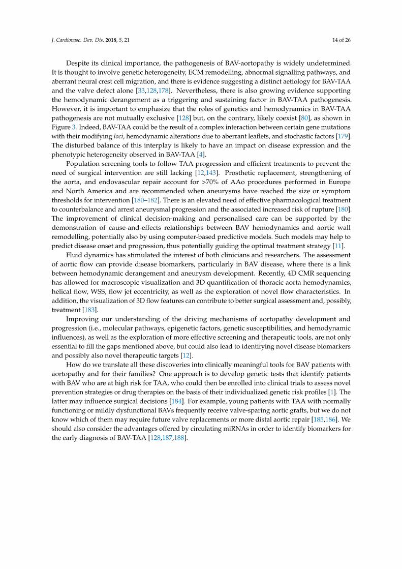
Despite its clinical importance, the pathogenesis of BAV-aortopathy is widely undetermined.It is thought to involve genetic heterogeneity, ECM remodelling, abnormal signalling pathways, andaberrant neural crest cell migration, and there is evidence suggesting a distinct aetiology for BAV-TAAand the valve defect alone [33,128,178]. Nevertheless, there is also growing evidence supportingthe hemodynamic derangement as a triggering and sustaining factor in BAV-TAA pathogenesis.However, it is important to emphasize that the roles of genetics and hemodynamics in BAV-TAApathogenesis are not mutually exclusive [128] but, on the contrary, likely coexist [80], as shown inFigure 3. Indeed, BAV-TAA could be the result of a complex interaction between certain gene mutationswith their modifying loci, hemodynamic alterations due to aberrant leaflets, and stochastic factors [179].The disturbed balance of this interplay is likely to have an impact on disease expression and thephenotypic heterogeneity observed in BAV-TAA [4].
Population screening tools to follow TAA progression and efficient treatments to prevent theneed of surgical intervention are still lacking [12,143]. Prosthetic replacement, strengthening ofthe aorta, and endovascular repair account for >70% of AAo procedures performed in Europeand North America and are recommended when aneurysms have reached the size or symptomthresholds for intervention [180–182]. There is an elevated need of effective pharmacological treatmentto counterbalance and arrest aneurysmal progression and the associated increased risk of rupture [180].The improvement of clinical decision-making and personalised care can be supported by thedemonstration of cause-and-effects relationships between BAV hemodynamics and aortic wallremodelling, potentially also by using computer-based predictive models. Such models may help topredict disease onset and progression, thus potentially guiding the optimal treatment strategy [11].
Fluid dynamics has stimulated the interest of both clinicians and researchers. The assessmentof aortic flow can provide disease biomarkers, particularly in BAV disease, where there is a linkbetween hemodynamic derangement and aneurysm development. Recently, 4D CMR sequencinghas allowed for macroscopic visualization and 3D quantification of thoracic aorta hemodynamics,helical flow, WSS, flow jet eccentricity, as well as the exploration of novel flow characteristics. Inaddition, the visualization of 3D flow features can contribute to better surgical assessment and, possibly,treatment [183].
Improving our understanding of the driving mechanisms of aortopathy development andprogression (i.e., molecular pathways, epigenetic factors, genetic susceptibilities, and hemodynamicinfluences), as well as the exploration of more effective screening and therapeutic tools, are not onlyessential to fill the gaps mentioned above, but could also lead to identifying novel disease biomarkersand possibly also novel therapeutic targets [12].
How do we translate all these discoveries into clinically meaningful tools for BAV patients withaortopathy and for their families? One approach is to develop genetic tests that identify patientswith BAV who are at high risk for TAA, who could then be enrolled into clinical trials to assess novelprevention strategies or drug therapies on the basis of their individualized genetic risk profiles [1]. Thelatter may influence surgical decisions [184]. For example, young patients with TAA with normallyfunctioning or mildly dysfunctional BAVs frequently receive valve-sparing aortic grafts, but we do notknow which of them may require future valve replacements or more distal aortic repair [185,186]. Weshould also consider the advantages offered by circulating miRNAs in order to identify biomarkers forthe early diagnosis of BAV-TAA [128,187,188].
J. Cardiovasc. Dev. Dis. 2018, 5, 21 15 of 26
J. Cardiovasc. Dev. Dis. 2017, 4, x FOR PEER REVIEW 15 of 26
Figure 3. Schematic summary of the multiscale nature of the problem of BAV aortopathy and the
interplay of different dimensions. The blue arrows indicate the known or possible interplay between
genetics, hemodynamic alterations, and other factors underlying BAV aortopathy. These represent
stimulating areas of current and future research.
Figure 3. Schematic summary of the multiscale nature of the problem of BAV aortopathy and theinterplay of different dimensions. The blue arrows indicate the known or possible interplay betweengenetics, hemodynamic alterations, and other factors underlying BAV aortopathy. These representstimulating areas of current and future research.
J. Cardiovasc. Dev. Dis. 2018, 5, 21 16 of 26
8. Conclusions
Whilst advances in medical imaging render our insight into aortic hemodynamics muchmore refined by generating not just exquisite qualitative data but also quantitative hemodynamicmeasurements, the link between hemodynamic derangements due to different valve morphotypesand the underlying tissue biology remains to be fully demonstrated. As reminded by importantresearch efforts in the field [189], an association does not imply causality, nevertheless mergingmultimodality imaging with genotyping tools is an avenue of research that can lead to shedding lighton the pathological, phenotypic, and ultimately functional aspects of this problem.
Conflicts of Interest: The authors declare no conflict of interest.
References
1. Prakash, S.K.; Bosse, Y.; Muehlschlegel, J.D.; Michelena, H.I.; Limongelli, G.; Della Corte, A.; Pluchinotta, F.R.;Russo, M.G.; Evangelista, A.; Benson, D.W.; et al. A roadmap to investigate the genetic basis of bicuspidaortic valve and its complications: Insights from the international bavcon (bicuspid aortic valve consortium).J. Am. Coll. Cardiol. 2014, 64, 832–839. [CrossRef] [PubMed]
2. Della Corte, A.; Bancone, C.; Quarto, C.; Dialetto, G.; Covino, F.E.; Scardone, M.; Caianiello, G.; Cotrufo, M.Predictors of ascending aortic dilatation with bicuspid aortic valve: A wide spectrum of disease expression.Eur. J. Cardio-Thorac. Surg. 2007, 31, 397–405.
3. Vallely, M.P.; Semsarian, C.; Bannon, P.G. Management of the ascending aorta in patients with bicuspidaortic valve disease. Heart Lung Circ. 2008, 17, 357–363. [CrossRef] [PubMed]
4. Padang, R.; Bannon, P.G.; Jeremy, R.; Richmond, D.R.; Semsarian, C.; Vallely, M.; Wilson, M.; Yan, T.D.The genetic and molecular basis of bicuspid aortic valve associated thoracic aortopathy: A link to phenotypeheterogeneity. Ann. Cardiothorac. Surg. 2013, 2, 83–91. [PubMed]
5. Losenno, K.L.; Goodman, R.L.; Chu, M.W. Bicuspid aortic valve disease and ascending aortic aneurysms:Gaps in knowledge. Cardiol. Res. Pract. 2012, 2012, 145202–145218. [CrossRef] [PubMed]
6. Siu, S.C.; Silversides, C.K. Bicuspid aortic valve disease. J. Am. Coll. Cardiol. 2010, 55, 2789–2800. [CrossRef][PubMed]
7. Isselbacher, E.M.; Lino Cardenas, C.L.; Lindsay, M.E. Hereditary influence in thoracic aortic aneurysm anddissection. Circulation 2016, 133, 2516–2528. [CrossRef] [PubMed]
8. Kuijpers, J.M.; Mulder, B.J. Aortopathies in adult congenital heart disease and genetic aortopathy syndromes:Management strategies and indications for surgery. Heart 2017, 103, 952–966. [CrossRef] [PubMed]
9. Phillippi, J.A.; Green, B.R.; Eskay, M.A.; Kotlarczyk, M.P.; Hill, M.R.; Robertson, A.M.; Watkins, S.C.;Vorp, D.A.; Gleason, T.G. Mechanism of aortic medial matrix remodeling is distinct in patients with bicuspidaortic valve. J. Thorac. Cardiovasc. Surg. 2014, 147, 1056–1064. [CrossRef] [PubMed]
10. Davies, R.R.; Kaple, R.K.; Mandapati, D.; Gallo, A.; Botta, D.M., Jr.; Elefteriades, J.A.; Coady, M.A. Naturalhistory of ascending aortic aneurysms in the setting of an unreplaced bicuspid aortic valve. Ann. Thorac. Surg.2007, 83, 1338–1344. [CrossRef] [PubMed]
11. Atkins, S.K.; Sucosky, P. Etiology of bicuspid aortic valve disease: Focus on hemodynamics. World J. Cardiol.2014, 6, 1227–1233. [CrossRef] [PubMed]
12. Li, Y.; Maegdefessel, L. Non-coding rna contribution to thoracic and abdominal aortic aneurysm diseasedevelopment and progression. Front. Physiol. 2017, 8, 429. [CrossRef] [PubMed]
13. Martin, L.J.; Ramachandran, V.; Cripe, L.H.; Hinton, R.B.; Andelfinger, G.; Tabangin, M.; Shooner, K.;Keddache, M.; Benson, D.W. Evidence in favor of linkage to human chromosomal regions 18q, 5q and 13qfor bicuspid aortic valve and associated cardiovascular malformations. Hum. Genet. 2007, 121, 275–284.[CrossRef] [PubMed]
14. Cripe, L.; Andelfinger, G.; Martin, L.J.; Shooner, K.; Benson, D.W. Bicuspid aortic valve is heritable. J. Am.Coll. Cardiol. 2004, 44, 138–143. [CrossRef] [PubMed]
15. Milewicz, D.M.; Regalado, E. Heritable thoracic aortic disease overview. In Genereviews®; Adam, M.P.,Ardinger, H.H., Pagon, R.A., Wallace, S.E., Bean, L.J.H., Mefford, H.C., Stephens, K., Amemiya, A.,Ledbetter, N., Eds.; Genereviews: Seattle, WA, USA, 1993.
J. Cardiovasc. Dev. Dis. 2018, 5, 21 17 of 26
16. Martin, L.J.; Hinton, R.B.; Zhang, X.; Cripe, L.H.; Benson, D.W. Aorta measurements are heritable andinfluenced by bicuspid aortic valve. Front. Genet. 2011, 2, 61. [CrossRef] [PubMed]
17. Mathieu, P.; Bosse, Y.; Huggins, G.S.; Corte, A.D.; Pibarot, P.; Michelena, H.I.; Limongelli, G.; Boulanger, M.C.;Evangelista, A.; Bedard, E.; et al. The pathology and pathobiology of bicuspid aortic valve: State of the artand novel research perspectives. J. Pathol. Clin. Res. 2015, 1, 195–206. [CrossRef] [PubMed]
18. Yetman, A.T.; Graham, T. The dilated aorta in patients with congenital cardiac defects. J. Am. Coll. Cardiol.2009, 53, 461–467. [CrossRef] [PubMed]
19. Grewal, N.; DeRuiter, M.C.; Jongbloed, M.R.; Goumans, M.J.; Klautz, R.J.; Poelmann, R.E.; Gittenberger-deGroot, A.C. Normal and abnormal development of the aortic wall and valve: Correlation with clinicalentities. Neth. Heart J. 2014, 22, 363–369. [CrossRef] [PubMed]
20. Sawada, H.; Rateri, D.L.; Moorleghen, J.J.; Majesky, M.W.; Daugherty, A. Smooth muscle cells derived fromsecond heart field and cardiac neural crest reside in spatially distinct domains in the media of the ascendingaorta-brief report. Arterioscler. Thromb. Vasc. Biol. 2017, 37, 1722–1726. [CrossRef] [PubMed]
21. Harmon, A.W.; Nakano, A. Nkx2–5 lineage tracing visualizes the distribution of second heart field-derivedaortic smooth muscle. Genesis 2013, 51, 862–869. [CrossRef] [PubMed]
22. Jiao, J.; Xiong, W.; Wang, L.C.; Yang, J.; Qiu, P.; Hirai, H.; Shao, L.N.; Milewicz, D.; Chen, Y.E.; Yang, B.Differentiation defect in neural crest-derived smooth muscle cells in patients with aortopathy associatedwith bicuspid aortic valves. Ebiomedicine 2016, 10, 282–290. [CrossRef] [PubMed]
23. Mead, T.J.; Yutzey, K.E. Notch pathway regulation of neural crest cell development in vivo. Dev. Dyn. 2012,241, 376–389. [CrossRef] [PubMed]
24. Koenig, S.N.; Bosse, K.; Majumdar, U.; Bonachea, E.M.; Radtke, F.; Garg, V. Endothelial notch1 is required forproper development of the semilunar valves and cardiac outflow tract. J. Am. Heart Assoc. 2016, 5, e003075.[CrossRef] [PubMed]
25. McKellar, S.H.; Tester, D.J.; Yagubyan, M.; Majumdar, R.; Ackerman, M.J.; Sundt, T.M., III. Novelnotch1 mutations in patients with bicuspid aortic valve disease and thoracic aortic aneurysms. J. Thorac.Cardiovasc. Surg. 2007, 134, 290–296. [CrossRef] [PubMed]
26. Van de Pol, V.; Kurakula, K.; DeRuiter, M.C.; Goumans, M.J. Thoracic aortic aneurysm development inpatients with bicuspid aortic valve: What is the role of endothelial cells? Front. Physiol. 2017, 8, 938.[CrossRef] [PubMed]
27. Cheung, C.; Bernardo, A.S.; Trotter, M.W.B.; Pedersen, R.A.; Sinha, S. Generation of human vascular smoothmuscle subtypes provides insight into embryological origin-dependent disease susceptibility. Nat. Biotechnol.2012, 30, 165–173. [CrossRef] [PubMed]
28. Laforest, B.; Andelfinger, G.; Nemer, M. Loss of gata5 in mice leads to bicuspid aortic valve. J. Clin. Investig.2011, 121, 2876–2887. [CrossRef] [PubMed]
29. Padang, R.; Bagnall, R.D.; Richmond, D.R.; Bannon, P.G.; Semsarian, C. Rare non-synonymous variations inthe transcriptional activation domains of gata5 in bicuspid aortic valve disease. J. Mol. Cell. Cardiol. 2012, 53,277–281. [CrossRef] [PubMed]
30. Lee, T.C.; Zhao, Y.D.; Courtman, D.W.; Stewart, D.J. Abnormal aortic valve development in mice lackingendothelial nitric oxide synthase. Circulation 2000, 101, 2345–2348. [CrossRef] [PubMed]
31. Pisano, C.; Maresi, E.; Balistreri, C.R.; Candore, G.; Merlo, D.; Fattouch, K.; Bianco, G.; Ruvolo, G.Histological and genetic studies in patients with bicuspid aortic valve and ascending aorta complications.Interact. Cardiovasc. Thorac. Surg. 2012, 14, 300–306. [CrossRef] [PubMed]
32. Bonderman, D.; Gharehbaghi-Schnell, E.; Wollenek, G.; Maurer, G.; Baumgartner, H.; Lang, I.M. Mechanismsunderlying aortic dilatation in congenital aortic valve malformation. Circulation 1999, 99, 2138–2143.[CrossRef] [PubMed]
33. Tadros, T.M.; Klein, M.D.; Shapira, O.M. Ascending aortic dilatation associated with bicuspid aortic valve:Pathophysiology, molecular biology, and clinical implications. Circulation 2009, 119, 880–890. [CrossRef][PubMed]
34. Halushka, M.K.; Angelini, A.; Bartoloni, G.; Basso, C.; Batoroeva, L.; Bruneval, P.; Buja, L.M.; Butany, J.;d’Amati, G.; Fallon, J.T.; et al. Consensus statement on surgical pathology of the aorta from the society forcardiovascular pathology and the association for european cardiovascular pathology: Ii. Noninflammatorydegenerative diseases-nomenclature and diagnostic criteria. Cardiovasc. Pathol. 2016, 25, 247–257. [CrossRef][PubMed]
J. Cardiovasc. Dev. Dis. 2018, 5, 21 18 of 26
35. Nataatmadja, M.; West, M.; West, J.; Summers, K.; Walker, P.; Nagata, M.; Watanabe, T. Abnormal extracellularmatrix protein transport associated with increased apoptosis of vascular smooth muscle cells in marfansyndrome and bicuspid aortic valve thoracic aortic aneurysm. Circulation 2003, 108, 329–334. [CrossRef][PubMed]
36. Cotrufo, M.; Della Corte, A.; De Santo, L.S.; Quarto, C.; De Feo, M.; Romano, G.; Amarelli, C.; Scardone, M.; DiMeglio, F.; Guerra, G.; et al. Different patterns of extracellular matrix protein expression in the convexity andthe concavity of the dilated aorta with bicuspid aortic valve: Preliminary results. J. Thorac. Cardiovasc. Surg.2005, 130, 504–511. [CrossRef] [PubMed]
37. Matthias Bechtel, J.F.; Noack, F.; Sayk, F.; Erasmi, A.W.; Bartels, C.; Sievers, H.H. Histopathological gradingof ascending aortic aneurysm: Comparison of patients with bicuspid versus tricuspid aortic valve. J. HeartValve Dis. 2003, 12, 54–59. [PubMed]
38. Grewal, N.; Gittenberger-de Groot, A.C. Pathogenesis of aortic wall complications in marfan syndrome.Cardiovasc. Pathol. 2018, 33, 62–69. [CrossRef] [PubMed]
39. Grewal, N.; Gittenberger-de Groot, A.C.; Poelmann, R.E.; Klautz, R.J.; Lindeman, J.H.; Goumans, M.J.;Palmen, M.; Mohamed, S.A.; Sievers, H.H.; Bogers, A.J.; et al. Ascending aorta dilation in association withbicuspid aortic valve: A maturation defect of the aortic wall. J. Thorac. Cardiovasc. Surg. 2014, 148, 1583–1590.[CrossRef] [PubMed]
40. LeMaire, S.A.; Wang, X.; Wilks, J.A.; Carter, S.A.; Wen, S.; Won, T.; Leonardelli, D.; Anand, G.; Conklin, L.D.;Wang, X.L.; et al. Matrix metalloproteinases in ascending aortic aneurysms: Bicuspid versus trileaflet aorticvalves. J. Surg. Res. 2005, 123, 40–48. [CrossRef] [PubMed]
41. Bilen, E.; Akcay, M.; Bayram, N.A.; Kocak, U.; Kurt, M.; Tanboga, I.H.; Bozkurt, E. Aortic elastic propertiesand left ventricular diastolic function in patients with isolated bicuspid aortic valve. J. Heart Valve Dis. 2012,21, 189–194. [PubMed]
42. Nistri, S.; Grande-Allen, J.; Noale, M.; Basso, C.; Siviero, P.; Maggi, S.; Crepaldi, G.; Thiene, G. Aortic elasticityand size in bicuspid aortic valve syndrome. Eur. Heart J. 2008, 29, 472–479. [CrossRef] [PubMed]
43. Nathan, D.P.; Xu, C.; Plappert, T.; Desjardins, B.; Gorman, J.H., III; Bavaria, J.E.; Gorman, R.C.; Chandran, K.B.;Jackson, B.M. Increased ascending aortic wall stress in patients with bicuspid aortic valves. Ann. Thorac. Surg.2011, 92, 1384–1389. [CrossRef] [PubMed]
44. Meierhofer, C.; Schneider, E.P.; Lyko, C.; Hutter, A.; Martinoff, S.; Markl, M.; Hager, A.; Hess, J.; Stern, H.;Fratz, S. Wall shear stress and flow patterns in the ascending aorta in patients with bicuspid aortic valvesdiffer significantly from tricuspid aortic valves: A prospective study. Eur. Heart J. Cardiovasc. Imaging 2013,14, 797–804. [CrossRef] [PubMed]
45. Pasta, S.; Rinaudo, A.; Luca, A.; Pilato, M.; Scardulla, C.; Gleason, T.G.; Vorp, D.A. Difference in hemodynamicand wall stress of ascending thoracic aortic aneurysms with bicuspid and tricuspid aortic valve. J. Biomech.2013, 46, 1729–1738. [CrossRef] [PubMed]
46. Yang, P.; Schmit, B.M.; Fu, C.H.; DeSart, K.; Oh, S.P.; Berceli, S.A.; Jiang, Z.H. Smooth muscle cell-specifictgfbr1 deficiency promotes aortic aneurysm formation by stimulating multiple signaling events. Sci. Rep.2016, 6, 25444. [CrossRef] [PubMed]
47. Zhang, P.; Hou, S.Y.; Chen, J.C.; Zhang, J.S.; Lin, F.Y.; Ju, R.J.; Cheng, X.; Ma, X.W.; Song, Y.; Zhang, Y.Y.; et al.Smad4 deficiency in smooth muscle cells initiates the formation of aortic aneurysm. Circ. Res. 2016, 118,388–399. [CrossRef] [PubMed]
48. Ignatieva, E.; Kostina, D.; Irtyuga, O.; Uspensky, V.; Golovkin, A.; Gavriliuk, N.; Moiseeva, O.; Kostareva, A.;Malashicheva, A. Mechanisms of smooth muscle cell differentiation are distinctly altered in thoracic aorticaneurysms associated with bicuspid or tricuspid aortic valves. Front. Physiol. 2017, 8, 536. [CrossRef][PubMed]
49. Della Corte, A.; Quarto, C.; Bancone, C.; Castaldo, C.; Di Meglio, F.; Nurzynska, D.; De Santo, L.S.; De Feo, M.;Scardone, M.; Montagnani, S.; et al. Spatiotemporal patterns of smooth muscle cell changes in ascendingaortic dilatation with bicuspid and tricuspid aortic valve stenosis: Focus on cell-matrix signaling. J. Thorac.Cardiovasc. Surg. 2008, 135, 8–18. [CrossRef] [PubMed]
50. Gordon, K.J.; Blobe, G.C. Role of transforming growth factor-beta superfamily signaling pathways in humandisease. Biochim. Biophys. Acta 2008, 1782, 197–228. [CrossRef] [PubMed]
51. Kaartinen, V.; Warburton, D. Fibrillin controls tgf-beta activation. Nat. Genet. 2003, 33, 331–332. [CrossRef][PubMed]

J. Cardiovasc. Dev. Dis. 2018, 5, 21 19 of 26
52. Chaudhry, S.S.; Cain, S.A.; Morgan, A.; Dallas, S.L.; Shuttleworth, C.A.; Kielty, C.M. Fibrillin-1 regulates thebioavailability of tgfbeta1. J. Cell. Biol. 2007, 176, 355–367. [CrossRef] [PubMed]
53. Rocchiccioli, S.; Cecchettini, A.; Panesi, P.; Farneti, P.A.; Mariani, M.; Ucciferri, N.; Citti, L.; Andreassi, M.G.;Foffa, I. Hypothesis-free secretome analysis of thoracic aortic aneurysm reinforces the central role of tgf-betacascade in patients with bicuspid aortic valve. J. Cardiol. 2017, 69, 570–576. [CrossRef] [PubMed]
54. Fedak, P.W.; de Sa, M.P.; Verma, S.; Nili, N.; Kazemian, P.; Butany, J.; Strauss, B.H.; Weisel, R.D.; David, T.E.Vascular matrix remodeling in patients with bicuspid aortic valve malformations: Implications for aorticdilatation. J. Thorac. Cardiovasc. Surg. 2003, 126, 797–806. [CrossRef]
55. Leme, M.P.; Butany, T.E.D.J.; Bastos, D.B.E.S.; Feitosa, S.C.P.L.A.; Murad, H.; Magnanini, M.M.F. Molecularevaluation of the great vessels of patients with bicuspid aortic valve disease. Braz. J. Cardiovasc. Surg. 2003,18, 148–156. [CrossRef]
56. Grewal, N.; Franken, R.; Mulder, B.J.; Goumans, M.J.; Lindeman, J.H.; Jongbloed, M.R.; DeRuiter, M.C.;Klautz, R.J.; Bogers, A.J.; Poelmann, R.E.; et al. Histopathology of aortic complications in bicuspid aorticvalve versus marfan syndrome: Relevance for therapy? Heart Vessel. 2016, 31, 795–806. [CrossRef] [PubMed]
57. Saharinen, J.; Taipale, J.; Monni, O.; Keski-Oja, J. Identification and characterization of a new latenttransforming growth factor-beta-binding protein, ltbp-4. J. Biol. Chem. 1998, 273, 18459–18469. [CrossRef][PubMed]
58. Kurtovic, S.; Paloschi, V.; Folkersen, L.; Gottfries, J.; Franco-Cereceda, A.; Eriksson, P. Diverging alternativesplicing fingerprints in the transforming growth factor-beta signaling pathway identified in thoracic aorticaneurysms. Mol. Med. 2011, 17, 665–675. [CrossRef] [PubMed]
59. Paloschi, V.; Kurtovic, S.; Folkersen, L.; Gomez, D.; Wagsater, D.; Roy, J.; Petrini, J.; Eriksson, M.J.; Caidahl, K.;Hamsten, A.; et al. Impaired splicing of fibronectin is associated with thoracic aortic aneurysm formation inpatients with bicuspid aortic valve. Arterioscler. Thromb. Vasc. Biol. 2011, 31, 691–697. [CrossRef] [PubMed]
60. Folkersen, L.; Wagsater, D.; Paloschi, V.; Jackson, V.; Petrini, J.; Kurtovic, S.; Maleki, S.; Eriksson, M.J.;Caidahl, K.; Hamsten, A.; et al. Unraveling divergent gene expression profiles in bicuspid and tricuspidaortic valve patients with thoracic aortic dilatation: The asap study. Mol. Med. 2011, 17, 1365–1373. [CrossRef][PubMed]
61. Forte, A.; Della Corte, A.; Grossi, M.; Bancone, C.; Provenzano, R.; Finicelli, M.; De Feo, M.; De Santo, L.S.;Nappi, G.; Cotrufo, M.; et al. Early cell changes and tgfbeta pathway alterations in the aortopathy associatedwith bicuspid aortic valve stenosis. Clin. Sci. 2013, 124, 97–108. [CrossRef] [PubMed]
62. Jones, J.A.; Barbour, J.R.; Stroud, R.E.; Bouges, S.; Stephens, S.L.; Spinale, F.G.; Ikonomidis, J.S. Alteredtransforming growth factor-beta signaling in a murine model of thoracic aortic aneurysm. J. Vasc. Res. 2008,45, 457–468. [CrossRef] [PubMed]
63. Forte, A.; Bancone, C.; Cobellis, G.; Buonocore, M.; Santarpino, G.; Fischlein, T.J.M.; Cipollaro, M.; De Feo, M.;Della Corte, A. A possible early biomarker for bicuspid aortopathy: Circulating transforming growth factorbeta-1 to soluble endoglin ratio. Circ. Res. 2017, 120, 1800–1811. [CrossRef] [PubMed]
64. Girdauskas, E.; Borger, M.A.; Secknus, M.A.; Girdauskas, G.; Kuntze, T. Is aortopathy in bicuspid aorticvalve disease a congenital defect or a result of abnormal hemodynamics? A critical reappraisal of a one-sidedargument. Eur. J. Cardio-Thorac. 2011, 39, 809–814. [CrossRef] [PubMed]
65. Youssefi, P.; Gomez, A.; He, T.; Anderson, L.; Bunce, N.; Sharma, R.; Figueroa, C.A.; Jahangiri, M.Patient-specific computational fluid dynamics-assessment of aortic hemodynamics in a spectrum of aorticvalve pathologies. J. Thorac. Cardiovasc. Surg. 2017, 153, 8–20. [CrossRef] [PubMed]
66. Den Reijer, P.M.; Sallee, D., III; Van der Velden, P.; Zaaijer, E.R.; Parks, W.J.; Ramamurthy, S.; Robbie, T.Q.;Donati, G.; Lamphier, C.; Beekman, R.P.; et al. Hemodynamic predictors of aortic dilatation in bicuspidaortic valve by velocity-encoded cardiovascular magnetic resonance. J. Cardiovasc. Magn. Reson. 2010, 12, 4.[CrossRef] [PubMed]
67. Grewal, N.; Girdauskas, E.; DeRuiter, M.C.; Goumans, M.J.; Lindeman, J.H.; Disha, K.; Wolterbeek, R.;Schoor, D.I.E.; Klautz, R.J.M.; Poelmann, R.E.; et al. The effects of hemodynamics on the inner layers of theaortic wall in patients with a bicuspid aortic valve. Integr. Mol. Med. 2017, 5, 7. [CrossRef]
68. Sievers, H.H.; Schmidtke, C. A classification system for the bicuspid aortic valve from 304 surgical specimens.J. Thorac. Cardiovasc. Surg. 2007, 133, 1226–1233. [CrossRef] [PubMed]
J. Cardiovasc. Dev. Dis. 2018, 5, 21 20 of 26
69. Hope, M.D.; Hope, T.A.; Meadows, A.K.; Ordovas, K.G.; Urbania, T.H.; Alley, M.T.; Higgins, C.B. Bicuspidaortic valve: Four-dimensional mr evaluation of ascending aortic systolic flow patterns. Radiology 2010, 255,53–61. [CrossRef] [PubMed]
70. Schaefer, B.M.; Lewin, M.B.; Stout, K.K.; Gill, E.; Prueitt, A.; Byers, P.H.; Otto, C.M. The bicuspid aorticvalve: An integrated phenotypic classification of leaflet morphology and aortic root shape. Heart 2008, 94,1634–1638. [CrossRef] [PubMed]
71. Schaefer, B.M.; Lewin, M.B.; Stout, K.K.; Byers, P.H.; Otto, C.M. Usefulness of bicuspid aortic valve phenotypeto predict elastic properties of the ascending aorta. Am. J. Cardiol. 2007, 99, 686–690. [CrossRef] [PubMed]
72. Girdauskas, E.; Disha, K.; Borger, M.A.; Kuntze, T. Relation of bicuspid aortic valve morphology to thedilatation pattern of the proximal aorta: Focus on the transvalvular flow. Cardiol. Res. Pract. 2012, 2012,478259. [CrossRef] [PubMed]
73. Hope, M.D.; Hope, T.A.; Crook, S.E.; Ordovas, K.G.; Urbania, T.H.; Alley, M.T.; Higgins, C.B. 4D flow CMR inassessment of valve-related ascending aortic disease. JACC: Cardiovasc. Imaging 2011, 4, 781–787. [CrossRef][PubMed]
74. Ruzmetov, M.; Shah, J.J.; Fortuna, R.S.; Welke, K.F. The association between aortic valve leaflet morphologyand patterns of aortic dilation in patients with bicuspid aortic valves. Ann. Thorac. Surg. 2015, 99, 2101–2108.[CrossRef] [PubMed]
75. Jackson, V.; Petrini, J.; Caidahl, K.; Eriksson, M.J.; Liska, J.; Eriksson, P.; Franco-Cereceda, A. Bicuspidaortic valve leaflet morphology in relation to aortic root morphology: A study of 300 patients undergoingopen-heart surgery. Eur. J. Cardio-Thorac. 2011, 40, E118–E124. [CrossRef] [PubMed]
76. Sievers, H.H.; Stierle, U.; Hachmann, R.M.S.; Charitos, E.I. New insights in the association between bicuspidaortic valve phenotype, aortic configuration and valve haemodynamics. Eur. J. Cardio-Thorac. 2016, 49,439–446. [CrossRef] [PubMed]
77. Habchi, K.M.; Ashikhmina, E.; Vieira, V.M.; Shahram, J.T.; Isselbacher, E.M.; Sundt, T.M.; Shekar, P.;Muehlschlegel, J.D.; Body, S.C.; Consortium, B.A.V. Association between bicuspid aortic valve morphotypeand regional dilatation of the aortic root and trunk. Int. J. Cardiovasc. Imaging 2017, 33, 341–349. [CrossRef][PubMed]
78. McNally, A.; Madan, A.; Sucosky, P. Morphotype-dependent flow characteristics in bicuspid aortic valveascending aortas: A benchtop particle image velocimetry study. Front. Physiol. 2017, 8, 44. [CrossRef][PubMed]
79. Cao, K.; Atkins, S.K.; McNally, A.; Liu, J.; Sucosky, P. Simulations of morphotype-dependent hemodynamicsin non-dilated bicuspid aortic valve aortas. J. Biomech. 2017, 50, 63–70. [CrossRef] [PubMed]
80. Guzzardi, D.G.; Barker, A.J.; van Ooij, P.; Malaisrie, S.C.; Puthumana, J.J.; Belke, D.D.; Mewhort, H.E.;Svystonyuk, D.A.; Kang, S.; Verma, S.; et al. Valve-related hemodynamics mediate human bicuspidaortopathy: Insights from wall shear stress mapping. J. Am. Coll. Cardiol. 2015, 66, 892–900. [CrossRef][PubMed]
81. Bauer, M.; Pasic, M.; Meyer, R.; Goetze, N.; Bauer, U.; Siniawski, H.; Hetzer, R. Morphometric analysisof aortic media in patients with bicuspid and tricuspid aortic valve. Ann. Thorac. Surg. 2002, 74, 58–62.[CrossRef]
82. Ohno, M.; Cooke, J.P.; Dzau, V.J.; Gibbons, G.H. Fluid shear-stress induces endothelial transforminggrowth-factor-beta-1 transcription and production—Modulation by potassium channel blockade. J. Clin. Investig.1995, 95, 1363–1369. [CrossRef] [PubMed]
83. DeCampli, W.M. Ascending aortopathy with bicuspid aortic valve: More, but not enough, evidence for thehemodynamic theory. J. Thorac. Cardiovasc. Surg. 2017, 153, 6–7. [CrossRef] [PubMed]
84. Bissell, M.M.; Hess, A.T.; Biasiolli, L.; Glaze, S.J.; Loudon, M.; Pitcher, A.; Davis, A.; Prendergast, B.; Markl, M.;Barker, A.J.; et al. Aortic dilation in bicuspid aortic valve disease flow pattern is a major contributor anddiffers with valve fusion type. Circ.-Cardiovasc. Imaging 2013, 6, 499–507. [CrossRef] [PubMed]
85. Hope, T.A.; Markl, M.; Wigstrom, L.; Alley, M.T.; Miller, D.C.; Herfkens, R.J. Comparison of flow patterns inascending aortic aneurysms and volunteers using four-dimensional magnetic resonance velocity mapping. J.Magn. Reson. Imaging 2007, 26, 1471–1479. [CrossRef] [PubMed]
86. Atkins, S.K.; Cao, K.; Rajamannan, N.M.; Sucosky, P. Bicuspid aortic valve hemodynamics induces abnormalmedial remodeling in the convexity of porcine ascending aortas. Biomech. Model. Mechanobiol. 2014, 13,1209–1225. [CrossRef] [PubMed]
J. Cardiovasc. Dev. Dis. 2018, 5, 21 21 of 26
87. Atkins, S.K.; Moore, A.N.; Sucosky, P. Bicuspid aortic valve hemodynamics does not promote remodeling inporcine aortic wall concavity. World J. Cardiol. 2016, 8, 89–97. [CrossRef] [PubMed]
88. Borger, M.A.; Preston, M.; Ivanov, J.; Fedak, F.W.M.; Davierwala, P.; Armstrong, S.; David, T.E. Shouldthe ascending aorta be replaced more frequently in patients with bicuspid aortic valve disease? J. Thorac.Cardiovasc. Surg. 2004, 128, 677–683. [CrossRef] [PubMed]
89. Oliver, J.M.; Gallego, P.; Gonzalez, A.; Aroca, A.; Bret, M.; Mesa, J.M. Risk factors for aortic complications inadults with coarctation of the aorta. J. Am. Coll. Cardiol. 2004, 44, 1641–1647. [CrossRef] [PubMed]
90. Keane, M.G.; Wiegers, S.E.; Plappert, T.; Pochettino, A.; Bavaria, J.E.; Sutton, M.G. Bicuspid aortic valves areassociated with aortic dilatation out of proportion to coexistent valvular lesions. Circulation 2000, 102, 35–39.[CrossRef]
91. Marsalese, D.L.; Moodie, D.S.; Lytle, B.W.; Cosgrove, D.M.; Ratliff, N.B.; Goormastic, M.; Kovacs, A. Cysticmedial necrosis of the aorta in patients without marfan’s syndrome: Surgical outcome and long-termfollow-up. J. Am. Coll. Cardiol. 1990, 16, 68–73. [CrossRef]
92. Thanassoulis, G.; Yip, J.W.; Filion, K.; Jamorski, M.; Webb, G.; Siu, S.C.; Therrien, J. Retrospective study toidentify predictors of the presence and rapid progression of aortic dilatation in patients with bicuspid aorticvalves. Nat. Clin. Pract. Cardiovasc. Med. 2008, 5, 821–828. [CrossRef] [PubMed]
93. Valdar, W.; Solberg, L.C.; Gauguier, D.; Cookson, W.O.; Rawlins, J.N.; Mott, R.; Flint, J. Genetic andenvironmental effects on complex traits in mice. Genetics 2006, 174, 959–984. [CrossRef] [PubMed]
94. Pan, S.; Lai, H.; Shen, Y.; Breeze, C.; Beck, S.; Hong, T.; Wang, C.; Teschendorff, A.E. DNA methylome analysisreveals distinct epigenetic patterns of ascending aortic dissection and bicuspid aortic valve. Cardiovasc. Res.2017, 113, 692–704. [CrossRef] [PubMed]
95. Shah, A.A.; Gregory, S.G.; Krupp, D.; Feng, S.; Dorogi, A.; Haynes, C.; Grass, E.; Lin, S.S.; Hauser, E.R.;Kraus, W.E.; et al. Epigenetic profiling identifies novel genes for ascending aortic aneurysm formation withbicuspid aortic valves. Heart Surg. Forum 2015, 18, E134–E139. [CrossRef] [PubMed]
96. Gomez, D.; Coyet, A.; Ollivier, V.; Jeunemaitre, X.; Jondeau, G.; Michel, J.B.; Vranckx, R. Epigenetic control ofvascular smooth muscle cells in marfan and non-marfan thoracic aortic aneurysms. Cardiovasc. Res. 2011, 89,446–456. [CrossRef] [PubMed]
97. Nigam, V.; Sievers, H.H.; Jensen, B.C.; Sier, H.A.; Simpson, P.C.; Srivastava, D.; Mohamed, S.A. Alteredmicrornas in bicuspid aortic valve: A comparison between stenotic and insufficient valves. J. Heart Valve Dis.2010, 19, 459–465. [PubMed]
98. Nader, J.; Metzinger-Le Meuth, V.; Maitrias, P.; Humbert, J.R.; Brigant, B.; Tribouilloy, C.; Metzinger, L.;Caus, T. Mir-92a: A novel potential biomarker of rapid aortic valve calcification. J. Heart Valve Dis. 2017, 26,327–333. [PubMed]
99. Albanese, I.; Yu, B.; Al-Kindi, H.; Barratt, B.; Ott, L.; Al-Refai, M.; de Varennes, B.; Shum-Tim, D.; Cerruti, M.;Gourgas, O.; et al. Role of noncanonical wnt signaling pathway in human aortic valve calcification.Arterioscler. Thromb. Vasc. Biol. 2017, 37, 543–552. [CrossRef] [PubMed]
100. Yanagawa, B.; Lovren, F.; Pan, Y.; Garg, V.; Quan, A.; Tang, G.; Singh, K.K.; Shukla, P.C.; Kalra, N.P.;Peterson, M.D.; et al. Mirna-141 is a novel regulator of bmp-2-mediated calcification in aortic stenosis.J. Thorac. Cardiovasc. Surg. 2012, 144, 256–262. [CrossRef] [PubMed]
101. Song, R.; Fullerton, D.A.; Ao, L.; Zheng, D.; Zhao, K.S.; Meng, X. Bmp-2 and tgf-beta1 mediatebiglycan-induced pro-osteogenic reprogramming in aortic valve interstitial cells. J. Mol. Med. 2015, 93,403–412. [CrossRef] [PubMed]
102. Song, R.; Fullerton, D.A.; Ao, L.H.; Zhao, K.S.; Meng, X.Z. An epigenetic regulatory loop controlspro-osteogenic activation by tgf-beta 1 or bone morphogenetic protein 2 in human aortic valve interstitialcells. J. Biol. Chem. 2017, 292, 8657–8666. [CrossRef] [PubMed]
103. Kirk, M.D.; Kahn, A.J. Extracellular matrix synthesized by clonal osteogenic cells is osteoinductive in vivoand in vitro: Role of transforming growth factor-beta 1 in osteoblast cell-matrix interaction. J. Bone Miner. Res.1995, 10, 1203–1208. [CrossRef] [PubMed]
104. Kapinas, K.; Kessler, C.B.; Delany, A.M. Mir-29 suppression of osteonectin in osteoblasts: Regulation duringdifferentiation and by canonical wnt signaling. J. Cell. Biochem. 2009, 108, 216–224. [CrossRef] [PubMed]
105. Mizuno, Y.; Tokuzawa, Y.; Ninomiya, Y.; Yagi, K.; Yatsuka-Kanesaki, Y.; Suda, T.; Fukuda, T.; Katagiri, T.;Kondoh, Y.; Amemiya, T.; et al. Mir-210 promotes osteoblastic differentiation through inhibition of acvr1b.FEBS Lett. 2009, 583, 2263–2268. [CrossRef] [PubMed]
J. Cardiovasc. Dev. Dis. 2018, 5, 21 22 of 26
106. Mizuno, Y.; Yagi, K.; Tokuzawa, Y.; Kanesaki-Yatsuka, Y.; Suda, T.; Katagiri, T.; Fukuda, T.; Maruyama, M.;Okuda, A.; Amemiya, T.; et al. Mir-125b inhibits osteoblastic differentiation by down-regulation of cellproliferation. Biochem. Biophys. Res. Commun. 2008, 368, 267–272. [CrossRef] [PubMed]
107. Luzi, E.; Marini, F.; Sala, S.C.; Tognarini, I.; Galli, G.; Brandi, M.L. Osteogenic differentiation of humanadipose tissue-derived stem cells is modulated by the mir-26a targeting of the smad1 transcription factor.J. Bone Miner. Res. 2008, 23, 287–295. [CrossRef] [PubMed]
108. Kim, Y.J.; Bae, S.W.; Yu, S.S.; Bae, Y.C.; Jung, J.S. Mir-196a regulates proliferation and osteogenic differentiationin mesenchymal stem cells derived from human adipose tissue. J. Bone Miner. Res. 2009, 24, 816–825.[CrossRef] [PubMed]
109. Li, H.; Xie, H.; Liu, W.; Hu, R.; Huang, B.; Tan, Y.F.; Xu, K.; Sheng, Z.F.; Zhou, H.D.; Wu, X.P.; et al. A novelmicrorna targeting hdac5 regulates osteoblast differentiation in mice and contributes to primary osteoporosisin humans. J. Clin. Investig. 2009, 119, 3666–3677. [CrossRef] [PubMed]
110. Hu, R.; Liu, W.; Li, H.; Yang, L.; Chen, C.; Xia, Z.Y.; Guo, L.J.; Xie, H.; Zhou, H.D.; Wu, X.P.; et al.A Runx2/miR-3960/miR-2861 regulatory feedback loop during mouse osteoblast differentiation. J. Biol. Chem.2011, 286, 12328–12339. [CrossRef] [PubMed]
111. Di Gregoli, K.; Mohamad Anuar, N.N.; Bianco, R.; White, S.J.; Newby, A.C.; George, S.J.; Johnson, J.L.MicroRNA-181b controls atherosclerosis and aneurysms through regulation of TIMP-3 and elastin. Circ. Res.2017, 120, 49–65. [CrossRef] [PubMed]
112. Zhang, P.; Huang, A.; Ferruzzi, J.; Mecham, R.P.; Starcher, B.C.; Tellides, G.; Humphrey, J.D.; Giordano, F.J.;Niklason, L.E.; Sessa, W.C. Inhibition of microRNA-29 enhances elastin levels in cells haploinsufficientfor elastin and in bioengineered vessels—Brief report. Arterioscler. Thromb. Vasc. Biol. 2012, 32, 756–759.[CrossRef] [PubMed]
113. Zampetaki, A.; Attia, R.; Mayr, U.; Gomes, R.S.; Phinikaridou, A.; Yin, X.; Langley, S.R.; Willeit, P.; Lu, R.;Fanshawe, B.; et al. Role of mir-195 in aortic aneurysmal disease. Circ. Res. 2014, 115, 857–866. [CrossRef][PubMed]
114. Sudo, R.; Sato, F.; Azechi, T.; Wachi, H. MiR-29-mediated elastin down-regulation contributes to inorganicphosphorus-induced osteoblastic differentiation in vascular smooth muscle cells. Genes Cells 2015, 20,1077–1087. [CrossRef] [PubMed]
115. Du, J.; Zheng, R.; Xiao, F.; Zhang, S.; He, K.; Zhang, J.; Shao, Y. Downregulated microrna-195 in the bicuspidaortic valve promotes calcification of valve interstitial cells via targeting smad7. Cell. Physiol. Biochem. 2017,44, 884–896. [CrossRef] [PubMed]
116. Heath, J.M.; Fernandez Esmerats, J.; Khambouneheuang, L.; Kumar, S.; Simmons, R.; Jo, H. Mechanosensitivemicrorna-181b regulates aortic valve endothelial matrix degradation by targeting timp3. Cardiovasc. Eng. Technol.2017, 1–10. [CrossRef] [PubMed]
117. Wang, Y.; Chen, S.; Deng, C.; Li, F.; Wang, Y.; Hu, X.; Shi, F.; Dong, N. Microrna-204 targets runx2 to attenuatebmp-2-induced osteoblast differentiation of human aortic valve interstitial cells. J. Cardiovasc. Pharmacol.2015, 66, 63–71. [CrossRef] [PubMed]
118. Xu, R.; Zhao, M.; Yang, Y.; Huang, Z.; Shi, C.; Hou, X.; Zhao, Y.; Chen, B.; Xiao, Z.; Liu, J.; et al.Microrna-449c-5p inhibits osteogenic differentiation of human vics through smad4-mediated pathway.Sci. Rep. 2017, 7, 8740. [CrossRef] [PubMed]
119. Keramati, A.R.; Sadeghpour, A.; Farahani, M.M.; Chandok, G.; Mani, A. The non-syndromic familial thoracicaortic aneurysms and dissections maps to 15q21 locus. BMC Med. Genet. 2010, 11, 143. [CrossRef] [PubMed]
120. Barbour, J.R.; Spinale, F.G.; Ikonomidis, J.S. Proteinase systems and thoracic aortic aneurysm progression.J. Surg. Res. 2007, 139, 292–307. [CrossRef] [PubMed]
121. Patuzzo, C.; Pasquali, A.; Malerba, G.; Trabetti, E.; Pignatti, P.; Tessari, M.; Faggian, G. A preliminarymicrorna analysis of non syndromic thoracic aortic aneurysms. Balkan J. Med. Genet. 2012, 15, 51–55.[PubMed]
122. Cammaerts, S.; Strazisar, M.; De Rijk, P.; Del Favero, J. Genetic variants in microrna genes: Impact onmicrorna expression, function, and disease. Front. Genet. 2015, 6, 186. [CrossRef] [PubMed]
123. Duggirala, A.; Delogu, F.; Angelini, T.G.; Smith, T.; Caputo, M.; Rajakaruna, C.; Emanueli, C. Non codingrnas in aortic aneurysmal disease. Front. Genet. 2015, 6, 125. [CrossRef] [PubMed]
J. Cardiovasc. Dev. Dis. 2018, 5, 21 23 of 26
124. Borghini, A.; Foffa, I.; Pulignani, S.; Vecoli, C.; Ait-Ali, L.; Andreassi, M.G. Mirnome profiling in bicuspidaortic valve-associated aortopathy by next-generation sequencing. Int. J. Mol. Sci. 2017, 18, 2948. [CrossRef][PubMed]
125. Liao, M.F.; Zou, S.L.; Weng, J.F.; Hon, L.W.; Yang, L.; Zhao, Z.Q.; Bao, J.M.; Jing, Z.P. A microrna profilecomparison between thoracic aortic dissection and normal thoracic aorta indicates the potential role ofmicrornas in contributing to thoracic aortic dissection pathogenesis. J. Vasc. Surg. 2011, 53, 1341–1349.[CrossRef] [PubMed]
126. Jones, J.A.; Stroud, R.E.; O’Quinn, E.C.; Black, L.E.; Barth, J.L.; Elefteriades, J.A.; Bavaria, J.E.; Gorman, J.H., III;Gorman, R.C.; Spinale, F.G.; et al. Selective microrna suppression in human thoracic aneurysms: Relationshipof mir-29a to aortic size and proteolytic induction. Circ. Cardiovasc. Genet. 2011, 4, 605–613. [CrossRef][PubMed]
127. Boon, R.A.; Seeger, T.; Heydt, S.; Fischer, A.; Hergenreider, E.; Horrevoets, A.J.; Vinciguerra, M.; Rosenthal, N.;Sciacca, S.; Pilato, M.; et al. Microrna-29 in aortic dilation: Implications for aneurysm formation. Circ. Res.2011, 109, 1115–1119. [CrossRef] [PubMed]
128. Wu, J.; Song, H.F.; Li, S.H.; Guo, J.; Tsang, K.; Tumiati, L.; Butany, J.; Yau, T.M.; Ouzounian, M.; Fu, S.B.; et al.Progressive aortic dilation is regulated by mir-17-associated mirnas. J. Am. Coll. Cardiol. 2016, 67, 2965–2977.[CrossRef] [PubMed]
129. Pei, H.; Tian, C.; Sun, X.; Qian, X.; Liu, P.; Liu, W.; Chang, Q. Overexpression of microrna-145 promotesascending aortic aneurysm media remodeling through tgf-beta 1. Eur. J. Vasc. Endovasc. 2015, 49, 52–59.[CrossRef] [PubMed]
130. Albinsson, S.; Della Corte, A.; Alajbegovic, A.; Krawczyk, K.K.; Bancone, C.; Galderisi, U.; Cipollaro, M.;De Feo, M.; Forte, A. Patients with bicuspid and tricuspid aortic valve exhibit distinct regional micrornasignatures in mildly dilated ascending aorta. Heart Vessel. 2017, 32, 750–767. [CrossRef] [PubMed]
131. Elia, L.; Quintavalle, M.; Zhang, J.; Contu, R.; Cossu, L.; Latronico, M.V.; Peterson, K.L.; Indolfi, C.;Catalucci, D.; Chen, J.; et al. The knockout of mir-143 and -145 alters smooth muscle cell maintenanceand vascular homeostasis in mice: Correlates with human disease. Cell Death Differ. 2009, 16, 1590–1598.[CrossRef] [PubMed]
132. Licholai, S.; Blaz, M.; Kapelak, B.; Sanak, M. Unbiased profile of microrna expression in ascending aorticaneurysm tissue appoints molecular pathways contributing to the pathology. Ann. Thorac. Surg. 2016, 102,1245–1252. [CrossRef] [PubMed]
133. Jiang, W.; Zhang, Z.; Yang, H.; Lin, Q.; Han, C.; Qin, X. The involvement of mir-29b-3p in arterial calcificationby targeting matrix metalloproteinase-2. Biomed. Res. Int. 2017, 2017, 6713606. [CrossRef] [PubMed]
134. Albinsson, S.; Skoura, A.; Yu, J.; DiLorenzo, A.; Fernandez-Hernando, C.; Offermanns, S.; Miano, J.M.;Sessa, W.C. Smooth muscle mirnas are critical for post-natal regulation of blood pressure and vascularfunction. PLoS ONE 2011, 6, e18869. [CrossRef] [PubMed]
135. Albinsson, S.; Sessa, W.C. Can micrornas control vascular smooth muscle phenotypic modulation and theresponse to injury? Physiol. Genom. 2011, 43, 529–533. [CrossRef] [PubMed]
136. Albinsson, S.; Suarez, Y.; Skoura, A.; Offermanns, S.; Miano, J.M.; Sessa, W.C. Micrornas are necessary forvascular smooth muscle growth, differentiation, and function. Arterioscler. Thromb. Vasc. Biol. 2010, 30,1118–1126. [CrossRef] [PubMed]
137. Zhou, J.; Li, Y.S.; Nguyen, P.; Wang, K.C.; Weiss, A.; Kuo, Y.C.; Chiu, J.J.; Shyy, J.Y.; Chien, S. Regulation ofvascular smooth muscle cell turnover by endothelial cell-secreted microrna-126: Role of shear stress. Cir. Res.2013, 113, 40–51. [CrossRef] [PubMed]
138. Demolli, S.; Doddaballapur, A.; Devraj, K.; Stark, K.; Manavski, Y.; Eckart, A.; Zehendner, C.M.; Lucas, T.;Korff, T.; Hecker, M.; et al. Shear stress-regulated mir-27b controls pericyte recruitment by repressing sema6aand sema6d. Cardiovasc. Res. 2017, 113, 681–691. [CrossRef] [PubMed]
139. Hergenreider, E.; Heydt, S.; Treguer, K.; Boettger, T.; Horrevoets, A.J.; Zeiher, A.M.; Scheffer, M.P.;Frangakis, A.S.; Yin, X.; Mayr, M.; et al. Atheroprotective communication between endothelial cells andsmooth muscle cells through mirnas. Nat. Cell Biol. 2012, 14, 249–256. [CrossRef] [PubMed]
140. Martinez-Micaelo, N.; Beltran-Debon, R.; Baiges, I.; Faiges, M.; Alegret, J.M. Specific circulating micrornasignature of bicuspid aortic valve disease. J. Transl. Med. 2017, 15, 76. [CrossRef] [PubMed]
141. Van Rooij, E.; Kauppinen, S. Development of microrna therapeutics is coming of age. EMBO Mol. Med. 2014,6, 851–864. [CrossRef] [PubMed]
J. Cardiovasc. Dev. Dis. 2018, 5, 21 24 of 26
142. Xu, H.X.; Wang, Y.; Zheng, D.D.; Wang, T.; Pan, M.; Shi, J.H.; Zhu, J.H.; Li, X.F. Differential expression ofmicrornas in calcific aortic stenosis. Clin. Lab. 2017, 63, 1163–1170. [CrossRef] [PubMed]
143. Ikonomidis, J.S.; Ivey, C.R.; Wheeler, J.B.; Akerman, A.W.; Rice, A.; Patel, R.K.; Stroud, R.E.; Shah, A.A.;Hughes, C.G.; Ferrari, G.; et al. Plasma biomarkers for distinguishing etiologic subtypes of thoracic aorticaneurysm disease. J. Thorac. Cardiovasc. Surg. 2013, 145, 1326–1333. [CrossRef] [PubMed]
144. Oury, C.; Servais, L.; Bouznad, N.; Hego, A.; Nchimi, A.; Lancellotti, P. Micrornas in valvular heart diseases:Potential role as markers and actors of valvular and cardiac remodeling. Int. J. Mol. Sci. 2016, 17, 1120.[CrossRef] [PubMed]
145. Abdulkareem, N.; Smelt, J.; Jahangiri, M. Bicuspid aortic valve aortopathy: Genetics, pathophysiology andmedical therapy. Interact. Cardiovasc. Thorac. Surg. 2013, 17, 554–559. [CrossRef] [PubMed]
146. Verma, S.; Siu, S.C. Aortic dilatation in patients with bicuspid aortic valve. N. Engl. J. Med. 2014, 370,1920–1929. [CrossRef] [PubMed]
147. McGee, K.; Bollache, E.; Barker, A.J.; Carr, J.C.; Markl, M.; Kansal, P. Impact of beta-blocker, ace inhibitor,and arb therapy on thoracic aorta wall shear stress in bicuspid aortic valve patients. J. Cardiovasc. Magn.Reson. 2016, 18, 345. [CrossRef]
148. Groenink, M.; den Hartog, A.W.; Franken, R.; Radonic, T.; de Waard, V.; Timmermans, J.; Scholte, A.J.;van den Berg, M.P.; Spijkerboer, A.M.; Marquering, H.A.; et al. Losartan reduces aortic dilatation rate inadults with marfan syndrome: A randomized controlled trial. Eur. Heart J. 2013, 34, 3491–3500. [CrossRef][PubMed]
149. Takagi, H.; Yamamoto, H.; Iwata, K.; Goto, S.; Umemoto, T.; Group, A. Effects of statin therapy on abdominalaortic aneurysm growth: A meta-analysis and meta-regression of observational comparative studies. Eur. J.Vasc. Endovasc. Surg. 2012, 44, 287–292. [CrossRef] [PubMed]
150. Taylor, A.P.; McCarthy, P.; Andrei, A.C.; Li, Z.; Mcgee, E.; Malaisrie, S.C.; Clennon, C.; Puthumana, J. Statinuse and aneurysm risk in patients with bicuspid aortic valve disease. J. Am. Coll. Cardiol. 2014, 63, A492.[CrossRef]
151. Verma, S.; Yanagawa, B.; Kalra, S.; Ruel, M.; Peterson, M.D.; Yamashita, M.H.; Fagan, A.; Currie, M.E.;White, C.W.; Wai Sang, S.L.; et al. Knowledge, attitudes, and practice patterns in surgical management ofbicuspid aortopathy: A survey of 100 cardiac surgeons. J. Thorac. Cardiovasc. Surg. 2013, 146, 1033–1040.[CrossRef] [PubMed]
152. Erbel, R.; Aboyans, V.; Boileau, C.; Bossone, E.; Bartolomeo, R.D.; Eggebrecht, H.; Evangelista, A.; Falk, V.;Frank, H.; Gaemperli, O.; et al. 2014 esc guidelines on the diagnosis and treatment of aortic diseases:Document covering acute and chronic aortic diseases of the thoracic and abdominal aorta of the adult.The task force for the diagnosis and treatment of aortic diseases of the european society of cardiology (esc).Eur. Heart J. 2014, 35, 2873–2926. [PubMed]
153. Baumgartner, H.; Falk, V.; Bax, J.J.; De Bonis, M.; Hamm, C.; Holm, P.J.; Iung, B.; Lancellotti, P.; Lansac, E.;Munoz, D.R.; et al. 2017 ESC/EACTS guidelines for the management of valvular heart disease. Eur. Heart J.2017, 38, 2739–2791. [CrossRef] [PubMed]
154. Hiratzka, L.F.; Creager, M.A.; Isselbacher, E.M.; Svensson, L.G.; Nishimura, R.A.; Bonow, R.O.; Guyton, R.A.;Sundt, T.M., III. Surgery for aortic dilatation in patients with bicuspid aortic valves: A statement ofclarification from the american college of cardiology/american heart association task force on clinicalpractice guidelines. J. Am. Coll. Cardiol. 2016, 67, 724–731. [CrossRef] [PubMed]
155. Greenland, P.; Alpert, J.S.; Beller, G.A.; Benjamin, E.J.; Budoff, M.J.; Fayad, Z.A.; Foster, E.; Hlatky, M.A.;Hodgson, J.M.; Kushner, F.G.; et al. 2010 ACCF/AHA guideline for assessment of cardiovascularrisk in asymptomatic adults: Executive summary: A report of the american college of cardiologyfoundation/american heart association task force on practice guidelines. Circulation 2010, 122, 2748–2764.[CrossRef] [PubMed]
156. Wooten, E.C.; Iyer, L.K.; Montefusco, M.; Hedgepeth, A.K.; Payne, D.D.; Kapur, N.K.; Housman, D.E.;Mendelsohn, M.E.; Huggins, G.S. Application of gene network analysis techniques identifies axin1/pdia2and endoglin haplotypes associated with bicuspid aortic valve. PLoS ONE 2010, 5, 8830. [CrossRef] [PubMed]
157. Moore, J.H. From genotypes to genometypes: Putting the genome back in genome-wide association studies.Eur. J. Hum. Genet. 2009, 17, 1205–1206. [CrossRef] [PubMed]
J. Cardiovasc. Dev. Dis. 2018, 5, 21 25 of 26
158. Yang, B.; Zhou, W.; Jiao, J.; Nielsen, J.B.; Mathis, M.R.; Heydarpour, M.; Lettre, G.; Folkersen, L.; Prakash, S.;Schurmann, C.; et al. Protein-altering and regulatory genetic variants near gata4 implicated in bicuspidaortic valve. Nat. Commun. 2017, 8, 15481. [CrossRef] [PubMed]
159. Regalado, E.S.; Guo, D.C.; Villamizar, C.; Avidan, N.; Gilchrist, D.; McGillivray, B.; Clarke, L.; Bernier, F.;Santos-Cortez, R.L.; Leal, S.M.; et al. Exome sequencing identifies smad3 mutations as a cause of familialthoracic aortic aneurysm and dissection with intracranial and other arterial aneurysms. Circ Res. 2011, 109,680–686. [CrossRef] [PubMed]
160. Gillis, E.; Kumar, A.A.; Luyckx, I.; Preuss, C.; Cannaerts, E.; van de Beek, G.; Wieschendorf, B.; Alaerts, M.;Bolar, N.; Vandeweyer, G.; et al. Candidate gene resequencing in a large bicuspid aortic valve-associatedthoracic aortic aneurysm cohort: Smad6 as an important contributor. Front. Physiol. 2017, 8, 400. [CrossRef][PubMed]
161. Ku, C.S.; Vasiliou, V.; Cooper, D.N. A new era in the discovery of de novo mutations underlying humangenetic disease. Hum. Genom. 2012, 6, 27. [CrossRef] [PubMed]
162. Hitz, M.P.; Lemieux-Perreault, L.P.; Marshall, C.; Feroz-Zada, Y.; Davies, R.; Yang, S.W.; Lionel, A.C.;D’Amours, G.; Lemyre, E.; Cullum, R.; et al. Rare copy number variants contribute to congenital left-sidedheart disease. PLoS Genet. 2012, 8, e1002903. [CrossRef] [PubMed]
163. Guala, D.; Sonnhammer, E.L.L. A large-scale benchmark of gene prioritization methods. Sci. Rep. 2017, 7,46598. [CrossRef] [PubMed]
164. Luyckx, I.; Loeys, B.L. The genetic architecture of non-syndromic thoracic aortic aneurysm. Heart 2015, 101,1678–1684. [CrossRef] [PubMed]
165. Pannu, H.; Tran-Fadulu, V.; Papke, C.L.; Scherer, S.; Liu, Y.; Presley, C.; Guo, D.; Estrera, A.L.; Safi, H.J.;Brasier, A.R.; et al. Myh11 mutations result in a distinct vascular pathology driven by insulin-like growthfactor 1 and angiotensin ii. Hum. Mol. Genet. 2007, 16, 2453–2462. [CrossRef] [PubMed]
166. Zhu, L.M.; Vranckx, R.; Van Kien, P.K.; Lalande, A.; Boisset, N.; Mathieu, F.; Wegman, M.; Glancy, L.;Gasc, J.M.; Brunotte, F.O.; et al. Mutations in myosin heavy chain 11 cause a syndrome associating thoracicaortic aneurysm/aortic dissection and patent ductus arteriosus. Nat. Genet. 2006, 38, 343–349. [CrossRef][PubMed]
167. Kuang, S.Q.; Guo, D.C.; Prakash, S.K.; McDonald, M.L.N.; Johnson, R.J.; Wang, M.; Regalado, E.S.; Russell, L.;Cao, J.M.; Kwartler, C.; et al. Recurrent chromosome 16p13.1 duplications are a risk factor for aorticdissections. PLoS Genet. 2011, 7, e1002118. [CrossRef] [PubMed]
168. Pepe, G.; Nistri, S.; Giusti, B.; Sticchi, E.; Attanasio, M.; Porciani, C.; Abbate, R.; Bonow, R.O.; Yacoub, M.;Gensini, G.F. Identification of fibrillin 1 gene mutations in patients with bicuspid aortic valve (bav) withoutmarfan syndrome. BMC Med. Genet. 2014, 15, 23. [CrossRef] [PubMed]
169. Maron, B.J.; Thompson, P.D.; Puffer, J.C.; McGrew, C.A.; Strong, W.B.; Douglas, P.S.; Clark, L.T.; Mitten, M.J.;Crawford, M.D.; Atkins, D.L.; et al. Cardiovascular preparticipation screening of competitive athletes:Addendum—An addendum to a statement for health professionals from the sudden death committee(council on clinical cardiology) and the congenital cardiac defects committee (council on cardiovasculardisease in the young), american heart association. Circulation 1998, 97, 2294. [PubMed]
170. Staudt, D.; Stainier, D. Uncovering the molecular and cellular mechanisms of heart development using thezebrafish. Annu. Rev. Genet. 2012, 46, 397–418. [CrossRef] [PubMed]
171. Nigam, V.; Srivastava, D. Notch1 represses osteogenic pathways in aortic valve cells. J. Mol. Cell. Cardiol.2009, 47, 828–834. [CrossRef] [PubMed]
172. Ipe, J.; Swart, M.; Burgess, K.S.; Skaar, T.C. High-throughput assays to assess the functional impact ofgenetic variants: A road towards genomic-driven medicine. Cts-Clin. Transl. Sci. 2017, 10, 67–77. [CrossRef][PubMed]
173. Doyle, A.; McGarry, M.P.; Lee, N.A.; Lee, J.J. The construction of transgenic and gene knockout/knockinmouse models of human disease. Transgenic. Res. 2012, 21, 327–349. [CrossRef] [PubMed]
174. Igoucheva, O.; Alexeev, V.; Halabi, C.M.; Adams, S.M.; Stoilov, I.; Sasaki, T.; Arita, M.; Donahue, A.;Mecham, R.P.; Birk, D.E.; et al. Fibulin-4 e57k knock-in mice recapitulate cutaneous, vascular and skeletaldefects of recessive cutis laxa 1b with both elastic fiber and collagen fibril abnormalities. J. Biol. Chem. 2015,290, 21443–21459. [CrossRef] [PubMed]
J. Cardiovasc. Dev. Dis. 2018, 5, 21 26 of 26
175. Bick, A.G.; Flannick, J.; Ito, K.; Cheng, S.; Vasan, R.S.; Parfenov, M.G.; Herman, D.S.; DePalma, S.R.; Gupta, N.;Gabriel, S.B.; et al. Burden of rare sarcomere gene variants in the framingham and jackson heart studycohorts. Am. J. Hum. Genet. 2012, 91, 513–519. [CrossRef] [PubMed]
176. Greenway, S.C.; Pereira, A.C.; Lin, J.C.; DePalma, S.R.; Israel, S.J.; Mesquita, S.M.; Ergul, E.; Conta, J.H.;Korn, J.M.; McCarroll, S.A.; et al. De novo copy number variants identify new genes and loci in isolatedsporadic tetralogy of fallot. Nat. Genet. 2009, 41, 931–935. [CrossRef] [PubMed]
177. Jiang, J.M.; Wakimoto, H.; Seidman, J.G.; Seidman, C.E. Allele-specific silencing of mutant myh6 transcriptsin mice suppresses hypertrophic cardiomyopathy. Science 2013, 342, 111–114. [CrossRef] [PubMed]
178. Fedak, P.W.; Verma, S.; David, T.E.; Leask, R.L.; Weisel, R.D.; Butany, J. Clinical and pathophysiologicalimplications of a bicuspid aortic valve. Circulation 2002, 106, 900–904. [CrossRef] [PubMed]
179. Padang, R.; Bagnall, R.D.; Semsarian, C. Genetic basis of familial valvular heart disease. Circ.-Cardiovasc.Gene. 2012, 5, 569–580. [CrossRef] [PubMed]
180. Golledge, J.; Muller, J.; Daugherty, A.; Norman, P. Abdominal aortic aneurysm: Pathogenesis and implicationsfor management. Arterioscler. Thromb. Vasc. Biol. 2006, 26, 2605–2613. [CrossRef] [PubMed]
181. Schanzer, A.; Simons, J.P.; Flahive, J.; Durgin, J.; Aiello, F.A.; Doucet, D.; Steppacher, R.; Messina, L.M.Outcomes of fenestrated and branched endovascular repair of complex abdominal and thoracoabdominalaortic aneurysms. J. Vasc. Surg. 2017, 66, 687–694. [CrossRef] [PubMed]
182. Williams, C.R.; Brooke, B.S. Effectiveness of open versus endovascular abdominal aortic aneurysm repairin population settings: A systematic review of statewide databases. Surgery 2017, 162, 707–720. [CrossRef][PubMed]
183. Stankovic, Z.; Allen, B.D.; Garcia, J.; Jarvis, K.B.; Markl, M. 4D flow imaging with MRI. Cardiovasc. Diagn. Ther.2014, 4, 173–192. [PubMed]
184. Della Corte, A.; Body, S.C.; Booher, A.M.; Schaefers, H.J.; Milewski, R.K.; Michelena, H.I.; Evangelista, A.;Pibarot, P.; Mathieu, P.; Limongelli, G.; et al. Surgical treatment of bicuspid aortic valve disease: Knowledgegaps and research perspectives. J. Thorac. Cardiovasc. Surg. 2014, 147, 1749–1757. [CrossRef] [PubMed]
185. Park, C.B.; Greason, K.L.; Suri, R.M.; Michelena, H.I.; Schaff, H.V.; Sundt, T.M. Fate of nonreplaced sinuses ofvalsalva in bicuspid aortic valve disease. J. Thorac. Cardiovasc. Surg. 2011, 142, 278–284. [CrossRef] [PubMed]
186. Schafers, H.J.; Kunihara, T.; Fries, P.; Brittner, B.; Aicher, D. Valve-preserving root replacement in bicuspidaortic valves. J. Thorac. Cardiovasc. Surg. 2010, 140, S36–S40. [CrossRef] [PubMed]
187. Fesler, A.; Jiang, J.; Zhai, H.; Ju, J. Circulating microrna testing for the early diagnosis and follow-up ofcolorectal cancer patients. Mol. Diagn. Ther. 2014, 18, 303–308. [CrossRef] [PubMed]
188. Jin, H.; Li, C.; Ge, H.; Jiang, Y.; Li, Y. Circulating microrna: A novel potential biomarker for early diagnosis ofintracranial aneurysm rupture a case control study. J. Transl. Med. 2013, 11, 296. [CrossRef] [PubMed]
189. Michelena, H.I.; Prakash, S.K.; Della Corte, A.; Bissell, M.M.; Anavekar, N.; Mathieu, P.; Bosse, Y.;Limongelli, G.; Bossone, E.; Benson, D.W.; et al. Bicuspid aortic valve: Identifying knowledge gaps andrising to the challenge from the international bicuspid aortic valve consortium (bavcon). Circulation 2014,129, 2691–2704. [CrossRef] [PubMed]
© 2018 by the authors. Licensee MDPI, Basel, Switzerland. This article is an open accessarticle distributed under the terms and conditions of the Creative Commons Attribution(CC BY) license (http://creativecommons.org/licenses/by/4.0/).


![Low Cost Local Kinetic Architecture Technology to …...2018/11/30 · Design Methodology: Kinetic Architecture.egypt [4] M.C. Phocas 1 and T. Sophocleous-Lemonari 2 , The 14thWorld](https://img.pdfslide.us/doc/110x75/5f6dfd57a71dcd2db507f314/low-cost-local-kinetic-architecture-technology-to-20181130-design-methodology.jpg)


![Effect of Bicuspid Aortic Valve Cusp Fusion on Aorta Wall ...The congenital bicuspid aortic valve (BAV) is a valvular defect present in 1% - 2% of the general population[1]. While](https://img.pdfslide.us/doc/110x75/5f34ae6844f7a3568d255217/effect-of-bicuspid-aortic-valve-cusp-fusion-on-aorta-wall-the-congenital-bicuspid.jpg)