Embed Size (px)
Citation preview
32International Journal of Scientifi c Study | January 2014 | Vol 1 | Issue 4
Sonographic Evaluation of Salivary Gland Tumors – A Hospital Based Study
Vijai Pratap, S K Jain1 Associate Professor, Department of Radio-diagnosis, Teerthankar Mahaveer Medical College and Research Centre, Moradabad, India, 1Professor, Department of Anatomy, Teerthankar Mahaveer Medical College and Research Centre, Moradabad, India
Corresponding Author: Dr. S K Jain, Professor, Department of Anatomy, Teerthankar Mahaveer Medical College and Research Centre, Moradabad, India. Phone - +91-9997168754.E-mail: [email protected]
imaging modalities are available like Sialography, Computerized Tomography, MRI and Ultrasound. Ultrasound is the fi rst imaging modality of choice for the salivary gland swellings. The advantage of Ultrasound in salivary gland enlargements is that it is comparatively easy to use, non ionizing, & less expensive. In the present study, sonography based differentiation of benign and malignant salivary gland lesions is done.
Although benign and malignant salivary gland tumors often have a similar sonographic appearance, several sonographic features, including a heterogeneous echotexture, indistinct margins, regional lymph node enlargement, and absence of distal acoustic enhancement, have been reported to be more frequently associated with malignancy.5
MATERIAL & METHODS
This study is being carried out in the department of Radio-diagnosis Teerthankar Mahaveer Medical College & its associated
INTRODUCTION
There are three pairs of salivary glands, namely Parotid, Submandibular and Sublingual. Parotid gland is located in the retro-mandibular fossa, Submandibular under the body of the mandible, & the Sublingual in the sublingual space lying lateral to the genioglossus muscle.
Salivary gland tumors are predominantly benign (80%). About 70% of the tumors are located in the parotid gland, 10% in the submandibular gland, and the remainder in the sublingual salivary glands. The size of the salivary gland is inversely proportional to the tumor detected being malignant.1
On histological basis, some benign and malignant salivary gland tumors share overlapping cytological features.2-4
Identifying the nature of swelling benign or malignant is next to impossible clinically and to rule out any confusion various
Original Article
Abstract
Background: As stated anatomically there are three paired major Salivary glands, the Parotid, Submandibular and Sublingual. Including other diseases salivary glands are also prone for neoplastic involvement though rarely. As a rule smaller the gland the chances of malignancy are more there. Salivary gland tumors mostly emerge in Parotid gland. After clinical evaluation, ultrasound is the most preferred imaging modality to differentiate benign from malignant conditions. The aim of this study is to fi nd out the incidence of salivary gland tumors among various neck pathologies and the most preferred radio-imaging modality to differentiate between benign and neoplastic salivary gland tumors.
Methods: This study was carried out in hospital of Teerthankar Mahaveer Medical College & Research Centre, Moradabad, in which all group of patients were included, following total research protocol as admissible in the research and ethical divison of the institute. Ultrasound with frequency of 7–12 MHz, was employed for the study.
Result: Out of 40 patients with lumps in the neck 4 patients (10%) were found to have salivary gland tumors in the neck, out of which 5% were malignant and 5% were benign in nature as demonstrated by ultrasonography.
Conclusion: Ultrasonograpy is the most preferred choice of investigation for salivary gland tumors identifi cation, though MRI is the most preferred modality for staging of malignancies of salivary gland tumors.
Keywords: Salivary glands, Ultrasonography & Malignancy
Pratap and Jain: Sonographic Evaluation of Salivary Gland Tumors
33 International Journal of Scientifi c Study | January 2014 | Vol 1 | Issue 4
hospital. Forty patients were evaluated for neck swelling in the neck out of which four patients were identifi ed as having salivary gland swelling. A routine protocol was maintained while evaluating the salivary gland lesions, which included informed consent (in patients under 18 yrs of age consent was taken from guardians), presence of female attendant in case of examination of female subject, Institutional research and ethical committee approval was taken before hand.
Patients were subjected to routine laboratory investigations and then taken for Ultrasound examination with the help of Ultrasound system present in the department.
The ultrasound scanner was placed on the skin immediately below the mandible, allowing the visualization of the salivary glands.
Out of forty patients in all 22 patients were male and 18 females. Age group between 21-30 yrs was found to be most susceptible for neck swellings. Ultrasound was performed using linear-array broadband transducer with a frequency of 7–12 MHz.
Bilateral examination of salivary glands was done as it is must do protocol.
Sampling MethodConvenience sampling technique was used in this study.
Age and Sex distribution of patients with Neck Masses (Table 1 and Figure 1)
Table 1: Age and Sex distribution of patients with neck massesAge group (In years) Male Female Total0-10 2 1 311-20 1 3 421-30 4 8 1231-40 3 3 641-50 5 2 751-60 3 1 461-70 3 - 371-80 1 - 1Total 22 18 40
21
43
5
3 3
11
3
8
32
10 0
0123456789
0-10 11-20 21-30 31-40 41-50 51-60 61-70 71-80
MaleFemale
Figure 1: Age and sex distribution of patients with neck masses
RESULTS
Table 2: Distribution of neck Massses according to the nature of the lesionNature of the lesion No. of cases Percentage of total casesInfl ammatory
AbscessAdenopathy
16
17.5%2.5%15%
DevelopmentalBranchial CystRanulaLymphangioma
111
7.5%2.5%2.5%2.5%
Thyroid MassesBenignMalignant
84
30%20%10%
MesenchymalLipomaSarcoma
22
10%5%5%
NeuralSchwannomaNeurofi broma
11
5%2.5%2.5%
VascularHemangiomaCarotid body tumor
11
5%2.5%2.5%
BoneOsteomaMetastasis
11
5%2.5%2.5%
Lymphnode Masses (non infl ammatory)
LymphomaMetastasis
13
10%2.5%7.5%
Salivary Gland MassesBenignMalignant
22
10%5%5%
Total 40 100%
17.50%
7.50%
30%
10%
5% 5% 5%
10% 10%
0.00%
5.00%
10.00%
15.00%
20.00%
25.00%
30.00%
35.00%
Series 1
Figure 2: Distribution of various neck pathologies
15.0%
20.0%
5.0%5.0%7.5%
0.00%
5.00%
10.00%
15.00%
20.00%
25.00%
2.5% 2.5%2.5%2.5% 2.5% 2.5%2.5%2.5%2.5%2.5%2.5%2.5%5.0%5.0%
Figure 3: Showing occurrence of salivary gland tumors in range of 10%
Pratap and Jain: Sonographic Evaluation of Salivary Gland Tumors
34International Journal of Scientifi c Study | January 2014 | Vol 1 | Issue 4
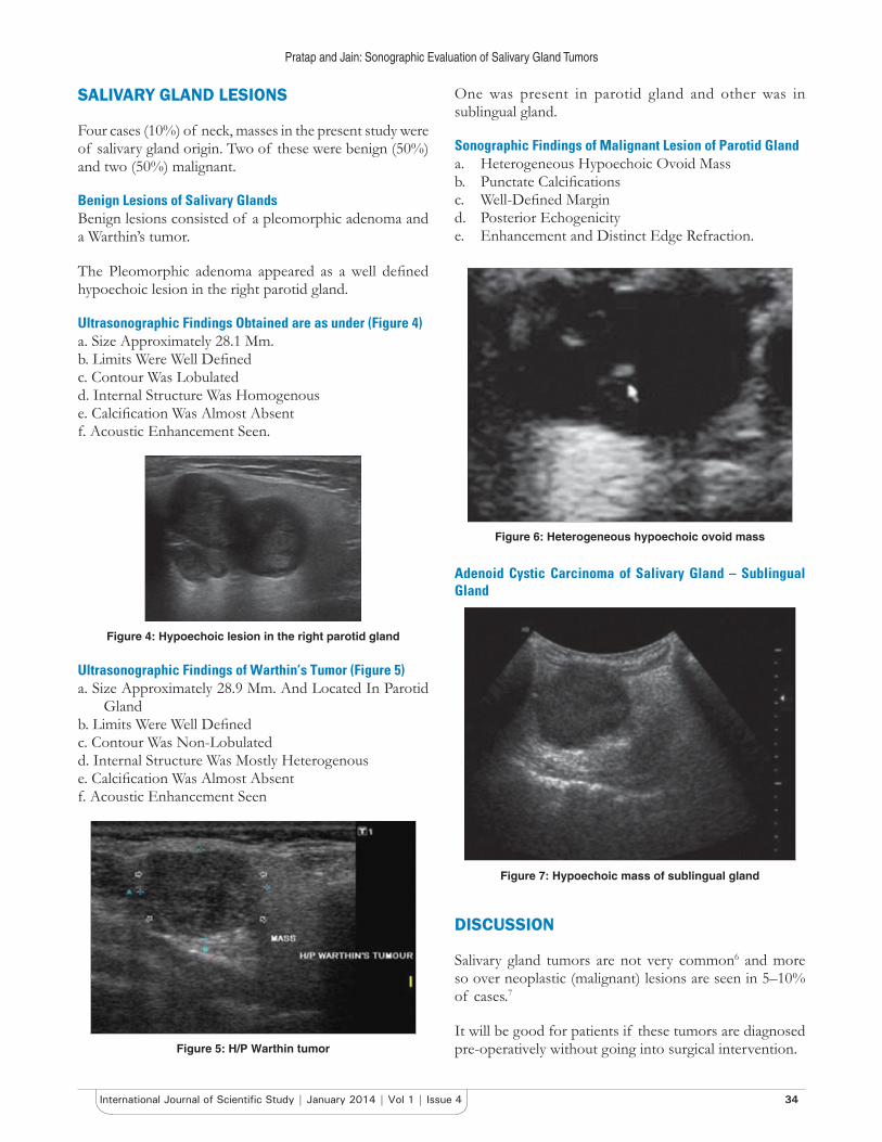
SALIVARY GLAND LESIONS
Four cases (10%) of neck, masses in the present study were of salivary gland origin. Two of these were benign (50%) and two (50%) malignant.
Benign Lesions of Salivary GlandsBenign lesions consisted of a pleomorphic adenoma and a Warthin’s tumor.
The Pleomorphic adenoma appeared as a well defi ned hypoechoic lesion in the right parotid gland.
Ultrasonographic Findings Obtained are as under (Figure 4)a. Size Approximately 28.1 Mm.b. Limits Were Well Defi nedc. Contour Was Lobulatedd. Internal Structure Was Homogenouse. Calcifi cation Was Almost Absentf. Acoustic Enhancement Seen.
Figure 4: Hypoechoic lesion in the right parotid gland
Ultrasonographic Findings of Warthin’s Tumor (Figure 5)a. Size Approximately 28.9 Mm. And Located In Parotid
Glandb. Limits Were Well Defi nedc. Contour Was Non-Lobulatedd. Internal Structure Was Mostly Heterogenouse. Calcifi cation Was Almost Absentf. Acoustic Enhancement Seen
Figure 5: H/P Warthin tumor
One was present in parotid gland and other was in sublingual gland.
Sonographic Findings of Malignant Lesion of Parotid Glanda. Heterogeneous Hypoechoic Ovoid Massb. Punctate Calcifi cations c. Well-Defi ned Margin d. Posterior Echogenicity e. Enhancement and Distinct Edge Refraction.
Figure 6: Heterogeneous hypoechoic ovoid mass
Adenoid Cystic Carcinoma of Salivary Gland – Sublingual Gland
Figure 7: Hypoechoic mass of sublingual gland
DISCUSSION
Salivary gland tumors are not very common6 and more so over neoplastic (malignant) lesions are seen in 5–10% of cases.7
It will be good for patients if these tumors are diagnosed pre-operatively without going into surgical intervention.
Pratap and Jain: Sonographic Evaluation of Salivary Gland Tumors
35 International Journal of Scientifi c Study | January 2014 | Vol 1 | Issue 4
Therefore, many clinical researchers have tried to evaluate the ability of sonography to differentiate benign and malignant tumors.
Sonography is a powerful tool for characterizing salivary gland tumors. Different imaging techniques are valuable in assessing salivary gland disease, out of which the choice of modality depends on local protocol, clinical features and, importantly, the site of suspected pathology. Technical advances, in many imaging centers have made ultrasound nowadays the investigation of choice for major salivary gland disease. It allows a quick, cheap and thorough assessment without the use of ionizing radiation. Ultrasound is able to simultaneously evaluate gland parenchyma and large ducts as well as demonstrate duct dilatation.
Tumors of the salivary glands are not com mon, representing about 3% of all head and neck tumors. Histopathology of salivary gland tumors is very varied, with a large number of both benign and malignant tumors. Out of this Pleomor-phic adenomas are the most common, representing 70-80% of all salivary gland tumors1 most frequently located in the parotid gland. Cytological examination often faces diffi culty in differentiating adenoid cystic carcinoma from Pleomorphic adenoma.7,8 It is seen histopathologically both lesions contain myxoid material.9-11 A number of ultrasonographic features are consid ered typical for pleomorphic adenomas: sharp borders, lobulations of the contour, homogeneous structure, poor vascularization, acoustic enhancement.12,13 which well correlates with the ultrasonographic pictures of our present study.
Warthin’s tumour is the second common salivary neoplasm, typically occurring in older male patients, with a propensity for smokers. It arises from parotid intraglandular lymphoid tissue, typically in the tail, and is multiple or bilateral in approximately 15% cases.
Ultrasound shows an ovoid hypoechoic mass. In our study it was present unilaterally and patient didn’t give history of smoking. Sublingual gland tumors are rare and account for only 0.4–2.6 of all salivary gland tumors.14,15
However, most of the recorded literature assert that Pleomorphic adenoma is more common than Adenolymphoma.7,16 Only Schick et al17 recorded an equal number of cases of Pleomorphic adenoma and Warthin’s tumour (7:7), which is also seen in our study.
The majority of sublingual gland tumors are malignant18
and ACC is the most common. As can be seen in our study out of two malignant lesions one is of Adenoid Cystic Carcinoma, which very well correlates with the study of Anderson LJ et al.19
CONCLUSION
Before going into any type of radiological investigation histological grading of salivary gland tumor is a preliminary step in clinical setting, though not alone.
A variety of radio-imaging modalities may be employed in salivary gland imaging in which Ultrasound has emerged as the technique of choice for major salivary gland disease and forms a useful aid for FNA/biopsy. MRI is of particular value for staging salivary gland malignancy.
As a simple guide If ultrasound is able to differentiate as a benign pathology there is no need to go further imaging.
Through our experience we now know that sonographic features are most accurate but we should keep other modalities in our mind for improving the diagnostic accuracy.
REFERENCES
1. Gritzmann N, Hollerweger A, Macheiner P, Rettenbacher T, Hubner E. Sonography of the salivary glands. European Radiology 2003; 13:364-375.
2. Elagoz S, Gulluoglu M, Yilmazbayhan D, Ozer H, Arslan I. The value of fi ne-needle aspiration cytology in salivary gland lesions, 1994-2004. ORL J Otorhinolaryngol Relat Spec 2007;69:516.
3. Stramandinoli RT, Sassi LM, Pedruzzi PA, Ramos GH, Oliveira BV, Ogata DC, Ioshii SO. Accuracy, sensitivity and specifi city of fi ne needle aspiration biopsy in salivary gland tumours: a retrospective study. Med Oral Patol Oral Cir Bucal 2010;15:e32-7.
4. Mihashi H, Kawahara A, Kage M, Kojiro M, Nakashima T, Umeno H, Sakamoto K, Chiziwa H. Comparison of preoperative fi ne-needle aspiration cytology diagnosis and histopathological diagnosis of salivary gland tumors. Kurume Med J 2006;53:23-7.
5. Kovacevic DO, Fabijanic I. Sonographic diagnosis of parotid gland lesions: correlation with the results of sonographically guided fi ne-needle aspiration biopsy. J Clin Ultrasound 2010;38:294-8.
6. Dumitriu D, Dudea S, Badea R, Botar-Jid C, Băciut G, Băciut M. B-mode and colour Doppler ultrasound features of salivary gland tumours. Med Ultrason 2008;10:31–37.
7. Bradley MJ, Durham LH, Lancer JM. The role of colour fl ow Doppler in the investigation of the salivary gland tumour. Clin Radiol 2000;55:759–762.
8. Stanley MW. Selected problems in fi ne needle aspiration of head and neck masses. Mod Pathol 2002;15:342-50.
9. Elsheikh TM, Bernacki EG. Fine needle aspiration cytology of cellular pleomorphic adenoma. Acta Cytol 1996;40:1165-75.
10. Cerulli G, Renzi G, Perugini M, Becelli R. Differential diagnosis between adenoid cystic carcinoma and pleomorphic adenoma of the minor salivary glands of palate. J Craniofac Surg 2004;15:1056-60.
11. Kapadia SB, Dusenbery D, Dekker A. Fine needle aspiration of pleomorphic adenoma and adenoid cystic carcinoma of salivary gland origin. Acta Cytol 1997;41:487-92.
12. Colella G, Cannavale R, Flamminio F, Foschini MP. Fineneedle aspiration cytology of salivary gland lesions: a systematic review. J Oral Maxillofac Surg 2010;68:2146-53.
13. Bialek E, Jakubowski W, Karpinska G. Role of ultrasonog raphy in diagnosis and differentiation of pleomorphic ade nomas. Arch Otolaryngol Head Neck Surg 2003; 129:929–933.
14. Luukka H, Klemi P, Leivo I, Koivunen P, Laranne J, Makitie A, et al. Salivary gland cancer in Finland 1991–96: and evaluation of 237 cases. Acta Otolaryngol 2005;125: 207–214.
Pratap and Jain: Sonographic Evaluation of Salivary Gland Tumors
36International Journal of Scientifi c Study | January 2014 | Vol 1 | Issue 4
15. Gurney TA, Eisele DW, Weinberg V, Shin E, Lee N. Adenoidcystic carcinoma of the major salivary glands treated with surgeryand radiation. Laryngoscope 2005;115: 1278–1282.
16. Mazaher H, Kashany SS, Sharifi an H. Diagnostic accuracy of triplex ultrasound in malignant parotid tumours. Iran J Radiol 2007;4:169–174.
17. Steiner E, Gahleitner A, Böhm P, Helbich T, Ba-Ssalamah A, et al. Differentiation of benign and malignant tumours of the parotid gland:
value of pulsed Doppler and colour Doppler sonography. Eur Radiol 1998;8:1462–1467.
18. Eneroth CM. Salivary gland tumors in the parotid gland, sub mandibular gland, and the palate region. Cancer 1971;27:1415–1418.
19. Anderson LJ, Therkildsen MH, Ockelmann HH, Bentzen JD, Schiodt T, Hansen HS. Malignant epithelial tumors in the minor salivary glands, the submandibular gland and the sublingual gland. Prognostic factors and treatment results. Cancer 1991;68:2431–2437
How to cite this article: Vijai Pratap, S K Jain. "Sonographic Evaluation of Salivary Gland Tumors – A Hospital Based Study". International Journal of Scientifi c Study. 2014;1(4):32-36.
Source of Support: Nil, Confl ict of Interest: None declared.