Embed Size (px)
Citation preview
©20
09 N
atu
re A
mer
ica,
Inc.
All
rig
hts
res
erve
d.
nature neurOSCIenCe advance online publication �
a r t I C l e S
Commissural axons of the developing spinal cord are guided to the ventral midline by a collaboration of chemoattractants (Netrin-1 and Shh) and chemorepellents (bone morphogenetic proteins) secreted by midline floor plate and roof plate cells, respectively. Once these axons reach the floor plate, they switch off their responsiveness to chemoat-tractants from the floor plate and become responsive to chemore-pulsive cues that are also expressed by the floor plate cells and the surrounding ventral gray matter, including members of the class 3 Semaphorins (Sema3B and Sema3F) and the Slit family proteins1–6. Neuropilin-2 mutant embryos have severe guidance defects, includ-ing stalling in the midline, overshooting to the contralateral side of the spinal cord and randomly projecting along the anterior-posterior axis4. These phenotypes are consistent with the proposed function of the floor plate–secreted chemorepellents to ensure proper midline exit and channel post-crossing commissural axons to turn into their correct (longitudinal) trajectory after exiting the midline. Nonetheless, the molecular mechanisms underlying changes in responsiveness to guidance cues, particularly the extrinsic signals that trigger these changes, are still largely unknown.
We studied the manner in which commissural axons gain respon-siveness to Semaphorin repulsion during midline crossing and found that Shh, a morphogen that is highly enriched in the floor plate, may act as an ‘on’ switch for Semaphorin repulsion in com-missural axons. The Shh receptors Patched-1 (Ptch1) and Smo were both necessary for this switch and for proper midline guidance of commissural axons. Shh probably sensitizes growth-cone respon-siveness to class 3 Semaphorins by downregulating the activity of the cAMP/protein kinase A (PKA) pathway. Therefore, we propose that the morphogen Shh is involved in regulating the responses of other guidance cues.
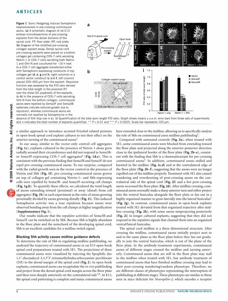
RESULTSShh activates Semaphorin repulsion in commissural axonsWe investigated the mechanisms by which Semaphorin repulsion in commissural axons is switched on during spinal cord midline path-finding. Cell bodies of many populations of commissural neurons are located in the dorsal spinal cord and initially extend axons to the ventral midline, the floor plate, over several days (from late embryonic day 8.9 (E8.9) and early E9.5 in mice and late E10 and E11 in rats until at least E12.5 in mice and E14 in rats). The commissural axons in the rostral (anterior) spinal cord differentiate earlier and their axons develop earlier than their caudal (posterior) counterparts. At each axis level, the more dorsal populations tend to differentiate and extend axons at a later time point than the ventral population (both in the dorsal spinal cord) during this stage of development. At E13 in rats, many rostral commissural axons have reached and crossed the midline while the more caudal populations are still growing toward the midline or are just starting to cross the floor plate. To obtain pre-crossing commissural axons, we used the dorsal-most margin of the caudal spinal cords (hindlimb level) from E13 rat embryos. These are the younger axons, which have not reached/contacted the floor plate. We used a modified pre-crossing explant assay in collagen gel to test whether any of the diffusible signaling molecules expressed in the floor plate could activate Semaphorin repulsion in pre-crossing commissural axons, which had not yet encountered the floor plate (Fig. 1). In this assay, the bottom collagen layer contains dispersed COS-7 cells expressing Netrin-1 alone or Netrin-1 and a secreted mol-ecule from the floor plate (Fig. 1b). In the top collagen gel, explants of dorsal commissural neurons from E13 rat embryos (equivalent to E11.5 mouse embryos) were embedded next to COS-7 cell aggre-gates (clumps) expressing Sema3B or Sema3F. We previously used
Neurobiology Section, Biological Sciences Division, University of California San Diego, La Jolla, California, USA. Correspondence should be addressed to Y.Z. ([email protected]).
Received 24 August; accepted 26 October; published online 29 November 2009; doi:10.1038/nn.2457
Sonic hedgehog induces response of commissural axons to Semaphorin repulsion during midline crossingLiseth M Parra & Yimin Zou
Pathfinding axons change responses to guidance cues at intermediate targets. During midline crossing, spinal cord commissural axons acquire responsiveness to class 3 Semaphorins and Slits, which regulate their floor plate exit and restrict their post-crossing trajectory into a longitudinal pathway. We found that Sonic Hedgehog (Shh) could activate the repulsive response of pre-crossing axons to Semaphorins. Blocking Shh function with a monoclonal antibody to Shh, 5E1, in ‘open-book’ explants or by expressing a dominant-negative form of Patched-1, Ptch1loop2, or a Smoothened (Smo) shRNA construct in commissural neurons resulted in severe guidance defects, including stalling and knotting inside the floor plate, recrossing, randomized anterior-posterior projection and overshooting after crossing, reminiscent of Neuropilin-2 mutant embryos. Enhancing protein kinase A activity in pre-crossing axons diminished Shh-induced Semaphorin repulsion and caused profound midline stalling and overshooting/wandering of post-crossing axons. Therefore, a morphogen, Shh, can act as a switch of axon guidance responses.
©20
09 N
atu
re A
mer
ica,
Inc.
All
rig
hts
res
erve
d.
� advance online publication nature neurOSCIenCe
a r t I C l e S
a similar approach to introduce secreted Frizzled-related proteins in open-book spinal cord explant cultures to test their effect on the anterior turning of the commissural axons7.
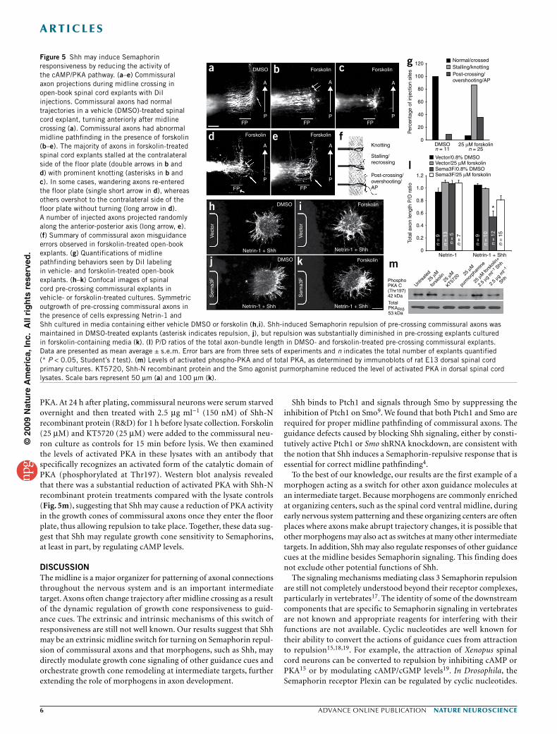
In our assay, similar to the vector-only control cell aggregates (Fig. 1c), explants cultured in the presence of Netrin-1 alone grew radially around their circumference and did not respond to Sema3B- or Sema3F-expressing COS-7 cell aggregates4 (Fig. 1d,e). This is consistent with the previous finding that Sema3B and Sema3F do not repel pre-crossing commissural axons. To our surprise, compared with the radial growth seen in the vector control in the presence of Netrin and Shh (Fig. 1f), pre-crossing commissural axons grown on top of collagen gel containing Netrin-1– and Shh-expressing cells were repelled by Sema3B- and Sema3F-secreting cell clumps (Fig. 1g,h). To quantify these effects, we calculated the total length of axons extending toward (proximal) or away (distal) from cell aggregates (Fig. 1b) in each experiment as the ratio of axons growing proximally divided by axons growing distally (Fig. 1i). This induced Semaphorin activity was a true repulsion because axons were observed bending away from the cell clumps at higher magnification (Supplementary Fig. 1).
Our results indicate that the repulsive activities of Sema3B and Sema3F can be switched on by Shh. Because Shh is highly abundant in the floor plate and the notochord of the developing spinal cord, Shh is an excellent candidate for a midline switch signal.
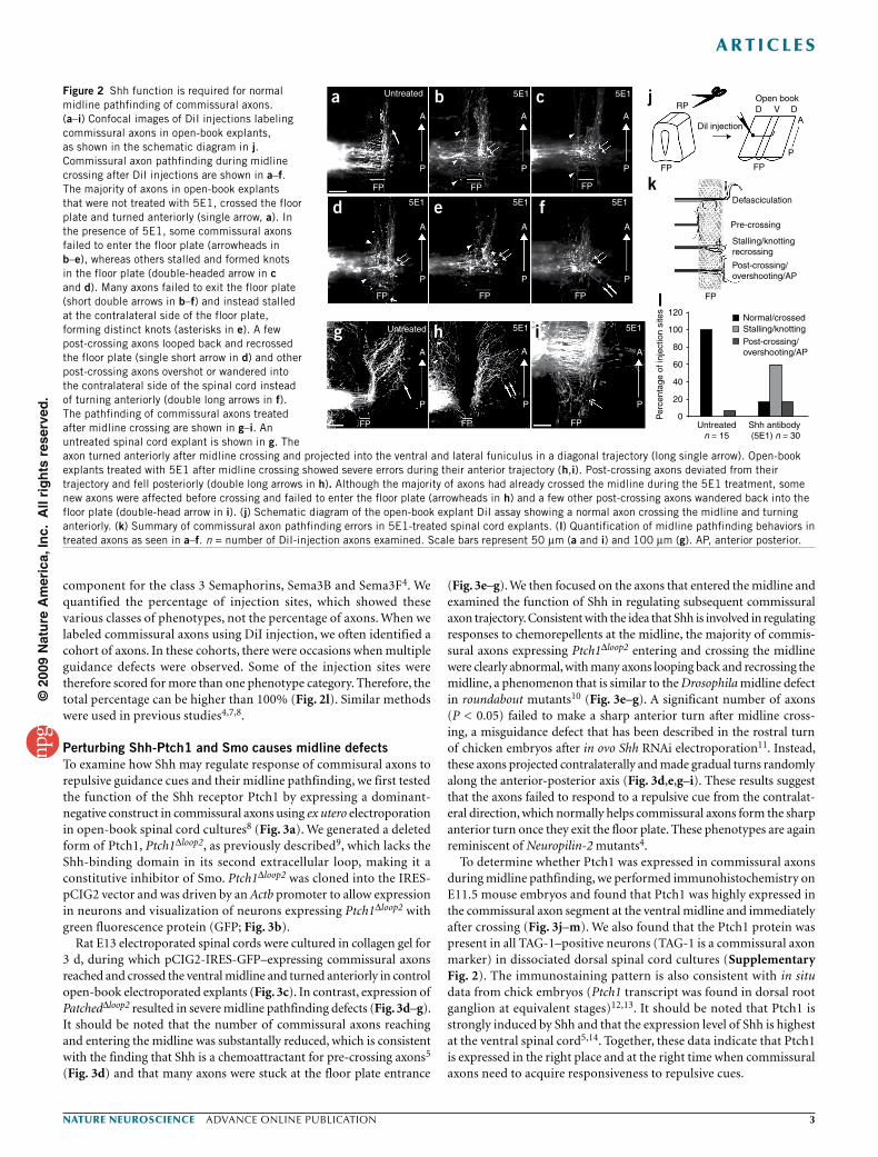
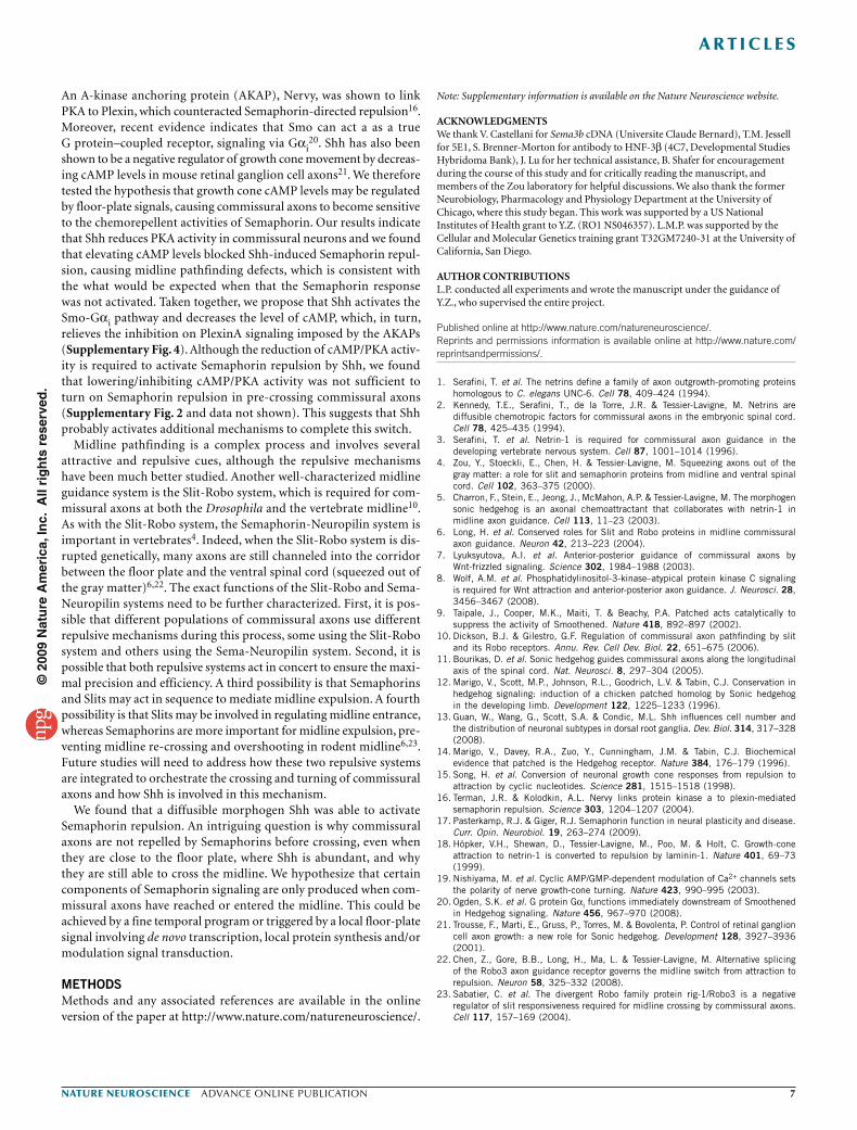
Blocking Shh activity causes midline guidance defectsTo determine the role of Shh in regulating midline pathfinding, we analyzed the trajectory of commissural axons in rat E13 open-book spinal cord preparations treated with 5E1. The projections of these commissural axons were visualized by injecting the lipophilic dye 1,1′-dioctadecyl-3,3,3′3′-tetramethylindocarbocyanine perchlorate (DiI) to the dorsal margin of the spinal cord (Fig. 2). In open-book explants, commissural axons can recapitulate the in vivo pathfinding and project from the dorsal spinal cord margin across the floor plate and then turn sharply anteriorly on the contralateral side7,8. At E13, the spinal cord patterning is complete and many commissural axons
have extended close to the midline, allowing us to specifically analyze the role of Shh on commissural axon midline pathfinding5.
Compared with untreated controls (Fig. 2a), when treated with 5E1, some commissural axons were blocked from extending toward the floor plate and projected along the anterior-posterior direction close to the ipsilateral border of the floor plate (Fig. 2b–e), consist-ent with the finding that Shh is a chemoattractant for pre-crossing commissural axons5. In addition, commissural axons stalled and knotted in the midline (Fig. 2c,d) and at the contralateral edge of the floor plate (Fig. 2b–f), suggesting that the axons were no longer expelled out of the midline properly. Treatment with 5E1 also caused wandering and overshooting of post-crossing axons on the con-tralateral side of the spinal cord (Fig. 2f) and a few post-crossing axons recrossed the floor plate (Fig. 2d). After midline crossing, com-missural axons normally make a sharp anterior turn and either project into the ventral funiculus alongside the floor plate or fan out in a highly organized manner to grow laterally into the lateral funiculus7 (Fig. 2g). In contrast, commissural axons in open-book explants treated with 5E1 deviated from that organized trajectory after mid-line crossing (Fig. 2h), with some axons misprojecting posteriorly (Fig. 2i) in longer cultured explants, suggesting that they did not respond to the repulsive signals that channel them into an organized ventral/lateral funiculus.
The spinal cord midline is a three-dimensional structure. After crossing the midline, commissural axons initially project next to and in the same plane as the floor plate before they fan out gradu-ally to join the ventral funiculus, which is out of the plane of the floor plate. In the antibody-treatment experiments, commissural axons of different stages crossed the midline and turning anteri-orly. Commissural axons that are still in the floor plate may stall in the midline when treated with 5E1, but antibody treatment of commissural axons that have finished midline crossing may lead to their post-crossing wandering/random phenotype. Therefore, we see different classes of phenotypes representing the interruption of pathfinding at different stages. These phenotypes are similar to those seen in mice deficient for Neuropilin-2, which encodes a receptor
Rat E13 spinal cord
a
b i
c d e
f g h
Collagen explant assay
CO
Scl
ump
RPOpen book
Vec
tor
Vec
tor
Pre-crossingexplant
FP FP
P
AD DV Netrin-1 Netrin-1
*
*****
*
Netrin-1
Netrin-1 + Shh
Sem
a3F
Vector
1.2
1.0
0.8
0.6
0.4
0.2P/D
rat
io o
f tot
al a
xon
leng
th
0
Sema3FSema3B
Sem
a3F
Sem
a3B
Sem
a3B
Netrin-1 + Shh
n =
8n
= 1
0n
= 1
0
n =
10
n =
11
n =
13
Netrin-1 + ShhNetrin-1 only
Netrin-1 + Shh
Figure 1 Sonic Hedgehog induces Semaphorin responsiveness in pre-crossing commissural axons. (a) A schematic diagram of rat E13 embryo microdissections of pre-crossing explants from the dorsal domains of the spinal cord. FP, floor plate. RP, root plate. (b) Diagram of the modified pre-crossing collagen explant assay. Dorsal spinal cord pre-crossing explants were placed on a bottom collagen gel containing COS-7 cells secreting Netrin-1 or COS-7 cells secreting both Netrin-1 and Shh-N and cocultured for ~16 h next to a COS-7 cell aggregate transfected either with Semaphorin-expressing constructs in top collagen gel (d, e, g and h, right columns) or a control vector construct (c and f, left column) placed 200–400 µm from the explant. Repulsive function was assessed by the P/D ratio derived from the total length in the proximal (P) over the distal (D) quadrants of the explants. (c–h) In the presence of COS-7 cells secreting Shh-N from the bottom collagen, commissural axons were repelled by Sema3F and Sema3B (asterisks indicate reduced growth due to repulsion), whereas commissural axons are normally not repelled by Semaphorins in the absence of Shh (top row in c). (i) Quantification of the total axon length P/D ratio. Graph shows means ± s.e.m. error bars from three sets of experiments and n indicates the total number of explants quantified. ** P < 0.01 and *** P < 0.0025. Scale bar represents 100 µm.
©20
09 N
atu
re A
mer
ica,
Inc.
All
rig
hts
res
erve
d.
nature neurOSCIenCe advance online publication �
a r t I C l e S
component for the class 3 Semaphorins, Sema3B and Sema3F4. We quantified the percentage of injection sites, which showed these various classes of phenotypes, not the percentage of axons. When we labeled commissural axons using DiI injection, we often identified a cohort of axons. In these cohorts, there were occasions when multiple guidance defects were observed. Some of the injection sites were therefore scored for more than one phenotype category. Therefore, the total percentage can be higher than 100% (Fig. 2l). Similar methods were used in previous studies4,7,8.
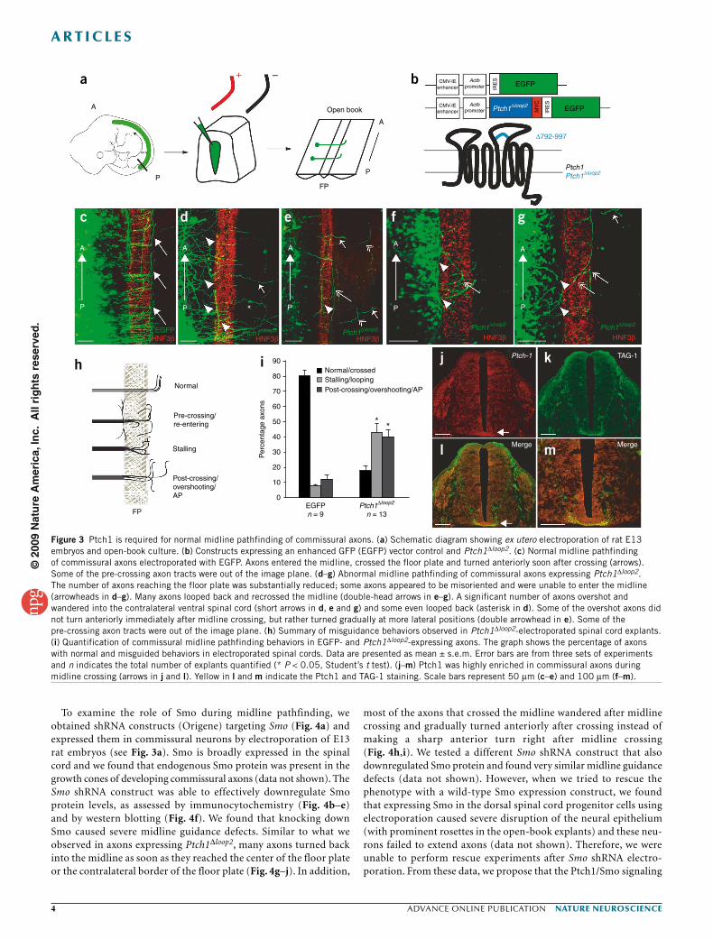
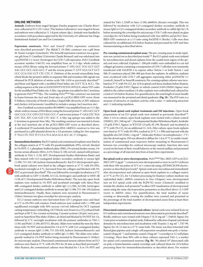
Perturbing Shh-Ptch1 and Smo causes midline defectsTo examine how Shh may regulate response of commisural axons to repulsive guidance cues and their midline pathfinding, we first tested the function of the Shh receptor Ptch1 by expressing a dominant- negative construct in commissural axons using ex utero electroporation in open-book spinal cord cultures8 (Fig. 3a). We generated a deleted form of Ptch1, Ptch1∆loop2, as previously described9, which lacks the Shh-binding domain in its second extracellular loop, making it a constitutive inhibitor of Smo. Ptch1∆loop2 was cloned into the IRES-pCIG2 vector and was driven by an Actb promoter to allow expression in neurons and visualization of neurons expressing Ptch1∆loop2 with green fluorescence protein (GFP; Fig. 3b).
Rat E13 electroporated spinal cords were cultured in collagen gel for 3 d, during which pCIG2-IRES-GFP–expressing commissural axons reached and crossed the ventral midline and turned anteriorly in control open-book electroporated explants (Fig. 3c). In contrast, expression of Patched∆loop2 resulted in severe midline pathfinding defects (Fig. 3d–g). It should be noted that the number of commissural axons reaching and entering the midline was substantally reduced, which is consistent with the finding that Shh is a chemoattractant for pre-crossing axons5 (Fig. 3d) and that many axons were stuck at the floor plate entrance
(Fig. 3e–g). We then focused on the axons that entered the midline and examined the function of Shh in regulating subsequent commissural axon trajectory. Consistent with the idea that Shh is involved in regulating responses to chemorepellents at the midline, the majority of commis-sural axons expressing Ptch1∆loop2 entering and crossing the midline were clearly abnormal, with many axons looping back and recrossing the midline, a phenomenon that is similar to the Drosophila midline defect in roundabout mutants10 (Fig. 3e–g). A significant number of axons (P < 0.05) failed to make a sharp anterior turn after midline cross-ing, a misguidance defect that has been described in the rostral turn of chicken embryos after in ovo Shh RNAi electroporation11. Instead, these axons projected contralaterally and made gradual turns randomly along the anterior-posterior axis (Fig. 3d,e,g–i). These results suggest that the axons failed to respond to a repulsive cue from the contralat-eral direction, which normally helps commissural axons form the sharp anterior turn once they exit the floor plate. These phenotypes are again reminiscent of Neuropilin-2 mutants4.
To determine whether Ptch1 was expressed in commissural axons during midline pathfinding, we performed immunohistochemistry on E11.5 mouse embryos and found that Ptch1 was highly expressed in the commissural axon segment at the ventral midline and immediately after crossing (Fig. 3j–m). We also found that the Ptch1 protein was present in all TAG-1–positive neurons (TAG-1 is a commissural axon marker) in dissociated dorsal spinal cord cultures (Supplementary Fig. 2). The immunostaining pattern is also consistent with in situ data from chick embryos (Ptch1 transcript was found in dorsal root ganglion at equivalent stages)12,13. It should be noted that Ptch1 is strongly induced by Shh and that the expression level of Shh is highest at the ventral spinal cord5,14. Together, these data indicate that Ptch1 is expressed in the right place and at the right time when commissural axons need to acquire responsiveness to repulsive cues.
Untreated
A A
P
A
P
A
P
A
P
A
P
A
P
A
P
P
A
P
FP
FPFPFP
FP FP FP
FPFP
5E1
5E1
Untreated
5E1
5E1 5E1
5E1
5E1RP
FP
Dil injection
Open book
Defasciculation
Pre-crossing
Stalling/knottingrecrossing
Post-crossing/overshooting/AP
FP
120
100
80
60
Per
cent
age
of in
ject
ion
site
s
40
20
0
Normal/crossed
Untreatedn = 15
Shh antibody (5E1) n = 30
Stalling/knottingPost-crossing/overshooting/AP
D V DA
P
FP
a b c j
k
l
d e f
g h i
Figure 2 Shh function is required for normal midline pathfinding of commissural axons. (a–i) Confocal images of DiI injections labeling commissural axons in open-book explants, as shown in the schematic diagram in j. Commissural axon pathfinding during midline crossing after DiI injections are shown in a–f. The majority of axons in open-book explants that were not treated with 5E1, crossed the floor plate and turned anteriorly (single arrow, a). In the presence of 5E1, some commissural axons failed to enter the floor plate (arrowheads in b–e), whereas others stalled and formed knots in the floor plate (double-headed arrow in c and d). Many axons failed to exit the floor plate (short double arrows in b–f) and instead stalled at the contralateral side of the floor plate, forming distinct knots (asterisks in e). A few post-crossing axons looped back and recrossed the floor plate (single short arrow in d) and other post-crossing axons overshot or wandered into the contralateral side of the spinal cord instead of turning anteriorly (double long arrows in f). The pathfinding of commissural axons treated after midline crossing are shown in g–i. An untreated spinal cord explant is shown in g. The axon turned anteriorly after midline crossing and projected into the ventral and lateral funiculus in a diagonal trajectory (long single arrow). Open-book explants treated with 5E1 after midline crossing showed severe errors during their anterior trajectory (h,i). Post-crossing axons deviated from their trajectory and fell posteriorly (double long arrows in h). Although the majority of axons had already crossed the midline during the 5E1 treatment, some new axons were affected before crossing and failed to enter the floor plate (arrowheads in h) and a few other post-crossing axons wandered back into the floor plate (double-head arrow in i). (j) Schematic diagram of the open-book explant DiI assay showing a normal axon crossing the midline and turning anteriorly. (k) Summary of commissural axon pathfinding errors in 5E1-treated spinal cord explants. (l) Quantification of midline pathfinding behaviors in treated axons as seen in a–f. n = number of DiI-injection axons examined. Scale bars represent 50 µm (a and i) and 100 µm (g). AP, anterior posterior.
©20
09 N
atu
re A
mer
ica,
Inc.
All
rig
hts
res
erve
d.
� advance online publication nature neurOSCIenCe
a r t I C l e S
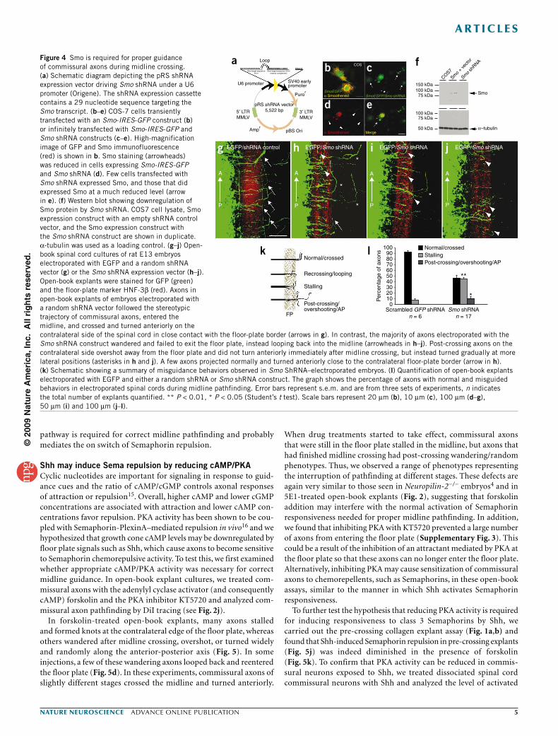
To examine the role of Smo during midline pathfinding, we obtained shRNA constructs (Origene) targeting Smo (Fig. 4a) and expressed them in commissural neurons by electroporation of E13 rat embryos (see Fig. 3a). Smo is broadly expressed in the spinal cord and we found that endogenous Smo protein was present in the growth cones of developing commissural axons (data not shown). The Smo shRNA construct was able to effectively downregulate Smo protein levels, as assessed by immunocytochemistry (Fig. 4b–e) and by western blotting (Fig. 4f). We found that knocking down Smo caused severe midline guidance defects. Similar to what we observed in axons expressing Ptch1∆loop2, many axons turned back into the midline as soon as they reached the center of the floor plate or the contralateral border of the floor plate (Fig. 4g–j). In addition,
most of the axons that crossed the midline wandered after midline crossing and gradually turned anteriorly after crossing instead of making a sharp anterior turn right after midline crossing (Fig. 4h,i). We tested a different Smo shRNA construct that also downregulated Smo protein and found very similar midline guidance defects (data not shown). However, when we tried to rescue the phenotype with a wild-type Smo expression construct, we found that expressing Smo in the dorsal spinal cord progenitor cells using electroporation caused severe disruption of the neural epithelium (with prominent rosettes in the open-book explants) and these neu-rons failed to extend axons (data not shown). Therefore, we were unable to perform rescue experiments after Smo shRNA electro-poration. From these data, we propose that the Ptch1/Smo signaling
A
P
A
P
A
P *
A
P
A
P
A
P
EGFPHNF3β HNF3β HNF3β
Open book
FP
P
A
CMV-IEenhancer EGFP
EGFP
Ptch1Ptch1∆loop2
IRE
S
IRE
S
MY
CCMV-IEenhancer
Actbpromoter
Actbpromoter Ptch1∆loop2
∆792-997
Ptch1∆loop2 Ptch1∆loop2 Ptch1∆loop2 Ptch1∆loop2
HNF3β HNF3β
a
c d e f g
b+ –
Ptch-1 TAG-1
MergeMerge
Normal
Pre-crossing/re-entering
Stalling
Post-crossing/overshooting/AP
FP
90
* *
80
70
60
50
40
30
20
10
0
Per
cent
age
axon
s
EGFPn = 9
Ptch1∆loop2
n = 13
h i j
l m
kNormal/crossedStalling/loopingPost-crossing/overshooting/AP
Figure 3 Ptch1 is required for normal midline pathfinding of commissural axons. (a) Schematic diagram showing ex utero electroporation of rat E13 embryos and open-book culture. (b) Constructs expressing an enhanced GFP (EGFP) vector control and Ptch1∆loop2. (c) Normal midline pathfinding of commissural axons electroporated with EGFP. Axons entered the midline, crossed the floor plate and turned anteriorly soon after crossing (arrows). Some of the pre-crossing axon tracts were out of the image plane. (d–g) Abnormal midline pathfinding of commissural axons expressing Ptch1∆loop2. The number of axons reaching the floor plate was substantially reduced; some axons appeared to be misoriented and were unable to enter the midline (arrowheads in d–g). Many axons looped back and recrossed the midline (double-head arrows in e–g). A significant number of axons overshot and wandered into the contralateral ventral spinal cord (short arrows in d, e and g) and some even looped back (asterisk in d). Some of the overshot axons did not turn anteriorly immediately after midline crossing, but rather turned gradually at more lateral positions (double arrowhead in e). Some of the pre-crossing axon tracts were out of the image plane. (h) Summary of misguidance behaviors observed in Ptch1∆loop2-electroporated spinal cord explants. (i) Quantification of commissural midline pathfinding behaviors in EGFP- and Ptch1∆loop2-expressing axons. The graph shows the percentage of axons with normal and misguided behaviors in electroporated spinal cords. Data are presented as mean ± s.e.m. Error bars are from three sets of experiments and n indicates the total number of explants quantified (* P < 0.05, Student’s t test). (j–m) Ptch1 was highly enriched in commissural axons during midline crossing (arrows in j and l). Yellow in l and m indicate the Ptch1 and TAG-1 staining. Scale bars represent 50 µm (c–e) and 100 µm (f–m).
©20
09 N
atu
re A
mer
ica,
Inc.
All
rig
hts
res
erve
d.
nature neurOSCIenCe advance online publication �
a r t I C l e S
pathway is required for correct midline pathfinding and probably mediates the on switch of Semaphorin repulsion.
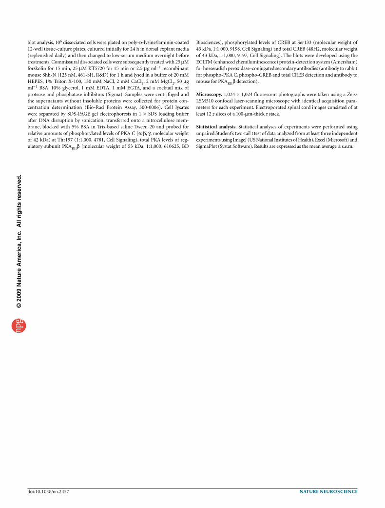
Shh may induce Sema repulsion by reducing cAMP/PKACyclic nucleotides are important for signaling in response to guid-ance cues and the ratio of cAMP/cGMP controls axonal responses of attraction or repulsion15. Overall, higher cAMP and lower cGMP concentrations are associated with attraction and lower cAMP con-centrations favor repulsion. PKA activity has been shown to be cou-pled with Semaphorin-PlexinA–mediated repulsion in vivo16 and we hypothesized that growth cone cAMP levels may be downregulated by floor plate signals such as Shh, which cause axons to become sensitive to Semaphorin chemorepulsive activity. To test this, we first examined whether appropriate cAMP/PKA activity was necessary for correct midline guidance. In open-book explant cultures, we treated com-missural axons with the adenylyl cyclase activator (and consequently cAMP) forskolin and the PKA inhibitor KT5720 and analyzed com-missural axon pathfinding by DiI tracing (see Fig. 2j).
In forskolin-treated open-book explants, many axons stalled and formed knots at the contralateral edge of the floor plate, whereas others wandered after midline crossing, overshot, or turned widely and randomly along the anterior-posterior axis (Fig. 5). In some injections, a few of these wandering axons looped back and reentered the floor plate (Fig. 5d). In these experiments, commissural axons of slightly different stages crossed the midline and turned anteriorly.
When drug treatments started to take effect, commissural axons that were still in the floor plate stalled in the midline, but axons that had finished midline crossing had post-crossing wandering/random phenotypes. Thus, we observed a range of phenotypes representing the interruption of pathfinding at different stages. These defects are again very similar to those seen in Neuropilin-2−/− embryos4 and in 5E1-treated open-book explants (Fig. 2), suggesting that forskolin addition may interfere with the normal activation of Semaphorin responsiveness needed for proper midline pathfinding. In addition, we found that inhibiting PKA with KT5720 prevented a large number of axons from entering the floor plate (Supplementary Fig. 3). This could be a result of the inhibition of an attractant mediated by PKA at the floor plate so that these axons can no longer enter the floor plate. Alternatively, inhibiting PKA may cause sensitization of commissural axons to chemorepellents, such as Semaphorins, in these open-book assays, similar to the manner in which Shh activates Semaphorin responsiveness.
To further test the hypothesis that reducing PKA activity is required for inducing responsiveness to class 3 Semaphorins by Shh, we carried out the pre-crossing collagen explant assay (Fig. 1a,b) and found that Shh-induced Semaphorin repulsion in pre-crossing explants (Fig. 5j) was indeed diminished in the presence of forskolin (Fig. 5k). To confirm that PKA activity can be reduced in commis-sural neurons exposed to Shh, we treated dissociated spinal cord commissural neurons with Shh and analyzed the level of activated
Smo target sequence29 nt
Loop
U6 promoter
ab
g h i j
k l
c
d e
f
5′ LTRMMLV
3′ LTRMMLV
Ampr
Puror
SV40 earlypromoter 150 kDa
Smo
α–tubulin
COS7
Smo
+ ve
ctor
Smo
shRNA
100 kDa75 kDa
50 kDa
100 kDa
Normal/crossed
Recrossing/looping
Stalling
Post-crossing/overshooting/AP
Normal/crossedStallingPost-crossing/overshooting/AP
**
FP
100908070
Per
cent
age
of a
xons
6050403020100
Scrambled GFP shRNAn = 6
Smo shRNAn = 17
75 kDa
COS
SmoEGFPα Smoothened
Merge
COS
α Smoothened
SmoEGFP/Smo shRNA
pBS Ori
EGFP/shRNA control EGFP/Smo shRNA EGFP/Smo shRNA
A
P
A
*
*
**
P
A
P
A
P
EGFP/Smo shRNA
pRS shRNA vector5,522 bp
Smo target sequence 29 ntreverse complement
*
Figure 4 Smo is required for proper guidance of commissural axons during midline crossing. (a) Schematic diagram depicting the pRS shRNA expression vector driving Smo shRNA under a U6 promoter (Origene). The shRNA expression cassette contains a 29 nucleotide sequence targeting the Smo transcript. (b–e) COS-7 cells transiently transfected with an Smo-IRES-GFP construct (b) or infinitely transfected with Smo-IRES-GFP and Smo shRNA constructs (c–e). High-magnification image of GFP and Smo immunofluorescence (red) is shown in b. Smo staining (arrowheads) was reduced in cells expressing Smo-IRES-GFP and Smo shRNA (d). Few cells transfected with Smo shRNA expressed Smo, and those that did expressed Smo at a much reduced level (arrow in e). (f) Western blot showing downregulation of Smo protein by Smo shRNA. COS7 cell lysate, Smo expression construct with an empty shRNA control vector, and the Smo expression construct with the Smo shRNA construct are shown in duplicate. α-tubulin was used as a loading control. (g–j) Open- book spinal cord cultures of rat E13 embryos electroporated with EGFP and a random shRNA vector (g) or the Smo shRNA expression vector (h–j). Open-book explants were stained for GFP (green) and the floor-plate marker HNF-3β (red). Axons in open-book explants of embryos electroporated with a random shRNA vector followed the stereotypic trajectory of commissural axons, entered the midline, and crossed and turned anteriorly on the contralateral side of the spinal cord in close contact with the floor-plate border (arrows in g). In contrast, the majority of axons electroporated with the Smo shRNA construct wandered and failed to exit the floor plate, instead looping back into the midline (arrowheads in h–j). Post-crossing axons on the contralateral side overshot away from the floor plate and did not turn anteriorly immediately after midline crossing, but instead turned gradually at more lateral positions (asterisks in h and j). A few axons projected normally and turned anteriorly close to the contralateral floor-plate border (arrow in h). (k) Schematic showing a summary of misguidance behaviors observed in Smo ShRNA–electroporated embryos. (l) Quantification of open-book explants electroporated with EGFP and either a random shRNA or Smo shRNA construct. The graph shows the percentage of axons with normal and misguided behaviors in electroporated spinal cords during midline pathfinding. Error bars represent s.e.m. and are from three sets of experiments, n indicates the total number of explants quantified. ** P < 0.01, * P < 0.05 (Student’s t test). Scale bars represent 20 µm (b), 10 µm (c), 100 µm (d–g), 50 µm (i) and 100 µm (j–l).
©20
09 N
atu
re A
mer
ica,
Inc.
All
rig
hts
res
erve
d.
� advance online publication nature neurOSCIenCe
a r t I C l e S
PKA. At 24 h after plating, commissural neurons were serum starved overnight and then treated with 2.5 µg ml−1 (150 nM) of Shh-N recombinant protein (R&D) for 1 h before lysate collection. Forskolin (25 µM) and KT5720 (25 µM) were added to the commissural neu-ron culture as controls for 15 min before lysis. We then examined the levels of activated PKA in these lysates with an antibody that specifically recognizes an activated form of the catalytic domain of PKA (phosphorylated at Thr197). Western blot analysis revealed that there was a substantial reduction of activated PKA with Shh-N recombinant protein treatments compared with the lysate controls (Fig. 5m), suggesting that Shh may cause a reduction of PKA activity in the growth cones of commissural axons once they enter the floor plate, thus allowing repulsion to take place. Together, these data sug-gest that Shh may regulate growth cone sensitivity to Semaphorins, at least in part, by regulating cAMP levels.
DISCUSSIONThe midline is a major organizer for patterning of axonal connections throughout the nervous system and is an important intermediate target. Axons often change trajectory after midline crossing as a result of the dynamic regulation of growth cone responsiveness to guid-ance cues. The extrinsic and intrinsic mechanisms of this switch of responsiveness are still not well known. Our results suggest that Shh may be an extrinsic midline switch for turning on Semaphorin repul-sion of commissural axons and that morphogens, such as Shh, may directly modulate growth cone signaling of other guidance cues and orchestrate growth cone remodeling at intermediate targets, further extending the role of morphogens in axon development.
Shh binds to Ptch1 and signals through Smo by suppressing the inhibition of Ptch1 on Smo9. We found that both Ptch1 and Smo are required for proper midline pathfinding of commissural axons. The guidance defects caused by blocking Shh signaling, either by consti-tutively active Ptch1 or Smo shRNA knockdown, are consistent with the notion that Shh induces a Semaphorin-repulsive response that is essential for correct midline pathfinding4.
To the best of our knowledge, our results are the first example of a morphogen acting as a switch for other axon guidance molecules at an intermediate target. Because morphogens are commonly enriched at organizing centers, such as the spinal cord ventral midline, during early nervous system patterning and these organizing centers are often places where axons make abrupt trajectory changes, it is possible that other morphogens may also act as switches at many other intermediate targets. In addition, Shh may also regulate responses of other guidance cues at the midline besides Semaphorin signaling. This finding does not exclude other potential functions of Shh.
The signaling mechanisms mediating class 3 Semaphorin repulsion are still not completely understood beyond their receptor complexes, particularly in vertebrates17. The identity of some of the downstream components that are specific to Semaphorin signaling in vertebrates are not known and appropriate reagents for interfering with their functions are not available. Cyclic nucleotides are well known for their ability to convert the actions of guidance cues from attraction to repulsion15,18,19. For example, the attraction of Xenopus spinal cord neurons can be converted to repulsion by inhibiting cAMP or PKA15 or by modulating cAMP/cGMP levels19. In Drosophila, the Semaphorin receptor Plexin can be regulated by cyclic nucleotides.
DMSO
*
*
Forskolin
DMSO
DMSO
Netrin-1 + Shh
Netrin-1 + Shh
Netrin-1 + Shh
Netrin-1 + Shh
Vec
tor
Sem
a3F
Sem
a3F
Vec
tor
Forskolin
Forskolin
Forskolin Forskolin
Forskolin120
Normal/crossedStalling/knottingPost-crossing/overshooting/AP
Per
cent
age
of in
ject
ion
site
s
100
80
60
40
20
0
1.2
Tota
l axo
n le
ngth
P/D
rat
io 1.0
0.8
0.6
0.4
0.2
n =
9n
= 1
3n
= 5
n =
7
n =
9
n =
10
n =
12
n =
15
0Netrin-1
Untre
ated
25 µM
forsk
olin25
µM
KT5720
25 µM
purm
orph
amine
25 µM
fors
kolin
+
2.5
µg m
l–1 S
hh
2.5
µg m
l–1
ShhPhosphoPKA C (Thr197)42 kDa
Total PKARIIβ53 kDa
Netrin-1 + Shh
DMSOn = 11
25 µM forskolinn = 25
Vector/0.8% DMSO
Sema3F/0.8% DMSO
*
Vector/25 µM forskolin
Sema3F/25 µM forskolin
PFP
FPFP
FPP
AA
P
A
P
A
P
A
FP
a b c
d
h i
l
g
mj k
eKnotting
Stalling/recrossing
Post-crossing/overshooting/AP
f
Figure 5 Shh may induce Semaphorin responsiveness by reducing the activity of the cAMP/PKA pathway. (a–e) Commissural axon projections during midline crossing in open-book spinal cord explants with DiI injections. Commissural axons had normal trajectories in a vehicle (DMSO)-treated spinal cord explant, turning anteriorly after midline crossing (a). Commissural axons had abnormal midline pathfinding in the presence of forskolin (b–e). The majority of axons in forskolin-treated spinal cord explants stalled at the contralateral side of the floor plate (double arrows in b and d) with prominent knotting (asterisks in b and c). In some cases, wandering axons re-entered the floor plate (single short arrow in d), whereas others overshot to the contralateral side of the floor plate without turning (long arrow in d). A number of injected axons projected randomly along the anterior-posterior axis (long arrow, e). (f) Summary of commissural axon misguidance errors observed in forskolin-treated open-book explants. (g) Quantifications of midline pathfinding behaviors seen by DiI labeling in vehicle- and forskolin-treated open-book explants. (h–k) Confocal images of spinal cord pre-crossing commissural explants in vehicle- or forskolin-treated cultures. Symmetric outgrowth of pre-crossing commissural axons in the presence of cells expressing Netrin-1 and Shh cultured in media containing either vehicle DMSO or forskolin (h,i). Shh-induced Semaphorin repulsion of pre-crossing commissural axons was maintained in DMSO-treated explants (asterisk indicates repulsion, j), but repulsion was substantially diminished in pre-crossing explants cultured in forskolin-containing media (k). (l) P/D ratios of the total axon-bundle length in DMSO- and forskolin-treated pre-crossing commissural explants. Data are presented as mean average ± s.e.m. Error bars are from three sets of experiments and n indicates the total number of explants quantified (* P < 0.05, Student’s t test). (m) Levels of activated phospho-PKA and of total PKA, as determined by immunoblots of rat E13 dorsal spinal cord primary cultures. KT5720, Shh-N recombinant protein and the Smo agonist purmorphamine reduced the level of activated PKA in dorsal spinal cord lysates. Scale bars represent 50 µm (a) and 100 µm (k).
©20
09 N
atu
re A
mer
ica,
Inc.
All
rig
hts
res
erve
d.
nature neurOSCIenCe advance online publication �
a r t I C l e S
An A-kinase anchoring protein (AKAP), Nervy, was shown to link PKA to Plexin, which counteracted Semaphorin-directed repulsion16. Moreover, recent evidence indicates that Smo can act a as a true G protein–coupled receptor, signaling via Gαi
20. Shh has also been shown to be a negative regulator of growth cone movement by decreas-ing cAMP levels in mouse retinal ganglion cell axons21. We therefore tested the hypothesis that growth cone cAMP levels may be regulated by floor-plate signals, causing commissural axons to become sensitive to the chemorepellent activities of Semaphorin. Our results indicate that Shh reduces PKA activity in commissural neurons and we found that elevating cAMP levels blocked Shh-induced Semaphorin repul-sion, causing midline pathfinding defects, which is consistent with the what would be expected when that the Semaphorin response was not activated. Taken together, we propose that Shh activates the Smo-Gαi pathway and decreases the level of cAMP, which, in turn, relieves the inhibition on PlexinA signaling imposed by the AKAPs (Supplementary Fig. 4). Although the reduction of cAMP/PKA activ-ity is required to activate Semaphorin repulsion by Shh, we found that lowering/inhibiting cAMP/PKA activity was not sufficient to turn on Semaphorin repulsion in pre-crossing commissural axons (Supplementary Fig. 2 and data not shown). This suggests that Shh probably activates additional mechanisms to complete this switch.
Midline pathfinding is a complex process and involves several attractive and repulsive cues, although the repulsive mechanisms have been much better studied. Another well-characterized midline guidance system is the Slit-Robo system, which is required for com-missural axons at both the Drosophila and the vertebrate midline10. As with the Slit-Robo system, the Semaphorin-Neuropilin system is important in vertebrates4. Indeed, when the Slit-Robo system is dis-rupted genetically, many axons are still channeled into the corridor between the floor plate and the ventral spinal cord (squeezed out of the gray matter)6,22. The exact functions of the Slit-Robo and Sema-Neuropilin systems need to be further characterized. First, it is pos-sible that different populations of commissural axons use different repulsive mechanisms during this process, some using the Slit-Robo system and others using the Sema-Neuropilin system. Second, it is possible that both repulsive systems act in concert to ensure the maxi-mal precision and efficiency. A third possibility is that Semaphorins and Slits may act in sequence to mediate midline expulsion. A fourth possibility is that Slits may be involved in regulating midline entrance, whereas Semaphorins are more important for midline expulsion, pre-venting midline re-crossing and overshooting in rodent midline6,23. Future studies will need to address how these two repulsive systems are integrated to orchestrate the crossing and turning of commissural axons and how Shh is involved in this mechanism.
We found that a diffusible morphogen Shh was able to activate Semaphorin repulsion. An intriguing question is why commissural axons are not repelled by Semaphorins before crossing, even when they are close to the floor plate, where Shh is abundant, and why they are still able to cross the midline. We hypothesize that certain components of Semaphorin signaling are only produced when com-missural axons have reached or entered the midline. This could be achieved by a fine temporal program or triggered by a local floor-plate signal involving de novo transcription, local protein synthesis and/or modulation signal transduction.
METHODSMethods and any associated references are available in the online version of the paper at http://www.nature.com/natureneuroscience/.
Note: Supplementary information is available on the Nature Neuroscience website.
AcknowledgmentSWe thank V. Castellani for Sema3b cDNA (Universite Claude Bernard), T.M. Jessell for 5E1, S. Brenner-Morton for antibody to HNF-3β (4C7, Developmental Studies Hybridoma Bank), J. Lu for her technical assistance, B. Shafer for encouragement during the course of this study and for critically reading the manuscript, and members of the Zou laboratory for helpful discussions. We also thank the former Neurobiology, Pharmacology and Physiology Department at the University of Chicago, where this study began. This work was supported by a US National Institutes of Health grant to Y.Z. (RO1 NS046357). L.M.P. was supported by the Cellular and Molecular Genetics training grant T32GM7240-31 at the University of California, San Diego.
AUtHoR contRIBUtIonSL.P. conducted all experiments and wrote the manuscript under the guidance of Y.Z., who supervised the entire project.
Published online at http://www.nature.com/natureneuroscience/. Reprints and permissions information is available online at http://www.nature.com/reprintsandpermissions/.
1. Serafini, T. et al. The netrins define a family of axon outgrowth-promoting proteins homologous to C. elegans UNC-6. Cell 78, 409–424 (1994).
2. Kennedy, T.E., Serafini, T., de la Torre, J.R. & Tessier-Lavigne, M. Netrins are diffusible chemotropic factors for commissural axons in the embryonic spinal cord. Cell 78, 425–435 (1994).
3. Serafini, T. et al. Netrin-1 is required for commissural axon guidance in the developing vertebrate nervous system. Cell 87, 1001–1014 (1996).
4. Zou, Y., Stoeckli, E., Chen, H. & Tessier-Lavigne, M. Squeezing axons out of the gray matter: a role for slit and semaphorin proteins from midline and ventral spinal cord. Cell 102, 363–375 (2000).
5. Charron, F., Stein, E., Jeong, J., McMahon, A.P. & Tessier-Lavigne, M. The morphogen sonic hedgehog is an axonal chemoattractant that collaborates with netrin-1 in midline axon guidance. Cell 113, 11–23 (2003).
6. Long, H. et al. Conserved roles for Slit and Robo proteins in midline commissural axon guidance. Neuron 42, 213–223 (2004).
7. Lyuksyutova, A.I. et al. Anterior-posterior guidance of commissural axons by Wnt-frizzled signaling. Science 302, 1984–1988 (2003).
8. Wolf, A.M. et al. Phosphatidylinositol-3-kinase–atypical protein kinase C signaling is required for Wnt attraction and anterior-posterior axon guidance. J. Neurosci. 28, 3456–3467 (2008).
9. Taipale, J., Cooper, M.K., Maiti, T. & Beachy, P.A. Patched acts catalytically to suppress the activity of Smoothened. Nature 418, 892–897 (2002).
10. Dickson, B.J. & Gilestro, G.F. Regulation of commissural axon pathfinding by slit and its Robo receptors. Annu. Rev. Cell Dev. Biol. 22, 651–675 (2006).
11. Bourikas, D. et al. Sonic hedgehog guides commissural axons along the longitudinal axis of the spinal cord. Nat. Neurosci. 8, 297–304 (2005).
12. Marigo, V., Scott, M.P., Johnson, R.L., Goodrich, L.V. & Tabin, C.J. Conservation in hedgehog signaling: induction of a chicken patched homolog by Sonic hedgehog in the developing limb. Development 122, 1225–1233 (1996).
13. Guan, W., Wang, G., Scott, S.A. & Condic, M.L. Shh influences cell number and the distribution of neuronal subtypes in dorsal root ganglia. Dev. Biol. 314, 317–328 (2008).
14. Marigo, V., Davey, R.A., Zuo, Y., Cunningham, J.M. & Tabin, C.J. Biochemical evidence that patched is the Hedgehog receptor. Nature 384, 176–179 (1996).
15. Song, H. et al. Conversion of neuronal growth cone responses from repulsion to attraction by cyclic nucleotides. Science 281, 1515–1518 (1998).
16. Terman, J.R. & Kolodkin, A.L. Nervy links protein kinase a to plexin-mediated semaphorin repulsion. Science 303, 1204–1207 (2004).
17. Pasterkamp, R.J. & Giger, R.J. Semaphorin function in neural plasticity and disease. Curr. Opin. Neurobiol. 19, 263–274 (2009).
18. Höpker, V.H., Shewan, D., Tessier-Lavigne, M., Poo, M. & Holt, C. Growth-cone attraction to netrin-1 is converted to repulsion by laminin-1. Nature 401, 69–73 (1999).
19. Nishiyama, M. et al. Cyclic AMP/GMP-dependent modulation of Ca2+ channels sets the polarity of nerve growth-cone turning. Nature 423, 990–995 (2003).
20. Ogden, S.K. et al. G protein Gαi functions immediately downstream of Smoothened in Hedgehog signaling. Nature 456, 967–970 (2008).
21. Trousse, F., Marti, E., Gruss, P., Torres, M. & Bovolenta, P. Control of retinal ganglion cell axon growth: a new role for Sonic hedgehog. Development 128, 3927–3936 (2001).
22. Chen, Z., Gore, B.B., Long, H., Ma, L. & Tessier-Lavigne, M. Alternative splicing of the Robo3 axon guidance receptor governs the midline switch from attraction to repulsion. Neuron 58, 325–332 (2008).
23. Sabatier, C. et al. The divergent Robo family protein rig-1/Robo3 is a negative regulator of slit responsiveness required for midline crossing by commissural axons. Cell 117, 157–169 (2004).
©20
09 N
atu
re A
mer
ica,
Inc.
All
rig
hts
res
erve
d.
nature neurOSCIenCe doi:10.1038/nn.2457
ONLINE METHODSAnimals. Embryos from staged Sprague-Dawley pregnant rats (Charles River) were collected at E13. CD-1 mice (The Jackson Laboratory) were staged in house and embryos were collected at 11. 5 d post coitum (dpc). Animals were handled in accordance with procedures approved by the University of California San Diego Institutional Animal Care and Use Committee.
expression constructs. Ntn1 and Sema3f cDNA expression constructs were described previously4. The Shh∆CT (N-Shh) construct was a gift from M. Tessier-Lavigne (Genentech). The Sema3B expression construct was a gener-ous gift from V. Castellani (Universite Claude Bernard) and was subcloned into a pcDNATM 3.1 vector (Invitrogen) for COS-7 cell expression. Ptch1 (GenBank accession number U46155) was amplified from an 11.5-dpc whole-embryo mouse cDNA library using the forward primer 5′-ATG GCC TCG GCT GGT AAC GCC GCC GGG GCC-3′ and the reverse primer 5′-GTT GGA GCT GCT CCC CCA CGG CCT CTC CTC-3′. Deletion of the second extracellular loop, which blocks the protein’s ability to sequester Shh and transduce Shh signals was obtained by PCR deletion of amino acids 768–1,026 as previously described13 and blunt-end ligated with a modified PmeI linker site (GTT TCA ACC). The coding sequence at the join is GLDLTDIVP/VST/LVCAVFLLN, where VST codes for the modified PmeI linker site. A Myc-tag epitope was added to the C terminus to generate Ptch1∆loop2Myc. The resulting Ptch1∆loop2Myc construct was inserted in frame between the AscI and XmaI sites of a pCIG2-IRES-GFP vector (from F. Polleux, University of North Carolina, Chapel Hill) driven by a CMV enhancer and chicken Actb promoter (modified to include a unique AscI insertion site).
Smo (GenBank accession number BC048091) was amplified from an 11.5-dpc whole-embryo mouse cDNA library using the forward primer 5′-ATG GCC GCT GGC CGC CCC GTG CGT GGG CCC-3′ and reverse primer 5′-GAA GTC TGA GTC TGC ATC CAA GAT CTC AGC-3′. A Myc-tag epitope was added to the C terminus to generate Smo-Myc. The resulting construct was inserted in frame at the AscI insertion site of the pCIG2-IRES-GFP vector as described above. A synthesized 29 nucleotide short hairpin RNA (shRNA) construct to Smo was purchased in a pRS plasmid driven by a U6 promoter coding for Smo sequence 5′-TGA CTC TGT TCT CCA TCA AGA GCA ACC AC-3′ (Origene).
Immunohistochemistry. Rat E13 spinal cord commissural explants were fixed in the collagen matrix at 37 °C with 4% paraformaldehyde (PFA, wt/vol), blocked in 5% PDT (1 × phosphate-buffered saline (PBS), 5% normal donkey serum, 1% bovine serum albumin (BSA), 0.1% Triton X-100, wt/vol), stained with primary antibody to TAG-1 (1:50, 4D7, Developmental Studies Hybridoma Bank) and then stained with Cy3-conjugated donkey secondary antibody to mouse IgM (1:300, 715-165-140, Jackson ImmunoResearch). Rat E13 electroporated open-book spinal cultures were fixed in the collagen matrix at 37 °C with 4% PFA, incubated overnight at 4 °C, harvested from the collagen and blocked with 5% PDT as previously described8. This was followed by overnight incubation at 4 °C with antibody to GFP (1:10,000, A11122, Invitrogen) and antibody to HNF-3β (1:50; 4C7, Developmental Studies Hybridoma Bank). The next day, open-book explants were washed in 1% PDT and incubated overnight with Alexa Fluor 488–conjugated donkey antibody to rabbit IgG (1:1,500, A21206, Invitrogen) and Cy3-conjugated donkey antibody to mouse IgG (1:300, 715-166-150, Jackson ImmunoResearch). Finally, these explants were mounted in Fluoromount G (Fisher) between two glass coverslips for microscopic analysis.
E11.5 mouse embryos were harvested from CD-1 pregnant mice and fixed at 4 °C in 4% PFA with rotation. Fixed embryos were washed with 1 × PBS and equilibrated overnight with 30% sucrose (wt/vol) followed by OCT (optimal cutting temperature) embedding. OCT blocks were frozen in a CO2/ethanol bath and kept at 80 °C for cryostat sectioning. Cryostat sections (20 µm) were proc-essed on Superfrost Plus slides (Fisher), air dried and blocked in 5% PDT for 1 h, followed by 4 °C overnight incubation with the primary antibodies to TAG-1 (1:50) and Ptch1 (1:100, ab53715, Abcam). Slides were washed the next day three times with 1% PDT and incubated for 1 h at 25 °C with Cy2-conjugated donkey antibody to mouse IgM (1:300, 715-226-020, Jackson ImmunoResearch) and Cy3-conjugated donkey antibody to rabbit IgG (1:300). The slides were subse-quently washed in 1% PDT before being mounted in Fluoromount G (Fisher) for microscopic analysis. Dissociated commissural neuron cultures from rat E13 embryos were fixed at 37 °C with 4% PFA for 20 min as described previously8. After fixation, the commissural cultures were blocked for 1 h in 1% PDT and
stained for TAG-1, DAPI or Smo (1:100, ab60016, Abcam) overnight. This was followed by incubation with Cy3-conjugated donkey secondary antibody to mouse IgM or Cy3-conjugated donkey secondary antibody to rabbit IgG for 2 h before mounting the coverslips for microscopy. COS-7 cells were plated on glass coverslips for 24 h before being transfected with Smo shRNA and pCIG2-Smo-IRES-GFP constructs at a 1:3 ratio using FuGENE 6 (Roche). Cells were then cultured for an additional 24 h before fixation and processed for GFP and Smo immunostaining as described above.
Pre-crossing commissural explant assay. The pre-crossing assay to study repul-sion was carried out as described previously3,4. Rat E13 spinal cords were isolated by microdissection and dorsal explants from the caudal most region of the spi-nal cord were collected. Explants (~200 µM width) were embedded in a rat-tail collagen gel matrix containing a resuspension of COS-7 cells that were transiently transfected with FuGENE 6 to express either Netrin-1 or both Netrin-1 and Shh-N constructs placed 200–400 µm from the explants. In addition, explants were cocultured with COS-7 cell aggregates expressing either pcDNATM 3.1 (control), Sema3F or Sema3B constructs. Pre-crossing explant cultures were incu-bated at 37 °C with 5% CO2 for 16 h in spinal cord dorsa medium before fixation. Forskolin (25 µM; F3917, Sigma) or vehicle control (0.8% DMSO, Sigma) were added to the culture medium 2 h after explants were embedded and cultured for a total of 14 h before fixation. For quantification, the total length of axon bundle was measured in the proximal and distal quadrants (Fig. 1b). The P/D ratio is a measure of attractive or repulsive activity, with a ratio >1 indicating attraction and <1 indicating repulsion.
open-book spinal cord explant treatments and diI injections. Open-book preparations of rat E13 spinal cords were isolated as previously described7,8. After 2–4 h in culture, open-book explants were treated with a vehicle control (DMSO), 5E1 (100 ng ml−1, Developmental Studies Hybridoma Bank), forskolin (25 µM; F3917, Sigma) or KT5720 (25 µM, EI-199, Biomol) and incubated at 37 °C with 5% CO2 for an additional 12–14 h. After culture, open-book explants were fixed at 37 °C with 4% PFA, washed in 25 °C 1 × PBS and injected with the lipophilic dye DiI (D282, 1 mg ml−1, Molecular Probes) via iontophoresis (~7 V) into the dorsal region. DiI was allowed to diffuse for 1 d at 37 °C to enable visual-ization of commissural axon trajectory before mounting in Fluoromount-G between two coverslips for confocal microscopy analysis. Injection sites were scored on the basis of their overall behavior at the ventral midline and presented as a percentage of all injected sites from at least three experiments.
Rat spinal cord ex utero electroporation. Ptch1∆loop2Myc-IRES-GFP or pCIG2-IRES-GFP (1 µg µl−1) constructs were electroporated ex utero in rat E13 embryos with three 100-ms pulses of 25 V at 1-s intervals using a BTX#ECM 830 electro-porator as described previously8. Spinal cords were microdissected immediately after electroporation and cultured as open-book explants in a collagen matrix at 37 °C at 5% CO2 for 3 d before processing for fixation (culture medium was replenished daily). shRNA constructs to Smo (Origene) were electroporated into rat E13 spinal cords with the EGFP-N2 vector (Clontech) modified to include the chicken Actb promoter8 to allow GFP visualization of electroporated axons using the same electroporation parameters as described above (1:3 GFP vector to shRNA ratio). For quantification, the total number of axons crossing or abnormally crossing the midline were counted and presented as the percentage of the total number of electroporated axons from at least three independent experiments.
dissociated commissural neuronal culture. Spinal cords were isolated from rat E13 embryos and commissural neurons were dissociated as previously described8. Briefly, embryos were treated with Dispase I (0.25 mg ml−1, D4818, Sigma) for 5 min prior to isolation of spinal cords. Followed by collection of spinal cord dorsal domains by microdissection and incubation with Trypsin (1 mg ml−1, T47-99, Sigma) for 10–12 min in a 37 °C water bath. The tissue was then triturated with flamed glass pipettes and a single-cell suspension was plated onto poly-d-lysine-coated (20 µg ml−1) and laminin-coated (10 µg ml−1) coverslips or 12-well plates (Corning). More than 90% cells in these cultures expressed TAG-1, a marker for spinal cord commissural neurons (Fig. 5b). We plated 105 dissociated cells on poly-d-lysine/laminin-coated coverslips and cultured them for 24 h before processing them for immunohistochemistry as described above. For western
©20
09 N
atu
re A
mer
ica,
Inc.
All
rig
hts
res
erve
d.
nature neurOSCIenCedoi:10.1038/nn.2457
blot analysis, 106 dissociated cells were plated on poly-d-lysine/laminin-coated 12-well tissue-culture plates, cultured initially for 24 h in dorsal explant media (replenished daily) and then changed to low-serum medium overnight before treatments. Commissural dissociated cells were subsequently treated with 25 µM forskolin for 15 min, 25 µM KT5720 for 15 min or 2.5 µg ml−1 recombinant mouse Shh-N (125 nM, 461-SH, R&D) for 1 h and lysed in a buffer of 20 mM HEPES, 1% Triton X-100, 150 mM NaCl, 2 mM CaCl2, 2 mM MgCl2, 50 µg ml−1 BSA, 10% glycerol, 1 mM EDTA, 1 mM EGTA, and a cocktail mix of protease and phosphatase inhibitors (Sigma). Samples were centrifuged and the supernatants without insoluble proteins were collected for protein con-centration determination (Bio-Rad Protein Assay, 500-0006). Cell lysates were separated by SDS-PAGE gel electrophoresis in 1 × SDS loading buffer after DNA disruption by sonication, transferred onto a nitrocellulose mem-brane, blocked with 5% BSA in Tris-based saline Tween-20 and probed for relative amounts of phosphorylated levels of PKA C (α β, γ; molecular weight of 42 kDa) at Thr197 (1:1,000, 4781, Cell Signaling), total PKA levels of reg-ulatory subunit PKARIIβ (molecular weight of 53 kDa, 1:1,000, 610625, BD
Biosciences), phosphorylated levels of CREB at Ser133 (molecular weight of 43 kDa, 1:1,000, 9198, Cell Signaling) and total CREB (48H2, molecular weight of 43 kDa, 1:1,000, 9197, Cell Signaling). The blots were developed using the ECLTM (enhanced chemiluminescence) protein-detection system (Amersham) for horseradish peroxidase–conjugated secondary antibodies (antibody to rabbit for phospho-PKA C, phospho-CREB and total CREB detection and antibody to mouse for PKARIIβ detection).
microscopy. 1,024 × 1,024 fluorescent photographs were taken using a Zeiss LSM510 confocal laser-scanning microscope with identical acquisition para-meters for each experiment. Electroporated spinal cord images consisted of at least 12 z slices of a 100-µm-thick z stack.
Statistical analysis. Statistical analyses of experiments were performed using unpaired Student’s two-tail t test of data analyzed from at least three independent experiments using ImageJ (US National Institutes of Health), Excel (Microsoft) and SigmaPlot (Systat Software). Results are expressed as the mean average ± s.e.m.