Embed Size (px)
Citation preview
Scanning Electron Microscopy Scanning Electron Microscopy
Volume 1986 Number 4 Article 17
9-19-1986
Some Thoughts about the Conservation of Scanning Electron Some Thoughts about the Conservation of Scanning Electron
Microscopic Preparations of Diatoms in a Museum Repository Microscopic Preparations of Diatoms in a Museum Repository
J. J. Lee American Museum of Natural History
C. W. Reimer Academy of Natural Sciences of Philadelphia
R. Mahoney Academy of Natural Sciences of Philadelphia
Follow this and additional works at: https://digitalcommons.usu.edu/electron
Part of the Life Sciences Commons
Recommended Citation Recommended Citation Lee, J. J.; Reimer, C. W.; and Mahoney, R. (1986) "Some Thoughts about the Conservation of Scanning Electron Microscopic Preparations of Diatoms in a Museum Repository," Scanning Electron Microscopy: Vol. 1986 : No. 4 , Article 17. Available at: https://digitalcommons.usu.edu/electron/vol1986/iss4/17
This Article is brought to you for free and open access by the Western Dairy Center at DigitalCommons@USU. It has been accepted for inclusion in Scanning Electron Microscopy by an authorized administrator of DigitalCommons@USU. For more information, please contact [email protected].
SCANNING ELECTRON MICROSCOPY /1986/IV (Pages 1403-1406) SEM Inc., AMF O'Hare (Chicago), IL 60666-0507 USA
0586-5581/86$1.00+0S
SOME THOUGHTS ABOUT THE CONSERVATION OF SCANNING ELECTRON MICROSCOPIC PREPARATIONS OF DIATOMS IN A MUSEUM REPOSITORY
* . l 1 J.J. Lee, C.W. Reimer, R. Mahoney
Dept. of Invertebrates, American Museum of Natural History, Central Park West at 79th St., New York, N. Y. 10024
1Diatom Herbarium, Academy of Natural Sciences of Philadelphia, Philadelphia, PA 19103
(Received for publication March 16, 1986, and in revised form September 19, 1986)
Abstract
The Scanning electron microscope (SEM) is now an indispensable tool for the study and the description of diatoms. Many new species have been described from SEM preparations and problems now arise with the preservation of designated types and other comparative material. Moisture contributes to the deterioration of diatom stubs. Special care must be taken to store stubs in vacuum desiccators in order to keep heavy metal coatings from peeling from the siliceous surfaces of diatoms. One alternative is to mount the designated type on a coverglass so that it can be inverted, mounted in Hyrax and preserved indefinitely for light microscopic observation. It is recommended that additional prepared slides and dried material be deposited in a museum repository, along with the designated type, so that it may be used for future SEM study.
Key Words: Diatom, Type specimens, Conservation of types, Specimen storage, Specimen deterioration, Repository specimens.
*Address for correspondence: For reprints and other information contact J.J. Lee at the address above.
Phone no.: (212) 690-8440
1403
Introduction
Although diatom taxonomy has been firmly rooted in specimens studied by conventional transmission light microscopy for more than a century, it is clear to all contemporary practicing diatom taxonomists that the SEM is now an indispensable tool for the description of diatoms. Since diatom taxonomy follows the botanical rules for nomenclature, description of species is based on type specimens. Type specimens of diatoms prepared over a century ago (e.g., Rabenhorst Exsicati & Kutzing Exsicati Collections) are still available today for examination and comparison in properly curated slides held in museum repositories (e.g., Philadelphia, Antwerp, London, Berlin). Since there has been an increasing trend toward the use of descriptions made by SEM observations, several new problems have been raised for those concerned with the long range stability of diatom taxonomy. Is it possible to re-examine preserved diatom type specimens in the SEM? Should repository curators risk experimentation with original specimens by dissolving mounting media on prepared slides and then coating the specimens with heavy metals for SEM observations? Equally important is the question: Can specimens on SEM stubs be preserved as types? What are the conditions for preservation and how long will they last? Answers to the latter questions would seem necessary before taking the risk of attempting to dissolve mounting media around curated type specimens.
In recent years we have collaboratively described a number of very small ( "'10pm) weakly silicified species of diatoms which are frustuleless as endosymbionts of larger foraminifera and which regain their ability to form frustules when they are cultured from ruptured hosts (Lee et al. 1980a,b; Lee and Reimer 1982; Reimer and Lee 1984). Mindful that the description of such small species has already led to the designation of photographic iconotypes (e.g., Hargraves and Guillard 1974) and the problems this may present to investigators in the future, we began to think about the types of materials which might be needed for comparative studies in the future. Although our particular concern is with tiny diatoms whose descriptions
J.J. Lee, C.W. Reimer, R. Mahoney
are based primarily on SEM observations, it was clear to us at the onset that there might be wider interest in the questions we were raising. To our knowledge this problem has not yet been addressed by other conservators.
Materials and Methods
The specimens we re-examined were endosymbiotic diatoms isolated in axenic culture from specimens of larger foraminifera collected in November 1980 and April 1980 from the Gulf of Elat, the Great Barrier Reef, Australia (in March 1981) and the Makapuu tide pool, Hawaii in April 1982. They were prepared approximately 4-6 weeks after isolation and were described at the 7th Diatom Symposium (Lee & Reimer 1982).
The diatoms from the culture were prepared by gentle oxidation in HzOz and then collected on the surface of either Millipore or Nuclepore membrane filters with the aid of a gentle vacuum. The filters were mounte<l on Al stubs with doublestick tape and then coated with a 10 nm thick Au-Pd mixture in a Polaron vacuum sputter coater. Specimens were initially examined on either a Cambridge Stereoscan Model S4-10 or a Model 250 (M-1) and re-examined on a Model 250 (M-3). All 3 instruments were equipped with La B
6 electron
emitters. Since we were dealing with very thin coating we kept acceleration voltage down to the 10-20 kV range. Photographs were taken on Polaroid type 55 positive/negative film and printed on Kodak polyprint paper.
Specimens were stored in SPI plastic SEM specimen boxes. Approximately half of the specimen boxes were stored in a desk drawer in our laboratory. The other half were placed under vacuum in a 250 mm Pyrex heavy wall vacuum desiccator (cat #3120) in the presence of silica gel (Davison Tel-Tale Grade 42). After 4 years the silica gel was still blue indicating that no significant amounts of moisture were present during the time the stubs were stored in the laboratory.
Results and Discussion
Almost invariable charging was observed on stubs stored under dust free, but otherwise normal, laboratory conditions. Charging was due to lifted, peeled, and curled coating. In most instances resolution was significantly impaired by the charging (Fig. lb). We did not attempt to recoat the deteriorated specimens because it did not seem logical to us that specimen integrity could be restored to near normal. After the examination of more than 1 x 10⁵ specimens, on 32 randomly selected stubs, prepared 4-5 years earlier, we concluded that storage of stubs under ordinary room conditions was not a reasonable herbarium option.
Much better preservation of specimens was found on stubs stored for the same time in a vacuum desiccator even though Murphy (1982) did not feel that it was necessary to have vacuum in the desiccator. Most of the specimens appeared normal (Figs. 2a,4). Careful searching of the stubs revealed that there were some
1404
specimens in which blistering of the heavy metal coating and/or peeling of the coat could be found (Figs. 2b, 3b, 3c, 5). In our examination of more than 1 X 10⁵ specimens on 40 stubs, we concluded that only a small proportion of the specimens, perhaps only 5%, showed obvious signs of the deterioration during 4-5 years of storage under vacuum. It would be very difficult to project the utility long term of stubs by this method.
From a curatorial point of view long term storage of stubs in vacuum dessiccator jars seems quite impracticable. Desiccator jars are quite bulky and hold relatively few stubs per unit volume. The length of preservation is uncertain at this time. Looking for materials which could be examined in the 22nd century and beyond we suggest one possible alternate, more conservative and workable solution to the preservation of type specimens and isotype-type materials. Type specimens can be prepared and mounted on microscope cover glasses. The opposite side of the glass can be attached to stubs by means of double-stick tape and specimens can be coated by conventional sputtercoating. After examination and photographing in the SEM, the cover glass can be loosened by applying acetone to the edge of the coverglass with the aid of a sable paint brush. The loosened cover glass can be removed with the aid of fine forceps without the risk of breaking (e.g., Dumont #5). The cover glass with the type specimen can then be inverted and mounted on a conventional microscope slide in Hyrax (Custom Research & Development, 8500 Mt. Vernon Road, Auburn, California 95603) a synthetic mounting medium with a high index of refraction. The 10 nm heavy metal coating on the specimen is transparent in both conventional light microscopy and phase contrast. Additional contrast is noted in the latter form of light microscopy. Slides with the type specimen and archivally treated prints of SEM observations can then be deposited in a herbarium. Since tiny species are so abundant in clone cultures, stubs with isotypes should also be deposited. We would further recommend that a small vial, containing the same cleaned material from which the type was taken, be deposited in the herbarium at the same time. The cleaned isotype material could be mounted on stubs and examined in the SEM at any indefinite future time. We believe that this procedure will preserve historically significant small diatom materials for future generations and will ease the transition of description as techniques available for observation undergo further refinement.
We are aware that our experience with an unusual group of weakly silicified and tiny diatoms may not have broad applications for more robust species. Perhaps deterioration of our specimens was accelerated by our preparative techniques (e.g., Rosowski et al., 1984). For example: the remaining organic material in the frustules may be hydroscopic; the double-stick tape used to hold the membrane filters may contribute to deterioration as solvents evaporate. The humidity and potentially
Conservation of Diatom Containing Stubs
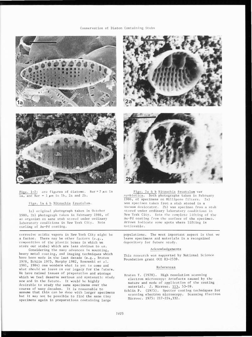
Figs. 1-2: la, and Bar
are figures of diatoms. 1 JJm in lb, 2a and 2b.
Bar = 2 ,um in
Figs. la & b Nitzschia frustulum.
la) original photograph taken in October 1980, lb) photograph taken in February 1986, of an organism on same stub stored under ordinary laboratory conditions in New York City. Note curling of Au-Pd coating.
corrosive acidic vapors in New York City might be a factor. There may be other factors (e.g., composition of the plastic boxes in which we store our stubs) which are less obvious to us.
Considering the many advances in mounting, heavy metal coating, and imaging techniques which have been made in the last decade (e.g., Braten 1978, Echlin 1975, Murphy 1982, Rosowski et al. 1981, 1984) one wonders what is yet to come and what should we leave as our legacy for the future. We have raised issues of preparation and storage which we feel deserve serious and systematic study now and in the future. It would be highly desirable to study the same specimens over the course of many decades. It is reasonable to assume that this can be done with larger specimens but it may not be possible to find the same tiny specimens again in preparations containing large
1405
Figs. 2a & b Nitzschia frustulum var symbiotica. Both photographs taken in February 1986, of specimens on Millipore filters. 2a) was specimen taken from a stub stored in a vacuum desiccator. 2b) was specimen from a stub stored under ordinary laboratory conditions in New York City. Note the complete lifting of the Au-Pd coating from the surface of the specimen. Arrows indicate some spots where lifting is noticeable.
populations. The most important aspect is that we leave specimens and materials in a recognized depository for future study.
Acknowledgements
This research was supported by National Science Foundation grant OCE 83-2539.
References
Braten T. (1978). High resolution scanning electron microscopy: Artefacts caused by the nature and mode of application of the coating material. J. Microsc. 113, 53-59.
Echlin P. (1975). SputtNcoating techniques for scanning electron microscopy. Scanning Electron Microsc. 1975: 217-224,332.
J.J. Lee, C.W. Reimer, R. Mahoney
Hargraves P, Guillard R. (1974). Structural and physiological observations on some small marine diatoms. Phycologia. 13, 163-172.
Lee J J, McEnery ME, Rottger R, Reimer CW. (1980a). The culture, isolation and identification of endosymbiotic diatoms for Heterostegina depressa d'Orbigny and Amphistegina lessonii d'Orbigny (larger foraminifera) from Hawaii. Botanica Marina Q, 297-302.
Lee J J, Reimer CW, McEnery ME. (1980b). The taxonomy of diatoms isolated as symbionts from the larger foraminifera from the Red Sea. Botanica Marina Q, 42-48.
Lee J J, Reimer CW. (1982). Isolation and identification of endosymbiotic diatoms, in Proceedings of the 7th Symposium on Recent and Fossil Diatoms, D. G. Mann (Ed), O. Koeltz, Koenigstein, W. Germany, 327-337.
Murphy J A. (1982). Considerations, materials and procedures for specimen mounting prior to scanning electron microscopic examination. Scanning Electron Microsc. 1982; II: 657-696.
Reimer CW, Lee J J. (1984). A new pennate diatom Protokeelia hottingerii, Gen. nov. ~ Nov., With a primitive keel. Proc. Acad. Nat. Sci. Philadelphia, 136, 194-199.
Rosowski JR, Hoagland K D, Roemer SC, Lee KW. (1981). Improving the image of delicate and complex biological surfaces. Scanning.'.'..• 181-187.
1406
5
Figs.
• • •
)
3-5:
•
' ' .-., ...
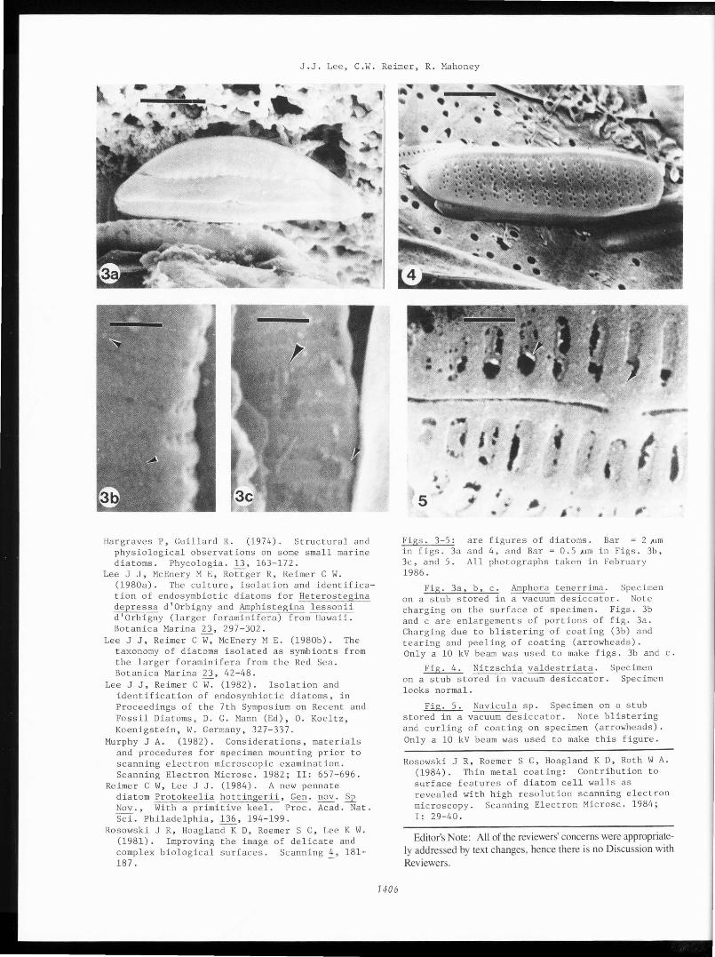
are in figs. 3a and 3c, and 5. All 1986.
•
! # f. r ·~ ~
I ,. ,·· • ,. .
figures of diatoms. Bar = 2 ,um 4, and Bar= 0.5 ,um in Figs. 3b, photographs taken in February
Fig. 3a, b, c. Amphora tenerrima. Specimen on a stub stored in a vacuum desiccator. Note charging on the surface of specimen. Figs. 3b and care enlargements of portions of fig. 3a. Charging due to blistering of coating (3b) and tearing and peeling of coating (arrowheads). Only a 10 kV beam was used to make figs. 3b and c.
Fig. 4. Nitzschia valdestriata. Specimen on a stub stored in vacuum desiccator. Specimen looks normal.
Fig. 5. Navicula sp. Specimen on a stub stored in a vacuum desiccator. Note blistering and curling of coating on specimen (arrowheads). Only a 10 kV beam was used to make this figure.
Rosowski JR, Roemer SC, Hoagland K D, Roth WA. (1984). Thin metal coating: Contribution to surface features of diatom cell walls as revealed with high resolution scanning electron microscopy. Scanning Electron Microsc. 1984; I: 29-40.
Editor's Note: All of the reviewers' concerns were appropriately addressed by text changes, hence there is no Discussion with Reviewers.