Embed Size (px)
Citation preview

This publication represents the views and expertopinions of an IARC Working Group on the
Identification of Carcinogenic Hazards to Humans,which met in Lyon, 5–11 November 2019
LYON, FRANCE - 2020
SOME INDUSTRIAL CHEMICAL INTERMEDIATES
AND SOLVENTS VOLUME 125
IARC MONOGRAPHS ON THE IDENTIFICATION
OF CARCINOGENIC HAZARDS TO HUMANS

143
1. Exposure Characterization
1.1 Identification of the agent
1.1.1 Nomenclature
Chem. Abstr. Serv. Reg. No.: 106-91-2Chem. Abstr. Serv. name: 2,3-epoxypropyl- methacrylateIUPAC systematic name: (oxiran-2-yl)methyl 2-methylprop-2-enoateSynonyms: glycidyl methacrylate; (RS)-2,3-epoxypropylmethacrylate; (±)-2,3-epoxypro-pylmethacrylate; 2-((methacryloxy)methyl)oxirane; 2-oxiranylmethyl ester; methacrylic acid-2,3-epoxypropylester; 2-propenoic acid, 2-methyl-; 1-propanol, 2,3-epoxy-, methacryl- ate.
1.1.2 Structural and molecular formula, and relative molecular mass
OO
O
Molecular formula: C7H10O3
Relative molecular mass: 142.15
1.1.3 Chemical and physical properties of the pure substance
Description: colourless, combustible liquid substance with a sweetish or fruity odour, which tends to polymerize spontaneously (HSDB, 2003)Boiling point: 189 °C (HSDB, 2003)Melting point: −41.5 °C (HSDB, 2003)Density: 1.04–1.07 g/cm3 (20 °C) (ECHA, 2019; IFA, 2019)Solubility: < 10–50 g/L (in water at 20–25 °C) (ECHA, 2019; IFA, 2019), very soluble in benzene, ethyl ether, and ethyl alcohol (HSDB, 2003)Vapour pressure: 4.2 hPa (25 °C) (ECHA, 2019)Flash point: 76–84 °C at 101.3 kPa (ECHA, 2019)Auto-ignition temperature: 389 °C at 101.3 kPa (ECHA, 2019)Vapour density: 4.91 (air = 1) (IFA, 2019)Octanol/water partition coefficient (P): log Kow = 0.96 (ILO, 2006)Conversion factor: 1 ppm = 5.91 mg/m3 (at 20 °C and 101.3 kPa).
GLYCIDYL METHACRYLATE

IARC MONOGRAPHS – 125
144
1.1.4 Technical grade and impurities
The purity of technical-grade glycidyl meth-acrylate is 92% (HSDB, 2003). Known poten-tial impurities of the technical product can be epichlorohydrin (0.02%) and polymerization inhibitors such as monomethyl ether hydro-quinone (≤ 0.01%) (HSDB, 2003; Dobrovolsky et al., 2016).
1.2 Production and use
1.2.1 Production process
Glycidyl methacrylate belongs to the group of substituted epoxides or substituted carboxylic acid esters and is produced by the esterifica-tion of methacrylic acid with either glycidol or epichlorohydrin (HSDB, 2003).
1.2.2 Production volume
Glycidyl methacrylate is listed as a High Production Volume chemical in the Screening Information Data Set (SIDS) of the Organisation for Economic Co-operation and Development (OECD, 2009). Currently, the majority of manu-facturing sites are located in the USA and Europe, with fewer sites being situated in Asia (Chem Sources, 2019). The European Chemicals Agency (ECHA) reported that 1000–10 000 tonnes of glycidyl methacrylate per year are currently manufactured and/or imported in the European Economic Area (ECHA, 2019). The aggregate production volume in the USA in 2014 and 2015 has been reported to be between 10 000 000 and 50 000 000 lb [between approximately 4500 and 23 000 tonnes] (US EPA, 2016). The production volume in Japan for glycidyl methacrylate in 1995 was approximately 3000 tonnes (OECD-SIDS, 2000).
1.2.3 Uses
Glycidyl methacrylate is mainly used as co-monomer for the production of various composite materials and epoxy polymers, such as bisphenol A-glycidyl methacrylate (BisGMA) and triethylene glycol-dimeth-acrylate (TEGDMA). These are used as dental sealants (Pulgar et al., 2000; Gioka et al., 2005; Vervliet et al., 2018), or bone adhesives and tissue (Palussière et al., 2005; Middleton et al., 2008; Sanus et al., 2008; Monmaturapoj et al., 2017). Glycidyl methacrylate is also used as an adhe-sion promotion/crosslinking co-monomer in the manufacture of vinyl and acrylic resins (HSDB, 2003). These resins are used as industrial powder and metal coatings for household appliances, facades, and automotives (Pietschmann, 2010). Glycidyl methacrylate, as an acrylic copolymer, has also been classified as a food contact material substance by the United States Food and Drug Agency (US FDA, 2018a) for aqueous and fatty foods (US FDA, 2018b, and for components of paper and paperboard in contact with dry food (US FDA, 2018b). Glycidyl methacrylate is also used for the manufacture of epoxy polymers, which are increasingly proposed for new medical applications such as hydrogel contact lenses, medical imaging, 3D-printing biomaterials and targeted drug delivery (Hagit et al., 2010; Hardy et al., 2015; Li et al., 2015; Abbadessa et al., 2016; Musgrave & Fang, 2019; Pei et al., 2019).
In the European Economic Area, the ECHA reported that glycidyl methacrylate has active registrations under the regulations of Registration, Evaluation, Authorisation and Restriction of Chemicals (REACH) and is used in articles, in formulation or re-packing, at industrial sites, and in manufacturing. Similarly, use as monomer in polymer synthesis has also been registered outside the European Union. The substance is used for the production of mixtures or articles by tabletting, compression, extru-sion, or pelletization. Specifically, the industrial

Glycidyl methacrylate
145
use of monomers occurs in the manufacture of thermoplastics and as a process regulator for polymerization processes in the production of resins, rubbers, and polymers. Consequently, glycidyl methacrylate-based polymers can be found in products with plastic materials, such as food packaging and storage devices, toys, and mobile phones. In addition, there is an imported polymer product registered in the European Union that can contain the monomer in or on the article (ECHA, 2019).
1.3 Methods of measurement and analysis
For personal air sampling of glycidyl meth-acrylate and its analysis, the use of an XAD2 sorbent tube at a flow rate of 1 L/minute for sample collection, butyl acetate for desorption, and gas chromatography with flame ionization detection (GC-FID) has been used previously (OECD-SIDS, 2000). [The Working Group noted that no further methodological details were mentioned in this report; however, the United States Environmental Protection Agency (US EPA) Compendium Method TO-15 for the analysis of volatile organic compounds in air, including the analysis of ethyl acrylate and methyl meth-acrylate, or the United States National Institute for Occupational Safety and Health (NIOSH) method 1614 for the analysis of ethylene oxide could possibly be adapted to the determination of glycidyl methacrylate.]
No methods have been published for the measurement of glycidyl methacrylate in other environmental media such as water, soil or waste matrices.
No analytical methods for biological moni-toring of glycidyl methacrylate in biological mate-rials such as blood or urine samples from exposed individuals were available [The Working Group noted that previously published methods on the determination of epoxides such as ethylene oxide,
i.e. measuring haemoglobin adducts in blood or mercapturic acids in urine, could possibly be adapted for glycidyl methacrylate.]
1.4 Exposure and occurrence
1.4.1 Environmental occurrence
Glycidyl methacrylate is not known to occur naturally in the environment. There are few data on the environmental occurrence of this chem-ical. On the basis of its low vapour pressure, glycidyl methacrylate is not expected to aero-solize readily (OECD-SIDS, 2000).
Glycidyl methacrylate can occur in the environment after release into waste water from chemical manufacturing; the amount released into air is negligible. It has been reported to be 100% biodegradable after 28 days using OECD 301C protocol and has a half-life of 3.66 days at pH 7 in water. On the basis of its low octanol/water partition coeffic-ient, bioaccumulation of glycidyl methacrylate is expected to be low. It was reported that 99.1% will be distributed into the water phase when discharged into water; the remainder will be distributed between soil (0.4%) and air (0.4%). In Japan, approximately 3.3 tonnes per year were reported to be released into rivers by one manufacturer, and 1.62 tonnes per year by a second manufacturer, while release into air was negligible. The higher of the two releases resulted in a local predicted environmental concentra-tion (PEClocal) of 8.9 × 10⁻³ mg/L as a worst-case scenario for water (OECD-SIDS, 2000).
1.4.2 Occupational exposure
Glycidyl methacrylate is manufactured in a closed system under well-controlled conditions, so air release is unlikely (OECD-SIDS, 2000). Some direct handling is required, such as during transfer at dedicated facilities and into small
IARC MONOGRAPHS – 125
146
containers, or laboratory work, when exposure can take place (ECHA, 2019).
The only sampling for occupational exposure available for glycidyl methacrylate was for Japan (OECD-SIDS, 2000). Glycidyl methacrylate was produced in a closed system. Sampling was conducted at two chemical-production sites for workers who were directly handling resin mate-rials during sampling, maintaining, can filling, filtering, analysing, and removing sludge. The tasks that did not involve direct handling were transferring and treating waste. The highest personal air concentration was 2.3 mg/m3 for filtration that was conducted three times per day and can filling that was conducted once every 7 days. For the other tasks, concentrations were below the limit of detection. Generally, dermal exposure, although short (5 minutes per day), was estimated to be 0.04 or 0.22 mg/kg body weight (bw) per day (OECD-SIDS, 2000).
Because glycidyl methacrylate is also used in the preparation of TEGDMA and BisGMA it can be assumed that workers preparing these dental and bone composite materials can also be poten-tially exposed (Olea et al., 1996). Specifically, some release of unreacted glycidyl methacrylate has been shown from a bone composite in an experimental setting, but the amount was not reported (Monmaturapoj et al., 2017). [The Working Group noted that short-term exposure to unreacted glycidyl methacrylate monomer might occur for workers during the preparation of dental and bone composite materials. Once the polymer is completely hardened, no exposure to glycidyl methacrylate is expected to occur. Hardening can take from a few minutes up to several days for some bone composites.]
Another study assessing dental-care personnel reported occupational exposure for respirable dust containing BisGMA and TEGDMA polymers, formed by reaction from bisphenol A and glycidyl methacrylate. The particles ranged in diameter from 6 nm to 5 µm and consisted of resin matrix. BisGMA
and TEGDMA monomers were released from the polymer by the grinding process. Glycidyl methacrylate itself was not measured (Cokic et al., 2017). [The Working Group noted that the glycidyl methacrylate monomer is not likely to be released from the grinding process.]
Additionally, an occupation of potential concern is work in a chemical laboratory. Matura et al. (1995) reported a case study of a female laboratory worker with confirmed allergic contact dermatitis after exposure to glycidyl methacrylate via compounded emulsions.
1.4.3 Exposure of the general population
Exposure for the general population has not been well documented. Glycidyl methacrylate has a low vapour pressure but inhalation may still be possible. Estimates of consumption of glycidyl methacrylate via drinking-water and fish for locations near to chemical-manufac-turing plants that produce or use this chem-ical are 2.97 × 10⁻⁴ mg/kg bw per day and 1.34 × 10⁻⁵ mg/kg bw per day, respectively, for an adult consuming 2 L per day of drinking-water or 90 g of fish, with a body weight of 60 kg (OECD-SIDS, 2000).
Patients, including young children, receive dental and bone composite materials containing TEGDMA and BisGMA (Olea et al., 1996; Nathanson et al., 1997; Pulgar et al., 2000; Gioka et al., 2005; Zimmerman-Downs et al., 2010; Vervliet at al., 2018). Bationo et al. (2016) reported use of monomers containing 3–5% glycidyl methacrylate to make an adhesive resin for orthodontic mineral fillers. The polymeriza-tion reaction for the dental resin occurs before the material is used in the patient, but often requires a blue visible light for a short time period to allow photo- or co-initiators to start the polymeriza-tion reaction. Curing time varies depending on the polymer, with some taking 20 seconds, while others, such as root canal sealer, taking 24 hours to set and 7 days to completely polymerize
Glycidyl methacrylate
147
(Vervliet et al., 2018), and bone composites taking as long as 10 days (Monmaturapoj et al., 2017). Release of bisphenol A, BisGMA, and TEDGMA was reported in many studies, but glycidyl meth-acrylate was not measured (Mair, 1994; Schmalz et al., 1999; Hagio et al., 2006; Lin et al., 2007).
[The Working Group noted that short-term exposure to unreacted glycidyl methacrylate monomer might occur for patients receiving these dental and bone composite materials while the polymerization process occurs. Once the polymer is completely hardened, no exposure to glycidyl methacrylate is expected to occur. Hardening can take few minutes up to several days for some bone composites.]
1.5 Regulations and guidelines
Glycidyl methacrylate has been listed by the ECHA as a carcinogen (Category 1B) and as a germ cell mutagen (Category 2) (ECHA, 2015a). An occupational exposure limit of 0.01 ppm [0.06 mg/m3] has been recommended by the Japan Society for Occupational Health (JSOH, 2018), whereas a short-term limit value of 5 mg/m3 is recommended in the People’s Republic of China (IFA, 2019).
2. Cancer in Humans
No data were available to the Working Group.
3. Cancer in Experimental Animals
See Table 3.1.
3.1 Mouse
Inhalation
In a study that complied with good laboratory practice (GLP), groups of 50 male and 50 female B6D2F1/Crlj [Crj:BDF1] mice (age, 5 weeks) were exposed by whole-body inhalation to clean air (control) or 2,3-epoxypropyl methacrylate [glycidyl methacrylate] (purity, > 99.7%) vapours at a concentration of 0.6, 2.5, or 10 ppm (v/v) for 6 hours per day, 5 days per week, for 104 weeks (JBRC, 2015a, b). The mice were observed daily for clinical signs and mortality. Survival rates of males at 2.5 and 10 ppm and of females at 0.6, 2.5, and 10 ppm were significantly lower than those of their respective controls (males, 26/50 controls, 26/50, 15/50, 14/50; females, 27/50 controls, 15/50, 19/50, 9/50). There was no signif-icant effect on body weight in exposed males and females. All mice underwent complete necropsy and histopathological examination.
In male and female mice, glycidyl meth-acrylate caused a significant increase in the inci-dence and/or a positive trend in the incidence of haemangiosarcoma of the nasal cavity in males (0/50 controls, 0/50, 1/50, 10/50; P < 0.01 at the highest dose, Fisher exact test; P < 0.01, Cochran–Armitage and Peto trend tests) and females (0/50 controls, 0/50, 1/50, 4/50; P < 0.01, Cochran–Armitage and Peto trend tests), and of haeman-gioma of the nasal cavity in males (0/50 controls, 0/50, 3/50, 8/50; P < 0.01 at the highest dose, Fisher exact test; P < 0.01, Cochran–Armitage and Peto trend tests) and females (0/50 controls, 0/50, 3/50, 7/50; P < 0.01 at high dose, Fisher exact test; P < 0.01, Cochran–Armitage and Peto trend tests). There was a significant increase in the inci-dence at the highest dose, and a positive trend in the incidence of haemangioma or haemangio-sarcoma (combined) of the nasal cavity in male and female mice.
There was a significant positive trend in the incidence of adenoma of the Harderian gland

IARC M
ON
OG
RAPH
S – 125
148 Table 3.1 Studies of carcinogenicity with glycidyl methacrylate in mice and rats exposed by inhalation (whole-body exposure)
Species, strain (sex) Age at start Duration Reference
Purity (vehicle) Dose(s) No. of animals at start No. of surviving animals
Incidence of tumours Significance Comments
Mouse, B6D2F1/Crlj (M) Age, 5 wk 104 wk JBRC (2015a, b)
Purity, > 99.7% (clean air) 0, 0.6, 2.5, 10 ppm (vapour) 6 h/day, 5 days/wk 50, 50, 50, 50 26, 26, 15, 14
Nasal cavity Principal strengths: multiple-dose study; males and females used, GLP study, covered most of lifespan Other comments: survival rates of males exposed at 2.5 and 10 ppm significantly decreased; incidence in historical controls for laboratory: nasal cavity adenoma, 2/2545 (range, 0.1%, 0–2%); and forestomach squamous cell papilloma, 7/2545 (range, 0.3%, 0–2%)
Haemangioma0/50, 0/50, 3/50, 8/50* P < 0.01 (Cochran–Armitage and
Peto trend tests), *P < 0.01 (Fisher exact test)
Haemangiosarcoma0/50, 0/50, 1/50, 10/50* P < 0.01 (Cochran–Armitage and
Peto trend tests), *P < 0.01 (Fisher exact test)
Haemangioma or haemangiosarcoma (combined)0/50, 0/50, 4/50, 16/50* P < 0.01 (Cochran–Armitage and
Peto trend tests), *P < 0.01 (Fisher exact test)
Adenoma0/50, 0/50, 0/50, 3/50 (6%) P < 0.01 (Cochran–Armitage and
Peto trend tests) Forestomach: squamous cell papilloma
0/50, 1/50, 0/50, 3/50 (6%) P < 0.05 (Cochran–Armitage trend test), P < 0.01 (Peto trend test)
Harderian gland: adenoma1/50, 1/50, 5/50, 5/50 P < 0.05 (Peto trend test)
Mouse, B6D2F1/Crlj (F) Age, 5 wk 104 wk JBRC (2015a, b)
Purity, > 99.7% (clean air) 0, 0.6, 2.5, 10 ppm (vapour) 6 h/day, 5 days/wk 50, 50, 50, 50 27, 15, 19, 9
Nasal cavity Principal strengths: multiple-dose study, males and females used, GLP study, covered most of lifespan Other comments: survival rates of all three groups of treated females significantly decreased; incidence in historical controls for the laboratory, histiocytic sarcoma of the uterus, 534/2545 (21.0%, 10–34%)
Haemangioma0/50, 0/50, 3/50, 7/50* P < 0.01 (Cochran–Armitage and
Peto trend tests), *P < 0.01 (Fisher exact test)
Haemangiosarcoma0/50, 0/50, 1/50, 4/50 P < 0.01 (Cochran–Armitage and
Peto trend tests)Haemangioma or haemangiosarcoma (combined)0/50, 0/50, 4/50, 11/50* P < 0.01 (Cochran–Armitage and
Peto trend tests), *P < 0.01 (Fisher exact test)

Glycidyl m
ethacrylate
149
Species, strain (sex) Age at start Duration Reference
Purity (vehicle) Dose(s) No. of animals at start No. of surviving animals
Incidence of tumours Significance Comments
Mouse, B6D2F1/Crlj (F) Age, 5 wk 104 wk JBRC (2015a, b)(cont.)
Lung: bronchioloalveolar carcinoma 0/50, 2/50, 0/50, 5/50* P < 0.01 (Cochran–Armitage and
Peto trend tests), *P < 0.05 (Fisher exact test)
Uterus: histiocytic sarcoma 11/50, 10/50, 12/50, 18/50 P < 0.05 (Cochran–Armitage trend
test), P < 0.01 (Peto trend test)
Harderian gland: adenoma1/50, 1/50, 2/50, 4/50 P < 0.05 (Peto trend test)
Rat, Wistar (M+F, combined) NR (weight, 200 ± 20 g) Age, 6 months Ouyang et al. (1990)
Purity, NR (air) 0, 15.3, 206 mg/m3 6 h/day, 6 days/wk 40, 40, 40 40, 40, 38
All sites Principal strengths: males and females used Principal limitations: limited experimental details, short duration of the study Other comments: groups of 20 males and 20 females at start
No significant increase in the incidence of tumour
Rat, F344/DuCrlCrlj (M) Age, 5 wk 104 wk JBRC (2015c, d)
Purity, > 99.7% (clean air) 0, 3.2, 8, 20 ppm (vapour) 6 h/day, 5 days/wk 50, 50, 50, 50 41, 44, 39, 9
Nasal cavity Principal strengths: males and females used, multiple-dose study, GLP study, study covered most of lifespan Other comments: survival rates of males at the highest dose significantly decreased
Squamous cell carcinoma0/50, 0/50, 0/50, 29/50* P < 0.01 (Cochran–Armitage and
Peto trend tests), *P < 0.01 (Fisher exact test)
Esthesioneuroepithelioma [neuroepithelial carcinoma]0/50, 0/50, 0/50, 7/50* P < 0.01 (Cochran–Armitage and
Peto trend tests), *P < 0.01 (Fisher exact test)
Adenoma 0/50, 7/50*, 9/50*, 0/50 *P < 0.01 (Fisher exact test)
Peritoneum: mesothelioma 1/50, 7/50*, 16/50**, 14/50**
P < 0.01 (Cochran–Armitage and Peto trend tests); *P < 0.05, **P < 0.01 (Fisher exact test)
Table 3.1 (continued)
IARC M
ON
OG
RAPH
S – 125
150
Species, strain (sex) Age at start Duration Reference
Purity (vehicle) Dose(s) No. of animals at start No. of surviving animals
Incidence of tumours Significance Comments
Rat, F344/DuCrlCrlj (M) Age, 5 wk 104 wk JBRC (2015c, d)(cont.)
Skin Basal cell epithelioma0/50, 1/50, 1/50, 4/50 P < 0.05 (Cochran–Armitage trend
test), P < 0.01 (Peto trend test) Keratoacanthoma
0/50, 4/50, 3/50, 3/50 P < 0.05 (Peto trend test) Subcutis: fibroma
5/50, 4/50, 4/50, 13/50* P < 0.01 (Cochran–Armitage and Peto trend tests), *P < 0.05 (Fisher exact test)
Rat, F344/DuCrlCrlj (F) Age, 5 wk 104 wk JBRC (2015c, d)
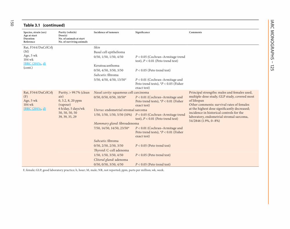
Purity, > 99.7% (clean air) 0, 3.2, 8, 20 ppm (vapour) 6 h/day, 5 days/wk 50, 50, 50, 50 39, 39, 35, 29
Nasal cavity: squamous cell carcinoma Principal strengths: males and females used, multiple-dose study, GLP study, covered most of lifespan Other comments: survival rates of females at the highest dose significantly decreased; incidence in historical controls for the laboratory, endometrial stromal sarcoma, 54/2846 (1.9%, 0–8%)
0/50, 0/50, 0/50, 10/50* P < 0.01 (Cochran–Armitage and Peto trend tests), *P < 0.01 (Fisher exact test)
Uterus: endometrial stromal sarcoma1/50, 1/50, 1/50, 5/50 (10%) P < 0.05 (Cochran–Armitage trend
test), P < 0.01 (Peto trend test)Mammary gland: fibroadenoma7/50, 14/50, 14/50, 23/50* P < 0.01 (Cochran–Armitage and
Peto trend tests), *P < 0.01 (Fisher exact test)
Subcutis: fibroma 0/50, 2/50, 2/50, 3/50 P < 0.05 (Peto trend test)Thyroid: C-cell adenoma
1/50, 1/50, 3/50, 4/50 P < 0.05 (Peto trend test) Clitoral gland: adenoma0/50, 0/50, 3/50, 4/50 P < 0.05 (Peto trend test)
F, female; GLP, good laboratory practice; h, hour; M, male; NR, not reported; ppm, parts per million; wk, week.
Table 3.1 (continued)

Glycidyl methacrylate
151
in male and female mice (P < 0.05, Peto trend test). In male mice, glycidyl methacrylate caused a significant positive trend in the incidence of adenoma of the nasal cavity (0/50 controls, 0/50, 0/50, 3/50; P < 0.01, Cochran–Armitage and Peto trend tests; with a historical control rate of 0.1% of 2545 male mice, range, 0–2%), and of squa-mous cell papilloma of the forestomach (0/50 controls, 1/50, 0/50, 3/50; P < 0.05, Cochran–Armitage trend test; P < 0.01, Peto trend test; with a historical control rate of 0.3% of 2545 male mice, range, 0–2%).
In female mice, glycidyl methacrylate caused a significant increase in the incidence at the highest dose (P < 0.05, Fisher exact test) and a positive trend in the incidence of bronchioloal-veolar carcinoma (P < 0.01, Cochran–Armitage and Peto trend tests), and a positive trend in the incidence of histiocytic sarcoma in the uterus (P < 0.05, Cochran–Armitage trend test; P < 0.01, Peto trend test).
Regarding non-neoplastic lesions, tran-sitional cell hyperplasia of the nasal cavity was observed in males and females exposed at 10 ppm and angiectasis of the nasal cavity was observed in females at 10 ppm (JBRC, 2015a, b). [The Working Group noted the strengths of this GLP study: the use of multiple doses and both males and females, while covering most of the lifespan.]
3.2 Rat
3.2.1 Inhalation
Three groups of 20 male and 20 female Wistar rats [age not reported; weight, 200 ± 20 g] were exposed to glycidyl methacrylate [purity not reported] at a concentration of 0 (control), 15.3, or 206 mg/m3 for 6 hours per day, 6 days per week, for 6 months. Two rats [sex unspec-ified] in the group at the highest dose died before the end of the study at 6 months. There was no significant increase in the incidence of
any tumour type in exposed rats (Ouyang et al., 1990). [The Working Group noted the limited experimental details and short duration of the study.]
In a study that complied with GLP, groups of 50 male and 50 female F344/DuCrlCrlj (Fischer) rats (age, 5 weeks) were exposed by whole-body inhalation to clean air (control) or 2,3-epoxypropyl methacrylate [glycidyl meth-acrylate] (purity, > 99.7%) vapours at a dose of 3.2, 8, or 20 ppm (v/v) for 6 hours per day, 5 days per week, for 104 weeks (JBRC, 2015c, d). The rats were observed daily for clinical signs and mortality. Survival rates of males and females exposed at 20 ppm were significantly lower than their respective controls (males, 41/50 controls, 44/50, 39/50, 9/50; females, 39/50 controls, 39/50, 35/50, 29/50). Body weights were significantly decreased in males at 20 ppm throughout the 2-year exposure period, and in females at 20 ppm during the last half (from 54 weeks) of the 2-year exposure period and females at 8 ppm during the late period (from 82 weeks) of the exposure, compared with their respective controls. All rats underwent complete necropsy and histopatho-logical examination.
In male and female rats, glycidyl methacrylate caused a significant increase in the incidence at the highest dose (P < 0.01, Fisher exact test), and a positive trend in the incidence of squamous cell carcinoma of the nasal cavity (P < 0.01, Cochran–Armitage and Peto trend tests).
In male rats, glycidyl methacrylate caused a significant increase in the incidence and a posi-tive trend in the incidence of esthesioneuroepi-thelioma [neuroepithelial carcinoma] of the nasal cavity (0/50 controls, 0/50, 0/50, 7/50; P < 0.01 at the highest dose, Fisher exact test; P < 0.01, Cochran–Armitage and Peto trend tests), meso-thelioma of the peritoneum (1/50 controls, 7/50, 16/50, 14/50; P < 0.05 at the lowest dose, P < 0.01 at the intermediate and highest doses, Fisher exact test; P < 0.01, Cochran–Armitage and Peto trend tests), subcutis fibroma (5/50 controls,
IARC MONOGRAPHS – 125
152
4/50, 4/50, 13/50; P < 0.05 at the highest dose, Fisher exact test; P < 0.01 Cochran–Armitage and Peto trend tests), and a significant positive trend in the incidence of basal cell epithelioma of the skin (P < 0.05, Cochran–Armitage trend test; P < 0.01, Peto trend test). There was also a signif-icant increase (P < 0.01, Fisher exact test) in the incidence of adenoma of the nasal cavity in the groups at the lowest and intermediate doses, and a significant positive trend (P < 0.05, Peto trend test) in the incidence of skin keratoacanthoma.
In female rats, glycidyl methacrylate caused a significant positive trend in the incidence of endometrial stromal sarcoma of the uterus (P < 0.05, Cochran–Armitage trend test; P < 0.01, Peto trend test), and a significant increase in the incidence at the highest dose (P < 0.01, Fisher exact test) and a positive trend in the incidence of fibroadenoma of the mammary gland (P < 0.01, Cochran–Armitage and Peto trend tests). There was also a significant positive trend (P < 0.05, Peto trend test) in the incidence of subcutis fibroma, thyroid C-cell adenoma, and adenoma of the clitoral gland.
Regarding non-neoplastic lesions in the nasal cavity, squamous cell hyperplasia with atypia in males and females at 20 ppm, squamous cell metaplasia in the respiratory epithelium in females at 3.2 ppm and in males and females at 8 and 20 ppm, squamous cell metaplasia with atypia in males at 8 ppm and in males and females at 20 ppm, and transitional epithelium hyper-plasia in males and females at 3.5 and 8 ppm and in females at 20 ppm, were observed in exposed groups (JBRC, 2015c, d). [The Working Group noted the strengths of this GLP study: the use of multiple doses and both males and females, while covering most of the lifespan.]
3.2.2 Oral administration (gavage)
In the study by Hadidian et al. (1968), five groups of three male and three female Fischer rats [age not reported, weanling] were given
glycidyl methacrylate by gavage at a dose of 0.001, 0.003, 0.01, 0.03, or 0.3 mg (in 0.5 mL steroid) per rat, five times per week for a total of 260 individual doses, for 52 weeks. A sixth group of 15 male and 15 female Fischer rats underwent similar treatment with doses of 0.1 mg per rat. Two groups of 30 male and 30 female Fischer rats served as vehicle or untreated concurrent controls. The rats were observed for six addi-tional months after treatment. At the end of the experiment (up to 600 days), full histopathology was performed. The pattern of tumour incidence observed with glycidyl methacrylate was similar to that observed in controls. [The Working Group noted the small number of animals per treated groups, the limited experimental details, the lack of statistics, and the limited reporting of results for controls. The Working Group judged the study inadequate for the evaluation.]
[The Working Group noted that the tumour site profile of glycidyl methacrylate in these studies is similar to that reported in carcino-genicity bioassays with glycidol. Specifically, in both male and female BDF1 mice exposed by inhalation, glycidol induced significant increases in the incidence of malignant tumours (haemangiosarcoma, and adenoma/adenocarci-noma) of the nasal cavity (JBRC, 2003a, b). In these female mice, squamous cell carcinoma of the nasal cavity, and malignant tumours of the uterus (histiocytic sarcoma) and mammary gland (adenocarcinoma) were also reported. In F344 rats exposed by inhalation, glycidol induced malignant tumours of the nasal cavity (adenoma or adenocarcinoma in males and females, and squamous cell carcinoma in males), peritoneum (mesothelioma in males), and uterus (endome-trial stromal sarcoma in females); other reported tumours in the rat were thyroid follicular cell carcinoma in males and splenic mononuclear cell leukaemia in females. In F344 rats treated with glycidol by gavage (IARC, 2000), there was an increased incidence of malignant tumours including of the peritoneum (males), mammary

Glycidyl methacrylate
153
gland (females), apocrine glands (males), brain (males and females), and gastrointestinal tract (males). In B6C3F1 mice treated with glycidol by gavage, there was an increased incidence of malignant tumours including of the uterus (female), Harderian gland (males and females), and mammary gland (females).]
4. Mechanistic Evidence
4.1 Absorption, distribution, metabolism, and excretion
4.1.1 Humans
(a) Exposed humans
No data were available to the Working Group.
(b) Human tissues in vitro
The metabolism of 14C-labelled glycidyl methacrylate (2 mM) was investigated in human liver homogenates over a 6-hour period (ECHA, 2015b). In the course of this study, the concentra-tion of a single metabolite that was formed was mass balanced with the concentration of glycidyl methacrylate. This metabolite was identified as glycidol based on retention time match with [14C]glycidol. [The Working Group considered such identification to be reasonable, taking also into account that maximum blood levels of glycidyl methacrylate were increased by 10-fold in rabbits in the presence of a carboxylesterase inhibitor (Shi et al., 1988); formation of both glycidol and methacrylic acid during ester cleavage remains to be definitely confirmed.]
4.1.2 Experimental systems
The intravenous administration of glycidyl methacrylate (200 mg/kg bw) in male rabbits resulted in the elimination of more than 95% of the compound from the rabbit blood within 10 minutes [The Working Group noted the lack
of experimental detail on sample collection]. In male rabbits treated with both glycidyl meth-acrylate (800 mg/kg bw, subcutaneously) and the carboxylesterase inhibitor tri-ortho-cresyl phos-phate (100 µg/kg bw), maximum blood levels of glycidyl methacrylate were increased by 10-fold compared with administration of glycidyl meth-acrylate alone, indicating that glycidyl meth-acrylate was most probably metabolized by carboxylesterase (Shi et al., 1988). [The Working Group noted that this study reported on first-order kinetics of the parent compound; no metabolites were specifically measured.]
First-order elimination of glycidyl meth-acrylate occurred in rabbit blood, plasma, and tissue homogenates, with half-lives of 4.6–22.2 minutes. Liver homogenate elimi-nated glycidyl methacrylate most effectively. Co-incubation of blood and plasma with glycidyl methacrylate and tri-ortho-cresyl phosphate (0.1 mM) decreased the elimination rate of glycidyl methacrylate by several fold. The micro-somal fraction of the rabbit liver homogenate eliminated glycidyl methacrylate faster than the mitochondrial and supernatant fractions. Co-incubation with tri-ortho-cresyl phosphate decreased the glycidyl methacrylate elimination rate in the supernatant, but not in the microsomes, indicating the role of microsomal oxidases in the biotransformation of glycidyl methacrylate (Shi et al., 1988).
The metabolism of [14C]glycidyl methacrylate (2 mM) was investigated using liver homogen-ates and nasal epithelial tissues from Fischer 344 (F344) rats and New Zealand rabbits (ECHA, 2015b). The half-life of glycidyl methacrylate hydrolysis in liver homogenates was faster in tissues from rats and rabbits (30 minutes) than in those from humans (2 hours). In the course of this study, the concentration of a single metabolite that was formed was mass balanced with the concentration of glycidyl methacrylate. This metabolite was identified as glycidol based on retention time match with [14C]glycidol

IARC MONOGRAPHS – 125
154
[see Working Group comment above, Section 4.1.1(b)].
Glycidyl methacrylate is likely to penetrate the skin in vivo given that the median lethal dose (LD50) values for dermal exposure (480 mg/kg bw in rabbits) are in the same range as the LD50 values for oral and intraperitoneal administra-tion (290–1050 mg/kg bw in various studies in mice, rats, and guinea-pigs) (ECHA, 2015a).
A proposed metabolic scheme for glycidyl methacrylate is presented in Fig. 4.1.
4.2 Evidence relevant to key characteristics of carcinogens
This section summarizes the evidence for the key characteristics of carcinogens (Smith et al., 2016), including whether glycidyl methacrylate is electrophilic or can be metabolically activated to an electrophile; is genotoxic; alters cell prolif-eration, cell death, or nutrient supply; induces epigenetic alterations; induces oxidative stress; or causes immortalization. Insufficient data were available for the evaluation of other key charac-teristics of carcinogens.
Fig. 4.1 Proposed metabolic pathways for glycidyl methacrylate in mammals
Glycidyl methacrylate
HO O
Glycidol
Methacrylic acid
Glycerol methacrylate
Epoxidehydrolase
CarboxyesteraseOO
OH3C
CH2
OO
OH
OHH3C
CH2
CH2
O
H3C
OH
Two metabolic pathways have been proposed, one mediated by carboxyesterase and a second mediated by epoxide hydrolase. [The Working Group noted that metabolites other than glycidol (see Working Group comment in Section 4.1.1) are putative.]Adapted with permission from ECHA (2015a). CLH report proposal for 2,3-epoxypropyl methacrylate (glycidyl methacrylate, GMA).

Glycidyl methacrylate
155
4.2.1 Is electrophilic or can be metabolically activated to an electrophile
No data on DNA adducts in humans or human systems were available.
Glycidyl methacrylate-specific DNA adducts were detected in the kidney, liver, testis and blood of rats treated with glycidyl methacrylate at a dose of 125 or 250 mg/kg bw using a nuclease P1-mediated 32P-postlabelling method (Fang et al., 1999a; Tan et al., 1999).
Glycidyl methacrylate (62.2 μM) induced a shift in the calf thymus DNA absorbance spectrum, indicating binding of glycidyl methacrylate to DNA (Xie et al., 1990a, 1992). After reaction of glycidyl methacrylate with deoxyadenosine monophosphate (dAMP), deoxycytidine monophosphate (dCMP), deoxy-guanosine monophosphate (dGMP), thymi-dine monophosphate (dTMP), and calf thymus DNA, covalent binding to all except dTMP at N6 of adenine or N3 of cytosine was observed, and a main DNA adduct in the reaction of glycidyl methacrylate with calf thymus DNA was N3-methacrylate-2-hydroxypropyl–dCMP (Fang et al., 1999b).
Glycidol, a metabolite of glycidyl meth-acrylate that has been identified with reasonable certainty, is a reactive epoxide that has been demonstrated to alkylate DNA in several studies in vitro (Hemminki, 1979, Hemminki et al., 1980; Hemminki, 1983; Djurič & Sinsheimer, 1984a, b; Djurič et al., 1986; Segal et al., 1990).
4.2.2 Is genotoxic
Studies on glycidyl methacrylate have been carried out in human cells in vitro, in non-human mammalian cells in vivo, in non-human mammalian cells in vitro, and in non-mammalian systems, as summarized in Table 4.1, Table 4.2, Table 4.3, and Table 4.4, respectively.
(a) Glycidyl methacrylate
(i) HumansSee Table 4.1.No studies in exposed humans were available
to the Working Group.In several studies in primary human cells
in vitro, induction of DNA strand breaks was reported after exposure to glycidyl methacrylate (Xie et al., 1990a; Poplawski et al., 2009; Styllou et al., 2015, 2017). Glycidyl methacrylate induced concentration-dependent increases in DNA double-strand breaks and single-strand breaks in human primary peripheral blood lympho-cytes as assessed using neutral and alkaline comet assays (Poplawski et al., 2009). A concen-tration-dependent increase in the number of foci containing both gamma-H2A histone family member X and tumour protein p53 binding protein 1 (γ-H2AX/53BP1) was seen in primary human gingival fibroblasts (Styllou et al., 2015). This effect was reduced by the antioxidant N-acetylcysteine (Styllou et al., 2017). Glycidyl methacrylate induced unscheduled DNA synthesis in lymphocytes (Xie et al., 1990a).
In cultured human lung fibroblast 2BS cells, glycidyl methacrylate induced a significant, concentration-dependent increase in DNA single-strand breaks, as measured by the alkaline comet assay Yin et al. (2003). The highest tested concentration of glycidyl methacrylate (5 μg/mL [35 µM]) induced significant DNA damage after as little as 1 hour. Glycidyl methacrylate induced a significant and concentration-dependent increase in mutant frequencies in the hypox-anthine-guanine phosphoribosyltransferase (HPRT) gene in the absence of metabolic acti-vation in cultured human lung fibroblasts (Yin et al., 2003). In transformed lung fibroblasts, mutations were observed in the TP53 gene (Tan et al., 1996) and the migration of the TP53 exon 8 amplicons was altered in the absence, but not presence, of metabolic activation (Tan et al., 1997). Glycidyl methacrylate induced phenotype

IARC M
ON
OG
RAPH
S – 125
156 Table 4.1 Genetic and related effects of glycidyl methacrylate in human cells in vitro
End-point Tissue, cell line Resultsa Concentration (LEC or HIC)
Comments Reference
Without metabolic activation
With metabolic activation
Unscheduled DNA synthesis Human lymphocytes (primary)
+ NT 5.2 mM [739 µg/mL] GMA and hydroxyurea
Purity is described as refractive index, nD
30 = 1.4494
Xie et al. (1990a)
DNA strand breaks (alkaline or neutral comet assay)
Lymphocytes (primary)
+ NT 0.3 mM [42.5 µg/mL] Purity, NR Poplawski et al. (2009)
DNA strand breaks (pulse-field electrophoresis)
Lymphocytes (primary)
+ NT 1.2 mM [224 µg/mL] Purity, NR Poplawski et al. (2009)
DNA strand breaks, γ-H2AX/53BP1 foci)
Gingival fibroblasts (primary)
+ NT 0.012 mM [1.7 µg/mL] Purity, NR Styllou et al. (2015)
DNA strand breaks (alkaline comet assay)
Lung fibroblast + NT 0.5 μg/mL [0.0035 mM] Yin et al. (2003)
Gene mutation, HPRT locus Lung fibroblast + NT 1.0 μg /mL [0.007 mM] Yin et al. (2003)Mutation of DNA repair genes (XRCC1, hMSH2, XPD, XRCC3)
Human bronchial epithelial cells, 16HBE
+ (only for hMSH2 gene)
NT 8 µg/mL [0.06 mM] Single dose tested Cytotoxicity, NR
Dong et al. (2009)
Mutation of TP53 gene Human embryonic lung fibroblasts (cell line)
+ NT 8 µg/mL [0.06 mM] Purity, NR Single dose tested Cytotoxicity, NR
Tan et al. (1996)
Chromosomal aberrations Human embryonic lung fibroblasts (cell line)
+ NT 1 µg/mL [0.007 mM] Purity, NR Cytotoxicity, NR
Tan et al. (1998)
Chromosomal aberrations Human bronchial epithelial cell line, 16HBE
+ NT Single exposure: 16 µg/mL [0.11 mM]
Wang et al. (2011)
+ NT Three rounds of exposure, 8 µg/mL
53BP1, P53 binding protein 1; GMA, glycidyl methacrylate; γ-H2AX, gamma-histone 2AX; HIC, highest ineffective concentration; HPRT, hypoxanthine-guanine phosphoribosyltransferase; LEC, lowest effective concentration; NR, not reported; NT, not tested.a +, positive.
Glycidyl m
ethacrylate
157
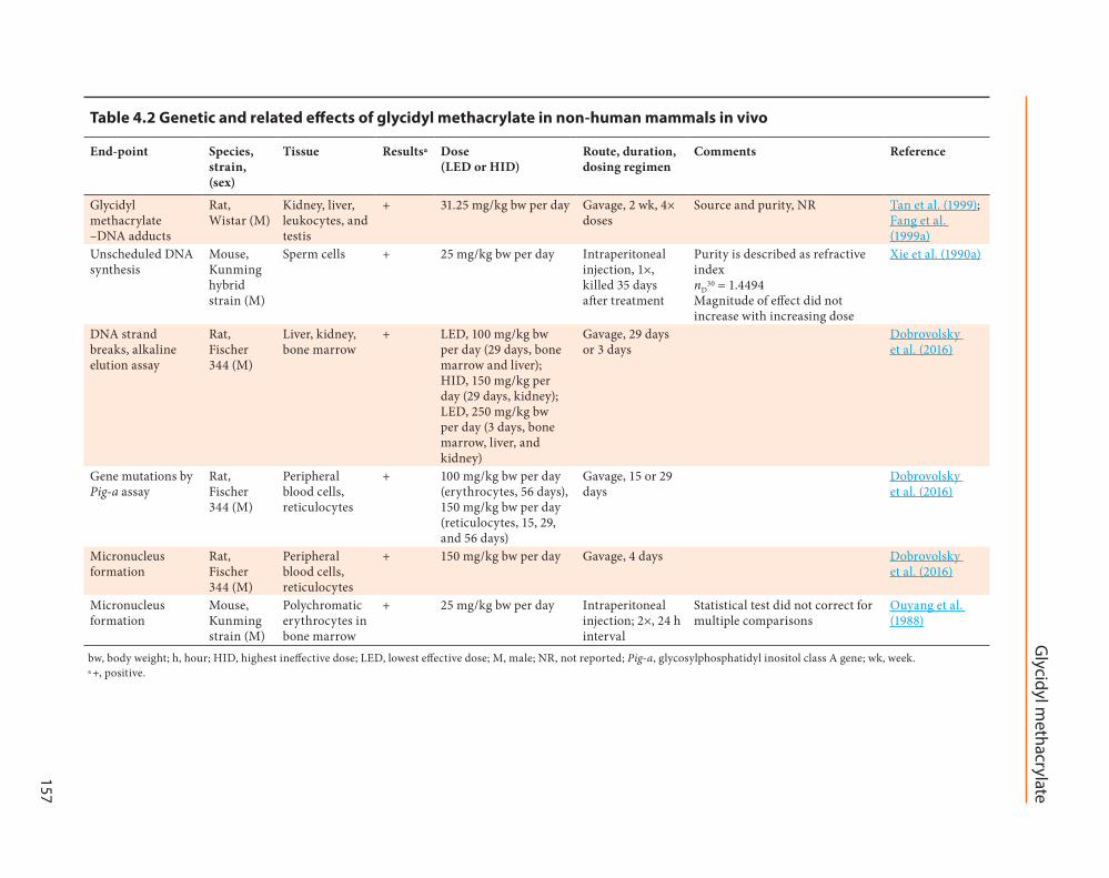
Table 4.2 Genetic and related effects of glycidyl methacrylate in non-human mammals in vivo
End-point Species, strain, (sex)
Tissue Resultsa Dose (LED or HID)
Route, duration, dosing regimen
Comments Reference
Glycidyl methacrylate –DNA adducts
Rat, Wistar (M)
Kidney, liver, leukocytes, and testis
+ 31.25 mg/kg bw per day Gavage, 2 wk, 4× doses
Source and purity, NR Tan et al. (1999); Fang et al. (1999a)
Unscheduled DNA synthesis
Mouse, Kunming hybrid strain (M)
Sperm cells + 25 mg/kg bw per day Intraperitoneal injection, 1×, killed 35 days after treatment
Purity is described as refractive index nD
30 = 1.4494 Magnitude of effect did not increase with increasing dose
Xie et al. (1990a)
DNA strand breaks, alkaline elution assay
Rat, Fischer 344 (M)
Liver, kidney, bone marrow
+ LED, 100 mg/kg bw per day (29 days, bone marrow and liver); HID, 150 mg/kg per day (29 days, kidney); LED, 250 mg/kg bw per day (3 days, bone marrow, liver, and kidney)
Gavage, 29 days or 3 days
Dobrovolsky et al. (2016)
Gene mutations by Pig-a assay
Rat, Fischer 344 (M)
Peripheral blood cells, reticulocytes
+ 100 mg/kg bw per day (erythrocytes, 56 days), 150 mg/kg bw per day (reticulocytes, 15, 29, and 56 days)
Gavage, 15 or 29 days
Dobrovolsky et al. (2016)
Micronucleus formation
Rat, Fischer 344 (M)
Peripheral blood cells, reticulocytes
+ 150 mg/kg bw per day Gavage, 4 days Dobrovolsky et al. (2016)
Micronucleus formation
Mouse, Kunming strain (M)
Polychromatic erythrocytes in bone marrow
+ 25 mg/kg bw per day Intraperitoneal injection; 2×, 24 h interval
Statistical test did not correct for multiple comparisons
Ouyang et al. (1988)
bw, body weight; h, hour; HID, highest ineffective dose; LED, lowest effective dose; M, male; NR, not reported; Pig-a, glycosylphosphatidyl inositol class A gene; wk, week.a +, positive.

IARC M
ON
OG
RAPH
S – 125
158 Table 4.3 Genetic and related effects of glycidyl methacrylate in non-human mammals in vitro
End-point Species, tissue, cell line
Resultsa Concentration (LEC or HIC)
Comments Reference
Without metabolic activation
With metabolic activation
Unscheduled DNA synthesis
Rat (strain and sex, NR), lymphocytes
+ NT 1300 µM [185 µg/mL] Purity is described as refractive index nD
30 = 1.4494 Cytotoxicity, NR
Xie et al. (1990a)
Gene mutation, Hprt locus
Chinese hamster lung fibroblast cells, V79
+ – –S9: 100 μM [14 µg/mL] (24 h), 200 μM [28 µg/mL] (4 h) +S9: 300 μM [42.5 µg/mL] (4 h)
Purity, NR Schweikl et al. (1998)
Mutation frequency
Mouse, embryonic fibroblasts, BALB/c 3T3 cells
+ NT 64 µg/mL Purity, NR Cytotoxicity, NR
Lei et al. (1998c)
Micronucleus formation
Chinese hamster lung fibroblast cells, V79
+ – –S9: 200 μM [28 µg/mL] (4 h), 100 μM [14 µg/mL] (24 h) +S9: 500 μM [71 µg/mL] (4 h)
Schweikl et al. (2001)
Micronucleus formation
Chinese hamster lung fibroblast cells, V79
+ NT 100 μM [14 µg/mL] Lee et al. (2006)
Sister-chromatid exchange
Chinese hamster lung fibroblast cells, V79
+ NT 78 μM [11 µg/mL] von der Hude et al. (1991)
h, hour; HIC, highest ineffective concentration; LEC, lowest effective concentration; NR, not reported; NT, not tested; S9, 9000 × g supernatant.a +, positive; –, negative.

Glycidyl m
ethacrylate
159
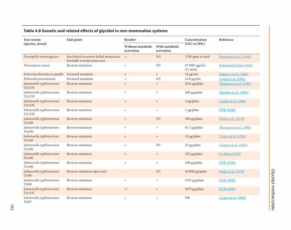
Table 4.4 Genetic and related effects of glycidyl methacrylate in non-mammalian systems
Test system (species, strain)
End-point Resultsa Concentration (LEC or HIC)
Comments on study quality
Reference
Without metabolic activation
With metabolic activation
Salmonella typhimurium TA100
Reverse mutation + + 250 µg/plate Purity, NR Schweikl et al. (1998)
Salmonella typhimurium TA100
Reverse mutation + + 112 µg/plate Source, NR Purity, 92% Cytotoxicity, NR
Ouyang et al. (1988)
Salmonella typhimurium TA98
Reverse mutation – – 5000 µg/plate Purity, NR Schweikl et al. (1998)
Salmonella typhimurium TA98
Reverse mutation – – NR Canter et al. (1986)
Salmonella typhimurium TA98
Reverse mutation – – 896 µg/plate Source, NR Purity, 92% Cytotoxicity, NR
Ouyang et al. (1988)
Salmonella typhimurium TA97a, TA102
Reverse mutation +/– +/– 250 µg/plate (TA97a) 500 µg/plate (TA102)
Purity, NR Schweikl et al. (1998)
Salmonella typhimurium TA97, 100, 1535
Reverse mutation + + 33 µg/plate for TA100 and TA1535 NR for TA97
Canter et al. (1986)
Klebsiella pneumoniae Mutation + NT 1 mM [142 µg/plate] Purity, 92% Cytotoxicity, NR
Voogd et al. (1981)
Escherichia coli PQ37 SOS chromotest
DNA damage + NT 0.3 mM [43 µg/plate] Purity, 97% von der Hude et al. (1990)
HIC, highest ineffective concentration; LEC, lowest effective concentration; NR, not reported; NT, not tested.a +, positive; –, negative; +/–, equivocal.
IARC MONOGRAPHS – 125
160
transformation and chromosomal aberrations in cultured embryonic lung fibroblasts (Tan et al., 1998). Chromosomal aberrations were detected in cultured bronchial epithelial cells treated with glycidyl methacrylate (Wang et al., 2011).
(ii) Experimental systems
Non-human mammals in vivoSee Table 4.2.In male F344 rats, oral exposure to glycidyl
methacrylate induced a dose-dependent increase in the percentage of tail DNA in bone marrow, liver, and kidney cells, as measured by the alka-line comet assay, and in gene mutation in eryth-rocytes and reticulocytes as measured by Pig-a assay (Dobrovolsky et al., 2016). Glycidyl meth-acrylate increased micronucleus formation in erythrocytes from peripheral blood in male F344 rats treated orally with a dose of 150 mg/kg bw per day for 4 days, and in polychromatic eryth-rocytes in bone marrow of male Kunming hybrid mice exposed to glycidyl methacrylate as two doses of 25 mg/kg bw per day by intraperitoneal injection at an interval of 24 hours (Ouyang et al., 1988; Dobrovolsky et al., 2016).
Unscheduled DNA synthesis was increased in the sperm of male Kunming hybrid mice 35 days after being exposed to glycidyl methacrylate at a dose of 25 mg/kg bw per day or higher as a single intraperitoneal injection (Xie et al., 1990a). [The Working Group noted that the magnitude of the effect did not increase with increasing dose.]
Non-human mammalian cells in vitroSee Table 4.3.Glycidyl methacrylate induced unscheduled
DNA synthesis and semi-conservative DNA replication in rat lymphocytes (Xie et al., 1990a). Glycidyl methacrylate induced a concentra-tion-dependent increase in hypoxanthine-gua-nine phosphoribosyltransferase (Hprt) mutation in V79 Chinese hamster lung fibroblast cells, an effect completely abolished by the addition of rat liver S9 microsomal fraction (Schweikl et al.,
1998). Glycidyl methacrylate induced a concen-tration-dependent increase in the frequencies of sister-chromatid exchange in Chinese hamster V79 cells (von der Hude et al., 1991). Schweikl et al. (2001) reported a dose-related increase in the frequencies of micronucleus formation in V79 Chinese hamster lung fibroblast cells exposed to glycidyl methacrylate (for 24 hours at 100, 150, or 200 μM or for 4 hours at 200, 300, 400, or 500 μM). The 24-hour exposure effects were reproduced by Lee et al. (2006). The effects disappeared in the presence of rat liver S9 micro-somal fraction (only 4-hour exposure tested) (Schweikl et al., 2001).
Non-mammalian experimental systemsSee Table 4.4.Glycidyl methacrylate induced reverse muta-
tions in Salmonella typhimurium strains TA97, TA100, TA1535, and TA102, but not TA98, both in the presence and in the absence of a rat liver S9 microsomal fraction (Canter et al., 1986; Ouyang et al., 1988; Schweikl et al., 1998). Glycidyl meth-acrylate was also mutagenic in Klebsiella pneumo-niae (Voogd et al., 1981). Glycidyl methacrylate induced DNA damage in the SOS chromotest using Escherichia coli PQ37 (von der Hude et al., 1990).
Acellular systemsGlycidyl methacrylate did not introduce
DNA breaks to isolated DNA, as assessed by the plasmid relaxation assay using pUC19 plasmids isolated from E. coli (Poplawski et al., 2009). E. coli HB101 transfected with glycidyl meth-acrylate-bound pBR322 was transformed to two stable and heritable mutants, from one of which deletion and insertion were detected in hot-spot regions (Xie et al., 1990b; Fang, 1991; Zuo et al., 1991; Zuo, 1991; Gao et al., 1994a, b).
(b) Glycidol
See Table 4.5, Table 4.6, Table 4.7, and Table 4.8.

Glycidyl m
ethacrylate
161
Table 4.5 Genetic and related effects of glycidol in human cells in vitro
End-point Tissue, cell line Resultsa Concentration (LEC or HIC)
Reference
Without metabolic activation
With metabolic activation
Unscheduled DNA synthesis
WI-38 cells – + 0.33 µg/mL Thompson et al. (1981)
Chromosomal aberrations
Lymphocytes (primary)
+ NT 29.6 µg/mL Norppa et al. (1981)
Sister-chromatid exchange
Lymphocytes (primary)
+ NT 3.7 µg/mL Norppa et al. (1981)
HID, highest ineffective dose; LED, lowest effective dose; NT, not tested.a +, positive; –, negative.
Table 4.6 Genetic and related effects of glycidol in non-human mammals in vivo
End-point Species, strain (sex) Tissue Resultsa Dose (LED or HID)
Route, duration, dosing regimen
Reference
Chromosomal aberrations
Rats, Sprague-Dawley (F) Bone marrow – 226 mg/kg bw per day Oral × 5 Thompson & Hiles (1981)
Chromosomal aberrations
Rats, Sprague-Dawley (F) Bone marrow – 145 mg/kg bw per day Intraperitoneal × 5 Thompson & Hiles (1981)
Micronucleus formation
Mouse, B6C3F1 (M) Polychromatic erythrocytes
+ 37.5 mg/kg bw per day Intraperitoneal × 2 NTP (1990)
Micronucleus frequency
Mouse, BalbC (M) Polychromatic erythrocytes
+ 120 mg/kg bw Intraperitoneal × 1 Aasa et al. (2017)
bw, body weight; F, female; HID, highest ineffective dose; LED, lowest effective dose; M, male. a +, positive; –, negative.

IARC M
ON
OG
RAPH
S – 125
162 Table 4.7 Genetic and related effects of glycidol in non-human mammals in vitro
End-point Species, tissue, cell line
Resultsa Concentration (LEC or HIC)
Comments Reference
Without metabolic activation
With metabolic activation
DNA damage (alkaline comet assay)
Chinese hamster ovary cells
+ NT 20 μg/mL Purity, NR El Ramy et al. (2007)
Gene mutation, Tk locus L5178Y mouse, lymphoma cells
+ + 8 μg/mL Thompson et al. (1981)
Gene mutation, Tk locus L5178Y mouse, lymphoma cells
+ NT 1.43 μg/mL NTP (1990)
Gene mutation, Hprt locus Chinese hamster ovary cells
+ NT 50 mMb [3.7 µg/mL] Aasa et al. (2016)
Gene mutation, 6-thioguanine resistance
Chinese hamster lung V79 cells
+ NT 0.15 μg/mL Smith et al. (1990)
Chromosomal aberrations Chinese hamster cells + + 12.5 μg/mL NTP (1990)Sister-chromatid exchange Chinese hamster cells + + 1.11 μg/mL NTP (1990)Sister-chromatid exchange Chinese hamster V79
cells+ NT 92.6 μg/mL von der Hude et al.
(1991)HIC, highest ineffective concentration; LEC, lowest effective concentration; NR, not reported; NT, not tested.a +, positive.b Value of LEC was not provided by the authors, but was estimated from the relevant figure in the publication.
Glycidyl m
ethacrylate
163
Table 4.8 Genetic and related effects of glycidol in non-mammalian systems
Test system (species, strain)
End-point Resultsa Concentration (LEC or HIC)
Reference
Without metabolic activation
With metabolic activation
Drosophila melanogaster Sex-linked recessive lethal mutations, heritable translocation test
+ NA 1230 ppm in feed Foureman et al. (1994)
Neurospora crassa Reverse mutation + NT 37 000b µg/mL (15 min)
Kølmark & Giles (1955)
Schizosaccharomyces pombe Forward mutation + + 74 µg/mL Migliore et al. (1982)Klebsiella pneumoniae Forward mutation + NT 14.8 µg/mL Voogd et al. (1981)Salmonella typhimurium TA1535
Reverse mutation + + 20.6 μg/plate Thompson et al. (1981)
Salmonella typhimurium TA1535
Reverse mutation + + 500 μg/plate Mamber et al. (1984)
Salmonella typhimurium TA1535
Reverse mutation + + 3 μg/plate Canter et al. (1986)
Salmonella typhimurium TA1535
Reverse mutation + + 1 μg/plate NTP (1990)
Salmonella typhimurium TA100
Reverse mutation + NT 100 μg/plate Wade et al. (1979)
Salmonella typhimurium TA100
Reverse mutation + + 61.7 μg/plate Thompson et al. (1981)
Salmonella typhimurium TA100
Reverse mutation + + 33 μg/plate Canter et al. (1986)
Salmonella typhimurium TA100
Reverse mutation + NT 25 μg/plate Claxton et al. (1991)
Salmonella typhimurium TA100
Reverse mutation + + 125 μg/plate De Flora (1979)
Salmonella typhimurium TA100
Reverse mutation + + 100 μg/plate NTP (1990)
Salmonella typhimurium TA98
Reverse mutation (spot test) – NT 10 000 μg/plate Wade et al. (1979)
Salmonella typhimurium TA98
Reverse mutation + + 3333 μg/plate NTP (1990)
Salmonella typhimurium TA1537
Reverse mutation +w + 1670 μg/plate NTP (1990)
Salmonella typhimurium TA97
Reverse mutation + + NR Canter et al. (1986)

IARC M
ON
OG
RAPH
S – 125
164
Test system (species, strain)
End-point Resultsa Concentration (LEC or HIC)
Reference
Without metabolic activation
With metabolic activation
Salmonella typhimurium TA97
Reverse mutation + + 1000 μg/plate NTP (1990)
Escherichia coli (Sd-4) Reverse mutation + NT 740 μg/mL Hussain & Osterman-Golkar (1984)
Escherichia coli PQ37 SOS chromotest
DNA strand breaks, cross-links or related damage
+ NT 244.5 µg/mL von der Hude et al. (1990)
Escherichia coli, rec assay Differential toxicity + NT NR Mamber et al. (1984)Prophage induction, SOS repair test
DNA strand breaks, cross-links or related damage
– NT 500 μg/plate Mamber et al. (1984)
HIC, highest ineffective concentration; LEC, lowest effective concentration; NA, not applicable; NT, not tested.a +, positive; –, negative; +w, weakly positive.b One dose tested; time-dependent response.
Table 4.8 (continued)
Glycidyl methacrylate
165
(i) HumansNo data on exposed humans were available to
the Working Group.Glycidol induced chromosomal aberrations
and sister-chromatid exchange in primary human lymphocytes in the absence of metabolic activation (Norppa et al., 1981), while unsched-uled DNA synthesis was induced in human fibroblast WI-38 cells only in the presence of metabolic activation (Thompson et al., 1981).
(ii) Experimental systemsGlycidol induced the formation of micronu-
clei in male mice B6C3F1 and BalbC mice after intraperitoneal administration (NTP, 1990; Aasa et al., 2017). It was without effect on chromosomal aberrations in rats after oral or intraperitoneal treatment (Thompson & Hiles, 1981).
Tests performed in experimental systems in vitro gave consistently positive results. Glycidol induced DNA damage in the alkaline comet assay in Chinese hamster ovary cells (El Ramy et al., 2007). Glycidol induced Tk gene mutation in mouse lymphoma L5178Y cells (Thompson et al., 1981; NTP, 1990), Hprt gene mutation in Chinese hamster ovary cells (Aasa et al., 2016), and gave positive results in the 6-thioguanine resistance test in Chinese hamster lung V79 cells (Smith et al., 1990). Glycidol also induced chromosomal aberrations and sister-chromatid exchange in Chinese hamster cells (NTP, 1990; von der Hude et al., 1991).
Glycidol gave uniformly positive results in several assays for reverse mutation in S. typhi-murium (De Flora, 1979; Wade et al., 1979; Thompson et al., 1981; Mamber et al., 1984; Canter et al., 1986; NTP, 1990; Claxton et al., 1991) and in two assays for mutation in fungi (Kølmark & Giles, 1955; Migliore et al., 1982). Glycidol was also mutagenic in K. pneumoniae (Voogd et al., 1981) and in E. coli (Hussain & Osterman-Golkar, 1984). Glycidol gave positive results in the assay for sex-linked recessive lethal mutation and in the heritable translocation test
in Drosophila melanogaster (Foureman et al., 1994). Glycidol gave positive results in the E. coli PQ37 SOS chromotest, and negative results in the prophage-induction SOS repair test (Mamber et al., 1984; von der Hude et al., 1990).
4.2.3 Alters cell proliferation, cell death, or nutrient supply
In B6D2F1/Crlj mice exposed to glycidyl methacrylate by inhalation for 104 weeks, increases in transitional cell hyperplasia of the nasal cavity were reported in males and females (JBRC, 2015a).
In F344/DuCrlCrlj rats exposed to glycidyl methacrylate by inhalation for 104 weeks, non-neoplastic lesions in the nasal cavity (squamous cell hyperplasia with atypia, squamous cell meta-plasia in respiratory epithelium, and squamous cell metaplasia with atypia) and transitional cell hyperplasia were reported in males and females (JBRC, 2015b).
Gap-junctional intercellular communication, as measured by the scrape-loading/dye-transfer technique, was significantly inhibited in trans-formed human lung fibroblast cells treated with glycidyl methacrylate (2.5 or 5 µg/mL) (Yin et al., 2003).
4.2.4 Induces oxidative stress
Glycidyl methacrylate induced oxidative damage to DNA in primary human lympho-cytes as assessed with the alkaline comet assay using the DNA repair enzymes endonuclease III (Endo III) and formamidopyrimidine-DNA glycosylase (Fpg) (Poplawski et al., 2009). The antioxidant N-acetylcysteine reduced the glycidyl methacrylate-induced double-strand breaks and nuclear chromatin condensation in primary human gingiva fibroblasts (Styllou et al., 2017). N-acetylcysteine reduced the frequency of micronucleus formation induced by glycidyl methacrylate in Chinese hamster lung fibroblast

IARC MONOGRAPHS – 125
166
V79 cells (Lee et al., 2006) (see Section 4.2.2(a)(ii)).
4.2.5 Evidence on other key characteristics of carcinogens
Glycidyl methacrylate (8.0 μg/mL [0.06 mM]) induced cell transformation of human lung fibroblasts (Yin et al., 2003). In addition, glycidyl methacrylate induced cell transformation of BALB/c mouse embryonic fibroblasts 3T3 cells or Golden Syrian hamster embryonic SHE cells in several studies (Xu et al., 1994; Yang et al., 1996; Zhang et al., 1996; Lei et al., 1998a, b).
In a series of studies, glycidyl methacrylate induced malignant transformation and methyl-ation of the promoter regions or overexpression of several genes in human bronchial epithelial cells or embryonic lung fibroblast (Tan et al., 1998; Xu et al., 2001; Dong et al., 2009; Yang et al., 2009; Dong et al., 2010; Hu et al., 2012). Glycidyl methacrylate (8 μg/mL) induced changes in DNA methylation in stages of malignant transforma-tion, as detected by a CpG (cytosine–phosphate–guanine) promoter methylation microarray (Wang et al., 2014), including of the opioid binding protein/cell adhesion molecule-like (OPCML) gene (Liu et al., 2015) in cultured human bronchial epithelial cells (16HBE).
Methylation of the P16 [CDKN2A] gene promoter was detected at the early stage and protophase stage in the process of malignant transformation of human bronchial epithelial cells exposed to glycidyl methacrylate (Hu et al., 2012).
4.3 Data relevant to comparisons across agents and end-points
The analysis of the bioactivity in vitro of the agents reviewed in IARC Monographs Volume 125 was informed by data from high-throughput screening assays generated by the Toxicity Testing in the 21st Century (Tox21) and Toxicity
Forecaster (ToxCast) research programmes of the Government of the USA (Thomas et al., 2018). Glycidyl methacrylate was one of thousands of chemicals tested across the large assay battery of the Tox21 and ToxCast research programmes as of 1 September 2019 (US EPA, 2019a). Detailed information about the chemicals tested, assays used, and associated procedures for data analysis is publicly available (US EPA, 2019a). [The Working Group noted that the metabolic capacity of the cell-based assays is variable, and generally limited, as acknowledged in Kavlock et al. (2012).]
Among the 402 assays in which glycidyl methacrylate (at concentrations up to 100 μM) was tested, it was found to be inactive in almost all assays. Active responses were observed in 8 assays (US EPA, 2019a). Effect on upregulation of nuclear factor erythroid 2-related factor 2 (NRF2) was reported at a half-maximal activity concen-tration (AC50) of 38 μM. For nuclear receptors, borderline activity (potency of > 50% or activity observed only at the highest concentration tested) was found for the mouse embryonic cell-based assay (SSH_3T3_GLI3) at an AC50 of 82 μM. For cell viability, glycidyl methacrylate was shown to be cytotoxic only in human T47D, HEK293, and HEPG2 cells at AC50s of 0.45–83 μM.
Glycidol was also tested (at concentrations up to 100 μM) in 673 assays and found to be inac-tive in almost all assays. Active responses were observed in 16 assays (US EPA, 2019b). Effects were observed in the form of TIMP metallo-peptidase inhibitor 1 downregulation, retinoid X receptor α agonism and androgen receptor antagonism at concentrations between 10 and 38.9 μM. These were noted as borderline effects (< 50% efficacy, only highest concentration above baseline). For cell viability, glycidol was shown to be cytotoxic only in human HEK293 and HEPG2 cells at AC50s of 33.6–65.5 μM.

Glycidyl methacrylate
167
5. Summary of Data Reported
5.1 Exposure characterization
Glycidyl methacrylate is a High Production Volume chemical that is mainly used as an inter-mediate in the production of epoxy polymers and vinyl and acrylic resins. These polymers are used in dental sealants, composites and adhe-sives; bone composite materials; powder coat-ings; and hydrogel lenses. There are emerging applications for the polymers in medical imaging and targeting drug delivery. Polymers formed of glycidyl methacrylate can also be used in food contact material. Occupational exposures by inhalation and dermal contact have been reported for chemical-production workers in a single study. Occupational exposure when preparing the dental and bone materials or expo-sure to patients receiving these materials may potentially occur, but has not been targeted for measurement. Exposure in the general popul-ation is not expected from use of the polymer-ized products.
5.2 Cancer in humans
No data were available to the Working Group.
5.3 Cancer in experimental animals
Glycidyl methacrylate was tested for carcin-ogenicity in one strain of male and female mice in one inhalation study, and in two different strains of male and female rats, respectively, in two inhalation studies, and one gavage study.
The inhalation study in mice was well-con-ducted under good laboratory practice, and resulted in a significant positive trend and increase in the incidence of haemangioma of the nasal cavity and of haemangioma or haem-angiosarcoma (combined) of the nasal cavity in males and females, and of nasal cavity
haemangiosarcoma in males and females (posi-tive trend only for females). In female mice, there was a significant positive trend and increase in the incidence of bronchioalveolar carcinoma. There was also a significant positive trend in the incidence of uterine histiocytic sarcoma, and adenoma of the Harderian gland in female mice. There was a significant positive trend in the inci-dence of adenoma of the nasal cavity, squamous cell papilloma of the forestomach, and adenoma of the Harderian gland in male mice.
One inhalation study in male and female rats was well-conducted under good laboratory prac-tice. In male and female rats, exposure to glycidyl methacrylate resulted in a significant positive trend and increase in the incidence of squamous cell carcinoma of the nasal cavity. In male rats, there was a significant positive trend and increase in the incidence of esthesioneuroepithelioma (neuroepithelial carcinoma) of the nasal cavity, and a significant increase in the incidence of adenoma of the nasal cavity. In male rats, there was also a significant positive trend and increase in the incidence of peritoneal mesothelioma and subcutis fibroma; significant positive trends in the incidence of skin basal cell epithelioma and skin keratoacanthoma were also observed. In female rats, exposure to glycidyl methacrylate resulted in a significant positive trend and increase in the incidence of fibroadenoma of the mammary gland; significant positive trends in the incidence of uterine endometrial stromal sarcoma, subcutis fibroma, thyroid C-cell adenoma, and adenoma of the clitoral gland were also observed.
The other inhalation study in rats gave nega-tive results.
The study in male and female rats treated by gavage was inadequate for the evaluation of the carcinogenicity of glycidyl methacrylate in experimental animals.

IARC MONOGRAPHS – 125
168
5.4 Mechanistic evidence
No data on the absorption, distribution, metabolism or excretion of glycidyl methacrylate in exposed humans were available. No direct data on the absorption or excretion of glycidyl meth-acrylate were available from mammalian species. Indirect evidence of dermal and oral absorption is provided by tests for acute toxicity in various rodent species, which suggest that dermal absorption of glycidyl methacrylate is likely to be as efficient as oral absorption. In one study in rabbits, a carboxyesterase inhibitor markedly reduced the decline of glycidyl methacrylate in the blood. Studies in vitro in human, rabbit, and rat tissue homogenates showed the formation over time of a metabolite that was reasonably identified as glycidol.
There is consistent and coherent evidence that glycidyl methacrylate exhibits key charac-teristics of carcinogens (is genotoxic) in human primary cells. In all available studies in human primary cells, glycidyl methacrylate induced DNA damage, including double-strand breaks and unscheduled DNA synthesis. In different cultured human cell lines, consistent findings of various types of damage to DNA, including DNA strand breaks, gene mutations, and chromo-somal aberrations were seen. In two studies in orally exposed rats, glycidyl methacrylate induced various types of damage to DNA, including strand breaks, gene mutations, and micronucleus formation. After intraperitoneal administration in mice, glycidyl methacrylate induced micronucleus formation and unsched-uled DNA synthesis in two studies. In several studies in rodent cells in vitro, glycidyl meth-acrylate induced Hprt gene mutation, micro-nucleus formation, sister-chromatid exchanges, and unscheduled DNA synthesis; the effects were abolished by metabolic activation in the available tests. In addition, glycidyl methacrylate gave generally positive results in tests for base-pair substitution mutation in bacteria, both in
the presence and in the absence of metabolic activation. Glycidyl methacrylate–DNA lesions in plasmid DNA yielded stable and heritable mutations.
Glycidol caused DNA damage, chromosomal aberrations, and sister-chromatid exchange in one study in human primary cells in vitro, and increased micronucleus formation in mice after intraperitoneal administration, but not chromo-somal aberrations in rats after intraperitoneal or oral administration. Glycidol gave consistently positive results in a substantial number of geno-toxicity assays in vitro. Glycidol gave positive results both in rodent cells, covering a range of end-points such as DNA damage, gene mutation and chromosomal aberrations, and in mutation tests in bacteria.
In experimental systems, there were consistent and coherent findings in male and female rats and mice exposed chronically by inhalation that glycidyl methacrylate alters cell proliferation, cell death, or nutrient supply. Glycidyl methacrylate induced a dose-related increase in the incidence of transitional cell hyperplasia of the nasal cavity in both sexes of mice and rats, and in squamous cell hyperplasia with atypia, squamous cell metaplasia in respira-tory epithelium, and squamous cell metaplasia with atypia in male and female rats.
There is suggestive evidence that glycidyl methacrylate is electrophilic, based on the forma-tion of DNA adducts in tissues of exposed rats and when glycidyl methacrylate was added to DNA or DNA bases, in the few available studies. In addition, glycidol is electrophilic. Glycidol is a reactive epoxide that alkylates DNA in vitro. There is also suggestive evidence from a narrow range of experiments in human and mammalian cells in vitro that glycidyl methacrylate induces oxidative stress or causes immortalization. Glycidyl methacrylate induced oxidative damage to DNA, and the antioxidant N-acetylcysteine reduced the effect of glycidyl methacrylate on several genotoxic end-points in human and

Glycidyl methacrylate
169
rodent cells. Glycidyl methacrylate also induced cell transformation in human and rodent cells in vitro.
Glycidyl methacrylate and glycidol were found to be mostly without effects in the assay battery of the Toxicity Testing in the 21st Century (Tox21) and Toxicity Forecaster (ToxCast) research programmes.
Overall, the mechanistic evidence is consistent and coherent that glycidyl meth-acrylate belongs, on the basis of mechanistic considerations, to a class of reactive glycidyl epoxides, for which one member has been classified as probably carcinogenic to humans. Glycidyl methacrylate bears structural similarity to other glycidyl epoxides, and there is close concordance with glycidol with respect to the genotoxicity profile as well as the tumour-site profile in chronic animal bioassays.
6. Evaluation and Rationale
6.1 Cancer in humans
There is inadequate evidence in humans regarding the carcinogenicity of glycidyl methacrylate.
6.2 Cancer in experimental animals
There is sufficient evidence in experimental animals for the carcinogenicity of glycidyl methacrylate.
6.3 Mechanistic evidence
There is strong evidence that glycidyl meth-acrylate belongs, on the basis of mechanistic considerations, to a class of reactive glycidyl epoxides for which one member, glycidol, has been classified as probably carcinogenic to humans (Group 2A). There is also strong evidence
in human primary cells that glycidyl methacrylate exhibits key characteristics of carcinogens.
6.4 Overall evaluation
Glycidyl methacrylate is probably carcino-genic to humans (Group 2A).
6.5 Rationale
The Group 2A evaluation for glycidyl meth-acrylate is based on sufficient evidence of cancer in experimental animals and strong mechanistic evidence. The evidence regarding cancer in humans was inadequate as no data were avail-able. The sufficient evidence of carcinogenicity in experimental animals is based on the induction of malignant neoplasms in two species.
There was strong mechanistic evidence, based on two distinct topics. There is strong evidence that glycidyl methacrylate belongs, based on mechanistic considerations, to a class of reac-tive glycidyl epoxides for which one member, glycidol, has been classified as probably carcino-genic to humans. Glycidyl methacrylate bears structural similarity to other members of this class, and there is close concordance with respect to the genotoxicity profile, and the target organs of carcinogenicity in chronic animal bioassays. There is also strong evidence in primary human cells that glycidyl methacrylate exhibits key char-acteristics of carcinogens; glycidyl methacrylate is genotoxic in all available tests in human primary cells, supported by consistent findings across several different test systems in various species. It also alters cell proliferation, cell death, or nutrient supply in experimental systems.
IARC MONOGRAPHS – 125
170
References
Aasa J, Abramsson-Zetterberg L, Carlsson H, Törnqvist M (2017). The genotoxic potency of glycidol estab-lished from micronucleus frequency and hemoglobin adduct levels in mice. Food Chem Toxicol. 100:168–74. doi:10.1016/j.fct.2016.12.022 PMID:28012894
Aasa J, Vare D, Motwani HV, Jenssen D, Törnqvist M (2016). Quantification of the mutagenic potency and repair of glycidol-induced DNA lesions. Mutat Res Genet Toxicol Environ Mutagen. 805:38–45. doi:10.1016/j.mrgentox.2016.05.011 PMID:27402481
Abbadessa A, Blokzijl MM, Mouser VH, Marica P, Malda J, Hennink WE, et al. (2016). A thermo-responsive and photo-polymerizable chondroitin sulfate-based hydrogel for 3D printing applications. Carbohydr Polym. 149:163–74. doi:10.1016/j.carbpol.2016.04.080 PMID:27261741
Bationo R, Jordana F, Boileau MJ, Colat-Parros J (2016). Release of monomers from orthodontic adhesives. Am J Orthod Dentofacial Orthop. 150(3):491–8. doi:10.1016/j.ajodo.2016.02.027 PMID:27585778
Canter DA, Zeiger E, Haworth S, Lawlor T, Mortelmans K, Speck W (1986). Comparative mutagenicity of aliphatic epoxides in Salmonella. Mutat Res. 172(2):105–38. doi:10.1016/0165-1218(86)90069-8 PMID:3531837
Chem Sources (2019). Chem Sources Online, Chemical Sources International, Inc. Glycidyl methacrylate. Available from: http://www.chemsources.com, accessed November 2019.
Claxton LD, Houk VS, Monteith LG, Myers LE, Hughes TJ (1991). Assessing the use of known mutagens to cali-brate the Salmonella typhimurium mutagenicity assay: I. Without exogenous activation. Mutat Res. 253(2):137–47. doi:10.1016/0165-1161(91)90127-T PMID:1922140
Cokic SM, Duca RC, Godderis L, Hoet PH, Seo JW, Van Meerbeek B, et al. (2017). Release of monomers from composite dust. J Dent. 60:56–62. doi:10.1016/j.jdent.2017.02.016 PMID:28257993
De Flora S (1979). Metabolic activation and deactivation of mutagens and carcinogens. Ital J Biochem. 28(2):81–103. PMID:399955
Djurič Z, Hooberman BH, Rosman L, Sinsheimer JE (1986). Reactivity of mutagenic propylene oxides with deoxy-nucleosides and DNA. Environ Mutagen. 8(3):369–83. doi:10.1002/em.2860080306 PMID:3519198
Djurič Z, Sinsheimer JE (1984a). Reactivity of propylene oxides towards deoxycytidine and identification of reaction products. Chem Biol Interact. 50(2):219–31. doi:10.1016/0009-2797(84)90098-X PMID:6744466
Djurič Z, Sinsheimer JE (1984b). Characterization and quantitation of 3-alkylthymidines from reactions of mutagenic propylene oxides with thymidine. Chem Biol Interact. 52(2):243–53. doi:10.1016/0009-2797(84)90077-2 PMID:6391705
Dobrovolsky VN, Pacheco-Martinez MM, McDaniel LP, Pearce MG, Ding W (2016). In vivo genotoxicity assess-ment of acrylamide and glycidyl methacrylate. Food Chem Toxicol. 87:120–7. doi:10.1016/j.fct.2015.12.006 PMID:26686995
Dong L, Hu J, Wang Q, et al. (2010). The altered expres-sion of relative genes in malignant transformation of human bronchial epithelial cells induced by glycidyl methacrylate. Carcinog Teratog Mutagen. 22(4):249–54.
Dong L, Wang Q, Sun J, et al. (2009). Point mutation of DNA repair genes of human bronchial epithelial cells malignant transformation induced by glycidyl meth-acrylate. Carcinog Teratog Mutagen. 21(1):1–5.
ECHA (2015a). CLH report. Proposal for Harmonised Classification and Labelling. Based on Regulation (EC) No. 1272/2008 (CLP Regulation), Annex VI, Part 2. Substance name: 2,3-epoxypropyl methacrylate (glycidyl methacrylate, GMA). European Chemicals Agency Database. Helsinki, Finland: European Chemicals Agency. Available from: https://echa.europa.eu/documents/10162/078acb8b-ad80-a408-efaa-20d084367e2e, accessed April 2020.
ECHA (2015b). Metabolism of glycidyl methacrylate in nasal epithelium and liver of Fischer 344 rats, New Zealand rabbits and humans. PTR# 35242-310-1. Report No. File HET K-031916-020. Study No. 981091. Domoradzki JY, et al., authors, 2004. Cited in: CLH report. Proposal for Harmonised Classification and Labelling. Based on Regulation (EC) No. 1272/2008 (CLP Regulation), Annex VI, Part 2. Substance name: 2,3-epoxypropyl methacrylate (glycidyl methacrylate, GMA). European Chemicals Agency Database. Helsinki, Finland: European Chemicals Agency. Available from: https://echa.europa.eu/documents/10162/078acb8b-ad80-a408-efaa-20d084367e2e, accessed April 2020.
ECHA (2019). 2,3-Epoxypropyl methacrylate. Information on Chemicals [online database]. Helsinki, Finland: European Chemicals Agency. Available from: https://echa.europa.eu/information-on-chemicals, accessed 12 March 2020.
El Ramy R, Ould Elhkim M, Lezmi S, Poul JM (2007). Evaluation of the genotoxic potential of 3-monochlo-ropropane-1,2-diol (3-MCPD) and its metabolites, glycidol and beta-chlorolactic acid, using the single cell gel/comet assay. Food Chem Toxicol. 45(1):41–8. doi:10.1016/j.fct.2006.07.014 PMID:16971032
Fang F (1991). [Plasmid pBR322 drug-resistance gene changes induced by glycidyl methacrylate]. Zhongguo Yi Xue Ke Xue Yuan Xue Bao. 13(4):309–12. [Chinese] PMID:1839530
Fang F, Jin Z, Haixin L, Mingjia T (1999b). Study on GMA-DNA adducts. Chin Med Sci J. 14(1):1–6. PMID:12899374
Fang FD, Lei HX, Zuo J, Tan MJ, Xu JN (1999a). Formation of glycidyl methacrylate-DNA adducts in vivo. Biomed Environ Sci. 12(2):95–102. PMID:10560534

Glycidyl methacrylate
171
Foureman P, Mason JM, Valencia R, Zimmering S (1994). Chemical mutagenesis testing in Drosophila. X. Results of 70 coded chemicals tested for the National Toxicology Program. Environ Mol Mutagen. 23(3):208–27. doi:10.1002/em.2850230310 PMID:8162896
Gao H, Zuo J, Xie D, Fang F (1994a). Molecular mutagen-esis induced by glycidyl methacrylate. Chin Med Sci J. 9(1):1–7. PMID:8086626
Gao HL, Zuo J, Fang FD (1994b). Spectrum of glycidyl methacrylate-induced mutation in plasmid-Esche-richia coli system. Biomed Environ Sci. 7(1):25–34. PMID:8024716
Gioka C, Bourauel C, Hiskia A, Kletsas D, Eliades T, Eliades G (2005). Light-cured or chemically cured orthodontic adhesive resins? A selection based on the degree of cure, monomer leaching, and cytotoxicity. Am J Orthod Dentofacial Orthop. 127(4):413–9, quiz 516. doi:10.1016/j.ajodo.2004.02.015 PMID:15821685
Hadidian Z, Fredrickson TN, Weisburger EK, Weisburger JH, Glass RM, Mantel N (1968). Tests for chemical carcinogens. Report on the activity of derivatives of aromatic amines, nitrosamines, quinolines, nitroal-kanes, amides, epoxides, aziridines, and purine antimetabolites. J Natl Cancer Inst. 41(4):985–1036. PMID:5686039
Hagio M, Kawaguchi M, Motokawa W, Miyazaki K (2006). Degradation of methacrylate monomers in human saliva. Dent Mater J. 25(2):241–6. doi:10.4012/dmj.25.241 PMID:16916224
Hagit A, Soenke B, Johannes B, Shlomo M (2010). Synthesis and characterization of dual modality (CT/MRI) core-shell microparticles for embolization purposes. Biomacromolecules. 11(6):1600–7. doi:10.1021/bm100251s PMID:20443579
Hardy N, Viola HM, Johnstone VP, Clemons TD, Cserne Szappanos H, Singh R, et al. (2015). Nanoparticle-mediated dual delivery of an antioxidant and a peptide against the L-type Ca2+ channel enables simulta-neous reduction of cardiac ischemia-reperfusion injury. ACS Nano. 9(1):279–89. doi:10.1021/nn5061404 PMID:25493575
Hemminki K (1979). Fluorescence study of DNA alkyla-tion by epoxides. Chem Biol Interact, 28(2–3):269–78. doi:10.1016/0009-2797(79)90167-4 PMID:549743
Hemminki K (1983). Nucleic acid adducts of chemical carcinogens and mutagens. Arch Toxicol. 52(4):249–85. doi:10.1007/BF00316495 PMID:6347126
Hemminki K, Paasivirta J, Kurkirinne T, Virkki L (1980). Alkylation products of DNA bases by simple epoxides. Chem Biol Interact. 30(3):259–70. doi:10.1016/0009-2797(80)90049-6 PMID:6966539
HSDB (2003). Glycidyl methacrylate. Hazardous Substances Data Bank. A Toxnet database. Bethesda (MD), USA: United States National Library of Medicine. Available from: https://pubchem.ncbi.nlm.nih.gov/soure/hsdb/494, accessed 12 March 2020.
Hu J, Wang QK, Wang AN, Dong L, Xu JN (2012). [Methylation status of P16 gene during malignant transformation of human bronchial epithelial cells induced by glycidyl methacrylate]. Zhonghua Lao Dong Wei Sheng Zhi Ye Bing Za Zhi. 30(7):521–3. [Chinese] PMID:22931820
Hussain S, Osterman-Golkar S (1984). Dose-response relationships for mutations induced in E. coli by some model compounds. With an addendum: Reaction kinetics in water of chloroethylene oxide, chloroacet-aldehyde, and chloroacetone. Hereditas. 101(1):57–68. doi:10.1111/j.1601-5223.1984.tb00448.x PMID:6386753
IARC (2000). Glycidol. Some industrial chemicals. IARC Monogr Eval Carcinog Risks Hum. 77:1–529. Available from: https://publications.iarc.fr/95 PMID:11236796
IFA (2019). Glycidyl methacrylate. GESTIS International Limit Values database. Germany: Institut für Arbeitsschutz der Deutschen Gesetzlichen Unfallversicherung (Institute for Occupational Safety and Health of the German Social Accident Insurance). Available from: https://www.dguv.de/ifa/gestis/gestis-internationale-grenzwerte-fuer-chemische-substanzen-limit-values-for-chemical-agents/index-2.jsp, accessed 16 March 2020.
ILO (2006). Glycidyl methacrylate. ICSC 1679. Inter-national Chemical Safety Cards database. Geneva, Switzerland: International Labour Organization. Available from: http://www.ilo.org/dyn/icsc/showcard.home.
JBRC (2003a). Summary of inhalation carcinogenicity study of glycidol in BDF1 mice. Study 0343. Available from: https://anzeninfo.mhlw.go.jp/user/anzen/kag/pdf/gan/Glycidol_Mice.pdf, accessed February 2020.
JBRC (2003b). Summary of inhalation carcinogenicity study of glycidol in F344 rats. Study 0342. Kanagawa, Japan: Japan Bioassay Research Center, Ministry of Health, Labour and Welfare of Japan. Available from: https://anzeninfo.mhlw.go.jp/user/anzen/kag/pdf/gan/Glycidol_Rats.pdf, accessed February 2020.
JBRC (2015a). Summary of inhalation carcinogenicity study of 2,3-epoxypropyl methacrylate in BDF1 mice. Study 0795. Japan Bioassay Research Center and Japan Industrial Safety Health Association. Ministry of Health, Labour and Welfare of Japan. Available from: http://anzeninfo.mhlw.go.jp/user/anzen/kag/pdf/gan/0795MAIN.pdf, accessed February 2020. [Japanese]
JBRC (2015b). 2,3-Epoxypropyl-methacrylate. Tables. Study 0795. Carcinogenicity test report by inhalation in BDF1 mice. Report prepared by Japan Bioassay Research Center and Japan Industrial Safety Health Association. Available from: http://anzeninfo.mhlw.go.jp/user/anzen/kag/pdf/gan/0795TABLE.pdf, accessed February 2020. [Japanese]
IARC MONOGRAPHS – 125
172
JBRC (2015c). Summary of inhalation carcinogenicity study of 2,3-epoxypropyl methacrylate in F344 rats. Study 0794. Japan Bioassay Research Center and Japan Industrial Safety Health Association. Ministry of Health, Labour and Welfare of Japan. Available from: http://anzeninfo.mhlw.go.jp/user/anzen/kag/pdf/gan/0794TABLE.pdf, accessed February 2020.
JBRC (2015d). 2.3-Epoxypropyl-methacrylate. Tables. Study 0794. Carcinogenicity test report by inhal-ation in F344 rats. Report prepared by Japan Bioassay Research Center and Japan Industrial Safety Health Association. Available from: http://anzeninfo.mhlw.go.jp/user/anzen/kag/pdf/gan/0794TABLE.pdf, accessed February 2020.
JSOH (2018). Recommendation of Occupational Expo-sure Limits. J Occup Health. 60(5):419–52. doi:10.1539/joh.ROEL2018 PMID:30259896
Kavlock R, Chandler K, Houck K, Hunter S, Judson R, Kleinstreuer N, et al. (2012). Update on EPA’s ToxCast program: providing high throughput decision support tools for chemical risk management. Chem Res Toxicol. 25(7):1287–302. doi:10.1021/tx3000939 PMID:22519603
Kølmark G, Giles NH (1955). Comparative studies of monoepoxides as inducers of reverse mutations in Neurospora. Genetics. 40(6):890–902. https://www.genetics.org/content/genetics/40/6/890.ful l .pdf PMID:17247598
Lee DH, Lim BS, Lee YK, Ahn SJ, Yang HC (2006). Involvement of oxidative stress in mutagenicity and apoptosis caused by dental resin monomers in cell cultures. Dent Mater. 22(12):1086–92. doi:10.1016/j.dental.2005.09.002 PMID:16376982
Lei H, Li Z, Xie D, Liu B (1998a). [Study on the relationship between deoxyribonucleoside triphosphate (dNTP) pools and cell transformation]. Wei Sheng Yan Jiu. 27(2):73–6. [Chinese] PMID:10682608
Lei HX, Li ZS, Xie DY, Liu BC, Fang FD (1998b). Role of deoxyribonucleoside triphosphates (dNTPs) in cell transformation. Biomed Environ Sci. 11(4):354–62. PMID:10095933
Lei HX, Li ZS, Xie DY, Liu BC, Zuo J, Fang FD (1998c). Role of dNTPs in mutagenesis. Biomed Environ Sci. 11(4):345–53. PMID:10095932
Li QL, Xu SH, Zhou H, Wang X, Dong B, Gao H, et al. (2015). pH and glutathione dual-responsive dynamic cross-linked supramolecular network on mesoporous silica nanoparticles for controlled anticancer drug release. ACS Appl Mater Interfaces. 7(51):28656–64. doi:10.1021/acsami.5b10534 PMID:26633741
Lin NJ, Bailey LO, Becker ML, Washburn NR, Henderson LA (2007). Macrophage response to methacrylate conversion using a gradient approach. Acta Biomater. 3(2):163–73. doi :10 .1016/j . ac tbio. 20 0 6 .10 .0 01 PMID:17140868
Liu H, Wang Q, Xie G, Li H, Wang A, Wen Y, et al. (2015). [Anti-oncogene of opioid binding protein/cell adhe-sion molecule-like methylation status in malignant transformation of human bronchial epithelial cells induced by glycidyl methacrylate]. Zhonghua Lao Dong Wei Sheng Zhi Ye Bing Za Zhi. 33(11):812–5. [Chinese] PMID:26887262
Mair LH (1994). Subsurface compression fatigue in seven dental composites. Dent Mater. 10(2):111–5. doi:10.1016/0109-5641(94)90050-7 PMID:7758846
Mamber SW, Bryson V, Katz SE (1984). Evaluation of the Escherichia coli K12 inductest for detection of poten-tial chemical carcinogens. Mutat Res. 130(3):141–51. doi:10.1016/0165-1161(84)90116-X PMID:6374442
Matura M, Poesen N, de Moor A, Kerre S, Dooms-Goossens A (1995). Glycidyl methacrylate and ethoxy-ethyl acrylate: new allergens in emulsions used to impregnate paper and textile materials. Contact Dermat. 33(2):123–4. doi:10.1111/j.1600-0536.1995.tb00514.x PMID:8549129
Middleton ET, Rajaraman CJ, O’Brien DP, Doherty SM, Taylor AD (2008). The safety and efficacy of vertebro-plasty using Cortoss cement in a newly established vertebroplasty service. Br J Neurosurg. 22(2):252–6. doi:10.1080/02688690701824354 PMID:18348022
Migliore L, Rossi AM, Loprieno N (1982). Mutagenic action of structurally related alkene oxides on Schizosaccharomyces pombe: the influence, ‘in vitro’, of mouse-liver metabolizing system. Mutat Res. 102(4):425–37. doi:10.1016/0165-1218(82)90105-7 PMID:7177150
Monmaturapoj N, Srion A, Chalermkarnon P, Buchatip S, Petchsuk A, Noppakunmongkolchai W, et al. (2017). Properties of poly(lactic acid)/hydroxyapa-tite composite through the use of epoxy functional compatibilizers for biomedical application. J Biomater Appl. 32(2):175–90. doi:10.1177/0885328217715783 PMID:28618978
Musgrave CSA, Fang F (2019). Contact lens materials: a materials science perspective. Materials (Basel). 12(2):E261. doi:10.3390/ma12020261 PMID:30646633
Nathanson D, Lertpitayakun P, Lamkin MS, Edalatpour M, Chou LL (1997). In vitro elution of leachable components from dental sealants. J Am Dent Assoc. 128(11):1517–23. doi:10.14219/jada.archive.1997.0091 PMID:9368436
National Toxicology Program (1990). NTP toxicology and carcinogenesis studies of glycidol (CAS No. 556-52-5) In F344/N rats and B6C3F1 mice (gavage studies). Natl Toxicol Program Tech Rep Ser. 374:1–229. PMID:12692647
Norppa H, Hemminki K, Sorsa M, Vainio H (1981). Effect of monosubstituted epoxides on chromosome aberra-tions and SCE in cultured human lymphocytes. Mutat Res. 91(3):243–50. doi:10.1016/0165-7992(81)90040-3 PMID:7242554

Glycidyl methacrylate
173
OECD (2009). The 2007 OECD list of high production volume chemicals. OECD Environment, Health and Safety Publications, Series on Testing and Assessment: No. 112. Dated 23 October 2009. ENV/JM/MONO(2009)40. Paris, France: Organisation for Economic Co-operation and Development. Available from: http://www.oecd.org/officialdocuments/public d i spl aydo c u ment p d f /?c ote =E N V/ J M /MONO (2009)40&doclanguage=en, accessed 26 June 2020.
OECD-SIDS (2000). Glycidyl methacrylate. SIDS Initial Assessment Report for the 10th SIAM (Japan, 15–17 March 2000. Available from: h t t p s : // h p vc h e m i c a l s . o e c d . o r g /u i / h a n d l e r .axd?id=44D6C34E-1889-4EFD-A534-47AE7B2986F5.
Olea N, Pulgar R, Pérez P, Olea-Serrano F, Rivas A, Novillo-Fertrell A, et al. (1996). Estrogenicity of resin-based composites and sealants used in dentistry. Environ Health Perspect. 104(3):298–305. doi:10.1289/ehp.96104298 PMID:8919768
Ouyang G, Hao A, Tang G (1990). Study on chronic toxicity of glycidyl methacrylate. Industrial Health and Occupational Diseases. 16(1):1–6.
Ouyang G, Xie D, Xu G, et al. (1988). Study on subacute toxicity mutagenesis and teratogenesis of glycidyl methacrylate. Wei Sheng Yen Chiu. 17(3):1–5.
Palussière J, Berge J, Gangi A, Cotten A, Pasco A, Bertagnoli R, et al. (2005). Clinical results of an open prospective study of a bis-GMA composite in percuta-neous vertebral augmentation. Eur Spine J. 14(10):982–91. doi:10.1007/s00586-003-0664-2 PMID:15931511
Pei M, Li G, Ma K, Li J, Wang Y, Liu P (2019). Polymeric prodrug microspheres with tumor intracellular microenvironment bioreducible degradation, pH-trig-gered “off-on” fluorescence and drug release for precise imaging-guided diagnosis and chemotherapy. Colloids Surf B Biointerfaces. 177:313–20. doi:10.1016/j.colsurfb.2019.02.002 PMID:30771583
Pietschmann J (2010). [Industrial powder coatings – basics, uses and procedures]. Industrielle Pulverbeschichtung – Grundlagen, Anwendungen, Verfahren; 3rd ed. Vieweg + Teubner Verlag (Springer Science and Business Media). [German]
Poplawski T, Pawlowska E, Wisniewska-Jarosinska M, Ksiazek D, Wozniak K, Szczepanska J, et al. (2009). Cytotoxicity and genotoxicity of glycidyl methacrylate. Chem Biol Interact. 180(1):69–78. doi:10.1016/j.cbi.2009.02.001 PMID:19428346
Pulgar R, Olea-Serrano MF, Novillo-Fertrell A, Rivas A, Pazos P, Pedraza V, et al. (2000). Determination of bisphenol A and related aromatic compounds released from bis-GMA-based composites and sealants by high performance liquid chromatography. Environ Health Perspect. 108(1):21–7. doi:10.1289/ehp.0010821 PMID:10620520
Sanus GZ, Tanriverdi T, Ulu MO, Kafadar AM, Tanriover N, Ozlen F (2008). Use of Cortoss as an alternative material in calvarial defects: the first clinical results in cranioplasty. J Craniofac Surg. 19(1):88–95. doi:10.1097/scs.0b013e31815c93fe PMID:18216670
Schmalz G, Preiss A, Arenholt-Bindslev D (1999). Bisphenol-A content of resin monomers and related degradation products. Clin Oral Investig. 3(3):114–9. doi:10.1007/s007840050088 PMID:10803121
Schweikl H, Schmalz G, Rackebrandt K (1998). The muta-genic activity of unpolymerized resin monomers in Salmonella typhimurium and V79 cells. Mutat Res. 415(1–2):119–30. doi:10.1016/S1383-5718(98)00067-9 PMID:9711268
Schweikl H, Schmalz G, Spruss T (2001). The induction of micronuclei in vitro by unpolymerized resin mono-mers. J Dent Res. 80(7):1615–20. doi:10.1177/00220345010800070401 PMID:11597020
Segal A, Solomon JJ, Mukai F (1990). In vitro reactions of glycidol with pyrimidine bases in calf thymus DNA. Cancer Biochem Biophys. 11(1):59–67. PMID:2337881
Shi T, Zhang BZ, , Yu TJ (1988). [Toxicokinetics of glycidyl methacrylate]. Zhongguo Yaolixue Yu Dulixue Zazhi/Chinese Journal of pharmacology and toxicology. 2:226–31. [Chinese]
Smith MT, Guyton KZ, Gibbons CF, Fritz JM, Portier CJ, Rusyn I, et al. (2016). Key characteristics of carcino-gens as a basis for organizing data on mechanisms of carcinogenesis. Environ Health Perspect. 124(6):713–21. doi:10.1289/ehp.1509912 PMID:26600562
Smith RA, Cohen SM, Lawson TA (1990). Acrolein muta-genicity in the V79 assay. Carcinogenesis. 11(3):497–8. doi:10.1093/carcin/11.3.497 PMID:2311195
Styllou M, Reichl FX, Styllou P, Urcan E, Rothmund L, Hickel R, et al. (2015). Dental composite components induce DNA-damage and altered nuclear morphology in gingiva fibroblasts. Dent Mater. 31(11):1335–44. doi:10.1016/j.dental.2015.08.156 PMID:26382061
Styllou P, Styllou M, Hickel R, Högg C, Reichl FX, Scherthan H (2017). NAC ameliorates dental compos-ite-induced DNA double-strand breaks and chromatin condensation. Dent Mater J. 36(5):638–46. doi:10.4012/dmj.2016-316 PMID:28747595
Tan M, Wang LE, Xie D, Li Y (1997). [Mutation of p53 gene in the transformed fibroblasts detected by nonra-dioisotopic single strand conformation polymorphism analysis]. Wei Sheng Yan Jiu. 26(3):159–61. [Chinese] PMID:10325625
Tan M, Xu J, Li Z, Lei H, Zuo J (1999). [Study on GMA-DNA adducts in vivo]. Zhongguo Yi Xue Ke Xue Yuan Xue Bao. 21(6):444–9. [Chinese] PMID:12567491
Tan M, Xu J, Li Z, Wang Y (1998). [Study on the trans-formation of mammal cells induced by glycidyl meth-acrylate in vitro]. Wei Sheng Yan Jiu. 27(3):145–8. [Chinese] PMID:10684116

IARC MONOGRAPHS – 125
174
Tan M, Xu J, Li Z, Xie D, Li Y, Fang F (1996). Alteration of P53 gene in the transformed fibroblasts induced by glycidyl methacrylate. Basic & Clinical Medicine. 16(4):48–52.
Thomas RS, Paules RS, Simeonov A, Fitzpatrick SC, Crofton KM, Casey WM, et al. (2018). The US Federal Tox21 Program: A strategic and operational plan for continued leadership. ALTEX. 35(2):163–8. doi:10.14573/altex.1803011 PMID:29529324
Thompson ED, Coppinger WJ, Piper CE, McCarroll N, Oberly TJ, Robinson D (1981). Mutagenicity of alkyl glycidyl ethers in three short-term assays. Mutat Res. 90(3):213–31. doi:10.1016/0165-1218(81)90002-1 PMID:7035937
Thompson ED, Hiles RA (1981). A method for deter-mining the maximum tolerated dose for in vivo cytogenetic analysis. Food Cosmet Toxicol. 19(3):347–51. doi:10.1016/0015-6264(81)90394-1 PMID:7262745
US EPA (2016). Chemical Data Reporting under the Toxic Substances Control Act [online database]. Washington DC, USA: United States Environmental Protection Agency. Available from: https://www.epa.gov/chemical-data-reporting, accessed 6 August 2019.
US EPA (2019a). Glycidyl methacrylate. Chemical activity summary. ToxCast & Tox21. Washington (DC): Office of Research and Development, United States Environmental Protection Agency. Available from: https://comptox.epa.gov/dashboard/dsstoxdb/results?search=DTXSID0025361#invitrodb-bioassays-toxcast-tox21.
US EPA (2019b). Glycidol. Chemical activity summary. ToxCast & Tox21. Washington (DC): Office of Research and Development, United States Environmental Protection Agency. Available from: https://comptox.epa.gov/dashboard/dsstoxdb/results?search=DTXSID4020666#invitrodb-bioassays-toxcast-tox21.
US FDA (2018a). 21CFR175.105. Adhesives. Code of Federal Regulations. United States Food and Drug Administration. Available from: https://www.accessdata.fda.gov/scripts/cdrh/cfdocs/cfcfr/cfrsearch.cfm?fr=175.105, accessed 8 July 2020.
US FDA (2018b). 21CFR176.170. Components of paper and paperboard in contact with aqueous and fatty foods. Code of Federal Regulations. United States Food and Drug Administration. Available from: https://www.accessdata.fda.gov/scripts/cdrh/cfdocs/cfcfr/cfrsearch.cfm?fr=176.170, accessed 8 July 2020.
Vervliet P, de Nys S, Boonen I, Duca RC, Elskens M, van Landuyt KL, et al. (2018). Qualitative analysis of dental material ingredients, composite resins and seal-ants using liquid chromatography coupled to quadru-pole time of flight mass spectrometry. J Chromatogr A. 1576:90–100. doi:10.1016/j.chroma.2018.09.039 PMID:30266234
von der Hude W, Carstensen S, Obe G (1991). Structure-activity relationships of epoxides: induction of sister-chromatid exchanges in Chinese hamster V79 cells. Mutat Res. 249(1):55–70. doi:10.1016/0027-5107(91)90132-8 PMID:2067543
von der Hude W, Seelbach A, Basler A (1990). Epoxides: comparison of the induction of SOS repair in Escherichia coli PQ37 and the bacterial mutagen-icity in the Ames test. Mutat Res. 231(2):205–18. doi:10.1016/0027-5107(90)90027-2 PMID:2200956
Voogd CE, van der Stel JJ, Jacobs JJ (1981). The mutagenic action of aliphatic epoxides. Mutat Res. 89(4):269–82. doi:10.1016/0165-1218(81)90108-7 PMID:7027032
Wade MJ, Moyer JW, Hine CH (1979). Mutagenic action of a series of epoxides. Mutat Res. 66(4):367–71. doi:10.1016/0165-1218(79)90047-8 PMID:379632
Wang AN, Wang QK, Yang M, Hu J, Dong L, Xu JN (2014). Experimental study on malignant transformation of human bronchial epithelial cells induced by glycidyl methacrylate and analysis on its methylation. Biomed Environ Sci. 27(7):523–30. PMID:25073911
Wang Q, Li Y, Dong L, Yang M, Hu J, Sun J, et al. (2011). [Chromosome damage of human bronchial epithelial cells induced by glycidyl methacrylate]. Wei Sheng Yan Jiu. 40(5):559–63. [Chinese] PMID:22043701
Xie DY, Gao HL, Zuo J, Zhang W, Li ZS, Yang HF, et al. (1990b). Analysis of the phenotype and the restriction enzyme mapping level of mutations induced by the new mutagen glycidyl methacrylate. Biomed Environ Sci. 3(2):146–55. PMID:2099788
Xie DY, Zhang W, Cao LF, Sun WQ, Li ZS, Gao Q, et al. (1990a). Studies of the genotoxicity of glycidyl meth-acrylate (GMA). Biomed Environ Sci. 3(3):281–9. PMID:2252547
Xie DY, Zhang W, Sun WQ, et al. (1992). The binding between glycidyl methacrylate and DNA. Carcinog Teratog Mutagen. 4(5):11–4.
Xu J, Xie D, Zhang S, Fang F (1994). Studies on carcino-genesis induced by glycidyl methacrylate. Basic Medical Sciences and Clinics. 14(5):40–2.
Xu J, Yin X, Wang Q, Li Z (2001). [Cloning of cNDAs involved in malignant transformation induced by glycidyl methacrylate]. Wei Sheng Yan Jiu. 30(1):14–7. [Chinese] PMID:11255752
Yang J, Li Z, Xu J, Xie D (1996). Transformation of golden syrian hamster embryo cells induced by glycidyl meth-acrylate (GMA). Wei Sheng Yan Jiu. 25(5):261–4.
Yang M, Xu JN, Wang QK, Li Y, Sun JX, Li JY, et al. (2009). [Study on malignant transformation of human bron-chial epithelial cells induced by glycidyl methacrylate]. Zhonghua Yu Fang Yi Xue Za Zhi. 43(3):187–92. [Chinese] PMID:19534922
Yin XJ, Fang FD, Xu JN, Zou CQ, He FS (2003). Genotoxic and nongenotoxic effects of glycidyl methacrylate on human lung fibroblast cells. Biomed Environ Sci. 16(3):283–94. PMID:14631834

Glycidyl methacrylate
175
Zhang S, Xu J, Xu F, Xie D (1996). Malignant transforma-tion inducing by glycidyl methacrylate in BALB/c 3T3 cells. Wei Sheng Yen Chiu. 25(3):129–33.
Zimmerman-Downs JM, Shuman D, Stull SC, Ratzlaff RE (2010). Bisphenol A blood and saliva levels prior to and after dental sealant placement in adults. J Dent Hyg. 84(3):145–50. PMID:20579427
Zuo J (1991). [New mutagen, glycidyl methacrylate (GMA) changed restriction enzyme map of plasmid]. Zhongguo Yi Xue Ke Xue Yuan Xue Bao. 13(2):144–7. [Chinese] PMID:1831713
Zuo J, Gao H, Xie D, Fang F (1991). Sequence analysis of gene mutation induced by glycidyl methacrylate. Carcinog Teratog Mutagen. 3(4):1–5.