Embed Size (px)
Citation preview
doi:10.1016/j.jmb.2008.11.025 J. Mol. Biol. (2009) 385, 1556–1567
Available online at www.sciencedirect.com
Somatostatin: A Novel Substrate and a Modulator ofInsulin-Degrading Enzyme Activity
Chiara Ciaccio1,2†, Grazia R. Tundo1,2†, Giuseppe Grasso3,Giuseppe Spoto3,4, Daniela Marasco5, Menotti Ruvo5, Magda Gioia1,Enrico Rizzarelli3,4 and Massimo Coletta1,2⁎
1Department of ExperimentalMedicine and BiochemicalSciences, University of RomaTor Vergata, Via Montpellier 1,I-00133 Roma, Italy2Interuniversity Consortium forthe Research on the Chemistry ofMetals in Biological Systems,P.za Umberto I 1, I-70100 Bari,Italy3Department of Chemistry,University of Catania,V.le A. Doria, Catania, Italy4Institute of Biostructureand Bioimaging, CNR,Catania, Italy5Institute of Biostructure andBioimaging CNR, Napoli, ItalyReceived 11 July 2008;received in revised form15 October 2008;accepted 17 November 2008Available online25 November 2008
*Corresponding author. DepartmenVergata, Via Montpellier 1, I-00133 R† C.C. and G.T. contributed equalAbbreviations used: IDE, insulin-
5-[(2-aminoethyl)-amino]naphthalenhydroxysuccinimide ester; Fmoc, fluAP-MALDI MS, atmospheric pressuplasmon resonance imaging; EDTA,phosphate-buffered saline; RF, radio
0022-2836/$ - see front matter © 2008 E
Insulin-degrading enzyme (IDE) is an interesting pharmacological target forAlzheimer's disease (AD), since it hydrolyzes β-amyloid, producing non-neurotoxic fragments. It has also been shown that the somatostatin levelreduction is a pathological feature of AD and that it regulates the neprilysinactivity toward β-amyloid.In this work, we report for the first time that IDE is able to hydrolyze
somatostatin [kcat (s−1)=0.38 (±0.05); Km (M)=7.5 (±0.9)×10−6] at the Phe6–
Phe7 amino acid bond. On the other hand, somatostatin modulates IDEactivity, enhancing the enzymatic cleavage of a novel fluorogenic β-amyloidthrough a decrease of the Km toward this substrate, which corresponds to the10–25 amino acid sequence of the Aβ(1–40). Circular dichroism spectroscopyand surface plasmon resonance imaging experiments show that somatostatinbinding to IDE brings about a concentration-dependent structural change ofthe secondary and tertiary structure(s) of the enzyme, revealing two possiblebinding sites. The higher affinity binding site disappears upon inactivation ofIDE by ethylenediaminetetraacetic acid, which chelates the catalytic Zn2+ ion.As a whole, these features suggest that the modulatory effect is due to anallosteric mechanism: somatostatin binding to the active site of one IDEsubunit (where somatostatin is cleaved) induces an enhancement of IDEproteolytic activity toward fluorogenic β-amyloid by another subunit.Therefore, this investigation on IDE–somatostatin interaction contributes toa more exhaustive knowledge about the functional and structural aspects ofIDE and its pathophysiological implications in the amyloid deposition andsomatostatin homeostasis in the brain.
© 2008 Elsevier Ltd. All rights reserved.
Keywords: insulin-degrading enzyme; kinetics; fluorogenic β-amyloidpeptide; somatostatin; circular dichroism
Edited by J. Weissmant of Experimental Medicine and Biochemical Sciences, University of Roma Toroma, Italy. E-mail address: [email protected] to this work.degrading enzyme; AD, Alzheimer's disease; TFA, trifluoroacetic acid; EDANS,e-1-sulfonic acid; Dabcyl-OSu, [4-((4-(dimethyl-amino) phenyl)azo)benzoic acid]-N-orenylmethoxycarbonyl; LC, liquid chromatography; MS, mass spectrometry;re matrix-assisted laser desorption/ionization mass spectrometry; SPRI, surfaceethylenediaminetetraacetic acid; FβA, fluorogenic β-amyloid; PBS,frequency; NCE, normalized collision energy; PDMS, poly(dimethylsiloxane).
lsevier Ltd. All rights reserved.
1557Somatostatin Interaction with Insulysin
Introduction
Insulin-degrading enzyme (IDE, insulysin) is a 110-kDa zinc-metalloprotease, involved in the hydrolysisof short polypeptides that vary significantly in se-quence, many of which (such as insulin, β-amyloid,amylin, glucagon, β-endorphin, and atrial natriureticpeptide) show a propensity to form under certainconditions β-sheet-rich amyloid fibrils.1 IDE expres-sion is ubiquitous in human tissue, being particularlyabundant in the brain, liver, and muscles, where it isfound primarily in the cytosol, peroxisomes, andendosomes; on the other hand, only a small fractionof the enzyme is located on the plasma membraneand in the mithocondria.2,3 Genetic studies indicatethat IDE region of chromosome 10q is associated tothe late-onset Alzheimer's disease (AD) and type IIdiabetes; furthermore, IDE knocked-out-based workshows glucose intolerance, hyperinsulinemia, andaccumulation of β-amyloid in the brain.4 These re-sults suggest that IDE may be involved in the patho-physiological pathways common to AD, type II dia-betes, and hyperinsulinemia.5–7
IDE appears to be a multisubunit protein, eachsubunit being formed by two domains, namely, (i)the N-terminal domain (IDE-N), where the catalyticsite is located, and (ii) the C-terminal domain (IDE-C). X-ray crystallography shows that the enzymelooks like a “clam with two valves”, where IDE-Nand IDE-C are kept together through a “latch”.8 Thelatch flexibility allows IDE to adopt two differentconformations, the “open” and the “closed” state.Only in the open conformation are substrates andreaction products free to go in and out of the activesite, favoring the enzymatic activity; on the otherhand, in the closed state, the active-site accessibilityis severely limited, being characterized by a lowenzymatic activity.9
Native IDE exists as a mixture of monomer, dimer,and tetramer, which are in equilibrium according tothe mass law. The dimeric form has been postulatedto be the most active one,10 although the evidence isquite indirect and not unequivocally proved. Thisfeature might be referable to a greater propensityof the dimeric species to adopt the open con-formation. Functional studies on IDE activity mod-ulation by metabolic peptides, such as dynorphins,have suggested an allosteric mechanism, accordingto which the binding of a peptide to a subunit bringsabout an alteration of the affinity and enzymaticactivity of IDE for substrates interacting with theadjacent one, likely shifting the equilibrium in
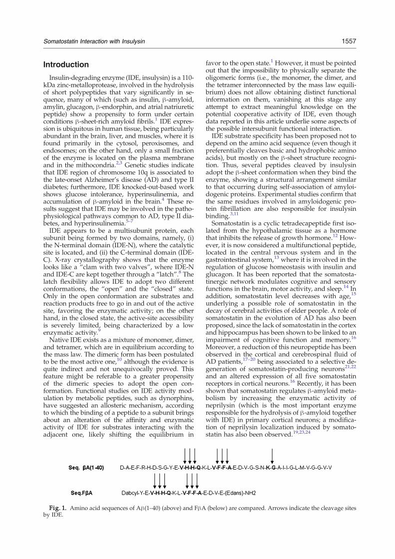
Fig. 1. Amino acid sequences of Aβ(1–40) (above) and FβAby IDE.
favor to the open state.1 However, it must be pointedout that the impossibility to physically separate theoligomeric forms (i.e., the monomer, the dimer, andthe tetramer interconnected by the mass law equili-brium) does not allow obtaining distinct functionalinformation on them, vanishing at this stage anyattempt to extract meaningful knowledge on thepotential cooperative activity of IDE, even thoughdata reported in this article underlie some aspects ofthe possible intersubunit functional interaction.IDE substrate specificity has been proposed not to
depend on the amino acid sequence (even though itpreferentially cleaves basic and hydrophobic aminoacids), but mostly on the β-sheet structure recogni-tion. Thus, several peptides cleaved by insulysinadopt the β-sheet conformation when they bind theenzyme, showing a structural arrangement similarto that occurring during self-association of amyloi-dogenic proteins. Experimental studies confirm thatthe same residues involved in amyloidogenic pro-tein fibrillation are also responsible for insulysinbinding.3,11
Somatostatin is a cyclic tetradecapeptide first iso-lated from the hypothalamic tissue as a hormonethat inhibits the release of growth hormone.12 How-ever, it is now considered a multifunctional peptide,located in the central nervous system and in thegastrointestinal system,13 where it is involved in theregulation of glucose homeostasis with insulin andglucagon. It has been reported that the somatosta-tinergic network modulates cognitive and sensoryfunctions in the brain, motor activity, and sleep.14 Inaddition, somatostatin level decreases with age,15
underlying a possible role of somatostatin in thedecay of cerebral activities of elder people. A role ofsomatostatin in the evolution of AD has also beenproposed, since the lack of somatostatin in the cortexand hippocampus has been shown to be linked to animpairment of cognitive function and memory.16
Moreover, a reduction of this neuropeptide has beenobserved in the cortical and cerebrospinal fluid ofAD patients,17–20 being associated to a selective de-generation of somatostatin-producing neurons21,22
and an altered expression of all five somatostatinreceptors in cortical neurons.16 Recently, it has beenshown that somatostatin regulates β-amyloid meta-bolism by increasing the enzymatic activity ofneprilysin (which is the most important enzymeresponsible for the hydrolysis of β-amyloid togetherwith IDE) in primary cortical neurons; a modifica-tion of neprilysin localization induced by somato-statin has also been observed.19,23,24
(below) are compared. Arrows indicate the cleavage sites
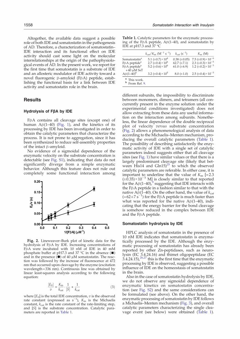
Table 1. Catalytic parameters for the enzymatic process-ing of the FβA peptide, Aβ(1–40), and somatostatin byIDE at pH7.3 and 37 °C
kcat/Km (M−1 s−1) kcat (s−1) Km (M)
Somatostatina 5.1 (±0.7)×104 0.38 (±0.05) 7.5 (±0.9)×10−6
FβA peptidea 2.7 (±0.4)×106 62.7 (±7.1) 2.3 (±0.3)×10−5
FβA peptidea
+40 μM SsT5.2 (±0.6)×106 61.0 (±6.9) 1.2 (±0.2)×10−5
Aβ(1–40)b 3.2 (±0.4)×105 8.0 (±1.0) 2.5 (±0.4)×10−5
a This work.b From Ref. 9.
1558 Somatostatin Interaction with Insulysin
Altogether, the available data suggest a possiblerole of both IDE and somatostatin in the pathogenesisof AD. Therefore, a characterization of somatostatin–IDE interaction and its functional effect on IDEactivity should cast some light on the molecularinterrelationships at the origin of the pathophysiolo-gical events of AD. In the present work, we report forthe first time that somatostatin is a substrate of IDEand an allosteric modulator of IDE activity toward anovel fluorogenic β-amyloid (FβA) peptide, estab-lishing the functional basis for a link between IDEactivity and somatostatin role in the brain.
Results
Hydrolysis of FβA by IDE
FβA contains all cleavage sites (except one) ofhuman Aβ(1–40) (Fig. 1), and the kinetics of itsprocessing by IDE has been investigated in order toobtain the catalytic parameters that characterize theprocess. It is not prone to aggregation, since it hasbeen synthesized to reduce self-assembly propertiesof the intact β-amyloid.No evidence of a sigmoidal dependence of the
enzymatic velocity on the substrate concentration isdetectable (see Fig. S1), indicating that data do notsignificantly diverge from a simple enzymaticbehavior. Although this feature does not rule outcompletely some functional interaction among
Fig. 2. Lineweaver–Burk plot of kinetic data for thehydrolysis of FβA by IDE. Increasing concentrations ofFβA were incubated with 10 nM of IDE in 40 mMphosphate buffer at pH7.3 and 37 °C in the absence (▪)and in the presence (●) of 40 μM somatostatin. The reac-tion was followed by the increase of fluorescence at 472nm that occurred upon cleavage by the enzyme (excitationwavelength=336 nm). Continuous line was obtained bylinear least-squares analysis according to the followingequation:
½E0�v
=Km
kcat� 1½S� +
1kcat
ð3Þ
where [E0] is the total IDE concentration, v is the observedrate constant (expressed as s−1), Km is the Michaelisconstant, kcat is the rate constant of the rate-limiting step,and [S] is the substrate concentration. Catalytic para-meters are reported in Table 1.
different subunits, the impossibility to discriminatebetween monomers, dimers, and tetramers (all con-currently present in the enzyme solution under theexperimental conditions investigated) does notallow extracting from these data any useful informa-tion on the interaction among subunits. Nonethe-less, the linear dependence of the double reciprocalplot of velocity versus substrate concentration(Fig. 2) allows a phenomenological analysis of dataaccording to the Michaelis–Menten mechanism, pro-ducing the overall catalytic parameters (Table 1).The possibility of describing satisfactorily the enzy-matic activity of IDE with a single set of catalyticparameters indeed suggests either that all cleavagesites (see Fig. 1) have similar values or that there is alargely predominant cleavage site (likely that bet-ween His14 and Gln15)25 to which the observedcatalytic parameters are referable. In either case, it isimportant to underline that the value of Km [=2.3(±0.35)×10− 5 M] is closely similar to that reportedfor the Aβ(1–40),9 suggesting that IDE interacts withthe FβA peptide in a fashion similar to that with thenative Aβ(1–40). On the other hand, the value of kcat(=62±7 s−1) for the FβA peptide is much faster thanwhat was reported for the native Aβ(1–40), indi-cating that the energy barrier for the bond cleavageis somehow reduced in the complex between IDEand the FβA peptide.
Somatostatin hydrolysis by IDE
HPLC analysis of somatostatin in the presence of10 nM IDE indicates that somatostatin is enzyma-tically processed by the IDE. Although the enzy-matic processing of somatostatin has already beenreported by other Zn-peptidases, such as neuro-lysin (EC 3.4.24.16) and thimet oligopeptidase (EC3.4.24.15),26,27 this is the first time that the enzymaticprocessing by IDE is observed, suggesting a possibleinfluence of IDE on the homeostasis of somatostatinin the brain.Also in the case of somatostatin hydrolysis by IDE,
we do not observe any sigmoidal dependence ofenzymatic kinetics on somatostatin concentra-tion (see Fig. S2) and the same considerations canbe formulated (see above). On the other hand, theenzymatic processing of somatostatin by IDE followsa Michaelis–Menten mechanism (Fig. 3), and overallcatalytic parameters characterizing the single clea-vage event (see below) were obtained (Table 1).
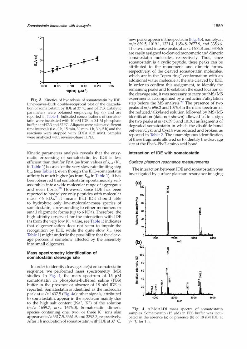
Fig. 3. Kinetics of hydrolysis of somatostatin by IDE.Lineweaver–Burk double-reciprocal plot of the degrada-tion of somatostatin by IDE at 37 °C and pH7.3. Catalyticparameters were obtained employing Eq. (3) and arereported in Table 1. Indicated concentrations of somatos-tatin were incubated with 10 nM IDE in 0.1 M phosphatebuffer at pH7.3 and 37 °C. Aliquots were taken at differenttime intervals (i.e., 0 h, 15 min, 30 min, 1 h, 3 h, 5 h) and thereactions were stopped with EDTA (0.5 mM). Sampleswere analyzed with reverse-phase HPLC.
Fig. 4. AP-MALDI mass spectra of somatostatinsamples. Somatostatin (15 μM) in PBS buffer was incu-bated in the absence (a) or presence (b) of 18 nM IDE at37 °C for 1 h.
1559Somatostatin Interaction with Insulysin
Kinetic parameters analysis reveals that the enzy-matic processing of somatostatin by IDE is lessefficient than that for FβA (as from values of kcat/Kmin Table 1) because of the very slow rate-limiting stepkcat (see Table 1), even though the IDE–somatostatinaffinity is much higher (as from Km in Table 1). It hasbeen observed that somatostatin spontaneously self-assembles into a wide molecular range of aggregatesand even fibrils.28 However, since IDE has beenreported to hydrolyze only peptides with molecularmass b6 kDa,9 it means that IDE should ableto hydrolyze only low-molecular-mass species ofsomatostatin, corresponding to either monomers orsmall oligomeric forms (up to 6 kDa). Therefore, thehigh affinity observed for the interaction with IDE(as from the very low Km value, see Table 1) indicatesthat oligomerization does not seem to impair therecognition by IDE, while the quite slow kcat (seeTable 1) might underlie the possibility that the cleav-age process is somehow affected by the assemblyinto small oligomers.
Mass spectrometry identification ofsomatostatin cleavage site
In order to identify cleavage site(s) on somatostatinsequence, we performed mass spectrometry (MS)studies. In Fig. 4, the mass spectrum of 15 μMsomatostatin in phosphate-buffered saline (PBS)buffer in the presence or absence of 18 nM IDE isreported. Somatostatin is identified as the molecularpeak at m/z 1637.5 (Fig. 4a); other signals, attributedto somatostatin, appear in the spectrum mainly dueto the high salt content (Na+, K+) of the solution(m/z 1659.7, m/z 1676.0). Somatostatin dimericspecies containing one, two, or three K+ ions alsoappear atm/z 3317.5, 3361.9, and 3393.3, respectively.After 1 h incubation of somatostatinwith IDE at 37 °C,
newpeaks appear in the spectrum (Fig. 4b), namely, atm/z 639.5, 1019.1, 1321.4, 1654.8, 2677.9, and 3356.6.The two most intense peaks atm/z 1654.8 and 3356.6are easily assigned to cleavedmonomeric and dimericsomatostatin molecules, respectively. Thus, sincesomatostatin is a cyclic peptide, these peaks can beattributed to the monomeric and dimeric forms,respectively, of the cleaved somatostatin molecules,which are in the “open ring” conformation with anadditional water molecule at the site cleaved by IDE.In order to confirm this assignment, to identify theremaining peaks and to establish the exact location ofthe cleavage site, itwas necessary to carry outMS/MSexperiments accompanied by a reduction/alkylationstep before the MS analysis.29 The presence of twopeaks atm/z 696.2 and 1076.3 in themass spectrumofthe reduced/alkylated solution followed by MS/MSidentification (data not shown) allowed us to assignthe two peaks atm/z 639.5 and 1019.1 as fragments ofdegraded somatostatin in which the disulfide bondbetween Cys3 and Cys14was reduced and broken, asreported in Table 2. The unambiguous identificationof these fragments allowed us to identify the cleavagesite at the Phe6–Phe7 amino acid bond.
Interaction of IDE with somatostatin
Surface plasmon resonance measurements
The interaction between IDE and somatostatinwasinvestigated by surface plasmon resonance imaging
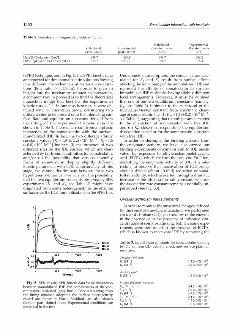
Table 2. Somatostatin fragments produced by IDE
Calculatedpeaks (m/z)
Experimentalpeaks (m/z)
Calculatedalkylated peaks
(m/z)
Experimentalalkylated peaks
(m/z)
HalaGlyCysLysAsnPheOH 639.7 639.5 696.7 696.2HPheTrpLysThrPheThrSerCysOH 1019.7 1019.1 1076.7 1076.3
1560 Somatostatin Interaction with Insulysin
(SPRI) technique, and in Fig. 5, the SPRI kinetic dataare reported for three somatostatin solutions flowinginto different microchannels at various concentra-tions (flow rate=50 μl/min). In order to give aninsight into the mechanism of such an interaction,a common way to proceed is to find the theoreticalinteraction model that best fits the experimentalkinetic curves.30,31 In our case, best results were ob-tained with an interaction model considering twodifferent sites to be present onto the interacting sur-face. Rate and equilibrium constants derived fromthe fitting of the experimental kinetic data areshown in Table 3. These data result from a biphasicinteraction of the somatostatin with the surface-immobilized IDE. In fact, the two different affinityconstant values [K1=4.9 (±2.2)×105 M−1, K2=1.6(±0.8)×105 M−1] indicate (i) the presence of twodifferent sites on the IDE surface, which are char-acterized by fairly similar affinities for somatostatin,and/or (ii) the possibility that various assemblyforms of somatostatin display slightly differentkinetic parameters with IDE. Unfortunately, at thisstage, we cannot discriminate between these twohypotheses; neither can we rule out the possibilitythat the two equilibrium constants observed by SPRexperiments (K1 and K2, see Table 3) might haveoriginated from some heterogeneity in the enzymesurface after the IDE immobilization on the SPR chip.
Fig. 5. SPRI results. SPRI kinetic data for the interactionbetween immobilized IDE and somatostatin at the con-centrations indicated (gray lines). Curves resulting fromthe fitting obtained adopting the surface heterogeneitymodel are shown in black. Residuals are also shown(bottom part, dotted lines). Experimental conditions aredescribed in the text.
Under such an assumption, the similar values calc-ulated for K1 and K2 result from surface effectsaffecting the functioning of the immobilized IDE andrepresent the affinity of somatostatin to surface-immobilized IDEmolecules having slightly differentlocal arrangements. However, it must be outlinedthat one of the two equilibrium constants (namely,K2, see Table 3) is similar to the reciprocal of theMichaelis–Menten constant from enzymatic cleav-age of somatostatin [i.e., 1/Km=1.3 (±0.2)×105 M−1,see Table 1], suggesting that (i) both parameters referto the interaction of somatostatin with free IDEand (ii) Km closely corresponds to the equilibriumdissociation constant for the somatostatin substratewith free IDE.In order to decouple the binding process from
the enzymatic activity, we have also carried outbinding experiments of somatostatin to IDE inacti-vated by exposure to ethylenediaminetetraaceticacid (EDTA), which chelates the catalytic Zn2+ ion,abolishing the enzymatic activity of IDE. It is inte-resting to observe that inactivation of IDE bringsabout a drastic (about 10-fold) reduction of soma-tostatin affinity, which is exerted through a dramaticincrease of the dissociation rate constant, whereasthe association rate constant remains essentially un-perturbed (see Fig. S3).
Circular dichroism measurements
In order to monitor the structural changes inducedby the somatostatin–IDE interaction, we performedcircular dichroism (CD) spectroscopy of the enzymein the absence or in the presence of indicated con-centrations of somatostatin (Fig. 6a). The same expe-riments were performed in the presence of EDTA,which is known to inactivate IDE by removing the
Table 3. Equilibrium constants for somatostatin bindingto IDE as from CD, activity effect, and surface plasmonresonance
Circular DichroismKa (M
−1) 1.2 (±0.2)×105
Kb (M−1) 4.9 (±0.5)×103
Activity effectK (M−1) 1.3 (±0.2)×105
Surface plasmon resonancek1a (M
−1 s−1) 3.4 (±1.4)×102
k1d (s−1) 7.0 (±1.5)×10−4
K1 (M−1) 4.9 (±2.2)×105
k2a (M−1 s−1) 2.4 (±1.3)×103
k2d (s−1) 1.5 (±0.2)×10−2
K2 (M−1) 1.6 (±0.8)×105
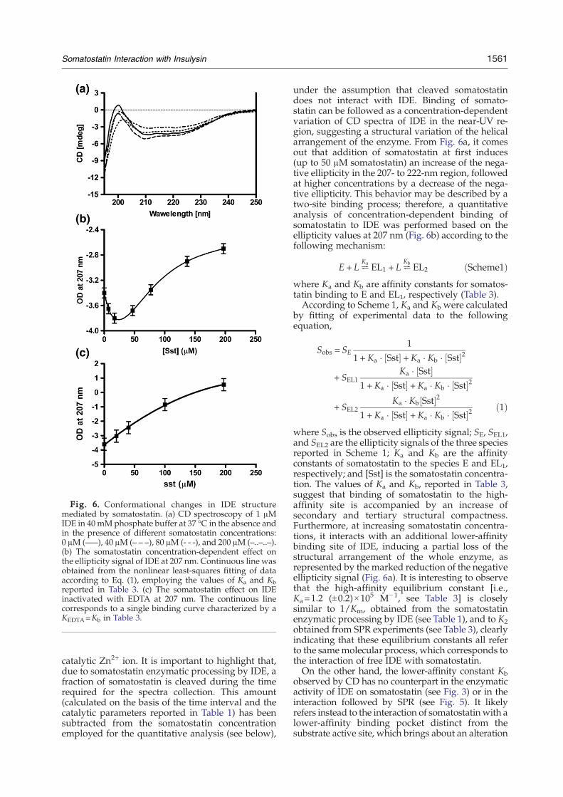
Fig. 6. Conformational changes in IDE structuremediated by somatostatin. (a) CD spectroscopy of 1 μMIDE in 40mMphosphate buffer at 37 °C in the absence andin the presence of different somatostatin concentrations:0 μM (—–), 40 μM (– – –), 80 μM (- - -), and 200 μM (–..–..–).(b) The somatostatin concentration-dependent effect onthe ellipticity signal of IDE at 207 nm. Continuous line wasobtained from the nonlinear least-squares fitting of dataaccording to Eq. (1), employing the values of Ka and Kbreported in Table 3. (c) The somatostatin effect on IDEinactivated with EDTA at 207 nm. The continuous linecorresponds to a single binding curve characterized by aKEDTA=Kb in Table 3.
1561Somatostatin Interaction with Insulysin
catalytic Zn2+ ion. It is important to highlight that,due to somatostatin enzymatic processing by IDE, afraction of somatostatin is cleaved during the timerequired for the spectra collection. This amount(calculated on the basis of the time interval and thecatalytic parameters reported in Table 1) has beensubtracted from the somatostatin concentrationemployed for the quantitative analysis (see below),
under the assumption that cleaved somatostatindoes not interact with IDE. Binding of somato-statin can be followed as a concentration-dependentvariation of CD spectra of IDE in the near-UV re-gion, suggesting a structural variation of the helicalarrangement of the enzyme. From Fig. 6a, it comesout that addition of somatostatin at first induces(up to 50 μM somatostatin) an increase of the nega-tive ellipticity in the 207- to 222-nm region, followedat higher concentrations by a decrease of the nega-tive ellipticity. This behavior may be described by atwo-site binding process; therefore, a quantitativeanalysis of concentration-dependent binding ofsomatostatin to IDE was performed based on theellipticity values at 207 nm (Fig. 6b) according to thefollowing mechanism:
E + L²Ka EL1 + L²
Kb EL2 ðScheme1Þwhere Ka and Kb are affinity constants for somatos-tatin binding to E and EL1, respectively (Table 3).According to Scheme 1, Ka and Kb were calculated
by fitting of experimental data to the followingequation,
Sobs = SE1
1 + Ka � Sst½ � +Ka � Kb � Sst½ �2
+ SEL1Ka � Sst½ �
1 +Ka � Sst½ � + Ka � Kb � Sst½ �2
+ SEL2Ka � Kb Sst½ �2
1 +Ka � Sst½ � + Ka � Kb � Sst½ �2 ð1Þ
where Sobs is the observed ellipticity signal; SE, SEL1,and SEL2 are the ellipticity signals of the three speciesreported in Scheme 1; Ka and Kb are the affinityconstants of somatostatin to the species E and EL1,respectively; and [Sst] is the somatostatin concentra-tion. The values of Ka and Kb, reported in Table 3,suggest that binding of somatostatin to the high-affinity site is accompanied by an increase ofsecondary and tertiary structural compactness.Furthermore, at increasing somatostatin concentra-tions, it interacts with an additional lower-affinitybinding site of IDE, inducing a partial loss of thestructural arrangement of the whole enzyme, asrepresented by the marked reduction of the negativeellipticity signal (Fig. 6a). It is interesting to observethat the high-affinity equilibrium constant [i.e.,Ka=1.2 (±0.2)×105 M−1, see Table 3] is closelysimilar to 1/Km, obtained from the somatostatinenzymatic processing by IDE (see Table 1), and to K2obtained from SPR experiments (see Table 3), clearlyindicating that these equilibrium constants all referto the samemolecular process, which corresponds tothe interaction of free IDE with somatostatin.On the other hand, the lower-affinity constant Kb
observed by CD has no counterpart in the enzymaticactivity of IDE on somatostatin (see Fig. 3) or in theinteraction followed by SPR (see Fig. 5). It likelyrefers instead to the interaction of somatostatin with alower-affinity binding pocket distinct from thesubstrate active site, which brings about an alteration
1562 Somatostatin Interaction with Insulysin
of the tertiary (and likely quaternary) structure ofIDE. This assumption is also supported by theevidence that IDE inactivated by EDTA displays thesame low-affinity equilibrium constant for somatos-tatin as the active IDE (i.e., K=Kb=4.9×10
3 M−1, seeTable 3), inducing only a loss of the structuralarrangement of the enzyme (Fig. 6c). As a whole,data by SPR and CD both seem to confirm that uponinactivation by EDTA, the high-affinity site forsomatostatin (possibly the active site) dramaticallyreduces its binding affinity, either becoming unableto bind somatostatin or displaying a value similar tothe low-affinity site.
Effect of somatostatin on IDE processing of theamyloid peptide
The effect of human somatostatin on the IDE-dependent degradation of fluorogenic substrate wasmeasured in reaction mixtures containing 5 μM FβApeptide and 2 nM IDE in the presence of indicatedconcentrations of somatostatin. It is important tounderline that, due to the somatostatin enzymaticprocessing by IDE, a fraction of somatostatin iscleaved during the time required for the fluorogenicassay. This amount (calculated on the basis of thetime interval and the catalytic parameters reportedin Table 1) has been subtracted from the somatos-tatin concentration employed for the quantitativeanalysis (see below), under the assumption thatcleaved somatostatin does not interact with IDE.The enzymatic activity on FβA as a function ofsomatostatin concentration is compared with valuesobtained in the absence of somatostatin (Fig. 7).Somatostatin significantly enhances IDE activity,with the maximum effect at about 40 μM, a behaviorperfectly consistent with the spectroscopic effectobserved by CD (Fig. 6), suggesting that this effectis related to the binding of somatostatin to freeIDE. It is interesting to outline that the same effecthas also been observed in the case of other IDEsubstrates, such as β-endorphin, bradykinin, andseveral dynorphins.10 Like for some of these subs-trates, at a much higher somatostatin concentration(N100 μM), an inhibitory effect is observed (Fig. 7),likely due to the presence in the FβA cleavage site ofa second somatostatin molecule, which becomes,in this way, a competitive inhibitor. It is worthoutlining that this concentration range is closelysimilar to that observed for the lower-affinity cons-tant observed by CD, suggesting that this interactionis also responsible for the progressive disappearanceof the somatostatin-linked activation process. How-ever, since the inhibitory effect appears at very highsomatostatin concentrations, we have only ana-lyzed the enhancing effect according to the follow-ing equation:
Aobs =1
1 + K � Sst½ � +ArK � Sst½ �
1 +K � Sst½ � ð2Þ
where Aobs is the observed relative activity withrespect to IDE in the absence of somatostatin, Ar is
the relative activity in the presence of a somatostatinconcentration needed to saturate the binding site, Kis the somatostatin affinity binding constant, and[Sst] is the somatostatin concentration. This equationunderlies the existence of only one activating bind-ing site for somatostatin, as it appears from our dataover the investigated concentration range.The close similarity of the somatostatin high-
affinity constant from CD experiments (i.e., Ka inTable 3, see Fig. 6) with both the equilibrium cons-tant for the second binding site observed by SPRIexperiments (i.e., K2 in Table 3, see Fig. 7) and thatresulting from the rate-enhancing effect (i.e., K inTable 3, see Fig. 7) indeed suggests that (i) the high-affinity site by CD (corresponding to the bindingsite characterized by K2 in SPRI experiments) is amodulatory cleft and (ii) the activity effect is relatedto the interaction of somatostatin with free IDE (i.e.,E in Scheme 1). In addition, the catalytic para-meters, obtained for FβA processing by IDE in thepresence of 40 μM somatostatin (see Fig. 2), showthat the somatostatin-linked effect is only exertedon Km, leaving kcat unchanged (see Table 1). There-fore, the rate-enhancing effect (see Fig. 7) is due toan allosteric mechanism, such that the binding ofsomatostatin to one site brings about a conforma-tional change of a second site, facilitating the FβAbinding. However, this conformational changeaffects the substrate affinity (as from the decreaseof Km for FβA upon somatostatin addition) but itdoes not change the cleavage rate kcat. This hasalready been observed for the processing of asynthetic substrate in the presence of dynorphinB-9,10 suggesting that cooperativity in IDE is likelyexpressed mostly through a change of substrateaffinity without affecting significantly the speed ofthe rate-limiting step.
Discussion
IDE is a Zn2+ metalloprotease characterized by thepresence of different quaternary structures (namely,monomeric, dimeric, and tetrameric forms), whichmight display different enzymatic activity towardamyloidogenic proteins.1,10 It has been shown thatIDE is able to enzymatically process the amyloidβ-protein,32 giving rise to new fragments that are notneurotoxic or that do not deposit on amyloidplaques25,33 and that this activity can be modulatedby metabolic peptides, such as dynorphins.1 As awhole, these findings render IDE a potentially inte-resting target for the design of anti-Alzheimer'sdrugs.34,35Furthermore, it is well known that somatostatin
performs crucial roles in the brain and that its dec-rease represents a pathological feature of AD, eventhough this reduction is not accompanied by analteration of proteolytic processing of peptideprecursors.36 Although the mechanism that couplesalterations of the somatostatinergic system to ADremains unclear, recently, it has been shown thatsomatostatin regulates β-amyloid metabolism
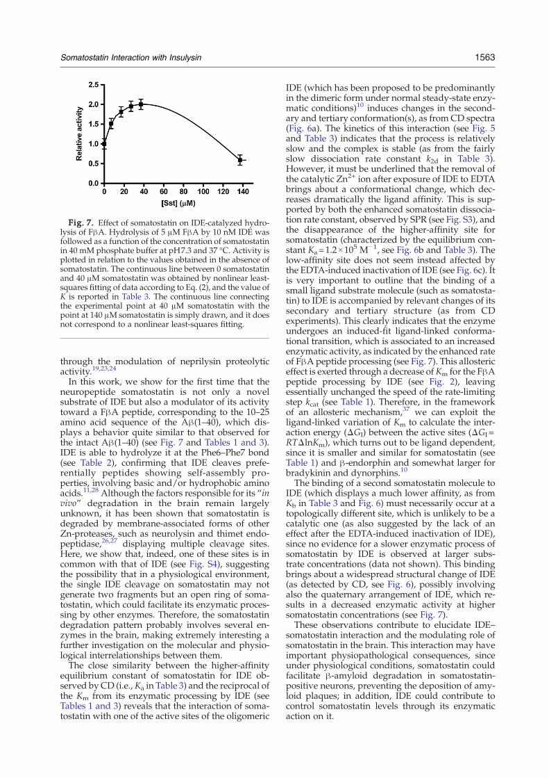
Fig. 7. Effect of somatostatin on IDE-catalyzed hydro-lysis of FβA. Hydrolysis of 5 μM FβA by 10 nM IDE wasfollowed as a function of the concentration of somatostatinin 40 mM phosphate buffer at pH7.3 and 37 °C. Activity isplotted in relation to the values obtained in the absence ofsomatostatin. The continuous line between 0 somatostatinand 40 μM somatostatin was obtained by nonlinear least-squares fitting of data according to Eq. (2), and the value ofK is reported in Table 3. The continuous line connectingthe experimental point at 40 μM somatostatin with thepoint at 140 μΜ somatostatin is simply drawn, and it doesnot correspond to a nonlinear least-squares fitting.
1563Somatostatin Interaction with Insulysin
through the modulation of neprilysin proteolyticactivity.19,23,24
In this work, we show for the first time that theneuropeptide somatostatin is not only a novelsubstrate of IDE but also a modulator of its activitytoward a FβA peptide, corresponding to the 10–25amino acid sequence of the Aβ(1–40), which dis-plays a behavior quite similar to that observed forthe intact Aβ(1–40) (see Fig. 7 and Tables 1 and 3).IDE is able to hydrolyze it at the Phe6–Phe7 bond(see Table 2), confirming that IDE cleaves prefe-rentially peptides showing self-assembly pro-perties, involving basic and/or hydrophobic aminoacids.11,28 Although the factors responsible for its “invivo” degradation in the brain remain largelyunknown, it has been shown that somatostatin isdegraded by membrane-associated forms of otherZn-proteases, such as neurolysin and thimet endo-peptidase,26,27 displaying multiple cleavage sites.Here, we show that, indeed, one of these sites is incommon with that of IDE (see Fig. S4), suggestingthe possibility that in a physiological environment,the single IDE cleavage on somatostatin may notgenerate two fragments but an open ring of soma-tostatin, which could facilitate its enzymatic proces-sing by other enzymes. Therefore, the somatostatindegradation pattern probably involves several en-zymes in the brain, making extremely interesting afurther investigation on the molecular and physio-logical interrelationships between them.The close similarity between the higher-affinity
equilibrium constant of somatostatin for IDE ob-served by CD (i.e.,Ka in Table 3) and the reciprocal ofthe Km from its enzymatic processing by IDE (seeTables 1 and 3) reveals that the interaction of soma-tostatin with one of the active sites of the oligomeric
IDE (which has been proposed to be predominantlyin the dimeric form under normal steady-state enzy-matic conditions)10 induces changes in the second-ary and tertiary conformation(s), as from CD spectra(Fig. 6a). The kinetics of this interaction (see Fig. 5and Table 3) indicates that the process is relativelyslow and the complex is stable (as from the fairlyslow dissociation rate constant k2d in Table 3).However, it must be underlined that the removal ofthe catalytic Zn2+ ion after exposure of IDE to EDTAbrings about a conformational change, which dec-reases dramatically the ligand affinity. This is sup-ported by both the enhanced somatostatin dissocia-tion rate constant, observed by SPR (see Fig. S3), andthe disappearance of the higher-affinity site forsomatostatin (characterized by the equilibrium con-stant Ka=1.2×10
5 M−1, see Fig. 6b and Table 3). Thelow-affinity site does not seem instead affected bythe EDTA-induced inactivation of IDE (see Fig. 6c). Itis very important to outline that the binding of asmall ligand substrate molecule (such as somatosta-tin) to IDE is accompanied by relevant changes of itssecondary and tertiary structure (as from CDexperiments). This clearly indicates that the enzymeundergoes an induced-fit ligand-linked conforma-tional transition, which is associated to an increasedenzymatic activity, as indicated by the enhanced rateof FβA peptide processing (see Fig. 7). This allostericeffect is exerted through a decrease ofKm for the FβApeptide processing by IDE (see Fig. 2), leavingessentially unchanged the speed of the rate-limitingstep kcat (see Table 1). Therefore, in the frameworkof an allosteric mechanism,37 we can exploit theligand-linked variation of Km to calculate the inter-action energy (ΔGI) between the active sites (ΔGI=RTΔlnKm), which turns out to be ligand dependent,since it is smaller and similar for somatostatin (seeTable 1) and β-endorphin and somewhat larger forbradykinin and dynorphins.10
The binding of a second somatostatin molecule toIDE (which displays a much lower affinity, as fromKb in Table 3 and Fig. 6) must necessarily occur at atopologically different site, which is unlikely to be acatalytic one (as also suggested by the lack of aneffect after the EDTA-induced inactivation of IDE),since no evidence for a slower enzymatic process ofsomatostatin by IDE is observed at larger subs-trate concentrations (data not shown). This bindingbrings about a widespread structural change of IDE(as detected by CD, see Fig. 6), possibly involvingalso the quaternary arrangement of IDE, which re-sults in a decreased enzymatic activity at highersomatostatin concentrations (see Fig. 7).These observations contribute to elucidate IDE–
somatostatin interaction and the modulating role ofsomatostatin in the brain. This interaction may haveimportant physiopathological consequences, sinceunder physiological conditions, somatostatin couldfacilitate β-amyloid degradation in somatostatin-positive neurons, preventing the deposition of amy-loid plaques; in addition, IDE could contribute tocontrol somatostatin levels through its enzymaticaction on it.
1564 Somatostatin Interaction with Insulysin
As a whole, this complex regulatory mechanismmay represent an elegant feedback network, whichmay play a central role in the homeostasis of somato-statin and β-amyloid. Thus, although a biologicalcounterproof is needed, we can hypothesize that thebrain levels of somatostatin and β-amyloid may bemutually balanced by the IDE enzymatic activity onboth of them and by their reciprocal modulatory roleon the IDE enzymatic activity. A pathological dec-rease of somatostatin synthesis may break this ba-lance and could trigger a down-regulation of IDE andneprilysin activity with a consequent accumulation ofβ-amyloid, leading to a vicious cycle that could becrucial for the onset of neurodegenerative processes.38
Materials and Methods
Materials
Recombinant IDE was obtained from Calbiochem.Human somatostatin, ethanol solution, ethanolamine–HCl (1 M), guanidine–HCl (8 M), and dithiobis(N)succinimidylpropionate (Lomant's reagent) were all pur-chased from Sigma-Aldrich (Milan, Italy). Dithiol tethersSPT-0013 and SPT-0014C were purchased from Sensopath.Gold substrates (GWC Technologies, USA) were obtainedby thermally evaporating a gold layer (450 Å) onto SF-10glass slides (Schott, USA). Chromium (50 Å) was used asthe adhesion layer. IDE preparations were in 40 mMphosphate buffer at pH7.3 and somatostatin prepara-tions were in 0.1 M phosphate buffer at pH7.3. Classicalfluorenylmethoxycarbonyl (Fmoc)-protected amino acidderivatives, the KA amide TentaGel resin, the protectedFmoc-L-Glu{5-[(2-aminoethyl)-amino]naphthalene-1-sul-fonic acid (EDANS)}-OH, and the [4-((4-(dimethyl-amino)phenyl)azo)benzoic acid]-N-hydroxysuccinimide ester(Dabcyl-OSu) for the synthesis of the peptide were fromNovabiochem (Laufelfingen, CH). Solvents were fromROMIL (Dublin, Ireland); other reagents for peptide syn-thesis, such as piperidine, activating agents, diisopropylethylamine, trifluoroacetic acid (TFA), and triisopropyl-silane were from Sigma-Aldrich.
FβA peptide synthesis
The internally quenched novel peptide,39 derived fromAβ(1–40), Dabcyl-Tyr-Glu-Val-His-His-Gln-Lys-Leu-Val-Phe-Phe-Ala-Glu-Asp-Val-Gly-Glu(EDANS)-NH2 (FβA),and containing residues 10–25, was prepared on a 433AApplied Biosystems automatic synthesizer (synthesisscale: 0.1 mmol), following standard Fmoc/tBu protocolsof solid-phase peptide synthesis.40 Briefly, the peptide wasassembled on a RINK amide PEGA resin onto whichFmoc-L-Glu(EDANS)-OH was previously anchored. Allamino acid derivatives were preactivated with 1-H-benzotriazolium, 1-[bis(dimethylamino)methylene]-hexa-fluorophosphate(1-),3-oxide/N-hydroxybenzotriazoleand coupled for 30 min. Deprotection of the Fmoc groupwas achieved by treatment with 30% piperidine indimethyl formamide for 20 min. Following the insertionof the N-terminal residue and deprotection, the Dabcylgroup was introduced by treatment of the resin withDabcyl-OSu (10 eq.) in dimethyl formamide (0.5 M) in thepresence of diisopropyl ethylamine (20 eq.). The peptidewas cleaved from the resin by treatment with a TFA–H2O–
triisopropylsilane (90:5:5, v/v/v) mixture for 4 h at roomtemperature, precipitated in cold diethylether, and lyo-philized from a 50:50 H2O–CH3CN solution. The crudeproduct was purified to homogeneity by semipreparativeRP-HPLC using a C18 50×2.2 cm ID column (Pheno-menex, Torrance, CA) equilibrated at 20 ml/min with 20%CH3CN in H2O and 0.1% TFA. A gradient from 20% to70% CH3CN and 0.1% TFAwas applied for 50 min to elutethe peptide. Fractions containing the purified productwere pooled, and the product was then characterized byliquid chromatography (LC)–MS on an LCQ Deca XP IonTrap mass spectrometer (ThermoElectron, Milan, Italy)equipped with an OPTON ESI source, operating at 4.2 kVneedle voltage and 320 °C and with a complete SurveyorHPLC system. Narrow-bore 50×2 mm C18 BioBasic LC–MS columns from ThermoElectron equilibrated at 0.2 ml/min with 20% CH3CN in H2O and 0.05% TFA were usedfor these analyses. A gradient from 20% to 70% of CH3CNand 0.05% TFA over 50 min was applied to elute thepeptide. The molecular weight was consistent with theexpected value within the limits of the experimental error(MWExp/Theor: 2545.6/2545.72 amu).The FβAwas dissolved in 20 mM phosphate buffer and
50% dimethyl sulfoxide, pH7.3. Undissolved species wereremoved by centrifuging freshly dissolved peptide at10,000g for 2 h at 4 °C, and the resulting supernatant wasstored at −20 °C until use.41 Peptide concentrations weredetermined using EDANS's extinction coefficient of5.9 mM−1 cm−1 at 335 nm.
Methods
HPLC analysis
Reaction mixtures containing 10 nM IDE and increasingconcentrations of somatostatin were incubated at 37 °C for5 h in 0.1 M phosphate buffer, pH7.3. Aliquots were takenat different time intervals (i.e., 0 h, 15 min, 30 min, 1 h, 3 h,5 h), and the reaction was stopped by addition of 0.5 mMEDTA. Samples were applied to a C4 reverse-phase HPLCcolumn (Surveyor, Thermo Finnigan), and the elution wasperformed at a flow rate of 1 ml/min using a linear gra-dient: 95% eluent A (H2O+0.1% TFA) and 5% eluent B(CH3CN+0.1% TFA), with absorbance monitored at 220and 254 nm.
Mass spectrometry: AP-MALDI MS experiments
Reaction mixtures containing 18 μM somatostatin in thepresence or absence of 10 nM IDE in 0.1 M phosphatebuffer (pH7.3) were incubated at 37 °C for 1 h and ana-lyzed with atmospheric pressure matrix-assisted laserdesorption/ionization (AP-MALDI) MS.All the AP-MALDI MS experiments were carried out by
using a Finnigan LCQ Deca XP PLUS (Thermo ElectronCorporation, USA) ion trap spectrometer that was fittedwith a MassTech Inc. (USA) AP-MALDI pulsed dynamicfocusing source. The latter consists of a flange containing acomputer-controlled X–Y positioning stage and a digitalcamera and is powered by a control unit that includes apulsed nitrogen laser (wavelength, 337 nm; pulse width,4 ns; pulse energy, 300 μJ; repetition rate up to 10 Hz) anda pulsed dynamic focusingmodule that imposes a delay of25 μs between the laser pulse and the application of thehigh voltage to the AP-MALDI target plate. Laser powerwas attenuated to about 55%. The target plate voltage was1.8 kV. The ion trap inlet capillary temperature was 220 °C.Capillary and tube lens offset voltages of 30 and 15 V,respectively, were applied. Other mass spectrometer
1565Somatostatin Interaction with Insulysin
parameters were as follows: multipole 1 offset at −3.75 V,multipole 2 offset at −9.50 V, multipole radio-frequency(RF) amplitude 400 V, lens at −24.0 V, and entrance lens at−88.0 V. Automatic Gain Control was turned off andinstead the scan time was fixed by setting the injectiontime to 220 ms and using five microscans. Although thereis the risk of losing resolution, the latter experimentalconditions were chosen as sensitivity was the main goal inmost experiments. For the same reason, although 1 minacquisition per sample was usually performed, in somecases, it was necessary that an acquisition up to 5 min anddifferent experiments were reproduced from three to fivetimes.Spectra of the studied solutions were acquired in a data-
dependent fashion by first acquiring full extended massrange from m/z 200 to 4000, followed by MS/MS scans ofthe most intense ions of the previous full MS scan. MS/MSscans were acquired using an isolation width of 5 m/z,activation qz of 0.250, activation time of 30 ms, and norm-alized collision energy (NCE) in the range 30–40%, depen-dent on the ion {NCE is the amplitude of the resonanceexcitation RF voltage scaled to the precursor mass basedon the following formula: RF amplitude=[NCE%/30%](precursor ion mass×tick amp slope+tick amp intercept),where the tick amp slope and tick amp intercept areinstrument-specific values. For our LCQ Deca, 35% NCEfor m/z 1000=1.8 V}.In order to unambiguously assign the somatostatin
fragments produced by IDE, a reduction/alkylation stepwas carried out in the reaction mixture prior to the MSanalysis without any purification step, according to theprocedure previously described.29
Fluorogenic assay for FβA hydrolysis by IDE
The hydrolysis of the fluorogenic peptide wasmeasuredat 37 °C following the increase in fluorescence at 472 nm(with excitation at 336 nm) after cleavage of the peptidebond by IDE and the separation of the quenching Dabcylgroup from the fluorescent EDANS group. Reactionmixtures contained the fluorogenic peptide substrate atthe indicated concentrations and 10 nM IDE.The effect of somatostatin on the IDE activity was
investigated following the hydrolysis of FβA at 37 °C in areaction mixture containing either (i) 2 nM IDE, 5 μMFβA,and various concentrations of human somatostatin or (ii)40 μM somatostatin, 2 nM IDE, and indicated FβA con-centrations. The reactions were followed on a Cary Eclipsefluorescence spectrofluorometer.
CD spectroscopy
IDE was diluted to 1 μM in 40 mM phosphate buffer atpH7.3 in the absence and in the presence of different con-centrations of somatostatin. The same experiments wereperformed in the presence of 50 μM EDTA. Samples wereexamined using 0.2-mm quartz cuvettes in a Jasco J-720CD spectropolarimeter (Tokyo, Japan). Spectra wererecorded at 37 °C and at 1 nm resolution with a scan rateof 20 nm/min. Eight scans were acquired and averaged foreach sample.
Poly(dimethylsiloxane) microfluidic devices fabrication
Microfluidic devices were made in poly(dimethylsilox-ane) (PDMS) polymer as described elsewhere.42 Briefly,PDMS microchannels were created by replication frommasters in polyvinyl chloride. Replicas were formed froma 1:10 mixture of PDMS curing agent and prepolymer
(Sylgard 184, Dow Corning, USA). The mixture was de-gassed under vacuum and then poured onto the master inorder to create a layer with a thickness of about 3–4 mm.The PDMS was then incubated for at least 2 h at 60 °Cbefore being removed from the masters. Microchannelswere 500 μmwide and 80 μm in height. At the ends of eachchannel, there were circular reservoirs (diameter, 400 μm).PEEK tubes (Upchurch Scientific) were inserted in suchreservoirs in order to connect the PDMS microfluidic cellto an Ismatec IP-N (Ismatec SA, Switzerland) peristalticpump.
SPRI measurements
The SPRI apparatus (GWC Technologies, USA) and themicrofluidic system used were the same as reported insome of our previous works.43 A six-microchannel micro-fluidic device was used in this case to follow the inter-action between IDE and somatostatin at differentconcentrations.SPR images were analyzed by using the V++ software
(version 4.0, Digital Optics Limited, New Zealand) and thesoftware package Image J 1.32j (National Institutes ofHealth, USA). SPRI provides data as pixel intensity units(0–255 scale). Data were converted in percentage ofreflectivity (%R) by using the formula:
kR = 1004 0:85 Ip=Is� �
where Ip and Is refer to the reflected light intensitydetected using p- and s-polarized light, respectively.Experiments were carried out by sequentially acquiring
15 frame-averaged SPR images with 5 s time delaybetween them. Kinetic data were obtained by plottingthe difference in percent reflectivity (%R) from selectedregions of interest of the SPR images as a function of time.All the SPRI experiments were carried out at room tem-perature. The rate constants ka and kd were calculatedby fitting adsorption/desorption kinetics data throughnumerical integration analysis.44
Immobilization of IDE on gold surface
Two different immobilization procedures were scruti-nized for IDE and positive results were obtained in bothcases. Specifically, similar SPRI signals were indeedregistered after a 40-min injection at 5 μl/min of a 36-nMIDE solution into a microchannel in contact with a goldsurface previously functionalized with (a) Lomant's re-agent45 and (b) dithiol tethers (SPT-0013:SPT-0014C=10:1mixed ethanol solution).46 The same procedure wasfollowed with IDE previously inactivated by EDTA.We found that the pH of the PBS buffer used for sample
dilution is crucial for a positive result of the activity mea-surements and a pH of 7.3 was chosen for all experiments.Ethanolamine–HCl (1 M) was used for deactivation of theunreacted NHS groups, while 5 min injection at 5 μl/minof guanidine–HCl (8 M) was used for the denaturation ofIDE. This last procedure was undertaken to avoid thepossibility that observed signals were due to unspecificinteractions between the somatostatin molecules and thefunctionalized SPRI chips. Thus, a large difference in theSPRI signal was recorded according to whether the soma-tostatin solutions were injected into microchannels wherethe immobilized IDE was in the native form or it had beenpreviously denatured (see Fig. S5). This result reinforcesthe conclusion that the kinetic data recorded in the case ofactive IDE–somatostatin are due to a real interactionbetween somatostatin and IDE rather than to trivial
1566 Somatostatin Interaction with Insulysin
somatostatin unspecific interactions with the functiona-lized SPRI chip.
Acknowledgements
The authors thankfully acknowledge useful dis-cussions with Prof. S. Marini and Drs. G. F.Fasciglione and D. Di Pierro during the early stagesof the project. The financial contribution from theItalian Ministry of University and Research (MiURFIRB RBNE03PX83 to M.R., E.R., and M.C.) is grate-fully acknowledged.
Supplementary Data
Supplementary data associated with this articlecan be found, in the online version, at doi:10.1016/j.jmb.2008.11.025
References
1. Song, E.-S. & Hersh, L. B. (2004). Insulysin. J. Mol.Chem. 5, 201–220.
2. Duckeorth, W. C., Bennett, R. G. & Hamel, F. G. (1998).Insulin degradation: progress and potential. Endocr.Rev. 19, 608–624.
3. Leissring, M. A., Farris, W., Wu, X., Cristodoulou,D. C., Haigis, M. C., Guarente, L. & Selkoe, D. J.(2004). Alternative translation initiation generates anovel isoform of insulin-degrading enzyme targetedto mitochondria. Biochem. J. 383, 439–446.
4. Qiu, W. Q. & Folstein, M. F. (2006). Insulin, insulin-degrading enzyme and amyloid-β peptide in Alzhei-mer's disease: review and hypothesis. Neurobiol.Aging, 27, 190–198.
5. Bertram, L., Blacker, D., Mullin, K., Keeney, D., Jones,J., Basu, S. et al. (2000). Evidence for genetic linkage ofAlzheimer's disease to chromosome 10q. Science, 290,2302–2303.
6. Ghosh, S., Watanabe, R. M., Valle, T. T., Hauser, E. R.,Magnuson, V. L., Langefeld, C. D. et al. (2000). TheFinland–United States investigation of non-insulin-dependent diabetes mellitus genetics (FUSION)study. I. An autosomal genome scan for genes thatpredispose to type 2 diabetes. Am. J. Hum. Genet. 67,1174–1185.
7. Meigs, J. B., Panhuysen, C. I., Myers, R. H., Wilson,P. W. & Cupples, L. A. (2002). A genome-wide scanfor loci linked to plasma levels of glucose and HbA(1c)in a community-based sample of Caucasian pedigrees:the Framingham offspring study.Diabetes, 53, 833–840.
8. Shen, Y., Joachimiak, A., Rosner, M. R. & Tang, W. J.(2006). Structure of human insulin-degrading enzymereveals a new substrate recognition mechanism.Nature, 443, 870–874.
9. Im, H., Manolopoulou, M., Malito, E., Shen, Y., Zhao,J., Fery, M. N. et al. (2007). Structure of substrate-freehuman insulin-degrading enzyme and biophysicalanalysis of ATP-induced conformational switch ofIDE. J. Biol. Chem. 282, 25453–25463.
10. Song, E.-S., Juliano, M. A., Juliano, L. & Hersh, L. B.(2003). Substrate activationof insulin-degrading enzyme
(Insulysin). A potential target for drug development.J. Biol. Chem. 278, 49789–49794.
11. Kuoroichkin, I. V. (2001). Insulin-degrading enzyme:embarking on amyloid destruction. Trends Biochem.Sci. 26 , 421–425.
12. Burgus, R., Ling, N., Butcher, M. & Guillemin, R. (1973).Primary structure of somatostatin, a hypothalamicpeptide that inhibits the secretion of pituitary growthhormone. Proc. Natl Acad. Sci. USA, 70, 684–688.
13. Reichlin, S. (1983). Somatostatin. N. Engl. J. Med. 309,1556–1563.
14. Viollet, M., Lepousez, G., Loudes, C., Videau, C.,Simon, A. & Epelbaum, J. (2007). Somatostatinergicsystems in brain: networks and functions. Mol. Cell.Endocrinol. 10, 1016–1028.
15. Hayashi, M., Yamashita, A. & Shimizu, K. (1997).Somatostatin and brain-derived neurotrophic factormRNA expression in the primate brain: decreasedlevels of mRNA during aging. Brain Res. 749, 283–289.
16. Van Uden, E., Veinbergs, I., Mallory, M., Orlando, R. &Masliah, E. (1999). A novel role for receptor-associatedprotein in somatostatin modulation: implications forAlzheimer's disease. Neuroscience, 88, 687–700.
17. Davis, K. L., Mohs, R. C., Marin, D. B., Purohit, D. P.,Perl, D. P., Lantz, M. et al. (1999). Neuropeptideabnormalities in patients with early Alzheimer dis-ease. Arch. Gen. Psychiatry, 56, 981–987.
18. Nemeroff, C. B. (1999). The preeminent role of neuro-peptide systems in the early pathophysiology ofAlzheimer's disease: up with corticotrophin-releasingfactor, down with acetylcholine. Arch. Gen. Psychiatry,56, 991–992.
19. Geci, C., How, J., Alturaihi, H. & Kumar, U. (2007). βamyloid increases somatostatin expression in culturedcortical neurons. J. Neurochem. 101 , 664–673.
20. Iwata, N., Tsubuki, S. & Takaki, T. (2000). Identifica-tion of the major Aβ1–42-degrading catabolic path-way in brain parenchyma: suppression leads tobiochemical and pathological deposition. Nat. Med.6, 1146–1150.
21. Kumar, U. (2005). Expression of somatostatin receptorsubtypes (SSTR1–5) in Alzheimer's disease brain: animmunohistochemical analysis. Neuroscience, 134,525–538.
22. Cervia, D. & Bagnoli, P. (2007). An update on soma-tostatin receptor signaling in native systems and newinsights on their pathophysiology. Pharmacol. Ther.116, 322–324.
23. Saito, T., Iwata, N., Tsubuki, S., Takaki, T., Takano, J.,Huang, S. M. et al. (2005). Somatostatin regulates brainamyloid β peptide Aβ42 through modulation of pro-teolytic degradation. Nat. Med. 11, 434–439.
24. Iwata, N., Tsubuki, S. & Takaki, T. (2001). Metabolicregulation of brain Aβ by neprilysin. Science, 292,1550–1552.
25. Chesneau, V., Vekrellis, K., Rosner, M. R. & Selkoe, D.L. (2000). Purified recombinant insulin-degradingenzyme degrades amyloid β-protein but does notpromote its oligomerization. Biochem. J. 351, 509–516.
26. Dahms, P. & Mentlein, R. (1992). Purification of themain somatostatin-degrading proteases from rat andpig brains, their action on other neuropeptides, andtheir identification as endopeptidases 24.15 and 24.16.Eur. J. Biochem. 208, 145–154.
27. Lucius, R. & Mentlein, R. (1991). Degradation of theneuropeptide somatostatin by cultivated neuronaland glial cells. J. Biol. Chem. 266, 18907–18913.
28. Van Grondelle, W., Iglesias, C. L., Coll, E., Artzner, F.,Paternostre, M., Lacombe, F. et al. (2007). Spontaneous
1567Somatostatin Interaction with Insulysin
fibrillation of the native neuropeptide hormoneSomatostatin-14. J. Struct. Biol. 160, 211–223.
29. Grasso, G., Rizzarelli, E. & Spoto, G. (2007). AP-MALDI/MS complete characterization of insulinfragments produced by the interaction of IDE withbovine insulin. J. Mass Spectrom. 42, 1590–1598.
30. Honjo, E., Watanabe, K. & Tsukamoto, T. (2002). Real-time kinetic analyses of the interaction of ricin toxinA-chain with ribosomes prove a conformationalchange involved in complex formation. J. Biochem.131, 267–275.
31. Yowler, B. C. & Schengrund, C. L. (2004). Botulinumneurotoxin A changes conformation upon binding toganglioside GT1b. Biochemistry, 43, 9725–9731.
32. Qiu,W. Q.,Walsh, D.M., Ye, Z., Vekrellis, K., Zhang, J.,Podlisny, M. B. et al. (1998). Insulin-degrading enzymeregulates extracellular levels of amyloid β-protein bydegradation. J. Biol. Chem. 273, 32730–32738.
33. Mukherjee, A., Song, E.-S., Kihiko-Ehmann, M.,Goodman, J. P., Jr, Pyrek, J. S., Estus, S. & Hersh,L. B. (2000). Insulysin hydrolyzes amyloid β peptidesto products that are neither neurotoxic nor deposit onamyloid plaques. J. Neurosci. 20, 8745–8749.
34. Farris, W., Mansourian, S., Chang, Y., Lindsley, L.,Eckman, E. A., Frosch, M. P. et al. (2003). Insulin-degrading enzyme regulates the levels of insulin,amyloid β-protein and the β-amyloid precursor pro-tein intracellular domain in vivo. Proc. Natl Acad. Sci.USA, 100, 4162–4167.
35. Miller, B. C., Eckman, E. A., Sambamurti, K., Dobbs,N., Chow, K. M., Eckman, C. B. et al. (2003). Amyloid-β peptide levels in brain are inversely correlated withinsulysin activity levels in vivo. Proc. Natl Acad. Sci.USA, 100, 6221–6226.
36. Winsky-Sommerer, R., Grouselle, D., Rougeot, C.,Laurent, V., David, J. P., Delacourte, A. et al. (2003).The proprotein convertase PC2 is involved in thematuration of prosomatostatin to somatostatin-14 butnot in the somatostatin deficit in Alzheimer's disease.Neuroscience, 122, 437–447.
37. Wyman, J., Jr (1964). Linked functions and reciprocaleffects in haemoglobin: a second look. Adv. ProteinChem. 19, 223–286.
38. Hama, E. & Saido, T. C. (2005). Etiology of sporadicAlzheimer's disease: somatostatin, neprilysin andamyloid β peptide. Med. Hypotheses, 65, 498–500.
39. Yaron, A., Carmel, A. & Katchalski-Katzir, E. (1972).Intramolecularly quenched fluorogenic substrates forhydrolytic enzymes. Anal. Biochem. 95, 228–235.
40. Fields, G. B. & Noble, R. L. (1990). Solid phase peptidesynthesis utilizing 9-fluorenylmethoxy-carbonil aminoacids. Int. J. Pept. Protein Res. 35, 161–214.
41. Leissring,M. A., Lu, A., Condron, M.M., Teplow, D. B.,Stein, R. L., Farris, W. & Selkoe, D. J. (2003). Kinetics ofamyloid β-protein degradation determined by novelfluorescence- and fluorescence polarization-basedassays. J. Biol. Chem. 278, 37314–37320.
42. Grasso, G., Fragai, M., Rizzarelli, E., Spoto, G. & Yeo,K. J. (2006). In situ AP-MALDI characterization ofanchored MMPs. J. Mass Spectrom. 41, 1561–1569.
43. D'Agata, R., Grasso, G., Iacono, G., Spoto, G. &Vecchio, G. (2006). Lectin recognition of a new SODmimic bioconjugate studied with surface plasmonresonance imaging. Org. Biomol. Chem. 4, 610–612.
44. Myszka, D. G., He, X., Dembo, M., Morton, T. A. &Goldstein, B. (1998). Extending the range of rateconstants available from BIACORE: interpreting masstransport-influenced binding data. Biophys. J. 75,583–594.
45. Grasso, G., D'Agata, R., Rizzarelli, E., Spoto, G.,D'Andrea, L., Pedone, C. et al. (2005). Activity ofanchored human matrix metalloproteinase-1 catalyticdomain on Au (111) surfaces monitored by ESI-MS.J. Mass Spectrom. 40, 1565–1571.
46. Lahiri, J., Isaacs, L., Tien, J. & Whitesides, G. M.(1999). A strategy for the generation of surfacespresenting ligands for studies of binding based on anactive ester as a common reactive intermediate: asurface plasmon resonance study. Anal. Chem. 71,777–790.