Embed Size (px)
Citation preview

Soluble Fibrin Degradation Products PotentiateTissue Plasminogen Activator-induced Fibrinogen ProteolysisJeffrey 1. Weitz, Beverly Leslie, and Jeffrey GinsbergDepartment of Medicine, McMaster University, and Hamilton Civic Hospitals Research Centre, Hamilton, Ontario L8 V I C3 Canada
Abstract
Despite its affinity for fibrin, tissue plasminogen activator (t-PA) administration causes systemic fibrinogenolysis. To inves-tigate the mechanism, t-PA was incubated with plasma in thepresence or absence of a fibrin clot, and the extent of fibrino-genolysis was determined by measuring B,3142. In the pres-
ence of fibrin, there is a 21-fold increase in B,O142 levels. Thepotentiation of fibrinogenolysis in the presence of fibrin is me-
diated by soluble fibrin degradation products because (a) theextent of t-PA induced fibrinogenolysis and clot lysis are di-rectly related, (b) once clot lysis has been initiated, fibrinogen-olysis continues even after the clot is removed, and (c) lysates ofcross-linked fibrin clots potentiate t-PA-mediated fibrinogen-olysis. Fibrin degradation products stimulate fibrinogenolysisby binding t-PA and plasminogen because - 70% of the la-beled material in the clot lysates binds to both t-PA- and plas-minogen-Sepharose, and only the bound fractions have poten-tiating activity. The binding site for t-PA and plasminogen is on
the E domain because characterization of the potentiating frag-ments using gel filtration followed by PAGEand immunoblot-ting indicates that the major species is (DD)E complex,whereas minor components include high-molecular weight de-rivatives containing the (DD)E complex and fragment E. Incontrast, D-dimer is the predominant species found in the frac-tions that do not bind to the adsorbants, and it has no potentiat-ing activity. Thus, soluble products of t-PA-induced lysis ofcross-linked fibrin potentiate t-PA-mediated fibrinogenolysisby providing a surface for t-PA and plasminogen bindingthereby promoting plasmin generation. The occurrence of thisphenomenon after therapeutic thrombolysis may explain thelimited clot selectivity of t-PA. (J. Clin. Invest. 1991. 87:1082-1090.) Key words: thrombolysis * (DD)E complex * plasmingeneration
Introduction
Tissue-type plasminogen activator (t-PA)' converts plasmino-gen to plasmin, thereby initiating fibrinolysis (1). The propertythat distinguishes t-PA from urokinase, the other major endoge-
Address correspondence to Dr. Jeffrey Weitz, Henderson General Hos-pital, 711 Concession St., Hamilton, Ontario L8V 1C3 Canada.
Received for publication 20 August 1990 and in revised form 15October 1990.
1. Abbreviations used in this paper: DD, D-dimer; rt-PA, recombinanttissue-type plasminogen activator; t-PA, tissue-type plasminogen acti-vator.
nous plasminogen activator, is its relative fibrin selectivity.Both t-PA and plasminogen bind to fibrin (1-4) where theyundergo conformational changes (5, 6) which promote theirinteraction and enhance plasmin generation on the clot surface(7, 8), where it is relatively protected from inactivation by a2-antiplasmin (1, 4). Through this mechanism, t-PA has the po-tential to produce clot lysis without inducing a systemic lyticstate.
The clot-selective properties of t-PA provided the majorimpetus for the development of t-PA as a therapeutic agent.With the successful cloning and expression of t-PA cDNA (9),amounts of recombinant t-PA (rt-PA) sufficient for clinicalstudies have recently become available. Although rt-PA is anextremely effective thrombolytic agent, its use in patients withacute myocardial infarction (10-15) and venous thromboem-bolic disease ( 16-19) results in significant fibrinogen proteoly-sis, albeit to a lesser extent than that produced by streptokinase(12-15). Thus, large clinical studies have demonstrated that inpharmacologically effective doses, rt-PA is not fibrin-specific.
There are two potential reasons why rt-PA administrationis associated with systemic fibrinogenolysis despite the in vitroevidence that t-PA is clot-specific. One is based on theoreticalcalculations of the reported kinetic parameters for the interac-tion between plasminogen and t-PA. Using computer-assistedsimulation, these models predicted that thrombolytic doses ofrt-PA would produce systemic fibrinogenolysis (20, 21). An-other potential explanation for the limited clot selectivity ofrt-PA is that products of thrombolysis augment fibrinogen pro-teolysis. This concept is supported by the results of in vitrostudies demonstrating that fibrin fragments accelerate plas-minogen activation by t-PA in buffer systems (22-28). In thisinvestigation, we performed studies in a plasma system (a) todetermine whether soluble degradation products of cross-linked fibrin can potentiate rt-PA induced fibrinogenolysis,and (b) to explore the mechanism of potentiation.
Methods
Reagents. Predominantly single-chain human rt-PA (lot K905 1A6)was obtained from Genentech, Inc., San Francisco, CA. Fragment Ewas purchased from Diagnostica Stago, Asnieres, France. Plasminogenwith an amino-terminal glutamic acid residue (glu-plasminogen) andmouse monoclonal antibody against human t-PA (PAM-2) were pur-chased from American Diagnostica, NewYork, NY. A mouse mono-clonal antibody against D-dimer (DD), designated 8D3, was gener-ously provided by Dr. D. Collen, University of Leuven, Belgium. Ahorseradish peroxidase-conjugated monospecific goat IgG against hu-man fibrinogen was obtained from Cooper Biomedical, Inc., Malvern,PA. Preliminary immunoblotting studies with this antibody indicatedthat it recognizes intact fibrinogen and the plasmin-derived fragmentsX, Y, and D. It does not, however, react with fragment E. A monospe-cific rabbit antibody against fragment E was purchased from Diagnos-tica Stago. Immunoblotting studies using this antibody demonstratedthat it recognizes intact fibrinogen, and fragments X, Y, and E. It doesnot react with fragment D or DD.
1082 J. I. Weitz, B. Leslie, and J. Ginsberg
J. Clin. Invest.© The American Society for Clinical Investigation, Inc.0021-9738/91/03/1082/09 $2.00Volume 87, March 1991, 1082-1090

Preparation of '25I-labeledfibrinogen. Fibrinogen was precipitatedfrom barium sulfate-adsorbed plasma with 2 M,-alanine as describedin detail elsewhere (29). The isolated fibrinogen was then trace labeledwith 1251 (30) to a specific activity of 100±5 uCi/mg.
Preparation of '25-labeled cross-linked fibrin clots. Blood was col-lected from the antecubital veins of healthy volunteers into plastic sy-ringes prefilled with 1/10 vol of 3.8% trisodium citrate. After thoroughmixing with the anticoagulant, the red cells were sedimented by centrif-ugation at 1,700 g for 15 min at 4°C. The harvested plasma was supple-mented with '251I-labeled fibrinogen (- 120,000 cpm/ml) and 500-idaliquots were then transferred to polypropylene eppendorf tubes. La-beled cross-linked fibrin clots were formed around wire coils by theaddition of CaCl2 (final concentration, 25 mM). For some experi-ments, '25"-labeled fibrin clots of varying sizes were prepared by therecalcification of plasma in volumes ranging from 250 Ml to 1 ml. In allcases however, the clots were aged for 60 min at 37°C with constantagitation, and then washed three times with l-ml aliquots of 0.1 MNaCI buffered with 0.05 MTris-HCI, pH 7.4 (TBS) over the course of30 min. The washed clots were then counted for radioactivity for 1 minusing a Clinigamma counter (LKB Instruments, Inc., Gaithers-burg, MD).
Clots formed in this fashion are cross-linked because they remainintact after 24 h incubation in 2% acetic acid. Further, SDS-PAGEanalysis under reducing conditions of clots solubilized in SDS as de-scribed by Francis et al. (31) demonstrates bands corresponding to thef,, gamma-gammadimers, and a-polymer chains (data not shown).Non-cross-linked a- or gamma-chains are not present, thus indicatingvirtually complete cross-linking.
Quantification of rt-PA-inducedflbrin(ogen)olysis in the presenceor absence of '25I-labeledfibrin clots. rt-PA (at concentrations rangingfrom 0.125 to 2.0 MLg/ml) was incubated with 500-ul aliquots of freshcitrated plasma for 60 min at 37°C in the presence or absence of '251-la-beled fibrin clots, and clot lysis and fibrinogenolysis were then moni-tored as follows. (a) Clot lysis. The extent of rt-PA-induced clot lysiswas quantified (i) by monitoring the time-course of release of '251-la-beled fibrin degradation products, and (ii) by removing the fibrin clotsfrom the rt-PA containing plasma, and after washing three times with500-,d aliquots of TBS, counting the residual radioactivity for 1 min.The difference between the radioactivity originally incorporated in theclot and the residual radioactivity was then expressed as a percentage ofthe initial radioactivity. (b) Fibrinogenolysis. The extent of rt-PA-in-duced fibrinogenolysis was determined by monitoring plasma levels ofBO1-42. At intervals, 1 00--l aliquots of plasma were removed, andunreacted fibrinogen was precipitated by the addition of 300 AL ofchilled ethanol followed by centrifugation at 15,000 g for 5 min. Theethanol supernatants were then evaporated to dryness in a Speed-VacConcentrator (Savant Instruments, Inc., Farmingdale, NY), reconsti-tuted to original volume with distilled water, and assayed for BOB1-42.In some experiments, immunoblot analysis was used to directly visual-ize the fibrinogen degradation products. For these studies, 20-Ml ali-quots were removed at intervals, diluted in an equal volume of 60 mMTris-HCl containing 2% SDS, 5% glycerol, and 0.00 1%bromophenolblue, heated at 100°C for 5 min, and then stored at -70°C until ana-lyzed as described below.
Radioimmunoassay of B(31-42. The fibrinogen-derived fragmentB1Ol-42 was measured by radioimmunoassay as previously describedusing a specific antibody that does not cross-react with fibrinopeptide Bor B,B15-42 (32).
Preparation of clot lysates. To prepare clot lysates, '251-labeledcross-linked fibrin clots were incubated for 60 min at 37°C in 500-,ulaliquots of TBS containing 4 ug/ml rt-PA and 0.02% Tween 80. At theend of the incubation period, the residual clots were removed, and theclot lysates were then pooled and immunodepleted of rt-PA by affinitychromatography. A mouse monoclonal IgG against the kringle- 1 re-gion (33) of human t-PA (PAM-2, American Diagnostica) was coupledto cyanogen bromide activated CH Sepharose 4B (Pharmacia FineChemicals, Piscataway, NJ) at a concentration of 20 mg/ml. The clot
lysates (10 ml) and anti-t-PA IgG coupled to CHSepharose 4B (750 M1of a 50%suspension) were mixed in a tube and agitated for 1 h at 23°C.After centrifugation at 10,000 g for 5 min, the supernatants were care-fully removed, and the immunodepletion procedure was then repeatedfor two additional 1-h cycles. The final material contained < 50 ng oft-PA as measured antigenically using an enzyme-linked immunosor-bent assay kit from American Diagnostica. The lysates were then con-centrated 10-fold using an ultrafiltration cell (series 8050, Amicon Di-vision, W. R. Grace & Co., Danvers, MA) fitted with a 5,000-mol wtcut-off membrane (YM5 disc membrane, Amicon), counted for radio-activity for 1 min, and stored in aliquots at -70°C.
The effect of clot lysates on rt-PA-mediated fJbrinogenolysis. Todetermine whether soluble fibrin degradation products potentiate rt-PA-mediated fibrinogenolysis, varying amounts of clot lysate or buffercontrol were incubated in 500-gl aliquots of citrated plasma for 60 minat 37°C in the presence or absence of rt-PA (at a concentration of 1 or 2Ag/ml). At intervals, 100-Ml aliquots were removed, and the unreactedfibrinogen was precipitated with 300 Al of chilled ethanol followed bycentrifugation at 15,000 g for 5 min. The ethanol supernatants werethen evaporated to dryness, reconstituted to original volume with dis-tilled water, and assayed for B,B1-42.
Adsorption of '25-labeledfibrin degradation products to plasmino-gen-Sepharose or rt-PA-Sepharose. Glu-plasminogen and rt-PA wereeach coupled to cyanogen bromide-activated CHSepharose 4B (Phar-macia Fine Chemicals) at a concentrations of 5 and 1 mg/ml, respec-tively. Lysates of '25W-labeled fibrin clots were then subjected to affinitychromatography at 23°C on columns (5 x 0.7 cm) containing eitherglu-plasminogen or rt-PA coupled to CHSepharose 4B. I-ml fractionswere collected and their radioactivity and absorbance at 280 nmweredetermined. After extensive washing of the columns with TBS, mate-rial bound to plasminogen-Sepharose was eluted with TBS containing0.05 Mlysine, whereas that bound to rt-PA-Sepharose was eluted with0.4 MNaCl buffered to pH 4.0 with 0.1 Mammonium acetate. Again,1 -ml fractions were collected, and their radioactivity and absorbance at280 nm were determined. After confirming the protein concentrationusing the method of Lowry (34), peak protein-containing fractionswere pooled, concentrated using Centricon- 10 ultrafiltration cartridges(Amicon Division, W. R. Grace & Co.), and then tested for their abilityto potentiate rt-PA-induced fibrinogenolysis in plasma using themethod described above.
Gelfiltration of clot lysates. Crude lysates of '251I-labeled fibrin clotsand material eluted from either the plasminogen-Sepharose or the rt-PA-Sepharose columns were subjected to chromatography at 23°C ona column (90 x 1.6 cm) of Sephacryl S-300 HR(Pharmacia Fine Chem-icals) using TBS at a rate of 50 ml/h. For preparative procedures, Dex-tran blue (Sigma Chemical Co., St. Louis, MO) was used to determinethe void volume, and the column was calibrated with thyroglobulin,gamma-globulin, ovalbumin, myoglobulin, and cyanocobalamin withmolecular weights of 670,000, 158,000, 44,000, 17,000, and 1,350 D,respectively. 2-ml fractions were collected and their radioactivity andabsorbance at 280 nm were measured. After confirming the proteinconcentration using the method of Lowry (34), peak-protein contain-ing fractions were pooled, concentrated using Centricon- 10 ultrafiltra-tion cartridges (Amicon Division, W. R. Grace & Co.), and then testedfor their ability to potentiate rt-PA-induced fibrinogenolysis in plasmausing the method described above.
PAGEand immunoblot analysis offibrin(ogen) degradation prod-ucts. Plasma incubated with rt-PA in the presence or absence of fibrinclots, crude clot lysates, the fibrin degradation products that bound andthose that did not bind to plasminogen-Sepharose or to rt-PA-Sepha-rose, and the peak protein-containing fractions recovered from the gelfiltration column were characterized using a combination of PAGEand immunoblot analysis. Samples were diluted in an equal volume of60 mMTris-HCI containing 0.00 1%bromphenol blue with or without2% SDS and 5% glycerol. Two electrophoretic systems were used: a7.5% polyacrylamide slab gel (4% stacking gel) containing 0.1% SDSusing a modified Laemmli discontinuous buffer system (35) under
Fibrin Degradation Products Potentiate Fibrinogenolysis 1083

nonreducing conditions and a 7.5% polyacrylamide gel (4% stackinggel) under nondisocciating conditions according to the method of Da-vis (36). The gels were either fixed in 40% methanol/ 10% acetic acidand stained with Coomasie Blue, or the separated proteins were electro-phoretically transferred onto nitrocellulose membranes. After blockingin 5% (wt/vol) fat-free milk diluted in 0.15 MNaCl, the membraneswere washed and incubated for 90 min with either a 1:3,000 dilution ofhorseradish peroxidase-conjugated goat anti-human fibrinogen IgG(Cooper Biomedical, Inc.), or a 1:1,000 dilution of rabbit antibodyagainst Fragment E (Diagnostica Stago) or mouse monoclonal IgGagainst DD(8D3). In the case of the latter two antibodies, the washedmembranes were incubated for 60 min with a 1:1,000 dilution of horse-radish peroxidase-conjugated goat anti-rabbit or anti-mouse IgG (Bio-Rad Laboratories, Inc., Richmond, CA), and all the membranes werethen developed with chloronaphthol followed by hydrogen peroxide.
Results
Formation of '25_-labeled fibrin clots. The recalcification of500-,ul aliquots of plasma containing '251-labeled fibrinogen re-sults in the formation of fibrin clots of standard size. - 95%ofthe radiolabeled fibrinogen is incorporated into the clots so thatthe radioactivity is 56,943±1,180 cpm (mean±SD).
Increased rt-PA-mediated fibrinogenolysis in the presenceoffibrin clots. To determine the influence of fibrin on rt-PA-mediated fibrinogenolysis, plasma containing increasing con-centrations of rt-PA was incubated in the presence or absenceof an '25W-labeled cross-linked fibrin clot, and B 13-42 was mea-sured as an index of fibrinogenolysis. As illustrated in Table I,in the absence of fibrin, rt-PA produces concentration-depen-dent generation of small amounts of Bf31-42 that represent< 1% of the total releasable B31-42 (as calculated from theplasma fibrinogen concentration of 6.2 ,M). In contrast, in thepresence of fibrin there is 2 1-fold increase in the amount ofB, 1-42 produced by each concentration of rt-PA. This increaseis not due to the release of trapped B,B 1-42 from within the clotsbecause < 10 nMB, 1-42 is recovered when clots suspended inbuffer are completely lysed by rt-PA (data not shown).
Table l. rt-PA-mediated Bfi1-42 Release in Citrated Plasmain the Presence and Absence ofa Fibrin Clot
B/I-42
rt-PA No clot Clot
pg/mI nM
None 0.6±0.1 2.3±0.90.125 5.4±0.8 113.3±10.60.250 13.9±2.6 269.2±15.20.500 21.9±4.8 393.5±21.31.000 56.3±9.8 1786.3±31.22.000 91.2±10.1 2243.3±38.9
Citrated plasma was incubated with increasing concentrations of rt-PA for 60 min at 37°C in the presence or absence of a fibrin clot. Atthe end of incubation period, unreacted fibrinogen was precipitatedwith ethanol, and the ethanol supernatants were assayed for BI3 1-42.The results illustrated represent the mean±SDof three separate ex-periments.
0 60 0 60 TIME (min) Figure 1. Immunoblot- + + Clot analysis of rt-PA-
induced fibrinogenolysisFibrinogen in the presence orX absence of a fibrin clot.
- y Citrated plasma wasincubated with 1 jg/ml
D of rt-PA for 60 min at37°C in the presence orabsence of a fibrin clot.At the times indicated,
aliquots of plasma were removed and subjected to PAGEandimmunoblot analysis as described in Methods.
That the increase in B,31-42 levels in the presence of a clotreflects enhanced fibrinogen proteolysis is confirmed by theresults of immunoblot analysis (Fig. 1). Marked fibrinogenproteolysis is produced by 1 jig/ml of rt-PA in the presence of afibrin clot but not in its absence.
Relationship between rt-PA-induced clot lysis andfibrino-genolysis. 1251I-labeled fibrin clots of different sizes were incu-bated for 60 min at 37°C in plasma containing 1 gg/ml rt-PA,and clot lysis and fibrinogenolysis were quantified. This con-centration of rt-PA was chosen because it represents a therapeu-tic level (37). To determine the extent of fibrinogenolysis, theamount of B# 1-42 generated was expressed as a percent of thetotal releasable B,B1-42 (as calculated from the plasma fibrino-gen concentration of 6.2 PM). As illustrated in Fig. 2, there is alinear relationship between the percent of B,B 1-42 released, andthe extent of clot lysis.
To investigate the mechanism for the relationship betweenclot lysis and fibrinogenolysis, 125I-labeled fibrin clots of a stan-dard size were incubated for 60 min at 37°C in 500-ll aliquotsof plasma containing 1 ug/ml rt-PA. At varying intervals, theclots were removed, and the time-course of clot lysis was moni-tored by measuring the release of 1251I-labeled fibrin degradation
aI)c) 20
1 o - r= 0.9987
m
0 20 40 60
Clot Lysis (%)
Figure 2. Relationship between the extent of B,B1-42 release and thepercentage of clot lysis. 2'I-labeled fibrin clots of different sizes wereincubated in plasma containing I tg/ml of rt-PA for 60 min at 37°C.At the end of the incubation period, the plasma levels of B,B1-42were assayed as an index of fibrinogenolysis, whereas the extent ofclot lysis was determined by counting the residual radioactivity of theclot. The amount of B,B 1-42 generated was then expressed as a percentof the total releasable B, 1-42 which was calculated from the plasmafibrinogen concentration of 6.2 jM. Using linear regression analysis,the correlation coefficient is 0.9987.
1084 J. I. Weitz, B. Leslie, and J. Ginsberg

A
x
E
0l2020
0ID
C
3,000-
300
B
2,000v
1,000-
0 20 40 60Time (min)
Figure 3. Time courses of concomitant rt-PA-induced release offibrin degradation products from '25I-labeled fibrin clots (A) andBoB1-42 generation (B). '25I-labeled fibrin clots were incubated inplasma containing 1 Ag/ml of rt-PA for 60 min at 370C. At the timesindicated, aliquots were removed and the concentrations of '251-la-beled fibrin degradation products and B,B1-42 were determined (o).To examine the effect of early removal of the clot on clot lysis andfibrinogenolysis, '251I-labeled clots incubating in plasma containing 1pg/ml of rt-PA were removed at the times indicated by the arrows,and the time courses of release of 1251-labeled fibrin degradationproducts and Bfi1-42 generation were then determined. The effect ofclot removal at 5 min (v), 10 min (o), or 20 min (A) incubation isillustrated. Each point represents the mean of three separate experi-ments, each done in duplicate.
products (Fig. 3 A), whereas fibrinogenolysis was followed bymeasuring the generation of Bj# 1-42 (Fig. 3 B). As indicated inFig. 3 A, the concentration of released 1251-labeled fibrin degra-dation products depends on the duration of exposure of the
clots to rt-PA. Once the clots are removed from the rt-PA con-taining plasma, release of '25I-labeled fibrin degradation prod-ucts ceases, whereas fibrinogenolysis continues (Fig. 3 B).Thus, even though removal of the clots after 20 min incubationin rt-PA-containing plasma results in only 25% clot lysis, thetime course of Bo 1-42 generation is almost identical to thatwhich occurs when clots undergo 63% clot lysis by exposure tort-PA for 60 min. Similarly, there is ongoing Bj3 1-42 generationwhen clots are removed after 10 min incubation with rt-PA,although only 13% clot lysis has occurred by this time. In con-trast, a 5-min exposure to rt-PA results in only 3% clot lysis,and there is little increase in the plasma levels of Bf1-42 if theclots are removed at this time. These findings indicate thatfibrinogenolysis cannot be ascribed to plasmin produced onthe clot surface because B3 1-42 generation continues after theclot is removed. Instead, the ongoing fibrinogen proteolysis isprimarily mediated by soluble fibrin degradation products be-cause once 25% lysis of the clot has been achieved by a 20-minexposure to rt-PA, the amount of B31-42 generated is almostthe same as that produced when clots are incubated with rt-PAfor 60 min.
Potentiation of rt-PA-induced fibrinogenolysis by clot ly-sates. To determine whether soluble fibrin degradation prod-ucts can potentiate rt-PA-mediated fibrinogenolysis, varyingamounts of clot lysate were incubated with rt-PA-containingplasma, and fibrinogenolysis was monitored by measuring thelevels of Bp1l-42. As illustrated in Fig. 4, the addition of clotlysate produces a concentration-dependent increase in theamount of Bo 1-42 that is generated. These studies thus indicatethat soluble fibrin degradation products can potentiate rt-PA-induced fibrinogenolysis.
Adsorption of I2SI-labeled fibrin degradation products toplasminogen-Sepharose or rt-PA-Sepharose. To explore themechanism by which soluble fibrin degradation products po-tentiate rt-PA-induced fibrinogenolysis, we set out to deter-mine whether these derivatives can bind plasminogen and/orrt-PA by examining the adsorption of these products to plas-minogen-Sepharose or rt-PA-Sepharose. - 72% of the '251-la-beled fibrin degradation products in the crude clot lysates bindsto both plasminogen-Sepharose and rt-PA-Sepharose (data notshown). The bound and unbound fractions were analyzed us-ing SDS-PAGEfollowed by autoradiography (Fig. 5). Similarfibrin-derived degradation products bind to both plasminogen-Sepharose and to rt-PA-Sepharose. In each case, two majorbands are visualized at - 195,000 and 60,000 D, respectively.
Figure 4. Potentiationof rt-PA-induced B,B1-42 generation by clot
600 lysates. Plasma was in-cubated with I gg/ml of
500 - rt-PA for 60 min atR 400 / 37°C in the absence (o)
or presence of 2.5 ul (o)300 or 5 gl (A) of clot lysate.
l / At the times indicated,m 200 aliquots were removed,
/00 / unreacted fibrinogen100 oo - t/ ,>fwas precipitated with
Z--> _ethanol, and the ethanol0 1 0 20 30 40 50 60 superatants were as-
TIME (miir) sayed for B#31-42.
Fibrin Degradation Products Potentiate Fibrinogenolysis 1085
1 2 3 4 5
° 195-x
3 60--
-52
Figure 5. Autoradiographic analysis of clot lysates. The crude clotlysate is shown in lane 1. Lanes 3 and 5 illustrate the fractions oflysate eluted from plasminogen-Sepharose and rt-PA-Sepharose,respectively, whereas lanes 2 and 4 show the lysate fractions that donot bind to these adsorbants.
In addition, higher molecular weight bands also are seen. SDS-PAGEanalysis of the material that does not bind to plasmino-gen-Sepharose or rt-PA-Sepharose shows only a faint band at195,000 D.
Potentiating activity of bound and unbound fractions. Thebound and unbound fractions also were tested for their abilityto potentiate rt-PA-induced Bo 1-42 generation in plasma, andtheir potentiating activity was then compared with that of thecrude lysate. Those fibrin degradation products that bind toplasminogen-Sepharose or to rt-PA-Sepharose potentiate Bf3l -42 generation to an extent similar to that produced by thecrude lysates (Table II). In contrast, the unbound fractions
Table II. Potentiation of rt-PA-induced Bf3l-42 Generationby Clot Lysates Before and After Adsorption with Plasminogen-Sepharose or rt-PA-Sepharose
Increase inAdsorbant B#3 1-42 B,3 1-42
nM
None 1346.5 14.7Plasminogen-Sepharose 4.2 <1t-PA-Sepharose 1.2 < IPlasminogen-Sepharose eluate 1234.5 13.5t-PA-Sepharose eluate 1263.2 13.8
rt-PA (2 ug/ml) was incubated in plasma for 60 min at 37°C in thepresence or absence of 10 pA of clot lysate. At the end of the incuba-tion period, unreacted fibrinogen was precipitated with ethanol, andthe ethanol supernatants were assayed for B31-42 as an index offibrinogenolysis. The B,B1-42 generated by rt-PA alone (91.6 nM)was subtracted so that the potentiating effect of the lysate could becalculated. This value divided by the basal Bo I-42 generation of 91.6nM represents the increase in B,B-42 produced by the lysate. Usingthe same assay system, the activity of the clot lysate was again deter-mined after adsorption with plasminogen-Sepharose or t-PA-Sepha-rose. The '251-labeled fibrin degradation products that bound to plas-minogen-Sepharose or t-PA-Sepharose were then eluted as describedin Methods, and the activity of 10 pl of each of the eluates was mea-sured. The results shown represent the mean of two separate experi-ments, each done in duplicate.
have virtually no potentiating activity. When the crude clotlysates or the fractions that bound to plasminogen-Sepharoseor rt-PA-Sepharose were concentrated by lyophylization ratherthan by ultrafiltration, their potentiating activity was lost, sug-gesting that the three-dimensional structure of these fragmentsis an important determinant of their activity.
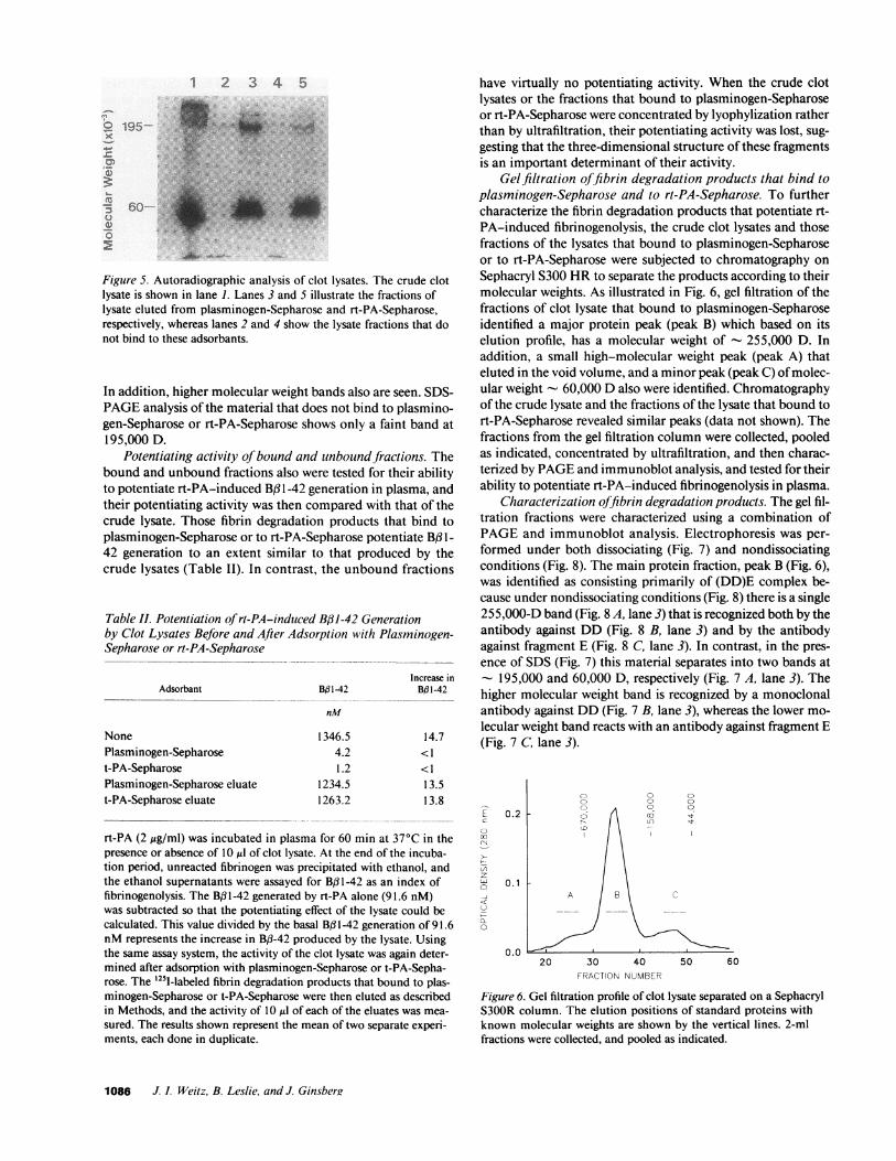
Gelfiltration offibrin degradation products that bind toplasminogen-Sepharose and to rt-PA-Sepharose. To furthercharacterize the fibrin degradation products that potentiate rt-PA-induced fibrinogenolysis, the crude clot lysates and thosefractions of the lysates that bound to plasminogen-Sepharoseor to rt-PA-Sepharose were subjected to chromatography onSephacryl S300 HRto separate the products according to theirmolecular weights. As illustrated in Fig. 6, gel filtration of thefractions of clot lysate that bound to plasminogen-Sepharoseidentified a major protein peak (peak B) which based on itselution profile, has a molecular weight of - 255,000 D. Inaddition, a small high-molecular weight peak (peak A) thateluted in the void volume, and a minor peak (peak C) of molec-ular weight - 60,000 Dalso were identified. Chromatographyof the crude lysate and the fractions of the lysate that bound tort-PA-Sepharose revealed similar peaks (data not shown). Thefractions from the gel filtration column were collected, pooledas indicated, concentrated by ultrafiltration, and then charac-terized by PAGEand immunoblot analysis, and tested for theirability to potentiate rt-PA-induced fibrinogenolysis in plasma.
Characterization offibrin degradation products. The gel fil-tration fractions were characterized using a combination ofPAGEand immunoblot analysis. Electrophoresis was per-formed under both dissociating (Fig. 7) and nondissociatingconditions (Fig. 8). The main protein fraction, peak B (Fig. 6),was identified as consisting primarily of (DD)E complex be-cause under nondissociating conditions (Fig. 8) there is a single255,000-D band (Fig. 8 A, lane 3) that is recognized both by theantibody against DD (Fig. 8 B, lane 3) and by the antibodyagainst fragment E (Fig. 8 C, lane 3). In contrast, in the pres-ence of SDS (Fig. 7) this material separates into two bands at
- 195,000 and 60,000 D, respectively (Fig. 7 A, lane 3). Thehigher molecular weight band is recognized by a monoclonalantibody against DD(Fig. 7 B, lane 3), whereas the lower mo-lecular weight band reacts with an antibody against fragment E(Fig. 7 C, lane 3).
0c
0-
F--
z
0
0.2
0.1
0.0
o 0o 0
000
20 30 40FRACTION NUMBER
60
Figure 6. Gel filtration profile of clot lysate separated on a SephacrylS300R column. The elution positions of standard proteins withknown molecular weights are shown by the vertical lines. 2-mlfractions were collected, and pooled as indicated.
1086 J. I. Weitz, B. Leslie, and J. Ginsbere

B
1 23 4
omm
C
1 2 3 4 Figiure 7. SDS-PAGEand immunoblot analysis of
the degradation products of cross-linked fibrin.
In A4, the gels were stained with Coomasie blue.
The separated proteins also were
electrophoretically transferred to nitrocellulose
membranes, which were then reacted with a
monoclonal antibody against DD (B) or a
monospecific antibody against fragment E (C).
Lane shows the crude lysate, whereas lanes 2-4
represent the gel filtration peaks A. B, and C,
respectively, that are shown in Fig. 6.
PAGEanalysis under dissociating conditions (Fig. 7 A, lane
2) of the peak that elutes in the void volume of the gel filtration
column (Fig. 6, peak A) reveals several high-molecular weight
bands. In addition, there are higher molecular weight deriva-
tives that do not enter the 4% stacking gel. Those fragments
that enter the gel are recognized by the antibody against DD
(Fig. 7 B, lane 2). Although not visualized on the gel stained
with Coomasie blue (Fig. 7 A, lane 2), these derivatives also
contain material that migrates at -~ 60,000 Dand is recognized
by the antibody against fragment E (Fig. 7 C, lane 2). Under
dissociating conditions a band of molecular weight greater than
the (DD)E complex is visualized (Fig. 8 A, lane 2). This band is
recognized both by an antibody against DD (Fig. 8 B, lane 2)
and by an antibody against fragment E (Fig. 8 C, lane 2). Thus,
these species represent high molecular weight derivatives that
contain the (DD)E complex.
The lowest molecular weight peak recovered from the gel
filtration column (Fig. 6, peak C) migrates as a major band of
-60,000 D under dissociating conditions (Fig. 7 A, lane 4)
that is recognized by the antibody against fragment E (Fig. 7 C,
lane 4) but not by the antibody against DD(Fig. 7 B, lane 4). In
addition, at least two higher molecular weight bands that are
recognized by the antibody against DDalso are visualized (Fig.
7 B, lane 4). These bands are of lower molecular weight than
intact DD, and probably represent products of its digestion by
plasmin. Under nondissociating conditions, peak C migrates as
a minor band at -60,000 D and a major band at -~ 55,000 D
(Fig. 8 A, lane 4). These bands are recognized by the antibody
against fragment E (Fig. 8 C, lane 4) and not by the antibody
against DD (Fig. 8 B, lane 4), indicating that they represent
fragment E species.
The fibrin degradation products that do not bind to plas-
minogen-Sepharose or rt-PA-Sepharose (Fig. 5, lanes 2 and 4,
respectively) also were characterized by SDS-PAGEand im-
munoblot analysis (data not shown). This material consists pri-
marily of DDbecause the 195,000-D band visualized in Fig. 5
is recognized by an antibody against DD. Little or no fragment
E is present because a 60,000-D band is not seen in Fig. 5 nor is
a distinct band visualized on immunoblot analysis using the
antibody against fragment E.
Potentiation of rt-PA-indlucedJibrinogenolvsis by isolated
degradation produicts of cross-linked fibrin. The fractions repre-
senting peaks A, B, and C were tested for their ability to poten-
tiate rt-PA-induced Bf3 -42 generation in plasma, and their
activity was compared with that of the starting material (Table
III). All of the plasmic degradation products of cross-linked
fibrin that bind to plasminogen-Sepharose or to rt-PA-Sepha-
rose have the potential to augment rt-PA-induced fibrinogen-
olysis and over 75% of the activity of the starting material is
recovered (data not shown). On a gravimetric basis, however,
(DD)E complex is the most potent stimulator (Table III). This
finding may explain the substantial fibrinogenolysis that ac-
companies rt-PA-induced clot lysis because (DD)E complex is
the maj.or derivative found in the clot lysates. In contrast, DD,
which is the predominant species in those fractions that do not
bind to plasminogen-Sepharose or to rt-PA-Sepharose (Fig. 5)
does not have potentiating activity (Table II).
Discussion
The activation of plasminogen by t-PA is accelerated in the
presence of fibrin (7, 8). Although fibrinogen has little effect on
the rate of t-PA-induced plasminogen activation (7, 8), frag-
ments of fibrinogen or fibrin can mimic the accelerating effect
of intact fibrin (22-28). Rate-enhancing fragments can be gen-
erated by digesting fibrin(ogen) with plasmin or with cyanogen
A
1 23 4
DD.E-UW P"YU~-:
B
1 23 4
C
1
DD-E-11 fri DD-E-.
kAL:..I I
3 4 Figutre 8. PAGEand immunoblot analysis of the
degradation products of cross-linked fibrin done
~under nondissociating conditions. In panel A, the
gels were stained with Coomasie blue. The
separated proteins also were electrophoretically
transferred to nitrocellulose membranes which
were then reacted with a monoclonal antibody
against DD(B) or a monospecific antibody against
fragment E (C). Lane I shows the crude lysate,
whereas lanes 2-4 represent the gel filtration peaks
A, B, and C, respectively, that are shown in
Fig. 6.
Fihrin Degradation Produicts Pot entiate Fibrinogeno/i'sis 1087
A1 2 34
,7f n
E- 0 ..... P.' t'" t".

Table III. Potentiation of rt-PA-induced Bf31-42 Generationby Isolated Fibrin Degradation Products
Increase in BPA142 forSpecies each ug protein
nM
Plasminogen-Sepharose eluate 206±32High-molecular weight derivatives 18±5(DD)E Complex 110±28Fragment E 32±8
The potentiating activity of the fraction of the crude clot lysates thatbound to plasminogen-Sepharose was determined by incubating fourdifferent concentrations of this material with plasma for 60 min at37°C in the presence or absence of 1 ,g/ml of rt-PA. At the end of theincubation period, unreacted fibrinogen was precipitated with eth-anol, and the ethanol supernatants were assayed for B3 1-42. The in-crease in rt-PA-induced generation of B,B1-42 produced by the plas-minogen-Sepharose eluate was then calculated. The eluate was thensubjected to chromatography on Sephacryl S300HRas described inMethods, and the fractions pooled as indicated in Fig. 6. The poten-tiating activity of peaks A, B, and C which represent high-molecularweight derivatives that contain the (DD)E complex, (DD)E complexitself, and fragment E, respectively, was then tested as describedabove. The values illustrated represent the mean±SDof two differentexperiments each done in duplicate.
bromide. The physiologic significance of these observations isuncertain however, because many of these fragments are notproduced in vivo. In addition, the studies reported to date haveonly examined the accelerating effect of fibrin(ogen) fragmentsin buffer systems, and the potential modulating effects of anti-proteinases have not been considered. In the present study wehave extended the observations made in buffer systems by (a)demonstrating that the physiologic products of rt-PA-inducedlysis of cross-linked fibrin can potentiate rt-PA-mediated fi-brinogenolysis in a plasma system, (b) identifying and charac-terizing those fibrin degradation products that have potentiat-ing activity, and (c) demonstrating that the products with po-tentiating activity bind both plasminogen and rt-PA, whichsuggests that they act by providing a surface on which the fibri-nolytic components can be assembled.
In the presence of fibrin there is an - 2 1-fold increase inthe amount of B, 1-42 generated by a wide range of rt-PA con-centrations (Table I). That the higher Bf 1-42 concentrations inthe presence of fibrin reflect increased fibrinogen proteolysis isconfirmed by the results of immunoblot analysis which permitsdirect visualization of the fibrinogen degradation products(Fig. 1). Whenclots of increasing size are exposed to the sameconcentration of rt-PA, there is a linear relationship betweenthe extent of clot lysis and that of fibrinogenolysis (Fig. 2). Thisphenomenon cannot be explained on the basis of the largerclots providing a greater surface on which plasmin can be gen-erated because there is ongoing Bfl 1-42 generation after theclots are removed from rt-PA-containing plasma (Fig. 3). Iffibrinogenolysis was mediated by plasmin bound to the clotsurface, B,3 1-42 production would cease when the clot is re-moved.
That soluble fibrin degradation products are responsible forthe ongoing fibrinogenolysis is suggested by the observation
that the extent of BJ3 1-42 generation that occurs after clot re-moval depends on the time that the clot is exposed to rt-PA,and is established by the demonstration that clot lysates en-hance rt-PA-mediated generation of B,#1-42 in a concentra-tion-dependent fashion (Fig. 4). The potentiating activity ofsoluble fibrin degradation products involves the binding of rt-PAand plasminogen because only the functionally active prod-ucts bind both to rt-PA-Sepharose and to plasminogen-Sepha-rose (Table II). Further, analysis of the bound products usingSDS-PAGEand autoradiography (Fig. 5) indicates that similarderivatives bind to both adsorbants. After separation of theseproducts by gel filtration (Fig. 6), the predominant 255,000-Dspecies (peak B) was identified as (DD)E complex by PAGEand immunoblot analysis (Figs. 7 and 8) consistent withprevious reports that this complex is the major product of plas-min degradation of cross-linked fibrin (38-42). The minorpeaks identified by gel filtration include a series of high-molecular weight species (peak A) that elutes in the void vol-umeof the column, some of which have a mol wt > 106 and donot enter the 4%stacking gel. Those products that enter the gelcontain epitopes that are recognized both by an antibodyagainst D-dimer (Fig. 7 B and 8 B, lane 2) and by an antibodyagainst fragment E (Fig. 7 Cand 8 C, lane 2). Thus, these largefragments contain the (DD)E complex and represent the high-molecular weight derivatives that have been extensively char-acterized by Francis et al. (31, 43). The other minor peak iden-tified by gel filtration (peak C) has a molecular weight of- 60,000 Dand represents predominantly fragment E. Analy-sis under nondissociating conditions indicates that more thanone fragment E species is present (Fig. 8 A, lane 4). Thesefindings are consistent with those of Olexa and Budzynski (42,44, 45) who identified three major fragment E species that weredesignated E 1, E2, and E3 according to their increasing electro-phoretic mobility. These three fragment E derivatives representsequential plasmic degradation products, and only El and E2are capable of binding to DD(44, 45).
Whereas high-molecular weight derivatives that containthe (DD)E complex, (DD)E complex itself, and fragment Ebind to both plasminogen-Sepharose and rt-PA-Sepharose,DDdoes not bind (Fig. 5). These findings suggest that a bindingsite for both plasminogen and rt-PA is located within the Edomain. Although the binding sites for t-PA have notpreviously been defined, plasminogen binding sites have beenidentified on the amino-terminal regions of the Aa- and B,3-chains of fragment E (46-48). Fibrinogen fragment Dalso con-
tains plasminogen binding sites (47, 49). The finding that DDdoes not bind to plasminogen-Sepharose suggests that thesesites are inaccessible when gamma-gammacross-linking of ad-jacent D domains results in DDformation.
Only the fibrin fragments that have affinity for plasmino-gen and rt-PA are capable of augmenting fibrinogenolysis (Ta-ble II) suggesting that these derivatives act by providing a sur-face on which the fibrinolytic components can be assembled.Plasmin generated at this site is relatively protected from inac-tivation by fluid-phase inhibitors because soluble fibrin degra-dation products potentiate rt-PA-induced B,31-42 generationin plasma (Fig. 4). Other investigators have demonstrated thathigh-molecular weight derivatives of cross-linked fibrin (27)and (DD)E complex (28) can accelerate rt-PA-induced activa-tion of plasminogen in a buffer system. Wehave extended these
1088 J. I. Weitz, B. Leslie, and J. Ginsberg
observations in three ways. First, we have shown that in addi-tion to these derivatives, fragment E also potentiates rt-PA-in-duced fibrinogenolysis (Table III). Quantitatively, however,(DD)E complex, the major product of plasmin degradation ofcross-linked fibrin, is the most potent stimulator of fibrinogen-olysis (Table III). Second, because our studies were done in aplasma system, they indicate that plasmin generated on thesurface of fibrin degradation products is relatively protectedfrom inactivation by fluid-phase antiplasmins. Third, the datapresented in Fig. 3 suggest that plasmin generated on the sur-face of fibrin degradation products is a more important media-tor of fibrinogenolysis than plasmin generated on the clot sur-face.
In summary, we have demonstrated that the products ofrt-PA-induced degradation of cross-linked fibrin can poten-tiate rt-PA-mediated fibrinogenolysis by providing a surfacefor t-PA and plasminogen binding thereby promoting plasmingeneration. The occurrence of this phenomenon after therapeu-tic thrombolysis may explain the limited fibrin selectivity ofrt-PA.
Acknowledgments
The authors wish to thank Dr. J. Hirsh and Dr. A. Budzynski for theirexcellent advice and encouragement, and S. Crnic for preparing thismanuscript.
This work was supported by grants from the Heart and StrokeFoundation of Ontario and the Medical Research Council of Canada.Dr. Weitz is a Scholar of the Heart and Stroke Foundation of Ontario.
References
1. Wiman, B., and D. Collen. 1978. Molecular mechanisms of physiologicfibrinolysis. Nature (Lond.). 272:549-550.
2. Lucas, M. A., L. J. Fretto, and P. A. McKee. 1983. The binding of humanplasminogen to fibrin and fibrinogen. J. Biol. Chem. 258:4249-4256.
3. Rakoczi, I., B. Wiman, and D. Collen. 1978. On the biological significanceof the specific interaction between fibrin, plasminogen and antiplasmin. Biochim.Biophys. Acta. 540:295-300.
4. Rijken, D. C., M. Hoylaerts, and D. Collen. 1982. Fibrinolytic properties ofone-chain and two-chain human extrinsic (tissue-type) plasminogen activators. J.Biol. Chem. 257:2920-2925.
5. Loscalzo, J. 1988. Structural and kinetic differences between single-chainand two-chain tissue plasminogen activator. J. Clin. Invest. 82:1391-1397.
6. Banyai, L., and L. Patthy. 1984. Importance of intramolecular interactionsin the control of the fibrin affinity and activation of human plasminogen. J. Biol.Chem. 259:6466-6471.
7. Ranby, M. 1982. Studies on the kinetics of plasminogen activation by tissueplasminogen activator. Biochim. Biophys. Acta. 704:461-469.
8. Hoylaerts, M., D. C. Rijken, H. R. Lijnen, and D. Collen. 1982. Kinetics ofthe activation of plasminogen by human tissue plasminogen activator. Role offibrin. J. Biol. Chem. 257:2912-2919.
9. Pennica, D., W. E. Holmes, W. J. Kohr, R. N. Harkins, G. A. Vehar, D. A.Ward, W. F. Bennett, E. Yelverton, P. H. Seeburg, H. L. Heyneker, D. V. Goed-del, and D. Collen. 1983. Cloning and expression of human tissue-type plasmino-gen activator cDNA in E. coli. Nature (Lond.). 301:214-22 1.
10. Topol, E. J., W. R. Bell, and M. L. Weisfeldt. 1985. Coronary thromboly-sis with recombinant tissue-type plasminogen activator. A hematologic and phar-macologic study. Ann. Intern. Med. 103:837-843.
11. Van de Werf, F., P. A. Ludbrook, S. R. Bergmann, A. J. Tiefenbrunn,K. A. Fox, H. de Geest, M. Verstraete, D. Collen, and B. E. Sobel. 1984. Coronarythrombolysis with tissue-type plasminogen activator in patients with evolvingmyocardial infarction. N. Engl. J. Med. 310:609-613.
12. TIMI Study Group. 1985. The thrombolysis in myocardial infarction(TIMI) trial: phase I findings. N. Engl. J. Med. 312:932-936.
13. Rao, A. K., C. Pratt, A. Berke, A. Jaffe, I. Ockene, T. L. Schreiber, W. R.Bell, and M. Terrin. 1988. Thrombolysis in myocardial infarction (TIMI) trial-phase 1: hemorrhagic manifestations and changes in plasma fibrinogen and the
fibrinolytic system in patients treated with recombinant tissue plasminogen acti-vator and streptokinase. J. Am. Coll. Cardiol. I 1:1-1 1.
14. Verstraete, M., M. Bory, D. Collen, R. Erbel, R. J. Lennane, D. G.Mathey, H. R. Michels, M. Schartl, R. Uebis, R. Bernard, R. W. Brower, D. P.deBono, W. Huhmann, J. Lubsen, J. Meyer, W. Rutsch, W. Schmidt, and R. vonEssen. 1985. Randomized trial of intravenous recombinant tissue type plasmino-gen activator versus intravenous streptokinase in acute myocardial infarction.Lancet. i:842-847.
15. Owen, J., K. D. Friedman, B. A. Grossman, C. Wilkins, A. D. Berke, andE. R. Powers. 1987. Quantitation of fragment X formation during thrombolytictherapy with streptokinase or tissue plasminogen activator. J. Clin. Invest.79:1642-1647.
16. Goldhaber, S. Z., D. E. Vaughan, J. E. Markis, A. P. Selwyn, M. E.Meyerovitz, J. Loscalzo, D. S. Kim, C. M. Kessler, D. L. Dawley, G. V. R. K.Sharma, A. Sasahara, E. B. Grossbard, and E. Braunwald. 1986. Acute pulmo-nary embolism treated with tissue plasminogen activator. Lancet. ii:886-889.
17. Verstraete, M., G. A. Miller, H. Bounameaux, B. Charbonnier, J. P. Colle,G. Lecorf, G. A. Marbet, P. Mombaerts, and C. G. Olsson. 1988. Intravenous andintrapulmonary recombinant tissue-type plasminogen activator in the treatmentof acute massive pulmonary embolism. Circulation. 77:353-360.
18. Levine, M. N., J. Hirsh, J. Weitz, M. Cruickshank, J. Neemeh, A. G. G.Turpie, M. Andrew, M. Klimek, and M. Gent. 1990. A randomized trial of asingle bolus dosage regimen of recombinant tissue plasminogen activator in pa-tients with acute pulmonary embolism. Chest. 98:1473-1479.
19. Turpie, A. G. G. 1987. Thrombolytic therapy in venous thromboeombo-lism. In Tissue Plasminogen Activator in Thrombolytic Therapy. B. E. Sobel, D.Collen, and E. B. Grossbard, editors. Marcel Dekker, Inc., NewYork. 131-146.
20. Sobel, B. E., R. W. Gross, and A. K. Robison. 1984. Thrombolysis, clotselectivity, and kinetics. Circulation. 70:160-164.
21. Noe, D. A., and W. R. Bell. 1987. A kinetic analysis of fibrinogenolysisduring plasminogen activator therapy. Clin. Pharmacol. Ther. 41:297-303.
22. Nieuwenhuizen, W., A. Vermond, M. Voskuilen, D. W. Traas, and J. H.Verheijen. 1983. Identification of a site in fibrin(ogen) which is involved in theacceleration of plasminogen activation by tissue-type plasminogen activator. Bio-chim. Biophys. Acta. 748:86-92.
23. Verheijen, J. H., W. Nieuwenhuizen, and G. Wijngaards. 1982. Activa-tion of plasminogen by tissue activator is increased specifically in the presence ofcertain soluble fibrin(ogen) fragments. Thromb. Res. 27:377-385.
24. Verheijen, J. H., W. Neiuwenhuizen, D. W. Traas, G. T. G. Chang, and E.Hoegee. 1983. Differences in effects of fibrin(ogen) fragments on the activation of1-glu-plasminogen and 442-val-plasminogen by tissue-type plasminogen activa-tor. Thromb. Res. 32:87-92.
25. Nieuwenhuizen, W., J. H. Verheijen, A. Vermond, G. T. G. Chang. 1983.Plasminogen activation by tissue activator is accelerated in the presence of fi-brin(ogen) cyanogen bromide fragment FCB-2. Biochim. Biophys. Acta.755:531-533.
26. Voskuilen, M., A. Vermond, G. H. Veeneman, J. H. Van Boom, E. A.Klasen, N. D. Zegers, and W. Neiuwenhuizen. 1987. Fibrinogen lysine residueAa157 plays a crucial role in the fibrin-induced acceleration of plasminogenactivation catalyzed by tissue-type plasminogen activator. J. Biol. Chem.262:5944-5946.
27. Wilhelm, 0. G., and N. U. Bang. 1988. The influence of diverse fibrinsand fibrin(ogen) fragments on the acceleration of tissue-plasminogen activator-mediated plasminogen activation. In Fibrinogen 3. Biochemistry, Biologic Func-tions, Gene Regulation and Expression. M. W. Mossesson, D. L. Amrani, K. R.Siebenlist, J. P. Diorio. editors. Elsevier Science Publishers, Amsterdam. 181-184.
28. Hasan, A. K., and A. Z. Budzynski. 1989. Binding of one-chain andtwo-chain t-PA to fibrin fragments. Circu/ation. 80:2552a. (Abstr.)
29. Ikeno, L. C., B. M. Bowen, and M. Der. 1981. Commercial production of'I-fibrinogen injection. J. Radioanal. Chem. 65:179-188.
30. McFarlane, A. S. 1965. Labelling of plasma proteins with radioactiveiodine. Biochem. J. 62:135-143.
31. Francis, C. W., V. J. Marder, and S. E. Martin. 1980. Plasmic degradationof crosslinked fibrin. 1. Structural analysis of the particulate clot and identifica-tion of new macromolecular-soluble complexes. Blood. 56:456-464.
32. Weitz, J. I., J. A. Koehn, R. E. Canfield, S. L. Landman, and R. Friedman.1986. Development of a radioimmunoassay for the fibrinogen-derived fragmentB#31-42. Blood. 67:1014-1022.
33. MacGregor, I. R., L. R. Micklem, K. James, and D. S. Pepper. 1985.Characterization of epitopes on human tissue plasminogen activator recognizedby a group of monoclonal antibodies. Thromb. Haemostasis. 53:45-50.
34. Lowry, 0. H., N. J. Rosebrough, A. L. Farr, and R. J. Randall. 1951.Protein measurement with the folin phenol reagent. J. Biol. Chem. 193:265-275.
35. Laemmli, U. K. 1970. Cleavage of structural proteins during the assemblyof the head of bacteriophage T4. Natuire (Lond.). 227:680-685.
36. Davis, B. J. 1964. Disc electrophoresis. 11. Method and application tohuman serum proteins. Ann. NY.4cad. Sci. 121:404-427.
37. Garabedian, H. D., H. K. Gold, R. C. Leinbach, J. A. Johns, T. Yasuda,
Fibrin Degradation Products Potentiate Fibrinogenolysis 1089

M. Kanke, and D. Collen. 1987. Comparative properties of two clinical prepara-tions of recombinant human tissue-type plasminogen activator in patients withacute myocardial infarction. J. Am. Coll. Cardiol. 9:599-607.
38. Gaffney, P. J., and M. Brasher. 1973. Subunit structure of the plasmin-in-duced degradation products of crosslinked fibrin. Biochim. Biophys. Acta.295:308-3 13.
39. Hudry-Clergeon, G., L. Paturel, and M. Suscillon. 1974. Identificationd'un complexe (D-D) . . . E dans les produits de degradation de la fibrine bovinestabilisee par le facteur XIII. Pathol. Biol. 22(Suppl.):47-52.
40. Kopec, M., E. Teisseyre, G. Dudek-Wojciechowska, M. Koczewiak, A.Pankiewicz, and Z. S. Latallo. 1973. Studies on the "Double D" fragment fromstabilized bovine fibrin. Thromb. Res. 2:283-29 1.
41. Pizzo, S. V., L. M. Taylor, Jr., M. L. Schwartz, R. L. Hill, and P. A.McKee. 1973. Subunit structure of fragment D from fibrinogen and crosslinkedfibrin. J. Biol. Chem. 248:4584-4590.
42. Olexa, S. A., and A. Z. Budzynski. 1979. Primary soluble plasmic degrada-tion product of human cross-linked fibrin. Isolation and stoichiometry of the(DD)E complex. Biochemistry. 18:991-995.
43. Francis, C. W., V. J. Marder, and G. H. Barlow. 1980. Plasmic degrada-
tion of crosslinked fibrin. Characterization of new macromolecular soluble com-plexes and a model of their structure. J. Clin. Invest. 66:1033-1043.
44. Olexa, S. A., and A. Z. Budzynski. 1979. Binding phenomena of isolatedunique plasmic degradation products of human cross-linked fibrin. J. Biol.Chem. 254:4925-4932.
45. Olexa, S. A., A. Z. Budzynski, R. F. Doolittle, B. A. Cottrell, and T. C.Greene. 1981. Structure of fragment E species from human cross-linked fibrin.Biochemistry. 20:6139-6145.
46. Cederholm-Williams, S. A., and S. J. Fennel. 1981. Binding of plas-min(ogen) to sepharose bound fibrin(ogen) alpha-chain. Thromb. Res. 21:503-506.
47. Varadi, A., and L. Patthy. 1983. Location of plasminogen-binding sites inhuman fibrin(ogen). Biochemistry. 22:2440-2446.
48. Varadi, A., and L. Patthy. 1984. Beta (Leul21-Lys122) segment of fibrino-gen is in a region essential for plasminogen binding by fibrin fragment E. Biochem-istry. 23:2108-2112.
49. Christensen, U. 1985. C-Terminal lysine residues of fibrinogen fragmentsessential for binding to plasminogen. FEBS(Fed. Eur. Biochem. Soc.) Lett.182:43-46.
1090 J. I. Weitz, B. Leslie, and J. Ginsberg


![Lipoprotein(a), Fibrin Binding, and Plasminogen Activation · Upoproteln(a) (Lp[a]> Is a complex plasma llpoproteln In which apollpoprotein (apo) B-100 Is covalerrtty linked by a](https://img.pdfslide.us/doc/110x75/5c8ba0db09d3f298038c9870/lipoproteina-fibrin-binding-and-plasminogen-activation-upoprotelna-lpa.jpg)