Embed Size (px)
Citation preview
Journal of Neirroclrrmisrn Raven Press, New York 0 1985 International Society for Neurochemistry
Solubilization of an Adenosine Uptake Site in Brain
Ajay Verma, Michelle Houston, and Paul J. Marangos
Abstract: Procedures are described for the solubilization of adenosine uptake sites in guinea pig and rat brain tissue. Using [3H]nitrobenzylthioinosine (13H]NBI) the solubilized site is characterized both kinetically and phar- macologically. The binding is dependent on protein con- centration and is saturable, reversible. specific. and high affinity in nature. The K , and B,,, of guinea pig extracts are 0.13 t 0.02 nM and 133 c 18 fmolimg protein. re- spectively, with linear Scatchard plots obtained routinely. Similar kinetic parameters are observed in rat brain. Adenosine uptake inhibitors are the most potent inhibi- tors of ['HINBI binding with the following order of po- tency, dilazep > hexobendine > dipyridamole. Adeno- sine receptor ligands are much less potent inhibitors of binding, and caffeine i s without effect. The solubilized adenosine uptake site is, therefore, shown to have vir- tually identical properties to the native membrane site. T h e binding of t h e adenos ine A , r e c e p t o r agonis t
[3H]cyclohexyladenosine ([3H]CHA) to the solubilized brain extract was also studied and compared with that of [ 'HINBI. In c o n t r a s t t o t h e [3H]NBI binding s i te [3H]CHA binds to two apparent populations of adenosine receptor, a high-affinity site with a K , of 0.32 * 0.06 nM and a B,,, of 105 -+ 30 f m o l h g protein ,and a lower- affinity site with a K , of 5.50 ? 0.52 nM and B,,, of 300 ? 5S fmol/mg protein. The pharmacology of the ['HICHA binding site is consistent with that of the adenosine re- ceptor and quite distinct from that of the uptake ([3H]NBI binding) site. Therefore, we show that the adenosine up- take site can be solubilized and that it retains both its binding and pharmacologic properties in the solubilized state. Key Words: Solubilized adenosine uptake sites- Nitrobenzylthioinosine-Adenosine receptors. Verrna A. et al. Solubilization of an adenosine uptake site in brain. J . N~itrochern. 45, 596-603 (1985).
Adenosine and several of its metabolically stable analogs, such as cyclohexyladenosine (CHA) and L-phenylisopropyladenosine (L-PIA). have been found to exert potent effects on nervous tissue ac- tivity. These compounds depress nerve cell firing (Phillis et a]., 1979), inhibit the stimulus-evoked re- lease of several neurotransmitters (Fredholm and Hedqvist, 1980). modulate adenylate cyclase ac- tivity (Daly, 1979; Van Calker et al., 1979; Patel et al., 1981), and induce sedation when administered in vivo (Haulica et al.. 1973; Crawley et al., 1982).
Adenosine is released in a depolarization-in- duced, calcium-dependent manner (Stone, 198 I ) and its effects are thought to be mediated by spe- cific, high-affinity, cell-surface recognition sites that have been described in brain using radiolabeled stable adenosine analogs (Bruns et al., 1980: Patel et al., 1982~). Termination of adenosine action is thought to be regulated primarily by a facilitated
reuptake mechanism involving specific elements in the cell membrane (Paterson, 1979; Bender et al., 1980; Barberis et al., 1981). The activity of this adenosine uptake mechanism is important since agents that inhibit this process have been shown to potentiate the neuromodulatory effects of adeno- sine (Huang and Daly, 1974; Phillis et al.. 1979; Crawley et al., 1983; Phillis and Wu, 1 9 8 3 ~ ) . Inhi- bition of adenosine uptake in peripheral tissue is thought to be the mechanism of action of vasoactive drugs such as dilazep and dipyridamole (Sano, 1974). Such an inhibition may also be partially re- sponsible for the actions of centrally active drugs such as benzodiazepines and phenothiazines (Phillis and Wu, 19830).
Recent investigations examining adenosine up- take sites directly have employed binding studies using [3H]nitrobenzylthioinosine ([3H]NBI), which is a potent, selective inhibitor of adenosine uptake
Received October 30, 1984; accepted February I I . 198.5. Address correspondence and reprint requests to Dr. P. J. Ma-
rangos at Unit on Neurochemistry, BPB. Bldg. 10. Room 3C- 210, Bethesda. Maryland 2020.5. U . S . A .
Abbrrricrrions usrd: CHA. cyclohexyladenosine; NBI, nitro- benzylthioinosine; L-PIA. L-phenylisopropyladenosine; PMSF, phenylmethylsufonyl fluoride.
596
SOLUBlLlZED ADENOSINE UPTAKE SITES IN BRAIN 597
into both nervous (Barberis et al., 1981) and non- nervous tissue (Pickard and Paterson, 1972; Bra- jeswar et al., 1975). [3H]NBI has been shown to bind specifically, reversibly, and wi th high affinity to adenosine uptake sites in erythrocytes (Jarvis and Young, 1980), HeLa cells (Lauzon and Paterson, 1977), and brain (Marangos et al., 1982). Hammond et al., in studies using human erythrocytes (Ham- mond et al., 1982) and guinea pig cortical tissue (Hammond and Clanachan, 1982, 1983), have shown that displacement of [3H]NBI binding by a test com- pound was indicative of that compound's ability to block adenosine uptake into these tissues. Thus, the measurement of site-specific binding of [3H]NB1 seems to offer a convenient probe for studying mo- lecular interactions at adenosine uptake sites just a s the stable rad ioac t ive adenos ine analogs d o for adenosine receptors.
O n e of the fundamental approaches in under- standing t h e b iochemis t ry of t hese membrane- bound sites is their solubilization in a functional s ta te . Severa l recent s tudies have used this ap - proach to examine adenos ine receptors in brain (Gavish e t al . , 1982; Bruns et al . , 1983; Nakata and Fuj i sawa, 1983). Using [3H]NBI a n d cyclo- he~y l [~H]adenos ine ([3H]CHA) as probes, we now report successful solubilization of both adenosine uptake sites and receptors from guinea pig brains. [3H]NBI-labeled uptake sites are characterized and are seen to retain pharmacological and binding ki- netic properties similar t o those described in mem- brane bound preparations.
MATERIALS AND METHODS
Membrane preparation Frozen rat or guinea pig whole brains were suspended
in 10 volumes of cold 50 mM Tris-HCI buffer (pH 7.5) containing 10 mM EDTA, 100 M phenylmethysulfonyl fluoride (PMSF), and 1 rnM ],lo-phenathroline using a Brinkman Polytron (speed 5 for 30 s). The suspension was centrifuged at 30,000 g for 20 min and the resulting pellets were suspended in 10 volumes of 50 rnM Tris-HCI (pH 7.5) and spun again as above. The following pellets were resuspended in Tris buffer and incubated with 2 Uiml of adenosine deaminase at room temperature for 30 min. The suspensions were centrifuged again and the final pel- lets wcrc stored at - 70°C. Pellets thus stored were found to be stable for several weeks.
Detergent solubilization Solubilization of membrane pellets was accomplished
using the method described by Bruns et al. for solubilizing various receptors (Bruns et al., 1983). Briefly, 1 g (tissue wet weight) of membrane pellet was thawed and sus- pended in 20 volumes of 50 mM Tris-HCI and centrifuged at 30,000 g for 20 min. Using a pasteur pipet, the washed pellet was finely resuspended in 2 ml of solubilization buffer containing 10 mM Tris-HCI, 1 mM dithiothreitol (DTT), and either 20% glycerol or no glycerol at 0°C. TWO milliliters of solubilization buffer with I % CHAPS deter-
gent was added, and the suspension was swirled gently for 30 s and centrifuged at 145,000 g for 1 h in a Beckman L8-55 ultracentrifuge using a 45-Ti rotor. The supernatant was removed with a pasteur pipet and passed through a Millipore 0.22 p.M filter. The filtrate was either assayed directly for binding or stored at -20°C. To test the effect of glycerol on stability of the solubilized extracts, the preparation extracted without any glycerol was adjusted to contain either 5 or 20% glycerol. Subsequently, all binding experiments were done with supernatant that had been adjusted to contain 20% glycerol after the extrac- tion.
[ 3H]NBI binding assay Solubilized extracts (0.3-0.4 rng of protein) were in-
cubated in the presence of [3H]NBI (17 Ci/mmol. Mo- ravek Biochem) in 50 mM Tris-HCI buffer (pH 7.4) (final volume of 0.5 ml) for 30 min at 22°C. Assays were ter- minated by rapid vacuum filtration through Whatman GF- B filters that had been presoaked for 1-24 h in 0.3% polyethylenimine (,Sigma). The filters were subsequently washed quickly with 4 x 3 ml washes of ice-cold Tris buffer. Filters were air-dried and counted in 10 ml of Redi-Solv scintillation fluid (Beckman). Nonspecific binding was determined by adding 5 p,M NBI (Calbio- chern) in the assay and routinely represented 10% or less of the total binding at 0.7 nM t3H]NBI. Specific binding was determined by subtracting the nonspecific binding values from total binding values. Scatchard analyses were performed by incubating the soluble extracts with [3H]NBI concentrations ranging from 0.05 to 2.5 nM. In- hibition studies were done by incubating the extracts in the presence of six different concentrations of inhibitor and 0.7 nM ['HINBI. All binding data are expressed as specific binding and all assays were performed in tripli- cate. K, values were calculated using the relationship K , = IC,Jl + [LIIK,).
[3H JCHA binding assay [3H]CHA binding to soluble extracts was measured in
a manner similar to that described above for [3H]NBI binding, except that the incubation was performed at 22°C for 2 h. Nonspecific binding was determined by incor- porating 50 K M CHA (Calbiochem). Scatchard analysis was performed by incubating extracts with [3H]CHA (25 Ci/rnmoI) concentrations ranging from 0.40 to 25.0 nM. Inhibition studies were done using 5 nM [3H]CHA. For calculating K , values the K , value for [3H]CHA binding was taken as 1 nM rather than using the component high- and low-affinity K , values. All protein determinations were done using the Biorad protein assay.
Veraparnil was a gift of Knoll Pharmaceuticals and ni- modipine of Miles Pharmaceuticals. In experiments uti- lizing nimodipine, procedures were performed in dark- ened rooms. Dilazep was a gift of Hoffmann La Roche (Nutley), CHA was obtained from Calbiochem, and L- PIA and all other chemicals used were obtained from Sigma.
RESULTS Binding characteristics
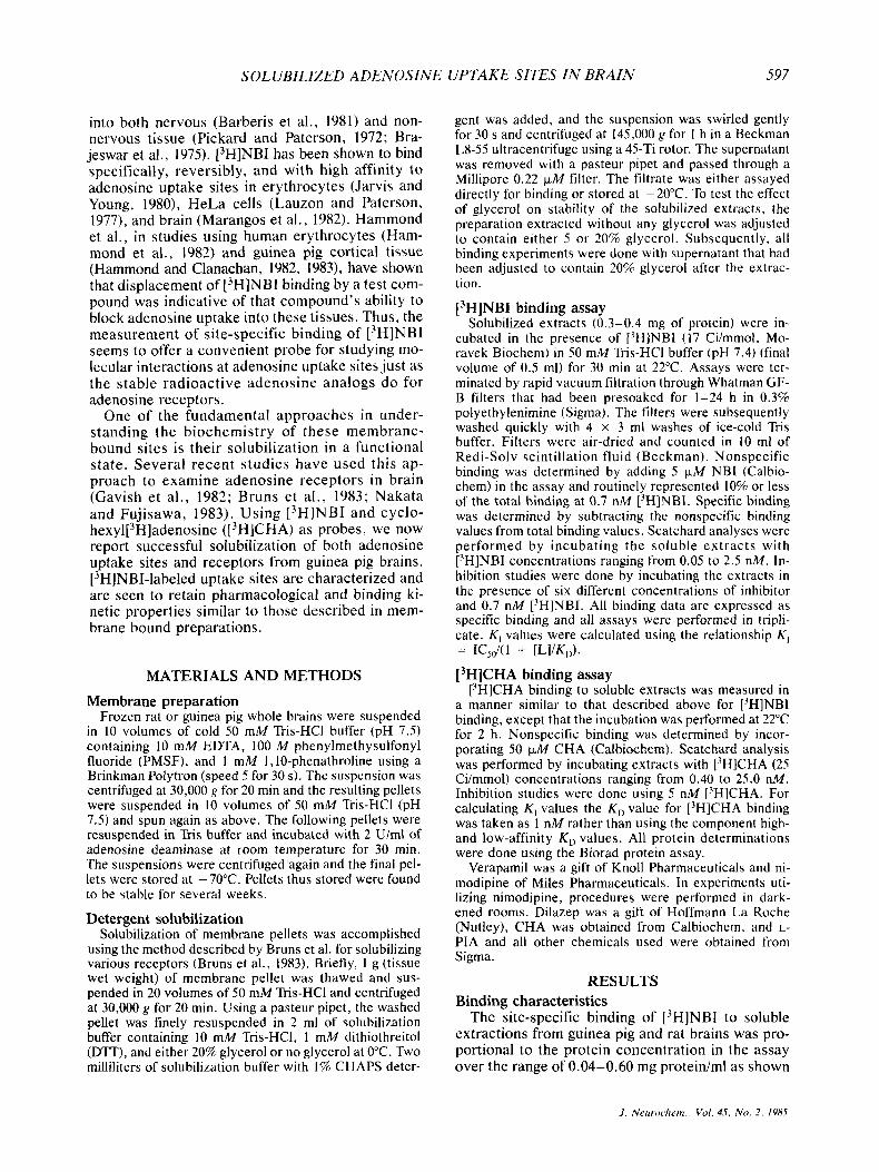
The site-specific binding of ['HINBI to soluble extractions from guinea pig and rat brains was pro- portional t o the protein concentration in the assay over the range of 0.04-0.60 rng protein/ml as shown
J . Neiiroclietn.. Vol . 45, N o . 2. 1985
598 A . VERMA ET A L .
in Fig. 1. Saturation of binding was achieved above a protein concentration of 0.60 mg/ml. In all exper- iments performed. final protein concentrations ranged between 0.30 and 0.40 mg/ml.
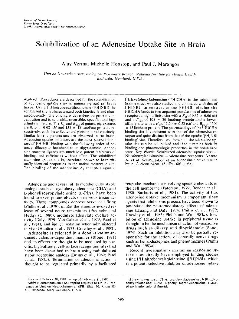
Figure 2 shows the effect of glycerol in stabilizing the activity of solubilized guinea pig extractions. Preparations extracted with 20%' glycerol in the sol- ubilization buffer and those that were reconstituted with 20% glycerol after extraction in glycerol-free buffer showed no loss of specific binding activity over a 2-week period. Reconstituting glyc- erol-free extractions with 5% glycerol led to a loss of about 30% of the initial specific binding after a 2-week period. In the absence of glycerol, prepa- rations were found to retain only 50% of the initial specific binding after 1 week and after 2 weeks al- most 90% of the activity was lost. Preparations ex- tracted with 20% glycerol included in the solubili- zation buffer were cloudy or translucent in appear- ance, even after filtration through 0 . 2 2 - ~ m filters whereas the preparations to which glycerol was added only after the extraction procedure were completely clear (data not shown).
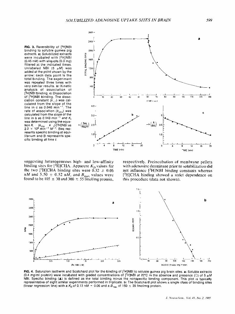
The time course for the association and dissocia- tion of [3H]NBI binding to guinea pig brain solubi- lized extractions is shown in Fig. 3a. The specific binding of 0.45 nM 13H]NBI was 50% complete after approximately 3 min and the binding reaction equi- librium was attained by 20 min. The observed for- ward rate constant (Aob,) calculated from the slope of the line in Fig. 3b was 0.143 min- ' and the dis- sociation rate constant ( k - obtained from the slope of the line in Fig. 3c was 0.046 min-l. K,, as determined using the equation K , = (k,,, - X - I)/ (['HINBI) (Williams et al., 1976), was 2 . 2 x lo8 min-' M - I . An estimate of the equilibrium disso- ciation constant (K,) obtained as the ratio k - , / k , was 0.21 nM.
The saturation isotherm and Scatchard analysis of [3H]NBI binding to soluble guinea pig brain ex-
r
0 2 4 6 8 10 12 14 16
TIME lcbysl
FIG. 2. Effect of glycerol on the stability of [3H]NBI binding to soluble guinea pig brain extracts. Preparations were ex- tracted with (A) and without 20% glycerol present in the sol- ubilization buffer. Those extracted without any glycerol were reconstituted with either 0% (O) , 5% (0), or 20% (A) glycerol after extraction and stored in aliquots at - 20°C. Assays for specific binding were done as described in Materials and Methods for 2 weeks. Each point reflects the percent initial (day 0) specific binding remaining after various storage times. The experiment was repeated twice with similar re- sults.
tracts are shown in Fig. 4. The specific binding was saturable with an apparent KD of 0.13 -+ 0.02 nM and B,,, value of 150 ? 35 fmol/mg protein. Similar plots were obtained in examining [3H]NBI binding to soluble extracts from rat brain with a K D value of 0.12 & 0.03 nM and B,,, of 133 ? 18 fmol/mg protein. All Scatchard plots performed for ['HINBI showed monophasic profiles indicating the pres- ence of only one class of binding sites.
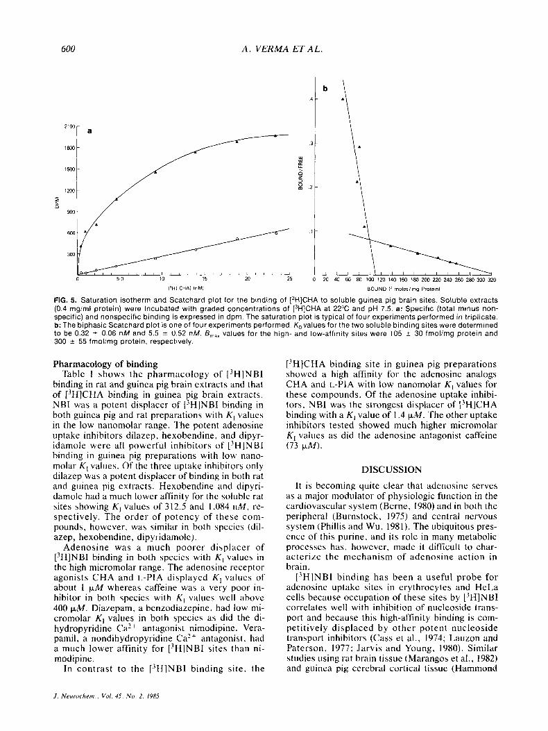
Kinetic analyses performed for the binding of the adenosine receptor agonist 13H]CHA to soluble guinea pig brain extracts are shown in Fig. 5. Sat- urable, specific binding of [3H]CHA, in contrast to ['HINBI binding, revealed biphasic Scatchard plot,
1800 -
1500 - D
m u 900-
I I I I 0 1 2 3 4 5 6 7 8 9 1 0
IPROTEINI lmgimll
FIG. 1. Effect of protein concentration on the site-specific binding of [3H]NBI to guinea pig sol- uble brain extracts. [3H]NBI (0.7 nM) was incu- bated for 30 min at 22°C in 50 mM Tris-HCI buffer (pH 7.5) adjusted to contain the indicated protein concentrations (abscissa) in the presence and ab- sence of 5 pM NBI. Site-specific binding of [3H]NBI (ordinate) was determined as described in Materials and Methods. Each point is the av- erage of two experiments performed in triplicate.
I I I I 7 8 9 1 0
IPROTEINI lmgimll
J . Nrurochrm., Vol. 45, N u . 2 , 1985
SOLUBILIZED ADENOSINE UPTAKE SITES I N BRAIN
4.0 - line in c as 0.046 m in - ' . The rate of association (k,b,) was calculated from the slope of the 3.0 -
was tion line determined K, in = b as (kobs 0.143 - using k-,)/[3H]NBI min- ' the and equa- as K, In($) 2.0 - I______I -I"(&) 0.5
1.0 -
2.2 x lo8 min-' M-' . Beq rep- resents specific binding at equi- l ibrium and B represents spe- cific binding at time t .
I I I I I , 1 I I
599
i B l 1 I I I I I I I
t
a
3000
C
a -
1 0 -
suggesting heterogeneous high- and low-affinity binding sites for [3H]CHA. Apparent K D values for the two [3H]CHA binding sites were 0.32 _t 0.06 nM and 5.50 & 0.52 nM, and B,,, values were found to be 105 * 30 and 300 5 55 fmol/mg protein,
respectively. Preincubation of membrane pellets with adenosine deaminase prior to solubilization did not influence L3H]NBI binding constants whereas [3H]CHA binding showed a strict dependence on this procedure (data not shown).
3000
1 5
a
10
b
w
B 0 - 3 -
m
0.5 - \
\
I 80 I
100 A 120 140 1Ml 180 ~i
200
['HI NBI InMl BOUND (fmoles'mg Protein1
FIG. 4. Saturation isotherm and Scatchard plot for the binding of [3H]NBI to soluble guinea pig brain sites. a: Soluble extracts (0.4 rng/ml protein) were incubated with graded concentrations of [3H]NBI at 22°C in the absence and presence (0) of 5 pM NBI. Specific binding (A) is defined as the total binding minus the nonspecific binding component. This plot is typically representative of eight similar experiments performed in triplicate. b: The Scatchard plot shows a single class of binding sites (linear regression line) with a & o f 0.13 nM 2 0.06 and a B,,, of 150 35 f m o l h g protein.
J . Nt~trrochrm.. V d . 4.5, N o . 2 . 1985
600 A . VERMA ET A L .
0 5 0 10 15 20 25 0 20 40 60 80 100 120 140 160 180 200 220 240 260 280 300 320
13Hi CHAl inMi BOUND if rnoleslmg Protein)
FIG. 5. Saturation isotherm and Scatchard plot for the binding of [3H]CHA to soluble guinea pig brain sites. Soluble extracts (0.4 mgiml protein) were incubated with graded concentrations of [3H]CHA at 22°C and pH 7.5. a: Specific (total minus non- specific) and nonspecific binding is expressed in dpm. The saturation plot is typical of four experiments performed in triplicate. b: The biphasic Scatchard plot is one of four experiments performed. K,values for the two soluble binding sites were determined to be 0.32 2 0.06 n M and 5.5 2 0.52 nM. B,,, values for the high- and low-affinity sites were 105 -c 30 fmol/mg protein and 300 -c 55 fmolimg protein, respectively.
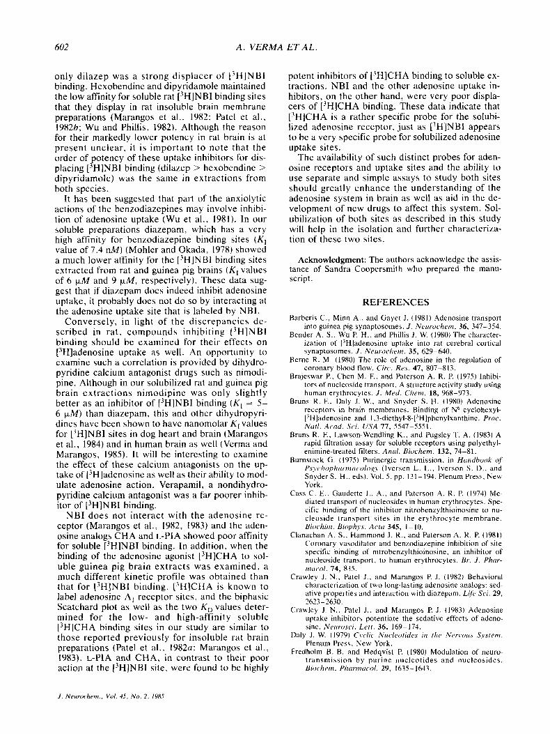
Pharmacology of binding Table 1 shows the pharmacology of [3H]NBI
binding in rat and guinea pig brain extracts and that of [3H]CHA binding in guinea pig brain extracts. NBI was a potent displacer of [3H]NBI binding in both guinea pig and rat preparations with K , values in the low nanomolar range. The potent adenosine uptake inhibitors dilazep. hexobendine. and dipyr- idamole were all powerful inhibitors of [3H]NBI binding in guinea pig preparations with low nano- molar K , values. Of the three uptake inhibitors only dilazep was a potent displacer of binding in both rat and guinea pig extracts. Hexobendine and dipyri- damole had a much lower affinity for the soluble rat sites showing K , values of 312.5 and 1,084 nM, re- spectively. The order of potency of these com- pounds, however, was similar in both spccies (dil- azep, hexobendine, dipyridamole).
Adenosine was a much poorer displacer of [3H1NBI binding in both species with K , values in the high micromolar range. The adenosine receptor agonists CHA and L-PIA displayed K , values of about 1 p M whereas caffeine was a very poor in- hibitor in both species with K , values well 'i ' b ove 400 F M . Diazepam, a benzodiazepine, had low mi- cromolar K , values in both species as did the di- hydropyridine Ca2 + antagonist nimodipine. Vera- pamil, a nondihydropyridine Ca'+ antagonist. had a much lower affinity for [3H]NBI sites than ni- modipine.
In contrast to the [3H1NBI binding site, the
[3H]CHA binding site in guinea pig preparations showed a high affinity for the adenosine analogs CHA and L-PIA with low nanomolar K , values for these compounds. Of the adenosine uptake inhibi- tors, NBI was the strongest displacer of 13H]CHA binding with a K , value of 1.4 p M . The other uptake inhibitors tested showed much higher micromolar K , values as did the adenosine antagonist caffeine (73 p M ) .
DISCUSSION
It is becoming quite clear that adenosine serves as a major modulator of physiologic function in the cardiovascular system (Berne, 1980) and in both the peripheral (Burnstock, 1975) and central nervous system (Phillis and Wu. 1981). The ubiquitous pres- ence of this purine, and its role in many metabolic processes has. however, made it difficult to char- acterize the mechanism of adenosine action in brain.
[3HlNBI binding has been a useful probe for adenosine uptake sites in erythrocytes and HeLa cells because occupation of these sites by L-'H]NBI correlates well with inhibition of nucleoside trans- port and because this high-affinity binding is com- petit ivel y displaced by other potent nucleoside transport inhibitors (Cass et al., 1974; Lauzon and Paterson. 1977: Jarvis and Young, 1980). Similar studies using rat brain tissue (Marangos et al., 1982) and guinea pig cerebral cortical tissue (Hammond
J . Nertrochem.. c'ol. 45. No. 2 . 1985
SOLUBILIZED ADENOSINE UPTAKE SITES IN BRAIN 601
TABLE 1. Pliartnncology of 13H]NBI arid [-'H]CHA binding to soluble rat arid guinea pig h i n extracts
Inhibitor
L3H1NBI (K, ) [3H]CHA (nM)
Rat Guinea pig Guinea pig
Adenosine uptake inhibitors NBI Dilazep Hexobendine Dipyridamole
Adenosine CHA L-PIA Caffeine
Benzodiazepines Diazepam
Ca" Antagonists Nimodipine Verapamil
Adenosine and analogs
0.59 t 0.33 37.5 -t 5.6
312.5 t 103.2 1.084 t 110
92,000 2 1,500 739.6 t 77.34 968.8 t 31.3
400.000
6,250 t 563
5.078 t 391 46.875 2 15,600
1.86 2 0.44 1,369 t 155 11.46 t 3.61 31.30 t 4.62 105,769 2 13.598 36.12 t 7.56
87.900 t 1.700 726.56 2 17.34 5.00 t 1.02
950 t 17.5 6.41 t 1.11
52,885 2 6.799
51,923 2 8,159
73.100 2 21,000 400,000
9,200 2 1,350
6,075 2 550 39,500 2 4,700
Inhibitory potency of various agents on the binding of [3H]NBI and ['HICHA to solubilized brain sites. Inhibition of [3H]NBI binding was performed in both rat and guinea pig preparations and that for ['HICHA in guinea pig alone. Six concentrations of each agent were tested for their effect on specific (total - nonspecific) binding of ['HINBI and ['HICHA. IC,, values were determined from semilog plots and the K , values derived as described in Materials and Methods. Each experiment was repeated three times with values representing means t SD.
and Clanachan, 1982, 1983) show ['HINBI to be a good probe for central adenosine uptake sites as well.
This study demonstrates that methods used to sol- ubilize brain adenosine receptors (Bruns et al., 1983) can also be used to solubilize adenosine up- take sites from brain using ['HINBI as a probe. These sites, solubilized from guinea pig and rat brains, retain characteristics similar to those re- ported for [3H]NBI binding sites in insoluble brain membrane preparations from these respective spe- cies (Hammond and Clanachan, 1982, 1983; Ma- rangos et al., 1982).
Using the procedure described by Bruns et al. we obtained good yields of solubilized adenosine re- ceptors and uptake sites. However, this procedure gave cloudy preparations due to the presence of glycerol in the solubilization buffer, and brought into question the solubility of the preparation. Even though our preparations were centrifuged at 145,000 g for I h, the density of glycerol in our samples affected the centrifugation process. Thus, it was un- certain whether the binding sites were solubilized. Removing glycerol from the solubilizing buffer gave us a clear preparation that still contained active binding sites but lost its activity rapidly. By ex- tracting the sites without any glycerol and then re- constituting the supernatant with glycerol after the extraction, we were able to obtain clear, soluble preparations that were also stable. To ensure solu- bility, all extractions were passed through a 0.22 p M filter before assaying. The use of glycerol in
this manner was necessary for stability and is thus recommended for such extractions.
In the present study, we found that [3H]NBI binding sites solubilized from rat and guinea pig brains had similar kinetic binding parameters. The binding was rapid in both cases and the KD value for [3H]NBI binding to rat brain extracts as deter- mined by Scatchard mass law analysis (KD = 0.12 nM) was similar to K , values for guinea pig soluble sites as determined separately by Scatchard (KD = 0.13 nM) and kinetic analyses (KD = 0.21 nM). The total number of binding sites was similar in both cases (rat, B,,, = 133 fmol/mg protein; guinea pig, B,,, = 150 fmol/mg protein) and linear Scatchard plots indicated the presence of only one class of binding site in both species.
In binding displacement studies NBI was found to be the most potent inhibitor of ['HINBI binding to soluble extracts from both species. In guinea pig preparations the coronary vasodilators dilazep, hexobendine, and dipyridamole were all very po- tent, competitive inhibitors of ['HINBI binding. These compounds produce their dilatory effect via an inhibition of adenosine uptake leading to the ac- cumulation of endogenous adenosine (Sano, 1974). The K , values reported here for soluble guinea pig sites are similar to those reported for inhibition of [3H]NBI binding in human erythrocytes (Clanachan et al., 1981), HeLa cells (Paterson et al., 1980). and dog heart and brain membrane preparations (Ma- rangos et al., 1984). In rat brain soluble extracts, however, of the three adenosine uptake inhibitors,
J . Neurochem., Vol. 45, No. 2, 1985
602 A . VERMA ET A L .
only dilazep was a strong displacer of ['HINBI binding. Hexobendine and dipyridamole maintained the low affinity for soluble rat ['HINBI binding sites that they display in rat insoluble brain membrane preparations (Marangos et at., 1982: Patel et al.. 1982b; Wu and Phillis, 1982). Although the reason for their markedly lower potency in rat brain is at present unclear, i t is important to note that the order of potency of these uptake inhibitors for dis- placing ['HINBI binding (dilazep > hexobendine > dipyridamole) was the same in extractions from both species.
It has been suggested that part of the anxiolytic actions of the benzodiazepines may involve inhibi- tion of adenosine uptake (Wu et al. , 1981). In our soluble preparations diazepam, which has a very high affinity for benzodiazepine binding sites (K, value of 7.4 nM) (Mohler and Okada, 1978) showed a much lower affinity for the ['HINBI binding sites extracted from rat and guinea pig brains (K , values of 6 p M and 9 p M , respectively). These data sug- gest that if diazepam does indeed inhibit adenosine uptake, it probably does not do so by interacting at the adenosine uptake site that is labeled by NBI.
Conversely, in light of the discrepancies de- scribed in rat , compounds inhibiting [3H]NBI binding should be examined for their effects on ["ladenosine uptake as well. An opportunity to examine such a correlation is provided by dihydro- pyridine calcium antagonist drugs such as nimodi- pine. Although in our solubilized rat and guinea pig brain extractions nimodipine was only slightly better as an inhibitor of [3H]NBI binding ( K , = 5- 6 p M ) than diazepam, this and other dihydropyri- dines have been shown to have nanomolar K , values for ['HINBI sites in dog heart and brain (Marangos et at., 1984) and in human brain as well (Verma and Marangos, 1985). I t will be interesting to examine the effect of these calcium antagonists on the up- take of ['Hladenosine as well as their ability to mod- ulate adenosine action. Verapamil, a nondihydro- pyridine calcium antagonist was a far poorer inhib- itor of ['HINBI binding.
NBI does not interact with the adenosine re- ceptor (Marangos et al., 1982, 1983) and the aden- osine analogs CHA and L-PIA showed poor affinity for soluble [3H]NBI binding. In addition. when the binding of the adenosine agonist ['HICHA to sol- uble guinea pig brain extracts was examined, a much different kinetic profile was obtained than that for [3H]NBI binding. ['HICHA is known to label adenosine A, receptor sites, and the biphasic Scatchard plot as well as the two K, values deter- mined for the low- and high-affinity soluble [3H]CHA binding sites in our study are similar to those reported previously for insoluble rat brain preparations (Patel et al., 1982a: Marangos et at., 1983). L-PIA and CHA, in contrast to their poor action at the ['HINBI site, were found to be highly
potent inhibitors of ['HICHA binding to soluble ex- tractions. NBI and the other adenosine uptake in- hibitors, on the other hand, were very poor displa- cers of ['HICHA binding. These data indicate that [3H]CHA is a rather specific probe for the solubi- lized adenosine receptor, just as ['HINBI appears to be a very specific probe for solubilized adenosine uptake sites.
The availability of such distinct probes for aden- osine receptors and uptake sites and the ability to use separate and simple assays to study both sites should greatly enhance the understanding of the adenosine system in brain as well as aid in the de- velopment of new drugs to affect this system. Sol- ubilization of both sites as described in this study will help in the isolation and further characteriza- tion of these two sites.
Acknowledgment: The authors acknowledge the assis- tance of Sandra Coopersmith who prepared the manu- script.
REFERENCES Barberis C., Minn A , , and Gayer J. (1981) Adenosine transport
into guinea pig synaptosomes. J. Neirrochetn. 36, 347-354. Bender A. S.. Wu P. H.. and Phillis J. W. (19x0) The character-
ization of [3H]adenosine uptake into rat cerebral cortical synaptosomes. J . Neirrochetn. 35, 629-640.
Berne R . M. (1980) The role of adenosine in the regulation of coronary blood flow. Circ. Res. 47, 807-813.
Brajeswar P.. Chen M. F.. and Paterson A. R. P. (1975) lnhibi- tors of nucleoside transport. A structure activity study using human erythrocytes. J . M e d . C/ieni. 18, 968-973.
Bruns R. E. Daly J. W.. and Snyder S. H. (1980) Adenosine receptors in brain membranes. Binding of N6 cyclohexyl- [?H]adenosine and I ,3-diethyI-8-['H]phenylxanthine. Proc. Ntrtl. Ac,od. Sci. U S A 77, 5547-5551.
Bruns R. F., Lawson-Wendling K., and Pugsley T. A. (1983) A rapid filtration assay for soluble receptors using polyethyl- enimine-treated filters. A n d . Biocliern. 132, 74-81.
Burnstock G. ( 1975) Purinergic transmission. in Htrtidbook qf f s ~ c ~ / i o p / i t r ~ . t ~ i t i ~ ~ o l o , ~ ~ (Iversen L. L . , Iverson S . D., and Snyder S. H.. eds). Vol. 5 . pp. 131-194. Plenum Press. New York.
Cass C . E . . Gaudette L. A,. and Paterson A. R. P. (1974) Me- diated transport of nucleosides in human erythrocytes. Spe- cific binding of the inhibitor nitrobenzylthioinosine to nu- cleoside transport sites in the erythrocyte membrane. Biodiiiti. B i o p l i . ~ ~ . Actti 345, 1- 10.
Clanachan A. S.. Hammond J. R., and Paterson A. R . P. (1981) Coronary vasodilator and benzodiazepine inhibition of site specific binding of nitrobenzylthioinosine, an inhibitor of nucleoside transport, to human erythrocytes. Br . J . Phcir- nitrc.ol. 74, 835.
Crawley J. N. , Patel J . . and Marangos P. J . (1982) Behavioral CharacteriLation of two long-lasting adenosine analogs: sed- ative properties and interaction with diazepam. Life Sci. 29, 2623-2630.
Crauley J. N.. Patel J . , and Marangos P. J. (1983) Adenosine uptake inhibitor, potentiate the sedative effects of adeno- sine. Nerirosci. L ~ t t . 36, 169-174.
Daly J. W. (1979) C\.clic Nircleotides iii the Ner iw t s System. Plenum Press, New York.
Fredholm B. B. and Hedqvist P. (1980) Modulation of neuro- transmission by purine nucleotides and nucleosides. Biiw/ieit i . Phtrrtncicol. 29, 1635- 1643.
J . Neurochem., Vol. 45, N o . 2 . 1985
SOL UBILIZED ADENOSINE UPTAKE SITES IN BRAIN 603
Gavish M.. Goodman R. R. , and Snyder S. H. (1982) Solubilized adenosine receptors in the brain: regulation by guanine nu-
' i leotides. Science 215, 1633- 1635. Hammond J. R. and Clanachan A. S. (1982) Benzodiazepines
inhibit the binding of nitrobenzylthioinosine. a nucleoside transport inhibitor. to CNS membranes. Br. J . Phurtnacol. 76, 301.
Hammond J. R. and Clanachan A. S. (1983) Inhibition of the site-specific binding of nitrobenzylthioinosine, an inhibitor of nucleoside transport, to C N S membranes, an Itrirrnn- tional Srunposiuni on Adenosine (Berne R. M. and Rubio R.. eds). Abstr. Martinus Nijhoff Medical Division. The Hague.
Hammond J. R.. Jarvis S . M., Paterson A. R. P.. and Clanachan A. S. (1983) Benzodiazepine inhibition of nucleoside trans- port in human erythrocytes. Biochetn. P/rcrrniucol. 32,1229- 1235.
Haulica T.. Aabei L. . Benisteanu D., and Topoliceanu F. (1973) Preliminary data on the possible hypogenic role of adeno- sine. J . Neirrochetn. 21, 1019- 1020.
Huang M. and Daly J. W. (1974) Adenosine-elicited accumula- tion of cyclic AMP in brain slices: potentiation by agents which inhibit uptake of adenosine. Life Sci. 14, 489-503.
Jarvis S. M. and Young, J. D. (1980) Nucleoside transport in human and sheep erythrocytes. evidence that nitrobenzyl- thioinosine binds specifically to functional nucleoside trans- port sites. Eiochem. J . 190, 377-383.
Lauzon G . J . and Paterson A. R. P. (1977) Binding of the nu- cleoside transport inhibitor nitrobenzylthioinosine to HeLa cells. Mol. Phunnncol. 13, 883-891.
Marangos P. J.. Patel J., Clark-Rosenberg R., and Martino A. M. (1982) [3H]Nitrobenzylthioinosine binding as a probe for the study of adenosine uptake sites in brain. J . N~rcroclietn. 39, 184- 191.
Marangos P. J . , Patel J.. Martino A. M.. Dilli M., and Boulenger J . P. (1983) Differential binding properties of adenosine re- ceptor agonists and antagonists in brain. J . Nectroclrern. 41, 361 -374.
Marangos P. J.. Finkel M. S. , Vernia A , , Maturi M. F., Patel J . , and Paterson R. E. (1984) Adenosine uptake sites in dog heart and brain; interaction with calcium antagonists. Lifi, Sci. 35, 1109-1116.
Mohler H. and Okada T. (1978) Biochemical identification of the s i te of ac t ion of benzodiazepines in human brain by [3HJdiazepam binding. Life Sci. 22, 985-996.
Nakata H. and Fujisawa H. (1983) Solubilization and partial characterization of adenosine binding sites from rat brain stem. FEBS Let t . 158, 93-97.
Patel J.. Newman M . . and Mcllwain H . (1981) Cyclic AMP binding capacities and histone kinase activities in subcel- M a r components of neocortical tissue. Biochetn. J . 194, 621-626.
Patel .I.. Marangos P. J.. Stivers .I., and Goodwin F. K . (1982~) Characterization of adenosine receptors in brain using N6 cyclohexyl['H]adenosine. BroI" ReJ. 237, 203-214.
Patel J., Marangos P. J . , Skolnick P.. Paul S. M . . and Martino A. M. (19826) Benzodiazepines are weak inhibitors of [3H]NBI binding to adenosine uptake sites in brain. N e w rosci. Le t t . 29, 79-82.
Paterson A. R. P. (1979) Adenosine transport, in Physiologictrl and Regiilatoty Fimctions of Adenosine und Adenine Ni t - cleotides (Baer H. P. and Drummond G. 1.. eds). pp. 305- 313. Raven Press, New York.
Paterson A. R. P., Lau E. Y., Ddhhg E., and Cass C. E. (1980) A common basis for inhibition of nucleoside transport by dipyridamole and nitrobenzylthioinosine'? M o l . P /Z~i I 'n7~~0/ . 18, 40-44.
Phillis J . W. and Wu P. H . (1981) The role of adenosine and its nucleotides in central synaptic transmission. Prog. Nerrro-
Phillis J. W., and Wu P. H. (1983~) Role of adenosine and ad- enine nucleotides, in the central nervou? system, in Pliysi- olog~j and Phnrmncology qf Adenosine Dc,ritwti\vs (Phillis J. W., Shimizu H. . and Ui M.. eds), pp. 219-237. Raven Press, New York.
Phillis J. W. and Wu P. H. (19836) Nitrobenzylthioinosine inhi- bition of adenosine uptake in guinea pig brain. J . Pkurtn. Pliirrmncol. 35, 540.
Phillis J. W., Edstrom J. P.. Kostopoulos G . K.. and Kirkpatrick J. R . (1979) Effects of adenosine and adenine nucleotides on synaptic transmission in the cerebral cortex. Con. J . Phy- siol. Plrarrnacol. 57, 1289-1312.
Pickard M. A. and Paterson A. R . P. (1972) Use of 4-nitro- benzyl-thioinosine in the measurement of rates of nucleo- side transport in human erythrocytes. Con. J . Bioc~lrem. 50, 839 - 840.
Sano N. (1974) Enhancement of coronary vasodilating actions of adenosine by dilazep and dipyridamole in the dog. J p n . J . Phnrmuc,ol. 24. 41 1 -478.
Stone T. W. (1981) Physiological roles for adenosine and aden- osine 5' triphosphate in the nervous system. Nerrro.rcirnce 6, 523-555.
Van Calken D., Muller M.. and Hamprecht B. (1979) Adenosine regulates via two different types of receptors-the accu- mulation of cyclic AMP in cultured brain cells. J . Neirro- clleln. 33, Y99- 1006.
Verma A , , and Marangos P. J. (1985) Nitrobenzylthioinosine binding in brain: an interspecies study. Life Sci. 36, 283- 291.
Williams L. T., Mullikin D. . and Leftkowitz R. J. (1976) Iden-
b i d . 16, 187-239.
wu
w u
tification of a-adrenergic receptors in uterine smooth muscle membranes by [3H]dihydroergocryptine binding. J . B i d . Clrem. 251, 6915-6923. P. H . and Phillis J. W. (1982) Nucleoside transport in rat cerebral cortical synaptosomal membrane: a high affinity probe study. Ini . J . Biochrni. 14, 1101-1105. P. H. , Phillis J. W.. and Bender A. S . (1981) Do benzodi- azepines bind at adenosine uptake sites in CNS? L$e Sci. 28, 1023-1031.
J . Nivrrochiwi., V d . 45, N o . 2. lY8.5