Embed Size (px)
Citation preview

1
3þ
This article was published in Water Research, 65 (15), 307-320, 2014
http://dx.doi.org/10.1016/j.watres.2014.07.037
Solar photocatalytic oxidation of recalcitrant natural
metabolic by-products of amoxicillin biodegradation
Joa~o H.O.S. Pereira a,1, Ana C. Reis b,1, Vera Homem b, Jos'e A. Silva b, Arminda
Alves b, Maria T. Borges c, Rui A.R. Boaventura a, Vı´tor J.P. Vilar a,*, Olga C.
Nunes b,**
a LSRE - Laboratory of Separation and Reaction Engineering, Associate Laboratory
LSRE/LCM, Faculdade de Engenharia, Universidade do Porto, Rua Dr. Roberto Frias,
4200-465 Porto, Portugal
b LEPABE - Laboratory of Process Engineering, Environment, Biotechnology and
Energy, Faculdade de Engenharia da Universidade do Porto, Rua Dr. Roberto Frias,
4200-465 Porto, Portugal
c CIMAR - Centre for Marine and Environmental Research, Universidade do Porto and
Departamento de Biologia, Faculdade de Ci encias da, Universidade do Porto, Rua
Campo Alegre, 4169-007 Porto, Portugal
Abstract
The contamination of the aquatic environment by non-metabolized and metabolized
antibiotic residues has brought the necessity of alternative treatment steps to current
water decontamination technologies. This work assessed the feasibility of using a multi-
stage treatment system for amoxicillin (AMX) spiked solutions combining: i) a biological
treatment process using an enriched culture to metabolize AMX, with ii) a solar photo-
catalytic system to achieve the removal of the metabolized transformation products
(TPs) identified via LC-MS, recalcitrant to further biological degradation. Firstly, a mixed
culture (MC) was obtained through the enrichment of an activated sludge sample
collected in an urban wastewater treatment plant (WWTP). Secondly, different aqueous
matrices spiked with AMX were treated with the MC and the metabolic
transformation products were identified. Thirdly, the efficiency of two solar assisted
photocatalytic processes (TiO2/UV or Fe3+/Oxalate/H2O2/UV-Vis) was assessed in
the degradation of the obtained TPs using a lab-scale prototype photoreactor
equipped with a compound parabolic collector (CPC). Highest AMX specific
biodegradation rates were obtained in buffer and urban wastewater (WW) media
(0.10 ± 0.01 and 0.13 ± 0.07 gAMX gbiomass-1h-1, respectively). The resulting TPs,
which no longer presented antibacterial activity, were identified as amoxicilloic acid
(m/ z = 384). The performance of the Fe /Oxalate/H2O2/UV-Vis system in the
2
removal of the TPs from WW medium was superior to the TiO2/UV process (TPs no
longer detected after 40 min (QUV = 2.6 kJ L-1), against incomplete TPs removal after
240 min (QUV = 14.9 kJ L-1), respectively).
1. Introduction
In the last decades, the use of antibiotics has evolved from preventing and treating
human and veterinary infections to other applications in diverse areas, such as
agriculture, aquaculture and animal husbandry (Martinez, 2009). To this date, -
lactams remain one of the most important and pre- scribed group of antibiotics (Sun
et al., 2012; Versporten et al., 2011), with amoxicillin (AMX), a broad-spectrum and
semi- synthetic penicillin, as one of most relevant of its class. AMX is poorly
metabolized in the organism and, consequently, around 80-90% is excreted in its
original form (Hirsch et al., 1999). Despite the high consumption, AMX is
infrequently detected in environmental samples. Raw wastewater con- centrations
can vary from 0.02 to 6.9 g L-1 and are easily eliminated by conventional
treatments (Michael et al., 2013). The infrequent detection of AMX in the
environment may be explained by low efficiency and sensitivity of extraction and
detection methods, combined with the highly reactive -lactam ring, which is
broken under environmental conditions, such as alkaline conditions (pH 7.5e9.0),
water hardness and by b-lactamase action (Babington et al., 2012; Deshpande et al.,
2004; Hirsch et al., 1999; Jerzsele and Nagy, 2009). Nevertheless, the low persistence
of AMX in water matrices may be compensated by its intensive use and continuous
discharge into the environment (Daughton and Ternes, 1999).
Amoxicillin has low toxicity and, for that reason, direct effects in the
environment are highly unlikely (Andreozzi et al., 2004). Nevertheless, indirect
effects, such as propagation of -lactam resistant bacteria (Martinez, 2009) and
the impact of metabolites are more pressing concerns. Several studies reported
an increasing proportion of b-lactam resistant organisms after wastewater
treatment, which suggests that conventional WWTP may promote the spread of
antibiotic resistance (Rizzo et al., 2013; Zhang et al., 2009). This could also explain
the frequent presence of resistance genes in different environmental
compartments such as surface and drinking water, a tendency reported for all
the major antibiotic classes, including penicillins (Vaz-Moreira et al., 2014).
Resistance against AMX and other penicillins can occur by different
mechanisms, and deactivation of this group of antibiotics through the action of
b-lactamases is one of the most common and relevant (Drawz and Bonomo,
2010). The products of AMX chemical or -lactamase hydrolysis are, to some
extent, more recalcitrant than the parent compound and for that reason, have
3
been frequently detected in both environ- mental samples and animal tissue
(Lamm et al., 2009; Pe'rez- Parada et al., 2011; Reyns et al., 2008). Amoxicilloic
acid is one of the major metabolites and in addition to its recalcitrant behaviour,
this compound retains allergenic properties that can cause adverse reactions
in sensitive individuals (Torres et al., 2010).
Given the aforementioned detrimental effects, the development of treatment
methods able to remove antibiotic residues and their by-products is henceforth
an environmental priority. Advanced oxidation processes (AOPs) are known as
highly efficient methods to treat otherwise recalcitrant organic pollutants. They
are characterized by different ways of generating the highly reactive and non-
selective hydroxyl radical (•OH) and other reactive oxygen species (Gogate and
Pandit, 2004a, b). As a way of reducing operating costs, recent research has been
focussing on the combination of AOPs able to use solar radiation as the source
of UV photons, such as TiO2/UV and the photo-Fenton process, with biological
degradation as a pre- or post-treatment stage (Elmolla and Chaudhuri, 2011;
Gonza'lez et al., 2008; Oller et al., 2011). Our research groups have previously
dealt with each of these processes separately. Barreiros et al. (2003) and Lopes et
al. (2013) successfully isolated bacteria commonly found in contaminated sites
and applied them, in situ, for the biodegradation of an organic pollutant.
Additionally, Pereira et al. (2013a, 2013b); Pereira et al. (2014) reported on the
removal of antibiotics such as Amoxicillin, Oxytetracycline and Oxolinic Acid
from aqueous solutions by solar TiO2-assisted photocatalysis and by the
ferrioxalate-mediated solar photo- Fenton process using a pilot-plant equipped
with compound parabolic collectors (CPCs). Although Amoxicillin has been
previously subject to several degradation studies via various AOPs (Ay and
Kargi, 2011; Dimitrakopoulou et al., 2012; Homem et al., 2010; Mavronikola et
al., 2009; Trovo' et al., 2011), none has focused on the removal of common
transformation products resulting from the slow transformation that the parent
compound undergoes in aquatic environ- ments (Gozlan et al., 2013; La€ngin
et al., 2009; Na€gele and Moritz, 2005). In this way, the aim of this study was to
evaluate a multistage treatment for AMX-spiked solutions combining: i) a
biological treatment step to metabolize the AMX molecule, with ii) a solar
photocatalytic system to achieve the mineralization of the transformation
products (TPs), recalcitrant to further biological removal. Firstly, a mixed culture
(MC) was obtained through the enrichment of an activated sludge sample
collected in a WWTP treating urban wastewater in order to optimize AMX
biotransformation. Secondly, different aqueous matrices spiked with AMX were
treated with the MC, which constituted a surrogate of activated sludge, and the
metabolic transformation products were identified. Thirdly, the efficiency of the
4
photocatalytic step was assessed in the removal of the metabolized TPs in a lab-
scale photoreactor prototype equipped with a compound parabolic collector
(CPC). The two proposed solar AOPs are the well-known photocatalytic system
mediated by TiO2 (TiO2/ UV) and a modification of the conventional photo-
Fenton process in order to work with near-neutral pH levels, the
Fe3+/Oxalate/H2O2/UV-Vis system. To the best of the authors' knowledge, this
is the first study dealing exclusively with the application of AOPs to remove
recalcitrant transformation products resulting from the natural degradation of
antibiotics, which are often overlooked.
2. Materials and methods
2.1. Reagents
Amoxicillin (MW: 365.4, CAS# 26787-78-0, HPLC-UV chromatogram and
MS/MS spectrum in Fig. 1a and b, respectively) was purchased from Sigma-
Aldrich. HPLC-grade methanol was from Prolabo, H3PO4 (85% p.a.), KH2PO4
and (NH4)2SO4 from Merck (analytical grade) and yeast extract (YE) from
Fisher Scientific. For the LC-MS analysis, methanol hyper- grade for LC-MS
(LiChrosolv®) from Merck, water LC-MS Chromasolv® and formic acid (98%
p.a.) from Fluka were used. Syringe filters with 0.2 mm nylon membranes were
purchased from Whatman and 0.45 mm nylon filter mem- branes from Supelco.
Titanium dioxide used in photocatalytic experiments was Degussa P25 (80%
anatase and 20% rutile). Photo-Fenton experiments were performed using
hydrogen peroxide (Quimite'cnica, S.A., 50% (w/v), 1.10 g cm-3), iron (III)
chloride hexahydrate (Merck) and oxalic acid dihydrate (VWR Prolabo, purity
98%). Sulfuric acid (Pronalab, 96%, 1.84 g cm-3) and sodium hydroxide (Merck)
were used for pH adjustment.
2.2. Microbial culture media
Mineral medium B (Barreiros et al., 2003) was used as microbial growth
medium, supplemented with 0.53 g L-1 (NH4)2SO4 and 1 g L-1 yeast extract
(YE) as nitrogen and carbon sources, respectively. This medium (pH = 7.2),
hereafter designated as enrichment medium (EM), was supplemented with
0.01-0.03 g L-1 of AMX. Antibiotic stock solutions were prepared weekly in
distilled water at a final concentration of 2 g L-1 and kept at -20 oC prior to
use.
5
2.3. Obtainment of a microbial culture enriched in AMX degraders
A sample from the aerobic activated sludge biological reactor collected in an
urban WWTP (Northern Portugal) was diluted 2-fold in EM initially spiked with
0.01 g L-1 AMX. The enrichment culture was successively transferred to fresh
medium with increasing AMX content, up to 0.03 g L-1, and with initial cell
densities corresponding to 0.02 g L-1 dry weight. Transferences were
performed for 24 weeks at intervals of 1e15 d. Cultures were incubated at 30 oC
and 120 rpm. The final culture obtained, which was used as a surrogate of
activated sludge in the combined treatment system, was named mixed culture
(MC).
2.4. Bacteria isolation, characterization and identification
The MC was serially diluted in sterile saline solution (0.85%, w/ v), spread on
Plate Count Agar (PCA) supplemented with AMX (0.03 g L-1) and incubated at
30 oC for 48 h. Individual colonies with distinct morphologies were purified by
subculturing on the same medium. Isolates were identified by 16S rRNA gene
sequence analysis. Amplification of the gene was performed by PCR using
universal primers 27F (5'-AGAGTTT- GATCMTGGCTCAG-3') and 1492R (5'-
TACCTTGTTACGACTT-3') (Lane, 1991). Nucleotide sequences were compared to
those available in public databases using Eztaxon server (http://
www.ezbiocloud.net/eztaxon) (Kim et al., 2012).
Resting cells assays, performed according to Barreiros et al. (2003), were carried
out with each distinct isolate to determine their role in AMX degradation.
2.5. Combined treatment process
In order to assess the influence of the aqueous matrix composition on the
efficiency of the treatment, assays were carried out in EM, saline solution
(NaCl, 0.85%, w/v), phosphate buffer (Buffer; 50 mM, pH 7.2), and wastewater
(WW) collected after secondary wastewater treatment in an urban WWTP (in
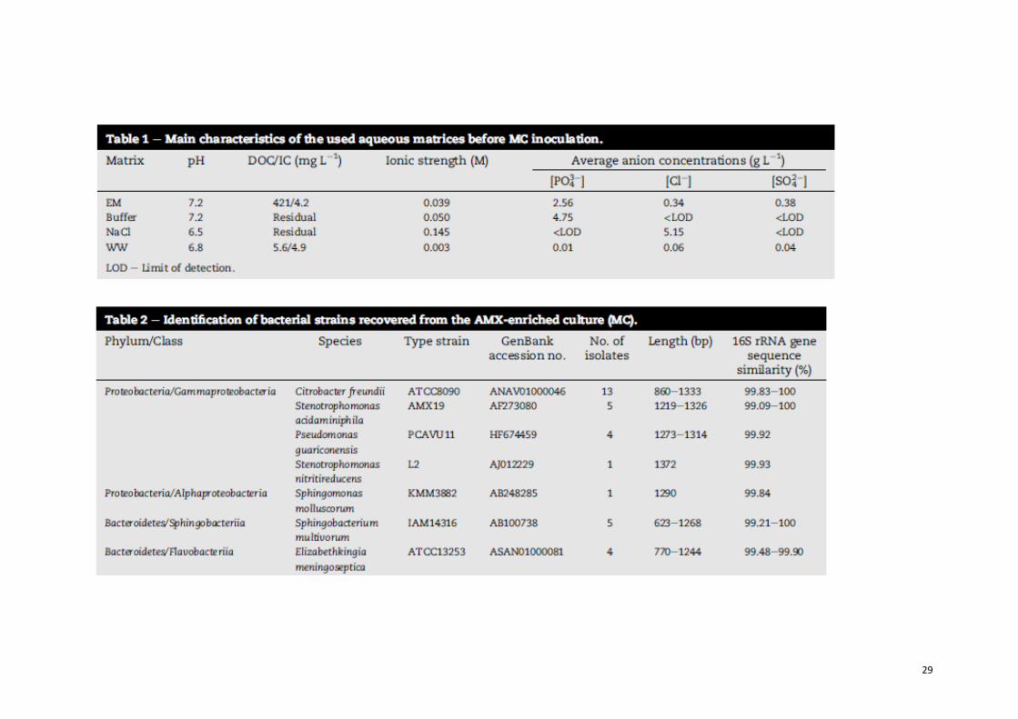
Northern Portugal). The main characteristics of each medium are presented
in Table 1. The high initial AMX concentration, 0.02 g L-1, was chosen to
properly follow the formation and depletion of the TPs by HPLC-UV/Vis
analysis.
2.5.1. Biological treatment
The biological treatment step was carried out in a batch sys- tem using a 5 L
6
Erlenmeyer flask, containing 1.2 L of the test matrix spiked with 0.02 g L-1 of
AMX, in parallel with a control flask to monitor abiotic degradation.
Inoculum was previously obtained by growing the MC in EM spiked with 0.02
g L-1 AMX. Cells were harvested at the late exponential phase, washed twice and
resuspended in sterile saline solution (0.85%, w/v). Given the inability of cells to
grow in the absence of macro- and micronutrients, decreasing complexity of the
tested matrices demanded increasing initial biomass concentration. For the
experiments carried out in EM, 0.03 g L-1 of cell dry weight was used, in
phosphate buffer, 0.11 g L-1, and for both saline solution and real wastewater,
0.26 g L-1. MC cells were separated from the respective medium by
centrifugation at 26,000 x g when AMX was found in residual concentrations
(0.57-1.97 g L-1). As expected due to the low organic carbon content (DOC
ranging between 10 and 25 mg L-1 after spiking with AMX and MC inoculation),
for the experiments in Buffer, NaCl and WW matrices, no bacterial growth was
observed, and AMX degradation followed zero order kinetics. The degradation
rate was obtained by simple linear regression, as expressed by Eq. (1):
where S represents the substrate content at time t (mg L-1), S0 the substrate
content at t = 0, t the time (h) and k the degradation rate (h-1). The half-life
(t1/2) and the specific degradation rates (ks) were calculated as follows (Eqs. (2)
and (3)):
where X represents the biomass content in cell dry weight (mg L-1).
2.5.2. Solar photocatalytic treatment
In all experiments, 1.1 L (Vt) of the resulting supernatant from the biological
treatment step was transferred into the recirculation glass vessel of the lab-scale
photoreactor provided with a sunlight simulator (SUNTEST). A schematic
7
2
representation and a detailed description can be consulted elsewhere (Soares et
al., 2014). Two AOPs, the TiO2/UV system or the Fe3+/Oxalate/H2O2/UV-Vis
process, were performed in order to assess their efficiency in the degradation
and mineralization of the AMX metabolic by-products. The pH was not
controlled in any of the reactions, in both AOP systems.
In the TiO2/UV experiments, 0.2 g L-1 of TiO2 was added to the bio-treated
wastewater, the pH was adjusted to 5.5 with sulfuric acid and the solution was
left to homogenize in darkness for 30 min. This TiO2 concentration was chosen
because it is considered to be the optimal concentration when working with the
CPC unit described in this work (internal diameter 46.4 mm) (Rodr´ıguez et al.,
2004), and is estimated to be able to absorb 100% of the incoming solar UV
photons (Colina-Ma'rquez et al., 2010). Before irradiation began, a sample
was taken to check initial AMX and DOC concentration and TPs' HPLC-UV
areas.
In the Fe3+/Oxalate/H2O2/UV-Vis experiments, oxalate was first added to the
supernatant in a 1:3 (NaCl matrix) or 1:9 (WW matrix) iron/oxalate molar ratio
dose and a sample was taken after 15 min. The pH was adjusted to 4.0 or 5.0 and
iron (III) chloride was then added to achieve a concentration of 2 mg L-1 Fe
(III), the total iron discharge limit into any water body, according to Portuguese
legislation (Decree-Law n.o 236/ 1998). Any marginal effect on pH was then
corrected, and the solution was left to homogenize for 15 min. Another sample
was then taken, and the initial iron concentration was also confirmed. The
solution was then pumped to the CPC unit (illuminated volume (Vi) = 270
mL; Vi/Vt ~ 0.24; ACPC = 0.025 m2 ) before the SUNTEST was turned on.
Irradiation was set as I = 500 WUV m-2, which is equivalent to 44 WUV
m-2 measured in the wavelength range from 280 to 400 nm. Immediately after
irradiation began, the initial H2O2 dose was added, while the pH of the solution
was left uncontrolled. To avoid excess H2O2 in solution, a controlled dosage
strategy was followed as suggested by Klamerth et al. (2010), starting with an
initial 30 mg L-1 (~0.9 mM) dose at the beginning of every experiment, with
supplementary 30 mg L-1 additions performed along the reaction whenever
the H2O2 concentration in solution approached 10 mg L-1.
Samples were taken during the experiments at pre-defined times and filtered
through 0.45 mm Nylon VWR membrane filters before analysis to evaluate the
photodegradation process. For HPLC analysis, samples were quenched with
methanol to stop any further reactions, while DOC analysis was performed
immediately, without pretreatment.
8
2.6. Analytical procedures
AMX concentration and transformation products (TPs) areas were obtained by
HPLC, in a system equipped with an UVeVis detector (Knauer), operating at 230
nm and using a 5 mm Superspher 100 RP-18e (125 x 4 mm) column from Merck.
For the mobile phase, a mixture of 96% (v/v) KH2PO4 buffer (50 mM) and
methanol acidified at pH 3 with ortophosphoric acid was used at 1 mL min-1.
Retention time for AMX was 11.8 ± 0.2 min. TP1, TP2, TP3 and TP4 were detected
at 8.3 ± 0.2, 6.80 ± 0.09, 4.10 ± 0.03 and 2.70 ± 0.01 min, respectively.
Identification of the TPs was performed using a Varian LC- MS system (Lake
Forest, USA), constituted by a ProStar 210 Binary Solvent Delivery Module and
a 500-MS LC Ion Trap Mass Spectrometer, equipped with an electrospray
ionization source (ESI). Data was acquired and processed by Varian MS
Workstation Version 6.9 software. A Pursuit® XRs Ultra C18 column (100 mm x
2.0 mm i.d., particle size: 2.8 m), in combination with a MetaGuard column
Pursuit® C18 (10 mm x 2.0 mm i.d., particle size: 5 m) were purchased from
Varian (Lake Forest, USA). The mobile phase was composed of 96% water with
0.1% formic acid and 4% methanol, running in isocratic mode at a flow rate of
0.2 mL min-1. The injection volume was 10 mL. The mass spectrometer had an
electrospray interface operated in positive mode using: capillary voltage, 69.0
V; shield voltage, 600 V; needle voltage, 3453 V; RF loading, 90%; drying gas
pressure, 15 psi; drying gas temperature, 250 oC; nebulizing gas pressure, 50
psi; multiplier offset, 300 V; MS scan range, 50-600 m/z.
Cell growth was measured by optical density (OD610) using a UV/Vis Unicam
Helios spectrophotometer and by dry weight using a calibration curve. The later
was obtained by filtering cell suspensions through 0.45 mm membranes
(Advantec), dried at 70 oC until constant weight.
Growth inhibition assays were carried out in 96-well microtiter plates using a
Synergy HT Multi-Mode Microplate Reader (Biotek Instruments, USA) in order to
test the toxicity of treated samples. Escherichia coli (DSM 1103) and Staphylo-
coccus aureus (DSM 1104) were used as test strains. Minimum inhibitory
concentration (MIC) for each strain is 4.0 and 0.25 mg AMX L-1, respectively
(Andrews, 2001). Each plate, in addition to the treated samples, contained both
negative and positive controls (inoculated media with and without 0.02
g L-1 AMX, respectively), and blanks (cell-free media). All wells were
supplemented with 2 g L-1 YE and the plates were then incubated at 30 oC,
continuously shaken for 20 h, with absorbance measurements at 610 nm every
30 min. Each sample was tested in triplicate and a first-order kinetic model was
9
used to fit the experimental data, being the specific growth rate (h-1)
normalized by that of the positive control. Comparison of means was made by a
one-way ANOVA test using R software (R Development Core Team, 2013).
Concentration of H2O2 was measured by the metavanadate method, according
to Nogueira et al. (2005). Dissolved iron concentration was determined by
colourimetry with 1,10- phenantroline, according to ISO 6332.
A detailed description of the analysis of dissolved organic carbon (DOC)
content (TC-TOC-TN analyzer), and low- molecular-weight carboxylate anions
(LMWCA, ion chromatography) can be consulted elsewhere (Pereira et al.,
2013b). Additional LMWCA analysis was performed in the HPLC-DAD system,
using a Rezex™ ROA-Organic Acid H+ (8%), LC Column 300 x 7.8 mm. The
isocratic method used 0.005 N H2SO4 delivered at a flow rate of 0.5 mL min-1.
Run time was 50 min, injection volume 10 L and the wavelength of the detector
was set at 210 nm. Sulfate, chloride and phosphate were quantified by ion
chromatography (Dionex ICS-2100; column AS 11-HC 4 x 250 mm; suppressor
ASRS 300 4 mm). Isocratic elution was performed using 30 mM NaOH at
a flow rate of 1.5 mL min-1.
Eq. (4) allows the calculation of the accumulated UV energy (QUV,n kJ L-1)
received on any surface in the same position with regard to the lamp, per unit of
volume of water inside the reactor, in the time interval t:
where tn is the experimental time of each sample (s), Vt the total reactor
volume (L), Ar the illuminated collector surface area (m2) and 𝑈𝑉G;n the
average solar ultraviolet radiation (W m-2) measured during the period tn (s).
3. Results and discussion
3.1. Characterization of the mixed culture (MC)
A mixed culture (MC) able to degrade AMX was obtained after enrichment of
activated sludge with this antibiotic. MC was able to grow unimpaired up to 0.45
g L-1 AMX in the presence of 1 g L-1 YE. However, no growth was observed
when AMX acted as a sole carbon source (data not shown). The spread plate
method revealed the presence of 33 different morphotypes, which were affiliated
10
to 6 distinct genera (Table 2).
All organisms were Gram-negative and the majority was affiliated to the phylum
Proteobacteria, known to be common inhabitants of activated sludge (Zhang et al., 2012).
Within this group a high percentage of isolates was closely related to Citrobacter freundii,
followed by Stenotrophomonas acid- aminiphila. Although in lower proportion, members
of the phylum Bacteroidetes were also isolated. When individually tested in phosphate
buffer (0.05 M, pH 7.2, 0.035 g L-1 AMX), resting cells of all isolates were able to degrade
AMX, albeit at different rates. The overall specific degradation rate of the MC (0.11 gAMX
gbiomass-1h-1) closely corresponded to that of C. freundii (0.09 gAMX gbiomass
-1h-1).
Other isolates exhibited either slower (Elizabethkingia meningoseptica, 0.03 gAMX g
gbiomass-1h-1), or higher (Stenotrophomonas nitritireducens, 2.95 gAMX gbiomass-1
h-1) degradation rates.
These results suggest that all the isolates express -lactamases. Indeed,
members of the same taxa have been described as carriers of different b-lactam
resistance genes (Gonzalez and Vila, 2012; Henriques et al., 2012; Literacka et
al., 2004; Vaz-Moreira et al., 2011). In addition, AmpC - lactamase, a class C
serine cephalosporinase, is frequently found in enterobacteria, such as C.
freundii (Drawz and Bonomo, 2010), while metallo--lactamases, class B zinc
-lactamases, are expressed by members of E. meningoseptica,
Stenotrophomonas sp. and Pseudomonas sp. (Drawz and Bonomo, 2010;
Galleni et al., 2001), which are able to degrade most -lactam antibiotics
including carbapenems (Walsh et al., 2005). Given their wide diversity (Drawz
and Bonomo, 2010), -lactamases may possess different affinities for AMX,
which may explain the significant differences in the degradation rates
observed in the present study. Because a mixed culture is a better surrogate of
activated sludge than axenic bacterial cultures, MC was further used in the
combined treatment.
3.2. Combined treatment for AMX removal
In order to assess the influence of the aqueous matrix composition in the
efficiency of the combined treatment, as- says were performed in EM, Buffer and
NaCl solution. Given our major aim was the assessment of the feasibility of using
this process in the decontamination of real waters, the efficiency of the combined
system in the removal of AMX and degradation products thereof was also
carried out in treated urban wastewater.
3.2.1. Biological degradation step performance
In all tested matrices, the biodegradation of AMX occurred concomitantly with
11
the accumulation of TP1, which subsequently yielded TP2, both detected by
HPLC analysis (HPLC- UV/Vis chromatograms in Fig. 1a). Further analysis of
these metabolites by LC-MS/MS indicated that they had similar mass spectra,
equivalent to that of amoxicilloic acid ([C16H21N3O6S + H]+, m/z = 384, Fig.
1c), the product of b- lactamase action (Deshpande et al., 2004; Na€gele and
Moritz, 2005). Although LC-MS/MS was unable to distinguish TP1 from TP2,
these products were, probably, the stereoisomers (5R,6R) and (5S,6R) of
amoxicilloic acid, identified before (Pe'rez-Parada et al., 2011). The results
obtained in the present study suggest that the enzymatic catalysis favours the
formation of one of the isomers. This behaviour was also observed by Lewis et
al. (1997), which confirmed the formation of the (5R,6R) isomer mediated by a
serine -lactamase and consequent epimerization to the (5S,6R) configuration by
non-enzymatic mechanisms. The hydrolysis of the b-lactam ring results in the
total loss of antimicrobial activity, which was herein confirmed by experimental
data. By the end of the biological step, the liquid phase no longer inhibited the
growth of E. coli, whereas its effect against S. aureus was greatly reduced, as
shown, as an example, in the experiment performed in the NaCl matrix (Fig. 2).
AMX hydrolysis by the MC followed a zero-order kinetic model, except in the
assay carried out in EM. In this particular case, the presence of YE resulted in
increasing biomass con- tent over time, with the degradation following first-
order kinetics, not allowing a direct comparison with the experiments carried
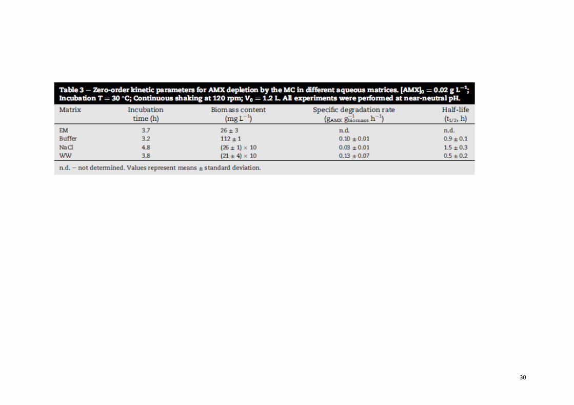
out in other matrices. AMX biodegradation was significantly faster (p < 0.05) in
Buffer and WW than in the NaCl matrix, while Buffer and WW matrices
displayed similar specific degradation rates (p > 0.05) (Table 3).
These differences can be explained by the distinct chemical composition and
pH values of each matrix. The comparatively higher ionic strength, lower pH
and lack of buffer capacity of the NaCl matrix have probably contributed for
these results. Indeed, NaCl is a weak inhibitor of OXA-type - lactamases (Afzal-
Shah et al., 2001). The high standard deviation observed for the WW experiments
may be explained by variations in its chemical and microbiological
composition. Abiotic degradation of the parent compound in the cell-free matrix
was negligible for all experiments (data not shown).
TP1/TP2 exhibited a recalcitrant behaviour, since the MC could not remove
them even after 46 d of incubation (data not shown). This observation is
supported by Gozlan et al. (2013) who detected amoxicilloic acid at relatively
high concentrations (mg L-1) in secondary effluents used for irrigation, as
well in the underground water collected in a well located in the irrigated field.
Given that TP1/2 result from the hydrolysis of the beta- lactam ring, the DOC
remained constant in all experiments, except in the EM matrix, where the
12
consumption of YE by the MC led to a 25% decrease in the initial carbon content
(Fig. 3a and b, left side). These results and observations highlight the need of
complementing AMX biodegradation with the proposed photocatalytic
methods for the removal and mineralization of the resulting transformation
products.
For the combined treatment assays, the endpoint of the biological step was set
between 90 and 95% of AMX depletion, which corresponded to incubation
periods ranging from 3.2 to
4.8 h, for Buffer and NaCl matrices, respectively (Table 3). However, possibly
given the high stability of the b-lactamases produced by the MC members and
extracellular release by leakage into the medium (Livermore, 1995), AMX trans-
formation was not hampered during the processing of the samples, and
transference of the supernatant into the photoreactor. Consequently, the AMX
concentration at the beginning of the AOPs treatments was below the
quantification limit (0.5 mg L-1) (Fig. 4a and b, before irradiation). As
expected, by the end of the biological degradation step, no significant DOC
reduction was observed in all the matrices with low initial DOC content (0 to
approximately 1 mg L-1 of DOC reduction). In opposition, the utilization of
yeast extract as carbon and energy source allowed a DOC reduction of about 25%
in the EM matrix.
3.2.2. TiO2/UV photocatalysis step performance
During preliminary TiO2/UV photocatalytic experiments, where the pH was
kept unchanged, the removal of TP1/TP2 was remarkably slow (data not shown),
with no apparent mineralization. To the best of our knowledge, no information
regarding the pKa constants of TP1/TP2 is available, thus not allowing any direct
conclusion regarding the role of electro- static attraction between TiO2 particles
and TP1/TP2 molecules. For that reason, all the matrices were acidified (5.5) prior
to photocatalytic treatment to take advantage of the positive effect of increased
catalyst surface when smaller TiO2 particle size is favoured under pH values
lower than the Point of Zero Charge of Degussa P25 (pHPZC = 6.7 (Malato et al.,
2009)), as recommended by Li et al. (2010).
The NaCl solution was the most favourable matrix, as TP1 and TP2 were no
longer detected after 90 min of illumination (QUV = 5.3 kJ L-1), followed by
the Buffer matrix, which required 180 min of illumination (QUV = 10.9 kJ L-1).
However, a third unidentified TP (TP3) accumulated in all matrices (Fig. 3a and
b). In the WW experiment, a fourth product also accumulated (TP4), albeit
transiently (Fig. 3b).
13
Differences in matrices complexity in EM and WW experiments seem to
account for the inability of completely removing TP1/TP2 under similar
photo-treatment periods. The pronounced amount of organic matter
compared to the simpler matrices may have not only competed with the
catalyst for absorption of incident UV radiation, but also with the TP1/TP2
attack by the generated •OH radicals. Controls were performed in the
absence of light and no considerable effects were found. Despite the initial
decreases in the DOC content due to the adsorption of organic matter onto
the surface of the catalyst, no notable DOC removal occurred during the
illumination period. This can be explained by the high concentrations of Cl-
(NaCl) and PO43- (Buffer) in the respective matrices, which might have
affected the mineralization process via competitive adsorption onto the TiO2
sur- face, their •OH radical scavenger properties, and finally, via the
formation of less reactive inorganic radicals, as suggested by Guillard et al.
(2005) and De Laat et al. (2004). The exception was the NaCl experiment, which
saw a 35% DOC reduction after 180 min, when compared to the initial value
before the catalyst addition step (Fig. 3b).
3.2.3. Photo-Fenton step performance
Given the apparent unsuitability of using the TiO2/UV system with the most
realistic matrix (WW), the Fe3+/Oxalate/H2O2/ UV-Vis system was tested as
an alternative suited to near- neutral pH levels. Beforehand, some
considerations about this treatment must be made. The conventional photo-
Fenton process, as simplified by Gogate and Pandit (2004b), comprises the
combination of ferrous iron (Fe2+) with hydrogen peroxide (H2O2) and (solar)
UV-Vis radiation resulting in the production of two moles of •OH per mole of
hydrogen peroxide (Eqs. (5) and (6)):
The solubility of Fe3+-hydroxy complexes, especially considering the molar
fraction of the most photoactive species, [Fe(OH)]2+ (with absorption bands
between 290 and 400 nm), is very limited for pH values above 3 (Pignatello et al.,
2006), which would demand additional costs of initial acidification and final
neutralization of the solution. However, this can be overcome by means of the
formation of Fe(III)- carboxylate complexes, which are able to extend the
solubility of iron to near neutral pH working conditions. At the same time, these
14
complexes present stronger radiation absorption at wavelengths up to 580 nm
and can increase the quantum yield of Fe2+ production according to Eq. (7)
(Jeong and Yoon, 2005; Pignatello et al., 2006):
For this reason, the Fe(III)-oxalate complex was chosen, as the quantum yield
of its two most photo-active species, Fe(C2O4)2- and Fe(C2O4)3
3-, were
reported by Faust and Zepp (1993) as being 1.0 and 0.6 (436 nm), respectively.
The working pH of the consequent experiments was decided according to the
distribution of these species in solution. In view of that, MINEQL þ software
was used to study the distribution of the Fe (III) complexes as a function of pH.
The presence of inorganic ions such as PO43-, SO42- and Cl- in the different
matrices (Table 1), which can compete with oxalate for available iron, thus
hindering the process performance (De Laat et al., 2004), was also considered.
The following oxalate and iron-oxalate equilibrium reactions were introduced in
MINEQL+, with dissociation constants (Panias et al., 1996) corrected using the
Davies equation for the approximate ionic strength of the corresponding matrix:
Notwithstanding the use of the iron(III)-oxalate complexes, the
solubility/concentration of iron (III) would be highly limited by the high
phosphate content of the Buffer and EM media, which would result in the
immediate precipitation of the mineral Strengite (FePO4.2H2O). Additionally,
the amount of organic carbon of the EM matrix would also be highly deleterious.
For these reasons, photo-Fenton experiments were not performed in these two
15
matrices.
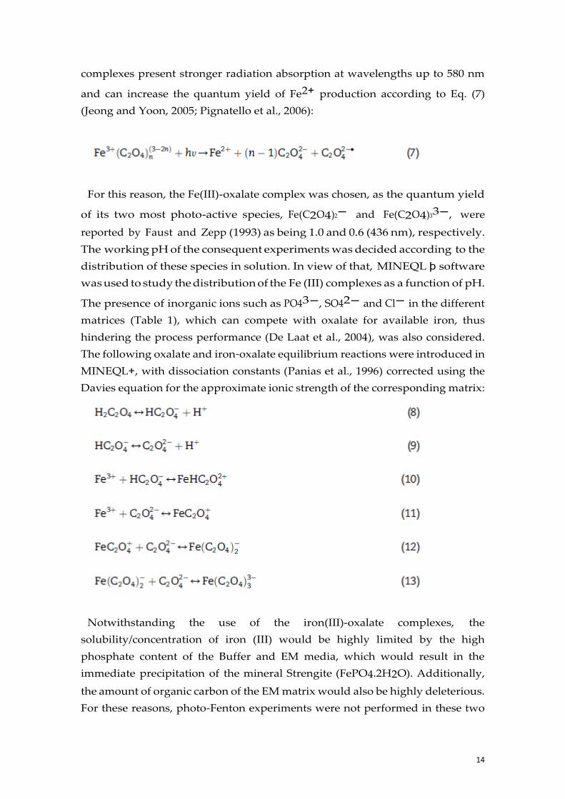
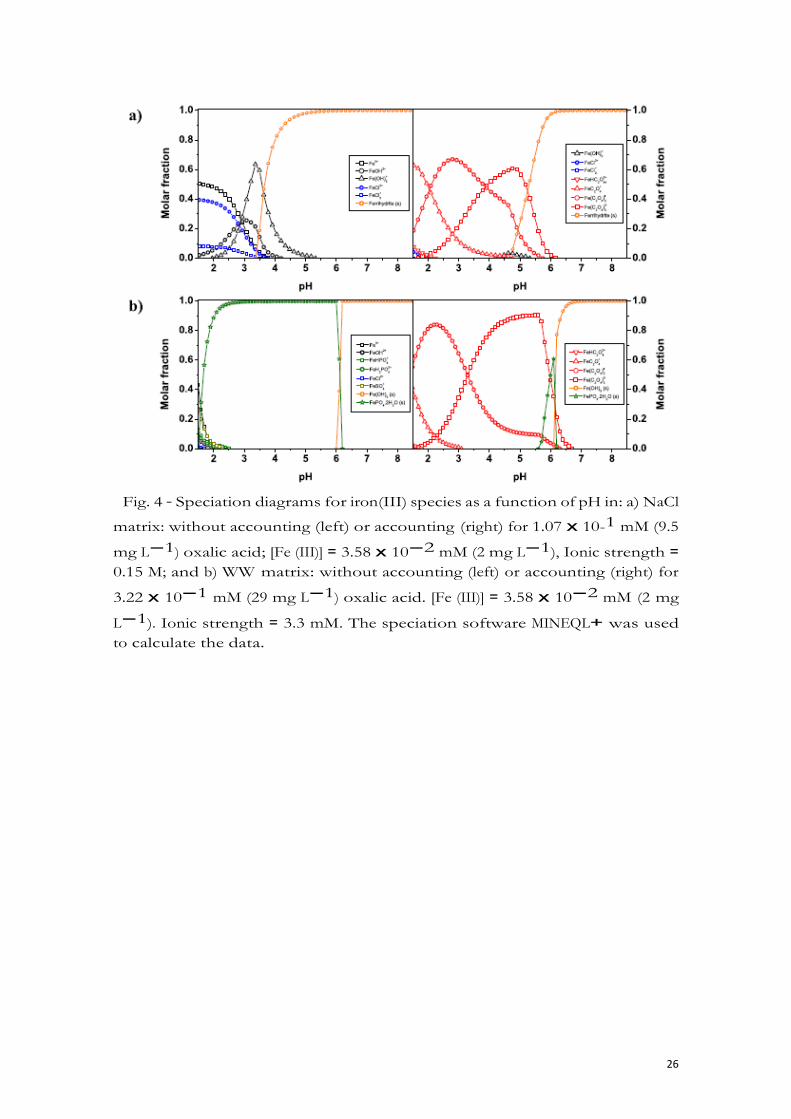
Fig. 4 a and b shows the calculated iron speciation diagrams according to the
molar concentration of each known component (Table 1) for NaCl and WW,
respectively, in the absence (left) or in the presence (right) of oxalate. Initially, a
1:3 Fe(III)-oxalate molar ratio was considered to ensure the formation of the
three photo-active complexes. The respective inorganic anion content of NaCl
and WW was not expected to interfere in the formation of the Fe(III)-oxalate
complexes. Despite this, WW experiments eventually required a higher
iron/oxalate molar ratio dose (1:9), than NaCl matrix (1:3), resulting in
remarkably different profiles.
In experiments performed with the initial 1:3 iron/oxalate molar ratio dose
(data not shown), the added oxalate was not sufficient to keep a satisfactory
Fe(III) concentration in solution along reaction time, due to the probable binding
of iron to natural dissolved organic matter contained in the WW matrix (Lindsey
and Tarr, 2000).
Ultimately, Fe3+/Oxalate/H O /UV-Vis experiments were performed in NaCl
and WW under two initial pH values, 4 and 5, as both Fe(C2O4)2- and
Fe(C2O4)33- are prominent in this pH range. Higher pH values were not
considered due to the fact that in NaCl matrix the Ferrihydrite (Fe(OH)3 (s))
fraction is shown to quickly rise near pH = 5.0, and becomes dominant at pH =
6.0 (Fig 4a), while in WW matrix, iron is expected to precipitate in the form of
Strengite (FePO4.2H2O) after pH = 5.5, and as Ferrihydrite (Fe(OH)3 (s)) after pH
= 6.0 (Fig. 4b).
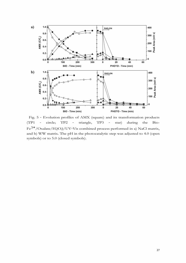
The results on TP1/TP2 removal from NaCl and WW matrix can be seen in Fig.
5a and b (right), respectively.
Compared to the TiO2/UV system, TP1/TP2 removal was clearly improved,
in both matrices and respective pH0 values. In both pH0 = 4.0 experiments,
TP1/TP2 were no longer detected after 20 min of illumination (QUV = 1.3 kJ
L-1), while the pH0 = 5.0 experiments required a longer period of illumination,
40 min (QUV = 2.6 kJ L-1). In WW experiments, a third unidentified TP (TP3)
appeared at 10 min of illumination, but it was no longer detected after 60 min
(QUV = 4.0 kJ L-1). Control experiments performed in darkness did not show
significant TP1/TP2 removal in the same period. The general influence of pH
on the experiments may be explained with the distribution of the above
mentioned Fe (III)-oxalate species. Even considering the differences in their
relative proportion over pH due to different iron/oxalate molar ratio doses,
the fraction of most photo-active species, Fe(C2O4)2- , tends to be higher
16
around pH = 4.0, compared to pH = 5.0. The higher consumption of H2O2
seems to attest to this fact.
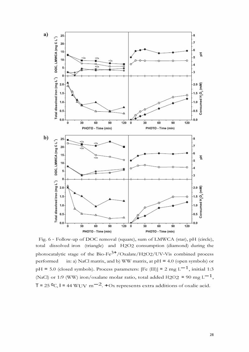
The process details for both matrices can be seen in Fig. 5a and b. By the end of
the first 30 min of illumination (QUV = 1.9 kJ L-1), an overall steady decrease
can be seen in total soluble iron concentration. This corresponded well with the
concomitant ongoing depletion of oxalate, apparent in HPLC analysis of low-
molecular-weight carboxylate anions (LMWCA). With dwindling amounts of
oxalate in solution, the distribution of iron (III) species seen in the right side of
Fig. 4a and b would progress into what can be seen in the left side of the same
Figure.
Up until this point in both NaCl matrix experiments, nevertheless, reduction of
DOC exceeded the oxalate depletion, and an average of 45% of the remaining
DOC was already in the form of tartronate, oxamate, malonate, glycolate,
formate and acetate, besides a lower amount of oxalate itself.
In both WW experiments, on the other hand, DOC decreased along with the
oxalate depletion, with vestigial amounts of other LMWCA.
Given the abovementioned fact that, in each matrix and tested pH0 level,
complete, or near complete TP1/TP2 removal occurred by 40 min of illumination,
the process was extended with additional dosages of oxalate (marked as +Ox in
Fig 6 a and b), in order to keep iron in solution and see the effects on the
remaining DOC content.
Notwithstanding the added carbon with each oxalate addition, and the
apparent levelling of DOC content in both NaCl and WW experiments, depletion
of oxalate was quick between additions. After 120 min of illumination (QUV =
8.3 kJ L-1) in NaCl experiments, an average of 55% of the final DOC content was
in the form of LMWCA. In WW medium, only the pH0 = 4.0 experiment benefited
with the additional oxalate, as it can be seen in the progressive decrease of DOC
content and its final 48% amount in the form of LMWCA, compared to 28% in
the case of pH0 = 5.0. In the same irradiation period, initial DOC concentration
was altogether reduced in the NaCl matrix by 54% (pH0 = 4.0) and 51% (pH0
= 5.0), while in the WW matrix it was reduced by 48% (pH0 = 4.0) and 21% (pH0
= 5.0).
4. Conclusions
In this work, a mixed culture able to degrade the antibiotic Amoxicillin in
different matrices was successfully obtained and characterized. AMX removal
was concomitant with the accumulation of transformation products, which were
17
3
putatively identified as stereoisomers of amoxicilloic acid, and did not show
antibacterial activity. However, they resisted further bacterial action, which
brought up the need of a further, more efficient treatment. In this way, a
complementary photo- catalytic oxidation step was proposed to enhance AMXC
degradation in a lab-scale apparatus simulating solar radiation, equipped with
a CPC photo-reactor. The first process proposed was a photocatalytic system
promoted by 0.2 g L-1 of TiO2 (TiO2/UV), carried out at pH0 = 5.5. In NaCl and
Buffer, the respective TP1/TP2 peaks were no longer detected after 90 (QUV = 5.3
kJ L-1) and 180 (QUV = 10.9 kJ L-1) min of illumination. In the most complex
matrices (EM and WW), however, TPs were still present after 180 min, the end
of the irradiance period. Additionally, none of the TiO2/UV experiments showed
considerable DOC removal (maximum of 35% during the NaCl experiment). The
second process proposed was the photo-Fenton reaction enhanced by the Fe(III)-
oxalate complex (Fe3+/Oxalate/H2O2/UV-Vis), carried out using 2 mg Fe(III)
L-1, a 1:3 (NaCl) or 1:9 (WW) iron/oxalate molar ratio and a total H2O2
addition of 90 mg L-1. Based on the distribution of the two most photo-active
species, Fe(C2O4)2- and Fe(C2O4)33-, experiments with NaCl and WW were
performed under two initial pH values, 4.0 and 5.0. Substantially lower amounts
of accumulated UV energy per litre TP1/TP2 of solution were required to remove
TP1/TP2, compared to the TiO2/UV ex- periments. With both pH0 = 4.0
experiments, 20 min (QUV = 1.3 kJ L-1) were necessary, while with pH0 = 5.0
experiments, 40 min (QUV = 2.6 kJ L-1) sufficed to remove TP1 and TP2.
Regardless of the need of supplementary oxalate additions to extend the process
beyond TP1/TP2 removal, the remaining carbon content was either converted to
easily biodegradable low-molecular-weight carboxylate anions or removed in
higher proportions after only 120 min (QUV = 8.3 kJ L-1), the end of the
photocatalytic step. The Fe3+/ Oxalate/H2O2/UV-Vis system should thus be
preferred for this kind of treatment, even at near neutral pH levels. Generally
speaking, research concerning treatment methods to remove pharmaceuticals
such as antibiotics from effluents mainly focuses on the parent compounds.
Nevertheless, importance must also be given to naturally occurring by-products,
which can also present environmental risks by themselves. In this way,
combined treatments, such as the one proposed in this work, should be further
developed for safe effluent discharge.
18
Acknowledgements
This work was co-financed by FCT and FEDER under Programe COMPETE
(Projects PEst-C/EQB/LA0020/2013 and PTDC/AAC- AMB/113091/2009) as also by
QREN, ON2 and FEDER through project NORTE-07-0162-FEDER-000050 and
AQUAPHOTOBIO - PP-IJUP-2011-180 by University of Porto. J.H.O.S. Pereira
and A.C. Reis acknowledge their doctoral fellowships (SFRH/BD/ 62277/2009 and
SFRH/BD/95814/2013) supported by FCT. V. Homem acknowledges her post-
doctoral fellowship (SFRH/ BDP/76974/2011) supported by FCT. V.J.P. Vilar
acknowledges the FCT Investigator 2013 Programme (IF/01501/2013).
References
Afzal-Shah, M., Woodford, N., Livermore, D.M., 2001. Characterization of OXA-
25, OXA-26, and OXA-27, molecular class D beta-lactamases associated
with carbapenem resistance in clinical isolates of Acinetobacter baumannii.
Antimicrob. Agents Chemother. 45 (2), 583-588.
Andreozzi, R., Caprio, V., Ciniglia, C., de Champdore', M., Lo Giudice, R.,
Marotta, R., Zuccato, E., 2004. Antibiotics in the environment: occurrence in
Italian STPs, fate, and preliminary assessment on algal toxicity of
amoxicillin.Environ. Sci. Technol. 38 (24), 6832-6838.
Andrews, J.M., 2001. Determination of minimum inhibitory concentrations. J.
Antimicrob. Chemother. 48 (Suppl. 1), 5-16.
Ay, F., Kargi, F., 2011. Effects of reagent concentrations on advanced oxidation
of amoxicillin by photo-Fenton treatment. J. Environ. Eng. 137 (6), 472-480.
Babington, R., Matas, S., Marco, M.P., Galve, R., 2012. Current bioanalytical
methods for detection of penicillins. Anal. Bioanal. Chem. 403 (6), 1549-1566.
Barreiros, L., Nogales, B., Manaia, C.M., Ferreira, A.C.S., Pieper, D.H., Reis,
M.A., Nunes, O.C., 2003. A novel pathway for mineralization of the
thiocarbamate herbicide molinate by a defined bacterial mixed culture.
Environ. Microbiol. 5 (10), 944-953.
Colina-Ma'rquez, J., MacHuca-Martı´nez, F., Puma, G.L., 2010. Radiation
absorption and optimization of solar photocatalytic reactors for
environmental applications. Environ. Sci. Technol. 44 (13), 5112-5120.
Daughton, C.G., Ternes, T.A., 1999. Pharmaceuticals and personal care products
in the environment: agents of subtle change? Environ. Health Perspect. 107
(Suppl. 6), 907-938.
19
De Laat, J., Truong Le, G., Legube, B., 2004. A comparative study of the effects
of chloride, sulfate and nitrate ions on the rates of decomposition of H2O2
and organic compounds by Fe(II)/H2O2 and Fe(III)/H2O2. Chemosphere 55
(5), 715-723.
236/98 Decreto-Lei n.o 236/98 de 1 de Agosto 1998, 1998. Dia'rio da Repu´ blica e
I Se'rie-A, Portugal, pp. 3676-3722.
Deshpande, A.D., Baheti, K.G., Chatterjee, N.R., 2004. Degradation of beta-
lactam antibiotics. Curr. Sci. 87 (12), 1684-1695.
Dimitrakopoulou, D., Rethemiotaki, I., Frontistis, Z., Xekoukoulotakis, N.P.,
Venieri, D., Mantzavinos, D., 2012. Degradation, mineralization and
antibiotic inactivation of amoxicillin by UVeA/TiO2 photocatalysis. J.
Environ. Manag. 98 (1), 168-174.
Drawz, S.M., Bonomo, R.A., 2010. Three decades of beta- lactamase inhibitors.
Clin. Microbiol. Rev. 23 (1), 160-201.
Elmolla, E.S., Chaudhuri, M., 2011. Combined photo-FentoneSBR process for
antibiotic wastewater treatment. J. Hazard. Mater. 192 (3), 1418-1426.
Faust, B.C., Zepp, R.G., 1993. Photochemistry of aqueous iron(III)-
polycarboxylate complexes: roles in the chemistry of atmospheric and
surface waters. Environ. Sci. Technol. 27 (12), 2517-2522.
Galleni, M., Lamotte-Brasseur, J., Rossolini, G.M., Spencer, J., Dideberg, O.,
Frere, J.M., 2001. Standard numbering scheme for class B beta-lactamases.
Antimicrob. Agents Chemother. 45 (3), 660-663.
Gogate, P.R., Pandit, A.B., 2004a. A review of imperative technologies for
wastewater treatment I: oxidation technologies at ambient conditions. Adv.
Environ. Res. 8 (3-4), 501-551.
Gogate, P.R., Pandit, A.B., 2004b. A review of imperative technologies for
wastewater treatment II: hybrid methods. Adv. Environ. Res. 8 (3-4), 553-
597.
Gonzalez, L.J., Vila, A.J., 2012. Carbapenem resistance in Elizabethkingia
meningoseptica is mediated by metallo-beta- lactamase BlaB. Antimicrob.
Agents Chemother. 56 (4), 1686-1692.
Gonza'lez, O., Esplugas, M., Sans, C., Esplugas, S., 2008. Biodegradation of
photo-fenton pre-treated solutions of sulfamethoxazole by aerobic
communities. Molecular biology techniques applied to the determination of
existing strains. J. Adv. Oxid. Technol. 11 (2), 238-245.
Gozlan, I., Rotstein, A., Avisar, D., 2013. Amoxicillin-degradation products
formed under controlled environmental conditions: identification and
determination in the aquatic environment. Chemosphere 91 (7), 985-992.
20
Guillard, C., Puzenat, E., Lachheb, H., Houas, A., Herrmann, J.M., 2005. Why
inorganic salts decrease the TiO2 photocatalytic efficiency. Int. J.
Photoenergy 7 (1), 1-9.
Henriques, I.S., Araujo, S., Azevedo, J.S., Alves, M.S., Chouchani, C., Pereira, A.,
Correia, A., 2012. Prevalence and diversity of carbapenem-resistant bacteria
in untreated drinking water in Portugal. Microb. Drug Resist. 18 (5), 531-537.
Hirsch, R., Ternes, T., Haberer, K., Kratz, K.L., 1999. Occurrence of antibiotics in
the aquatic environment. Sci. Total Environ. 225 (1-2), 109-118.
Homem, V., Alves, A., Santos, L., 2010. Amoxicillin degradation at ppb levels by
Fenton's oxidation using design of experiments. Sci. Total Environ. 408 (24),
6272-6280.
Jeong, J., Yoon, J., 2005. pH effect on OH radical production in photo/ferrioxalate
system. Water Res. 39 (13), 2893-2900.
Jerzsele, A., Nagy, G., 2009. The stability of amoxicillin trihydrate and potassium
clavulanate combination in aqueous solutions. Acta Veterinaria Hung. 57
(4), 485-493.
Kim, O.S., Cho, Y.J., Lee, K., Yoon, S.H., Kim, M., Na, H., Park, S.C., Jeon, Y.S.,
Lee, J.H., Yi, H., Won, S., Chun, J., 2012. Introducing EzTaxon-e: a
prokaryotic 16S rRNA gene sequence database with phylotypes that
represent uncultured species. Int. J. Syst. Evol. Microbiol. 62 (Pt 3), 716-721.
Klamerth, N., Rizzo, L., Malato, S., Maldonado, M.I., Agu¨ era, A., Ferna'ndez-
Alba, A.R., 2010. Degradation of fifteen emerging contaminants at mg L-1
initial concentrations by mild solar photo-Fenton in MWTP effluents. Water
Res. 44 (2), 545-554.
Lamm, A., Gozlan, I., Rotstein, A., Avisar, D., 2009. Detection of amoxicillin-
diketopiperazine-2', 5' in wastewater samples. J. Environ. Sci. Health Tox
Hazard Subst. Environ. Eng. 44 (14), 1512-1517.
Lane, D.J., 1991. 16S/23S rRNA Sequencing. Wiley, New York. La€ngin, A.,
Alexy, R., Ko€nig, A., Ku¨ mmerer, K., 2009. Deactivation and
transformation products in biodegradability testing of ß- lactams amoxicillin
and piperacillin. Chemosphere 75 (3), 347-354.
Lewis, E.R., Winterberg, K.M., Fink, A.L., 1997. A point mutation leads to altered
product specificity in beta-lactamase catalysis. Proc. Natl. Acad. Sci. U. S. A
94 (2), 443-447.
Li, G., Lv, L., Fan, H., Ma, J., Li, Y., Wan, Y., Zhao, X.S., 2010. Effect of the
agglomeration of TiO2 nanoparticles on their photocatalytic performance in
the aqueous phase. J. Colloid Interface Sci. 348 (2), 342-347.
21
Lindsey, M.E., Tarr, M.A., 2000. Inhibition of hydroxyl radical reaction with
aromatics by dissolved natural organic matter. Environ. Sci. Technol. 34 (3),
444-449.
Literacka, E., Empel, J., Baraniak, A., Sadowy, E., Hryniewicz, W., Gniadkowski,
M., 2004. Four variants of the Citrobacter freundii AmpC-Type
cephalosporinases, including novel enzymes CMY-14 and CMY-15, in a
Proteus mirabilis clone widespread in Poland. Antimicrob. Agents
Chemother. 48 (11), 4136-4143.
Livermore, D.M., 1995. beta-Lactamases in laboratory and clinical resistance.
Clin. Microbiol. Rev. 8 (4), 557-584.
Lopes, A.R., Danko, A.S., Manaia, C.M., Nunes, O.C., 2013. Molinate
biodegradation in soils: natural attenuation versus bioaugmentation. Appl.
Microbiol. Biotechnol. 97 (6), 2691-2700.
Malato, S., Ferna'ndez-Iba'n~ez, P., Maldonado, M.I., Blanco, J., Gernjak, W.,
2009. Decontamination and disinfection of water by solar photocatalysis:
recent overview and trends. Catal. Today 147 (1), 1-59.
Martinez, J.L., 2009. Environmental pollution by antibiotics and by antibiotic
resistance determinants. Environ. Pollut. 157 (11), 2893-2902.
Mavronikola, C., Demetriou, M., Hapeshi, E., Partassides, D., Michael, C.,
Mantzavinos, D., Kassinos, D., 2009.
Mineralisation of the antibiotic amoxicillin in pure and surface waters by
artificial UVA- and sunlight-induced fenton oxidation. J. Chem. Technol.
Biotechnol. 84 (8), 1211-1217.
Michael, I., Rizzo, L., McArdell, C.S., Manaia, C.M., Merlin, C., Schwartz, T.,
Dagot, C., Fatta-Kassinos, D., 2013. Urban wastewater treatment plants as
hotspots for the release of antibiotics in the environment: a review. Water
Res. 47 (3), 957-995.
Na€gele, E., Moritz, R., 2005. Structure elucidation of degradation products of
the antibiotic amoxicillin with ion trap MSn and accurate mass
determination by ESI TOF. J. Am. Soc. Mass Spectrom. 16 (10), 1670-1676.
Nogueira, R.F.P., Oliveira, M.C., Paterlini, W.C., 2005. Simple and fast
spectrophotometric determination of H2O2 in photo- Fenton reactions using
metavanadate. Talanta 66 (1), 86-91.
Oller, I., Malato, S., Sa'nchez-Pe'rez, J.A., 2011. Combination of advanced
oxidation processes and biological treatments for wastewater
decontamination-a review. Sci. Total Environ. 409 (20), 4141-4166.
Panias, D., Taxiarchou, M., Douni, I., Paspaliaris, I., Kontopoulos, A., 1996.
Thermodynamic analysis of the reactions of iron oxides: dissolution in oxalic
acid. Can. Metall. Q. 35 (4), 363-373.
22
Pereira, J.H.O.S., Reis, A.C., Nunes, O.C., Borges, M.T., Vilar, V.J.P., Boaventura,
R.A.R., 2013a. Assessment of solar driven TiO2- assisted photocatalysis
efficiency on amoxicillin degradation. Environ. Sci. Pollut. Res., 1-12.
Pereira, J.H.O.S., Reis, A.C., Queiro's, D., Nunes, O.C., Borges, M.T., Vilar, V.P.,
Boaventura, R.A.R., 2013b. Insights into solar TiO2- assisted photocatalytic
oxidation of two antibiotics employed in aquatic animal production, oxolinic
acid and oxytetracycline. Sci. Total Environ. 463-464, 274-283.
Pereira, J.H.O.S., Queiro's, D.B., Reis, A.C., Nunes, O.C., Borges, M.T.,
Boaventura, R.A.R., Vilar, V.J.P., 2014. Process enhancement at near neutral
pH of a homogeneous photo- Fenton reaction using ferricarboxylate
complexes: application to oxytetracycline degradation. Chem. Eng. J. 253 (0),
217-228.
Pe'rez-Parada, A., Agu¨ era, A., Go'mez-Ramos, M.d.M., Garcı´a- Reyes, J.F.,
Heinzen, H., Ferna'ndez-Alba, A.R., 2011. Behavior of amoxicillin in
wastewater and river water: identification of its main transformation
products by liquid chromatography/ electrospray quadrupole time-of-flight
mass spectrometry. Rapid Commun. Mass Spectrom. 25 (6), 731-742.
Pignatello, J.J., Oliveros, E., MacKay, A., 2006. Advanced oxidation processes for
organic contaminant destruction based on the Fenton reaction and related
chemistry. Crit. Rev. Environ. Sci. Technol. 36 (1), 1-84.
R Development Core Team, 2013. R: a Language and Environment for Statistical
Computing. R Foundation for Statistical Computing, Vienna, Austria.
Reyns, T., Cherlet, M., De Baere, S., De Backer, P., Croubels, S., 2008. Rapid
method for the quantification of amoxicillin and its major metabolites in pig
tissues by liquid chromatography- tandem mass spectrometry with
emphasis on stability issues. J. Chromatogr. B Anal. Technol. Biomed. Life
Sci. 861 (1), 108-116.
Rizzo, L., Manaia, C., Merlin, C., Schwartz, T., Dagot, C., Ploy, M.C., Michael, I.,
Fatta-Kassinos, D., 2013. Urban wastewater treatment plants as hotspots for
antibiotic resistant bacteria and genes spread into the environment: a review.
Sci. Total Environ. 447, 345-360.
Rodrı´guez, S.M., Ga'lvez, J.B., Rubio, M.I.M., Iba'n~ez, P.F., Padilla, D.A.,
Pereira, M.C., Mendes, J.F., De Oliveira, J.C., 2004. Engineering of solar
photocatalytic collectors. Sol. Energy 77 (5), 513-524.
Soares, P.A., Silva, T.F.C.V., Manenti, D.R., Souza, S.M.A.G.U., Boaventura,
R.A.R., Vilar, V.J.P., 2014. Insights into real cotton- textile dyeing wastewater
treatment using solar advanced oxidation processes. Environ. Sci. Pollut.
Res. 21 (2), 932-945.
23
Sun, L., Klein, E.Y., Laxminarayan, R., 2012. Seasonality and temporal
correlation between community antibiotic use and resistance in the United
States. Clin. Infect. Dis. 55 (5), 687-694.
Torres, M.J., Ariza, A., Fernandez, J., Moreno, E., Laguna, J.J., Montanez, M.I.,
Ruiz-Sanchez, A.J., Blanca, M., 2010. Role of minor determinants of
amoxicillin in the diagnosis of immediate allergic reactions to amoxicillin.
Allergy 65 (5), 590-596.
Trovo', A.G., Pupo Nogueira, R.F., Agu¨ era, A., Fernandez-Alba, A.R., Malato,
S., 2011. Degradation of the antibiotic amoxicillin by photo-Fenton process e
chemical and toxicological assessment. Water Res. 45 (3), 1394-1402.
Vaz-Moreira, I., Nunes, O.C., Manaia, C.M., 2011. Diversity and antibiotic
resistance patterns of Sphingomonadaceae isolates from drinking water.
Appl. Environ. Microbiol. 77 (16), 5697-5706.
Vaz-Moreira, I., Nunes, O.C., Manaia, C.M., 2014. Bacterial diversity and
antibiotic resistance in water habitats: searching the links with the human
microbiome. FEMS Microbiol. Rev. 38 (4), 761-778.
Versporten, A., Coenen, S., Adriaenssens, N., Muller, A., Minalu, G., Faes, C.,
Vankerckhoven, V., Aerts, M., Hens, N., Molenberghs, G., Goossens, H.,
Metz, S., Fluch, G., Vaerenberg, S., Goossens, M.M., Markova, B., Andra
sevic', A.T., Kontemeniotis, A., Vlc ek, J., Frimodt-Møller, N., Jensen, U.S.,
Rootslane, L., Laius, O., Vuopio-Varkila, J., Lyytikainen, O., Cavalie, P.,
Kern, W., Giamarellou, H., Antoniadou, A., Terna'k, G., Benko, R., Briem,
H., Einarsson, O., Cunney, R., Oza, A., Raz, R., Edelstein, H., Folino, P., Seilis,
A., Dumpis, U., Valinteliene, R., Bruch, M., Borg, M., Zarb, P., Natsch,
S., Kwint, M., Blix, H.S., Hryniewics, W., Olczak-Pienkowska, A., Kravanja,
M., Ozorowski, T., Ribeirinho, M., Caldeira, L., Bǎicus‚ , A., Popescu, G.,
Ratchina, S., Kozlov, R., Folta'n, V., C iz man, M., La'zaro, E., Campos, J., de
Abajo, F., Dohnhammar, U., Zanetti, G., Davey, P., Wickens, H., 2011.
European surveillance of antimicrobial consumption (ESAC): outpatient
penicillin use in Europe (1997e2009). J. Antimicrob. Chemother. 66 (Suppl.
6), vi13-vi23.
Walsh, T.R., Toleman, M.A., Poirel, L., Nordmann, P., 2005. Metallo-beta-
lactamases: the quiet before the storm? Clin. Microbiol. Rev. 18 (2), 306-325.
Zhang, Y., Marrs, C.F., Simon, C., Xi, C., 2009. Wastewater treatment contributes
to selective increase of antibiotic resistance among Acinetobacter spp. Sci.
Total Environ. 407 (12), 3702-3706.
Zhang, T., Shao, M.F., Ye, L., 2012. 454 pyrosequencing reveals bacterial
diversity of activated sludge from 14 sewage treatment plants. ISME J. 6 (6),
1137-1147.
24
Fig. 1 - a) Chromatograms obtained by HPLC-UV/Vis analysis at 230 nm.
Retention times of the compounds are: AMX: 12.1 min; TP1: 8.2 min and TP2:
6.9 min; b) MS/MS spectrum of Amoxicillin [m/z = 366]; c) MS/MS of
Amoxicilloic acid [m/z = 384].
Fig. 2 - Normalized growth rate in samples taken in the Bio-photo-Fenton
combined process in NaCl matrix, at pH0 = 5.0. Values represent means and
standard deviation (n = 3). Control (-) and Time 0.0 h are representative of the
beginning and the end of the biological step. Time 0.5 h and onwards
represent the photo-treatment period.
25
Fig. 3 - Follow-up of the Bio-TiO2 combined process on the degradation of
AMX (square), its resulting transformation products (TP1 e circle; TP2 e
triangle, TP3 e star, TP4 e diamond) and DOC (pentagon), using: a) EM (black
symbols) or Buffer (red symbols); b) WW (blue symbols) or Cl (orange symbols).
[AMX]0 = 20 mg L-1, Photocatalytic process parameters: [TiO2] = 0.2 g L-1,
pH0 = 5.5, T = 25 oC, I = 44 WUV m-2. (For interpretation of the references to
colour in this figure legend, the reader is referred to the web version of this
article.)
26
Fig. 4 - Speciation diagrams for iron(III) species as a function of pH in: a) NaCl
matrix: without accounting (left) or accounting (right) for 1.07 x 10⁻¹ mM (9.5
mg L-1) oxalic acid; [Fe (III)] = 3.58 x 10⁻² mM (2 mg L-1), Ionic strength =
0.15 M; and b) WW matrix: without accounting (left) or accounting (right) for
3.22 x 10⁻¹ mM (29 mg L-1) oxalic acid. [Fe (III)] = 3.58 x 10⁻² mM (2 mg
L-1). Ionic strength = 3.3 mM. The speciation software MINEQL+ was used
to calculate the data.
27
Fig. 5 - Evolution profiles of AMX (square) and its transformation products
(TP1 - circle; TP2 - triangle, TP3 - star) during the Bio-
Fe3+/Oxalate/H2O2/UV-Vis combined process performed in a) NaCl matrix,
and b) WW matrix. The pH in the photocatalytic step was adjusted to 4.0 (open
symbols) or to 5.0 (closed symbols).
28
Fig. 6 - Follow-up of DOC removal (square), sum of LMWCA (star), pH (circle),
total dissolved iron (triangle) and H2O2 consumption (diamond) during the
photocatalytic stage of the Bio-Fe3+/Oxalate/H2O2/UV-Vis combined process
performed in: a) NaCl matrix, and b) WW matrix, at pH = 4.0 (open symbols) or
pH = 5.0 (closed symbols). Process parameters: [Fe (III)] = 2 mg L-1, initial 1:3
(NaCl) or 1:9 (WW) iron/oxalate molar ratio, total added H2O2 = 90 mg L-1,
T = 25 oC, I = 44 WUV m-2. +Ox represents extra additions of oxalic acid.
29
30