Embed Size (px)
Citation preview

Amanda A. Muñoz, HMS IIIGillian Lieberman, MD
CT Imaging for Soft Tissue Tumors of the Neck
Amanda Muñoz, HMS IIIGillian Lieberman, MD

2
Amanda A. Muñoz, HMS IIIGillian Lieberman, MD Overview
• An introduction via the case of our patient CB• The Menu of Tests available to evaluate a neck mass• An overview of neck anatomy• The DDx of soft tissue masses in the neck• Radiographic characteristics of neck masses• Examples of various neck masses from different
patients• Examination of CB’s scan• A word about CB’s findings

3
Amanda A. Muñoz, HMS IIIGillian Lieberman, MD Beginning with “Our Patient” CB
• 51yo M h/o HTN, chol p/w R tongue lesion 1/04 • Bx 2/04 c/w metastatic clear cell renal carcinoma• CT of neck, thorax, abdomen performed for staging
– 12 x 10 cm R renal mass with R renal vein clot– 2 nodules in the L lingula– Enlarged lymph nodes: cervical, pretracheal, hilar, pericaval,
periaortic, retrocaval, retroperiotneal– Assymmetry of the oropharynx and hypopharynx w/o
definable mass• Normal liver, spleen, adrenals, pancreas• No bony lytic or sclerotic lesions• Brain MRI negative for any intracranial metastases
Courtesy Dr. David Hackney

4
Amanda A. Muñoz, HMS IIIGillian Lieberman, MD
• R radical nephrectomy and lymphadenectomy 2/04• pathology c/w clear cell carcinoma of the kidney• recovered well from surgery• presents to BIDMC 5/04 to explore treatment options• Denies recent F/C/NS, N/V/D, CP, SOB, flank pain, hematuria,
dysuria, edema, neurologic sx. Does report 8 lb weight loss over 1 month.
More about CB
Courtesy Dr. David Hackney

5
Amanda A. Muñoz, HMS IIIGillian Lieberman, MD
• no palpable LAD• Palpable mass in the R anterior neck• CN II-XII intact
CB’s Physical Exam
Courtesy Dr. David Hackney
CT scan performed to evaluate extent of current disease for therapy guidance

6
Amanda A. Muñoz, HMS IIIGillian Lieberman, MD The Menu of Tests for Neck Imaging
Eskey, Rad Clin N Am, p. 1091
Modality Utility
CTMost effective for tissue discrimination, spatial resolution, staging, mass effect, treatment planning, monitoring response
Ultrasound Characterize cystic or vascular masses
Angiography Pre-op embolization
PET/radio- nuclide scans Image neoplasms, thyroid nodules
MRI Difficult discrimination between neoplasm and normal tissue
CXR Not used for diagnosis
7
Amanda A. Muñoz, HMS IIIGillian Lieberman, MD Triangles for Clinicians
bjj.org/articles/ 971008-revive/
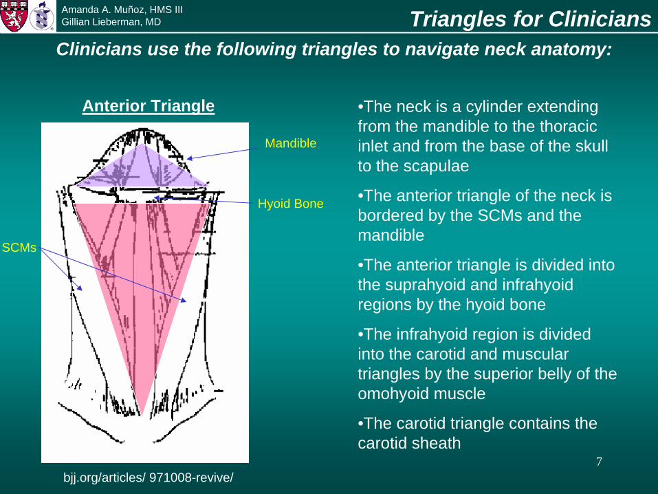
Clinicians use the following triangles to navigate neck anatomy:
Anterior Triangle
SCMs
Mandible
Hyoid Bone
•The neck is a cylinder extending from the mandible to the thoracic inlet and from the base of the skull to the scapulae
•The anterior triangle of the neck is bordered by the SCMs and the mandible
•The anterior triangle is divided into the suprahyoid and infrahyoid regions by the hyoid bone
•The infrahyoid region is divided into the carotid and muscular triangles by the superior belly of the omohyoid muscle
•The carotid triangle contains the carotid sheath
8
Amanda A. Muñoz, HMS IIIGillian Lieberman, MD
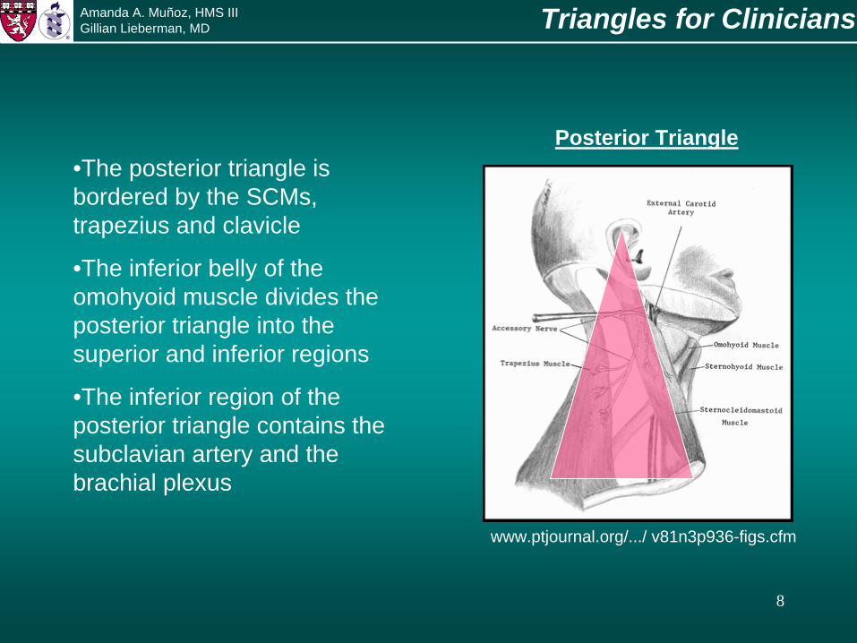
Posterior Triangle
Triangles for Clinicians
www.ptjournal.org/.../ v81n3p936-figs.cfm
•The posterior triangle is bordered by the SCMs, trapezius and clavicle
•The inferior belly of the omohyoid muscle divides the posterior triangle into the superior and inferior regions
•The inferior region of the posterior triangle contains the subclavian artery and the brachial plexus

9
Amanda A. Muñoz, HMS IIIGillian Lieberman, MD Spaces for Radiologists
Suprahyoid spaces• Submandibular –
below the mandible and floor of the mouth
• Visceral – containing the pharynx, larynx, trachea, glands
• Parapharyngeal – surrounding the pharynx
Radiologists use “spaces”, not triangles, to describe the neck
http://iris3.med.tufts.edu/headneck
•The spaces of the neck are defined by the cervical fascia
•The hyoid bone again divides the neck into two regions
Submandibular
Visceral
Parapharyngeal

10
Amanda A. Muñoz, HMS IIIGillian Lieberman, MD Spaces for Radiologists
Radiologists use “spaces”, not triangles, to describe the neck
Infrahyoid Spaces• Investing –
superficial, containing musculature
• Visceral – containing organs, larynx, pharynx, trachea, cartilages, glands
• Prevertebral – spinal column and surrounding musculature
http://iris3.med.tufts.edu/headneck
Investing
Visceral
Prevertebral
11
Amanda A. Muñoz, HMS IIIGillian Lieberman, MD Neck Masses: Types
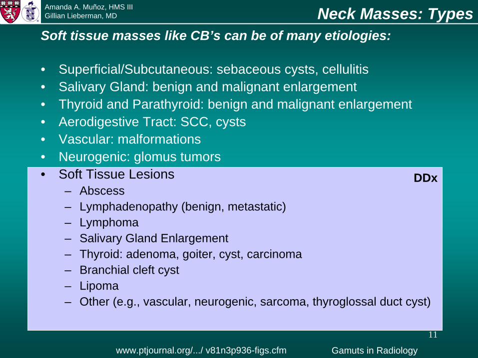
• Superficial/Subcutaneous: sebaceous cysts, cellulitis• Salivary Gland: benign and malignant enlargement• Thyroid and Parathyroid: benign and malignant enlargement• Aerodigestive Tract: SCC, cysts• Vascular: malformations• Neurogenic: glomus tumors• Soft Tissue Lesions
– Abscess– Lymphadenopathy (benign, metastatic)– Lymphoma– Salivary Gland Enlargement– Thyroid: adenoma, goiter, cyst, carcinoma– Branchial cleft cyst– Lipoma– Other (e.g., vascular, neurogenic, sarcoma, thyroglossal duct cyst)
Soft tissue masses like CB’s can be of many etiologies:
DDx
Gamuts in Radiologywww.ptjournal.org/.../ v81n3p936-figs.cfm

12
Amanda A. Muñoz, HMS IIIGillian Lieberman, MD
• Fat density• Fluid-fluid levels• Fascial planes• Size• Deep vs. superficial• Intramuscular• Necrosis• Hemorrhage• Margins
Neck Masses: Radiographic Characteristics
Eskey, Rad Clin N Am, p.1092
The following should be evaluated when imaging soft tissue neck masses:

13
Amanda A. Muñoz, HMS IIIGillian Lieberman, MD Some Examples from our differential….
• The following slides show examples of various types of neck masses
• Each mass should be evaluated according to the radiographic characteristics listed on the previous slide
• We will end with a look back at our patient CB’s CT of the neck to see if it shares any of the characteristics of the other examples of neck masses

14
Amanda A. Muñoz, HMS IIIGillian Lieberman, MD Abscess
http://rad.usuhs.mil/medpix/medpix.html?mode =geo_browse&table=card&expand=75
PACS, BIDMC
Normal CT Retropharyngeal Abscess
Abscess
•This mass is in the anterior triangle and the parapharyngeal space•It has no fat density, fluid-fluid levels, it is within a Fascial plane, of large Size, Deep, not intramuscular, does show evidence of Necrosis, no Hemorrhage•Difficult to see its margins because it is of similar density to the surrounding muscle

15
Amanda A. Muñoz, HMS IIIGillian Lieberman, MD Lymphadenopathy
PACS, BIDMC Courtesy Dr. Atif Zaheer
Necrotic Lymphadenitis
Lymph Node
Normal CT
•This mass is in the posterior triangle and the prevertebral space•It has no fat density, fluid-fluid levels, it is within a fascial plane, large Size, deep, not intramuscular, does show evidence of Necrosis with ? Extension into the temporalis, no Hemorrhage, Margins here appear visible

16
Amanda A. Muñoz, HMS IIIGillian Lieberman, MD Thyroid
Multi-Nodular Goiter
Thyroid Nodules
PACS, BIDMC PACS, BIDMC
Normal CT
•This mass is in the anterior triangle and the visceral space•It has no fat density, no fluid-fluid levels, it is within a fascial plane and limited to thyroid, enlarged Size, superficial, not Intramuscular, no Necrosis or Hemorrhage, good Margins. ? nodes. •well-enhancing due to vascular nature of gland, even w/o contrast due to Iodine content!

17
Amanda A. Muñoz, HMS IIIGillian Lieberman, MD Cystic
Courtesy Dr. Atif Zaheer
Branchial Cleft Cyst
Cyst
PACS, BIDMC
Normal CT
•This lesion is in the anterior triangle and the investing vs parapharyngeal space•It is well circumscribed, does not appear to have fat density, no fluid-fluid levels, is within a fascial plane, is superficial, large, and no evidence of necrosis or hemorrhage. HU = fluid.

18
Amanda A. Muñoz, HMS IIIGillian Lieberman, MD Lipomatous
www.aichi-gauin.ac.ip/.../NB122.htm
Submandibular Lipoma
Lipoma
PACS, BIDMC
Normal CT
•This lesion is in the Anterior triangle, investing space•Circumscribed, smooth, uniform attenutation. -65 to -125 HU, thin fibrous septa, does not enhance w/ IV contrast

19
Amanda A. Muñoz, HMS IIIGillian Lieberman, MD CB
Courtesy Dr. David Hackney
CB scan showed left thyroid enlargement and right-sided lymphadenopathy
Thyroid mass w/ tracheal compressionLymph nodes
•Anterior triangle, visceral space. Nodes are anterior and parapharyngeal.•left lobe enlargment to 48mm, appears hypodense for a thyroid though pt has not yet gotten contrast.•Mass is heterogenous with cyst formation on both sides, even though R is not as large as the left•R sided LAD (level 5), some with central hypodensity that may indicate necrosis, size and mutliplicity of nodes (also level 2 and 3)suggest malignancy•Displacement of trachea to L
20
Amanda A. Muñoz, HMS IIIGillian Lieberman, MD RCC and Thyroid Metastases
• Metastasis to thyroid gland is a very rare cause of thyroid enlargement• Vascular supply may result in entrapment of tumor emboli• Usually nodular, either single or multiple• Can mimic primary thyroid gland neoplasms or multinodular goiter• RCC is one of the most common types of cancer to metastasize to the
thyroid– Others include melanoma, breast, lung, colon– SCC local invasion
• When thyroid mets are the first clinical presentation, the primary is most often RCC
Hefess, Metastatic RCC
CT findings could be consistent with the following:
• Goiter with nodal mets• Primary thyroid malignancy w/ nodal mets• Metastatic disease involving both thyroid and nodes

21
Amanda A. Muñoz, HMS IIIGillian Lieberman, MD Summary
• CT scan is the modality of choice for tissue differentiation, staging and monitoring treatment of neck pathology
• Neck anatomy is divided into triangles or spaces• Neck masses may arise from many tissues of the
neck• Soft tissue neck masses have few specific
radiographic characteristics but many hints can point the radiologist toward a benign or malignant process
• CT may not be able to delineate the origin of a neck mass!

22
Amanda A. Muñoz, HMS IIIGillian Lieberman, MD References
• Branstetter, BF and Weissman JL. Normal Anatomy of the Neck with CT and MR Imaging Correlation. Radiologic Clinics of North America; Sept 2000 38:925-940.
• Eskey, CJ, Robson, CD, Weber AL. Imaging of Benign and Mailgnant Soft Tissue Tumors of the Neck. Radiologic Clinics of North America; Sept 200, 38:1091-1104.
• Gottlieb MD, Roland JT. Paradoxical spread of renal cell carcinoma to the head and neck. Laryngoscopy; Sept 1998: 1301-5.
• Hefess, CS, Wenig, BM, Thompson LD. Metastatic Renal Cell Carcinoma to the Thyroid Gland. Cancer; November 1 2002: 1869-1878
• Kazuhiro A, Hasegawa T, Onodera S, Oishi Y, Suzuki M. Renal Metastasis of Thyroid Carcinoma. International Journal of Urology; 2002 (9): 656-658.
• Larson SM, Robbins R. Positron emission tomography in thyroid cancer management. Seminars in Roentgenology; Apr 2002: 169-74.
• Lev S, Lev MH. Imaging of Cystic Lesions. Rad Clin N Am; Sept 2000; 38:1013-1029.• Pickhardt, PJ, Pickard RH. Sonography of delayed thyroid metastasis from renal cell
carcinoma with jugular vein extension. American Jounral or Roentgenology; Jul 2003: 272-4.• Sakai, O, et al. Lymph Node Pathology: Benign Proliferative, Lymphoma and Metastatic
Disease. Rad Clin N AM; Sept 2000; 38: 979-999.• Som, PM, et al. Head and Neck Imaging. Mosby; 1991: 498-669.• Weber, AL, Randolph G, Aksoy FG. The Thyroid and Parathyroid Glands: CT and MR Imaging
and Correlation with Pathology and Clinical Findings. Radiologic Clinics of North America; Sept 2000, 38:1105-1130.
• Weber, AL, Siciliano, A. CT and MR Imaging of Neck Infections with Clinical Correlations. Rad Clin of N Am; Sept 2000; 38:941-969.

23
Amanda A. Muñoz, HMS IIIGillian Lieberman, MD Acknowledgements
• My May 2004 Core Radiology Classmates• Pamela Lepkowski• Gillian Lieberman, MD• Dr. David Hackney• Dr. Michael Schuster• Dr. Atif Zaheer• Larry Barbaras our Webmaster• Michael Levinson