Embed Size (px)
Citation preview
Soft Tissue Techniques
Therese Obioha OMS V Sarah Watson OMSV
21 June 2013

Overview of Lab
Review of Screen, Scan, Segmental
Definition
Overview of Soft Tissue Techniques
Types of Soft Tissue Treatments
Practice Time
Use of Soft Tissue Treatment 2

Review of Screen, Scan, Segmental Definition
1. Screen◦ Is there a problem?◦ Look, listen, feel
2. Scan◦ Where is the problem?◦ One tissue scan + one confirmatory
motion scan Tiebreaker scan if needed
3. Segmental Definition◦ What is the problem?◦ Naming the dysfunction (in ease)
4. Diagnosis Treatment3

Screen...
Scan...
Segmental Definition...

Find Somatic Dysfunction on Your Partner
•Write down areas and remember the areas for later 5

Screen—Palpation
Axial Spine: Comparing Above with Below
Ask the question, “Is there a difference in tissue resistance?”
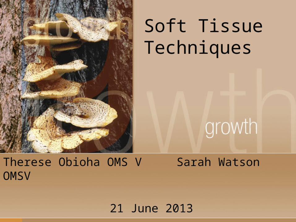
Screen—Gross Regional Motion
Cervical rotation Apply a slight
rotary force to the left/right
Cervical sidebending Tell patient to let
the head fall to the left/right

Screen—Gross Regional Motion
Thoracic rotation Apply a slight rotary
force to the left/right
Feel for that initial resistance to motion.
Thoracic sidebending Apply a slight
downward force using your body weight as leverage

Screen-Gross Regional Motion
Lumbar lordosis Patient *actively*
flexes (bends over) Lordosis should
flatten out Lumbar rotation
A notable prominence of left or right paraspinal tissues is abnormal
This test can be combined with the previous one.
Overview of Soft Tissue Treatments10

Overview of Soft Tissue Treatments: Causes of Dysfunction?
Irritation (somatic
dysfunction)
Psychogenic factors
overuse
trauma Infectious agents
Inactivity
Visceral Disease
11- Adapted from Dr. Eland

Soft Tissue – Circle of Irritation
Irritation
Pain
Increased muscle tension
Inflammation
Fibrous reaction
12
- Adapted from Dr. Eland
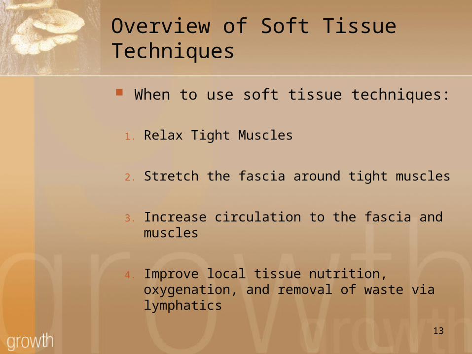
Overview of Soft Tissue Techniques
When to use soft tissue techniques:
1. Relax Tight Muscles
2. Stretch the fascia around tight muscles
3. Increase circulation to the fascia and muscles
4. Improve local tissue nutrition, oxygenation, and removal of waste via lymphatics 13
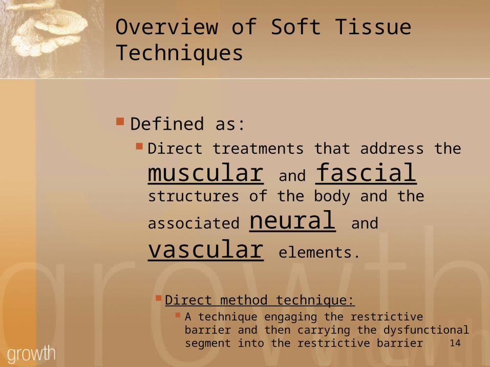
Overview of Soft Tissue Techniques
Defined as: Direct treatments that address the
muscular and fascial structures of the body and the
associated neural and
vascular elements.
Direct method technique: A technique engaging the restrictive barrier
and then carrying the dysfunctional segment into the restrictive barrier
14

Overview of Soft Tissue Techniques
Defined as:
Direct treatments that address the muscular and fascial structures of the
body and the associated neural and vascular elements.
Direct method technique:A technique to engage the restrictive barrier
(of a muscle or group of muscles)…and then focused on carrying the
dysfunctional segment TOWARDS the restrictive barrier
15
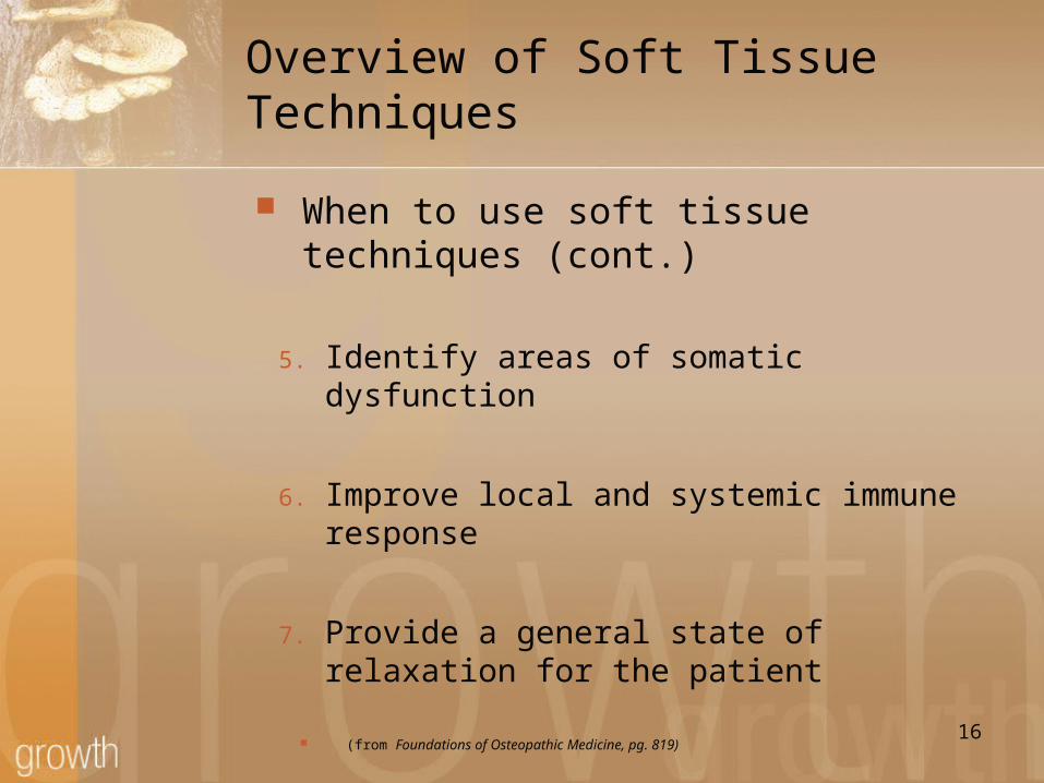
Overview of Soft Tissue Techniques
When to use soft tissue techniques (cont.)
5. Identify areas of somatic dysfunction
6. Improve local and systemic immune response
7. Provide a general state of relaxation for the patient
(from Foundations of Osteopathic Medicine, pg. 819) 16

Overview of Soft Tissue Techniques A Side Note!
Tissue Response and Treatment While you are doing these
techniques, you will feel changes in the tissues with your hands
Ex. Relaxation of muscles Increased temperature of tissue
(due to increased blood flow) A pulsation under your fingers
Related to Circulation and other natural body rhythms. 17

Types of Soft Tissue Treatment18
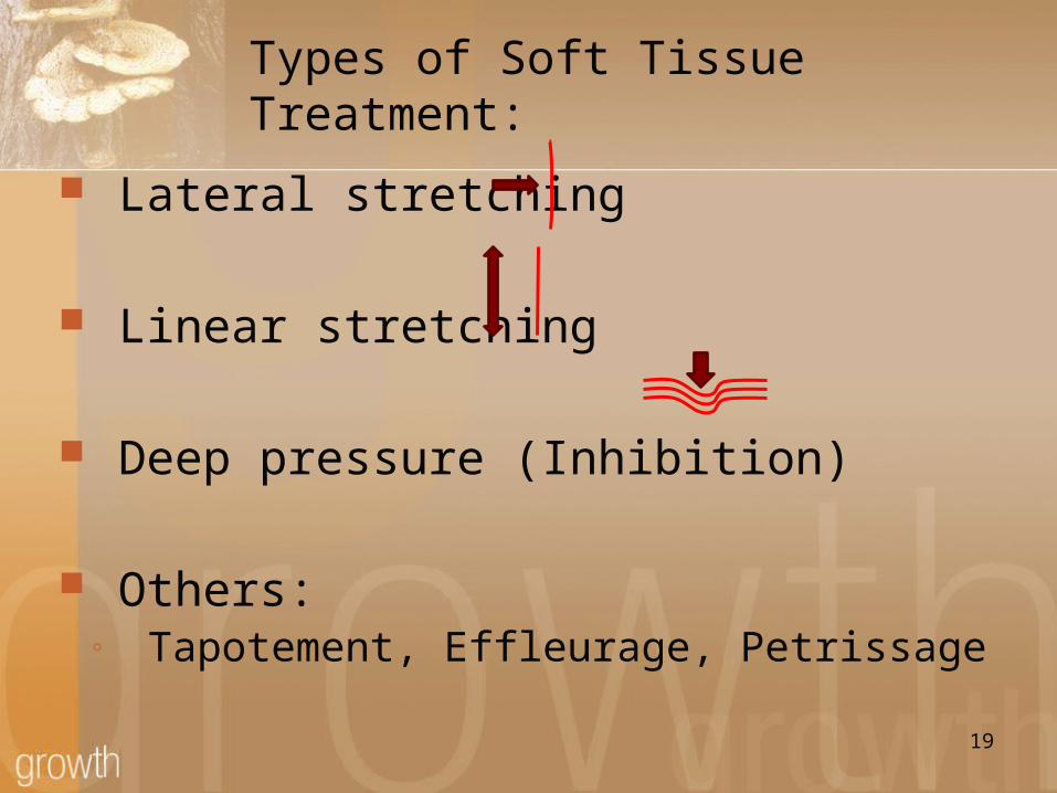
Types of Soft Tissue Treatment:
Lateral stretching
Linear stretching
Deep pressure (Inhibition)
Others:◦ Tapotement, Effleurage, Petrissage
19
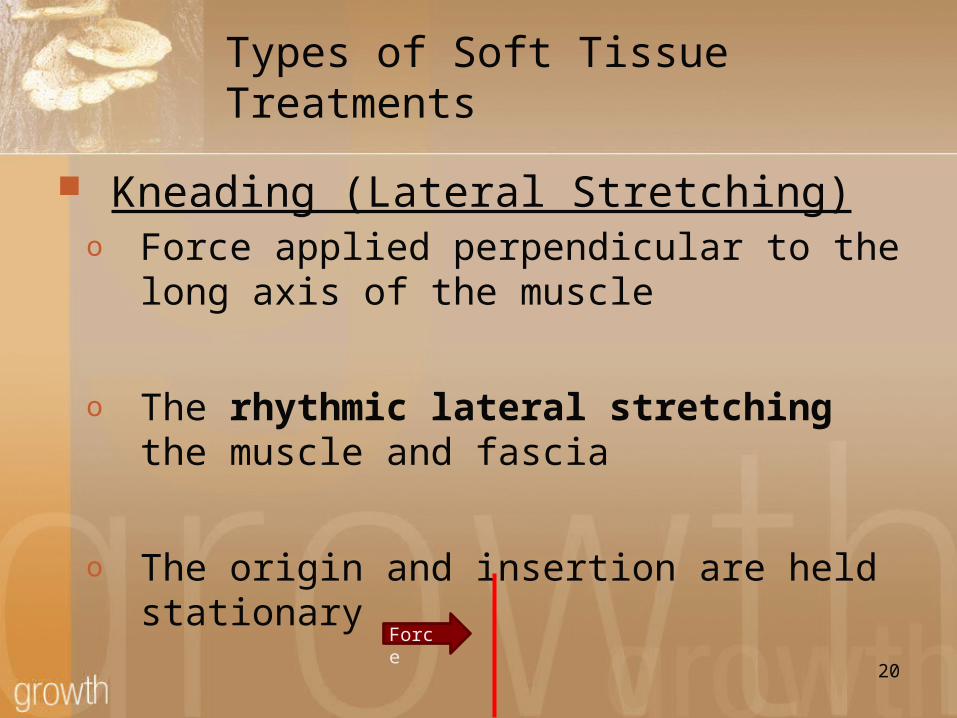
Types of Soft Tissue Treatments
Kneading (Lateral Stretching)o Force applied perpendicular to the long
axis of the muscle
o The rhythmic lateral stretching the muscle and fascia
o The origin and insertion are held stationary
20
Force
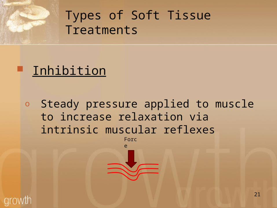
Types of Soft Tissue Treatments
Inhibition
o Steady pressure applied to muscle to increase relaxation via intrinsic muscular reflexes
21
Force

Types of Soft Tissue Treatments
Traction ( Linear Stretching)
o A linear force to draw structures apart
o The origin and insertion the muscle are pulled away from each
22

Types of Soft Tissue Treatments
Effleurage
o Light pressure to the skin to compress the underlying subcutaneous tissues
o This helps to move fluid along lymphatic channels
23

Types of Soft Tissue Treatments
Petrissage
o Deep kneading or squeezing action to remove swelling; more pressure than effleurage.
o This helps to break up adhesive bands from the skin to the deeper tissues
o Important to know WHERE you are directing this fluid
o Anatomy of lymph nodes and lymphatic flow24
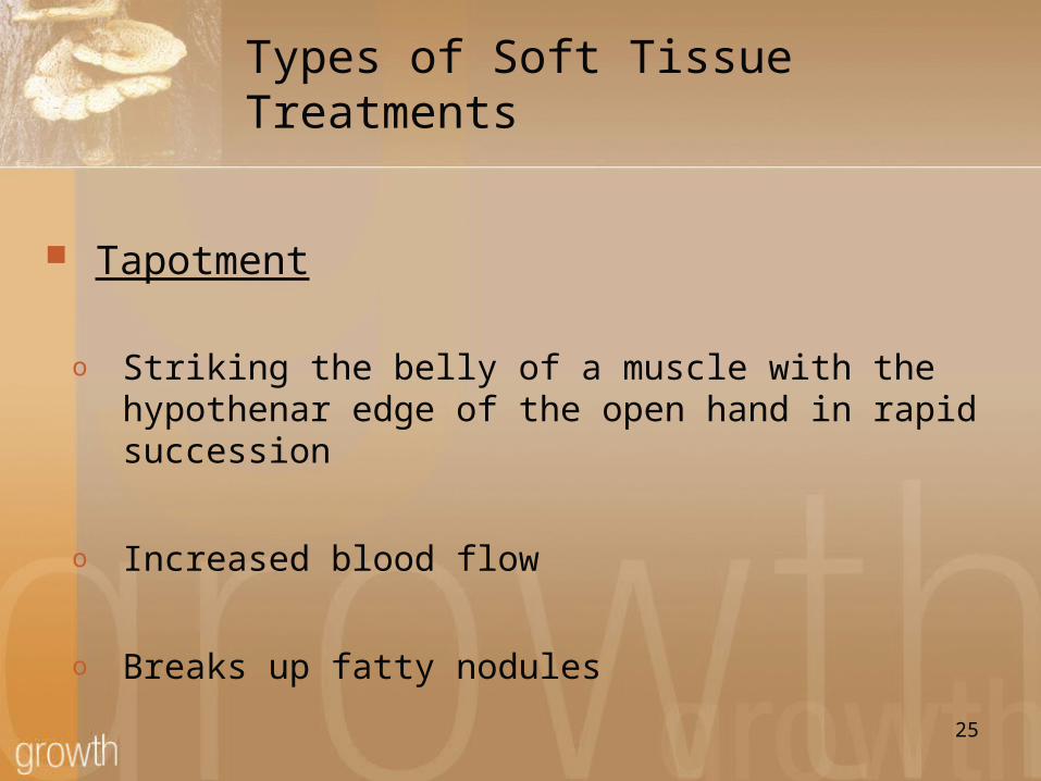
Types of Soft Tissue Treatments
Tapotment
o Striking the belly of a muscle with the hypothenar edge of the open hand in rapid succession
o Increased blood flow
o Breaks up fatty nodules25

Types of Soft Tissue Treatments
Deep Friction
o The application deep strokes of the thumb, knuckles, or elbow to small paraspinal muscles
o Helps to break up fibrotic changes in small paraspinal muscles
o Can also help to increase local circulation through release of local chemical mediators
26

Summary of Treatment Types
Most Commonly Used Kneading, Inhibition, Traction
To remove swelling Effleurage/Petrissage
To Increase Circulation Tapotement, Deep Friction
27
Time to Practice!28
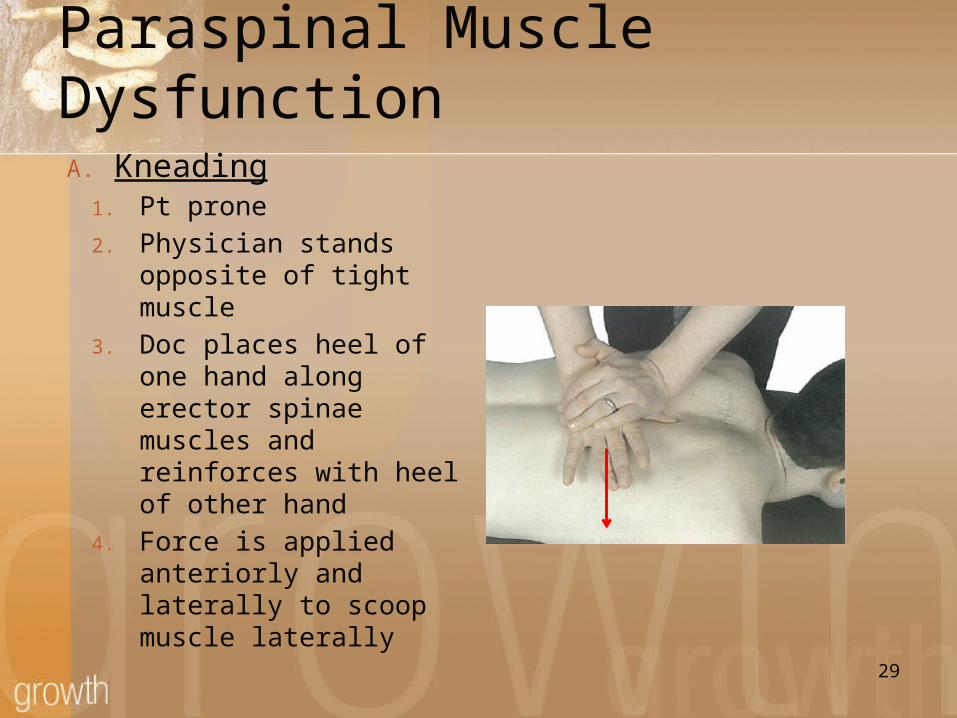
Paraspinal Muscle DysfunctionA. Kneading
1. Pt prone2. Physician stands opposite
of tight muscle3. Doc places heel of one
hand along erector spinae muscles and reinforces with heel of other hand
4. Force is applied anteriorly and laterally to scoop muscle laterally
29
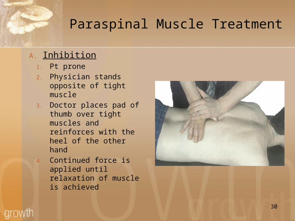
Paraspinal Muscle Treatment
A. Inhibition1. Pt prone2. Physician stands
opposite of tight muscle
3. Doctor places pad of thumb over tight muscles and reinforces with the heel of the other hand
4. Continued force is applied until relaxation of muscle is achieved
30
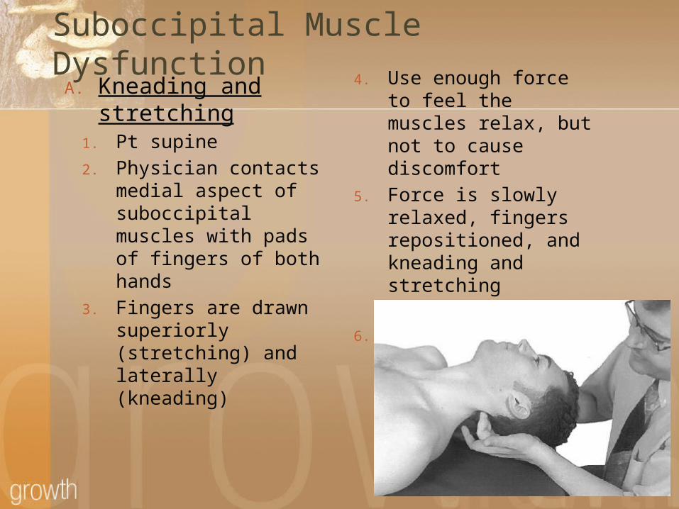
Suboccipital Muscle DysfunctionA. Kneading and
stretching1. Pt supine2. Physician contacts
medial aspect of suboccipital muscles with pads of fingers of both hands
3. Fingers are drawn superiorly (stretching) and laterally (kneading)
4. Use enough force to feel the muscles relax, but not to cause discomfort
5. Force is slowly relaxed, fingers repositioned, and kneading and stretching repeated
6. Recheck
31
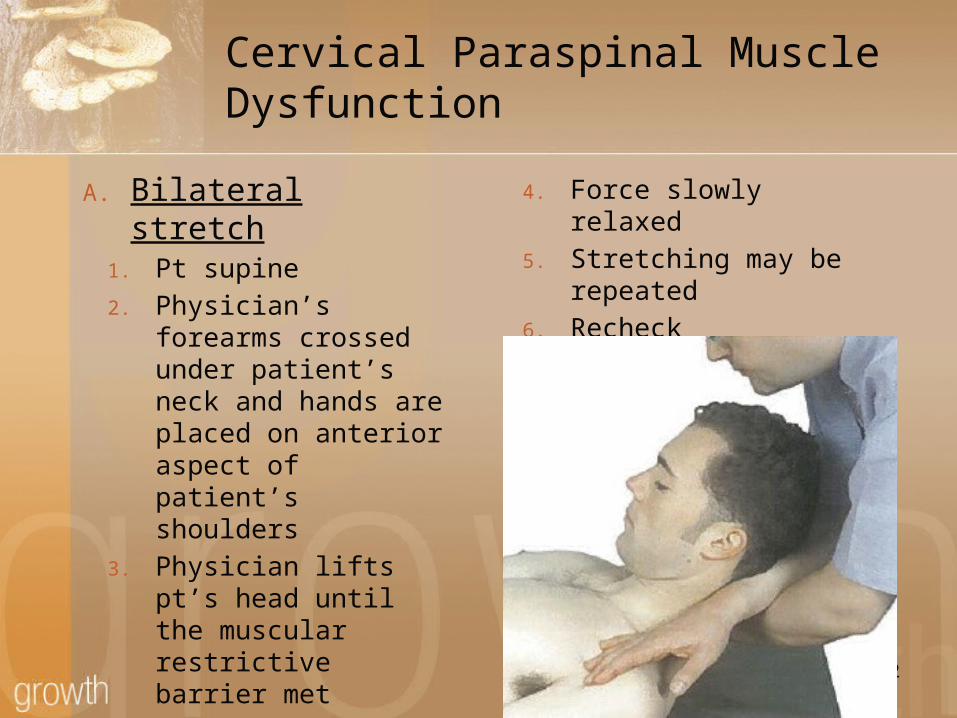
Cervical Paraspinal Muscle Dysfunction
A. Bilateral stretch1. Pt supine2. Physician’s forearms
crossed under patient’s neck and hands are placed on anterior aspect of patient’s shoulders
3. Physician lifts pt’s head until the muscular restrictive barrier met
4. Force slowly relaxed5. Stretching may be
repeated6. Recheck
32
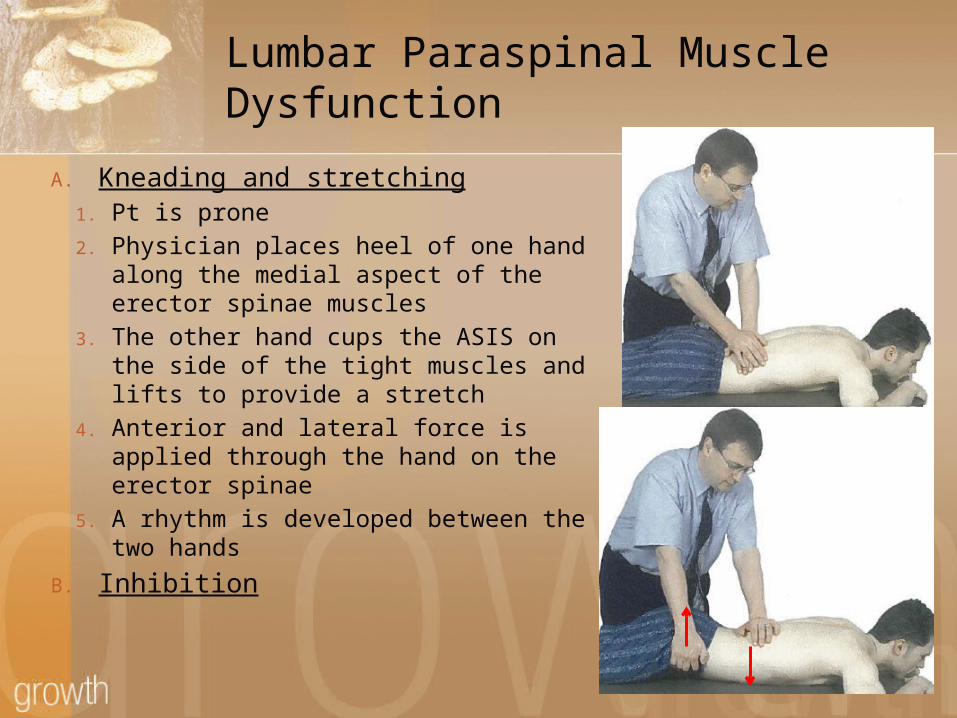
Lumbar Paraspinal Muscle Dysfunction
A. Kneading and stretching1. Pt is prone2. Physician places heel of one hand
along the medial aspect of the erector spinae muscles
3. The other hand cups the ASIS on the side of the tight muscles and lifts to provide a stretch
4. Anterior and lateral force is applied through the hand on the erector spinae
5. A rhythm is developed between the two hands
B. Inhibition
33

Upper Extremity Lymphatic CongestionA. Effleurage
1. Pt supine2. Physician lifts patient’s
arm vertically and grasps patient’s fingertip with one hand
3. Physician applies enough pressure to compress skin against the deep fascia and strokes the lateral side of finger from distal to proximal
4. Physician then applies enough pressure around the arm to compress skin against the deep fascia and strokes the arm from distal to proximal at various points on the circumference until the entire arm has been treated
34

Conclusion – Student Response
Types of soft tissue treatments
When to use soft tissue
Why would we use soft tissue treatments
35
(Some examples of) Clinical Application of Soft Tissue
Techniques
1. Ankle Sprain2. Low Back Pain3. Overuse
1. Acute2. Chronic
4. Tension Headache5. Other?
Next Lab!
Putting all the pieces together
OMM Applications for Headaches Dx, Tx, and Follow-Up Care…
37

Resources
DiGiovanna E, Shiowitz S, Dowling D. An Osteopathic Approach to Diagnosis and Treatment. 3rd ed. 2005: Philadelphia, PA. Lippincott Williams & Wilkins: 80-83
Ward, et al. Foundations of Osteopathic Medicine. 2nd ed. :glossary
Kimberly PE. Outline of Osteopathic Manipulative Procedure: The Kimberly Manual. Millenium ed. 2000; Marceline, MO. Walsworth Publishing Co: 37-59
38