Embed Size (px)
Citation preview
Soft Tissue Sarcoma Clinical Pathway
*Approved by the Sarcoma Specialty Treatment Team November 2017
The following pathway was developed through multidisciplinary efforts with physicians from the Mary Bird Perkins – Our Lady of the Lake Cancer Center. These pathways should be used as a supplemental guide for treatment for physicians at the Mary Bird Perkins –Our Lady of the Lake Cancer Center, and are not intended to replace the independent medical or professional judgment of physicians or other health care providers
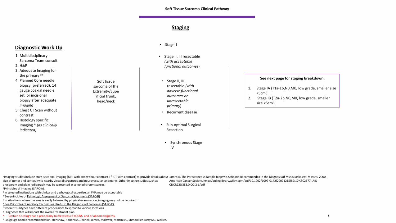
1. Multidisciplinary Sarcoma Team consult
2. H&P3. Adequate Imaging for
the primary ab
4. Planned Core needle biopsy (preferred), 14 gauge coaxial needle set or incisional biopsy after adequate imaging
5. Chest CT Scan without contrast
6. Histology specific Imaging * (as clinically indicated)
Diagnostic Work Up
Soft Tissue Sarcoma Clinical Pathway
Soft tissue sarcoma of the Extremity/Supe
rficial trunk, head/neck
• Stage 1
• Stage II, III resectable (with acceptable functional outcomes)
• Stage II, III resectable (with adverse functional outcomes or unresectable primary)
• Synchronous Stage IV
• Recurrent disease
Staging
1
aImaging studies include cross-sectional imaging (MRI with and without contrast +/- CT with contrast) to provide details about size of tumor and contiguity to nearby visceral structures and neurovascular landmarks. Other imaging studies such as angiogram and plain radiograph may be warranted in selected circumstances. bPrinciples of Imaging (SARC-A). c In selected institutions with clinical and pathological expertise, an FNA may be acceptabled See principles of Pathologic Assessment of Sarcoma Specimens (SARC-B) p In situations where the area is easily followed by physical examination, imaging may not be required.e See Principles of Ancillary Techniques Useful in the Diagnosis of Sarcomas (SARC-C) fDifferent subtypes have different propensities to spread to various locations. g Diagnoses that will impact the overall treatment plan• Certain histology has a propensity to metastasize to CNS and or abdomen/pelvis. * 14 gauge needle recommendation. Henshaw, Robert M., Jelinek, James, Malawer, Martin M., Shmookler Barry M., Welker,
James A. The Percutaneous Needle Biopsy is Safe and Recommended in the Diagnosis of Musculoskeletal Masses. 2000. American Cancer Society. http://onlinelibrary.wiley.com/doi/10.1002/1097-0142(20001215)89:12%3C2677::AID-CNCR22%3E3.0.CO;2-L/pdf
• Sub-optimal Surgical Resection
See next page for staging breakdown:
1. Stage IA (T1a-1b,N0,M0, low grade, smaller size <5cmj)
2. Stage IB (T2a-2b,N0,M0, low grade, smaller size <5cmj)
Soft Tissue Sarcoma Clinical Pathway
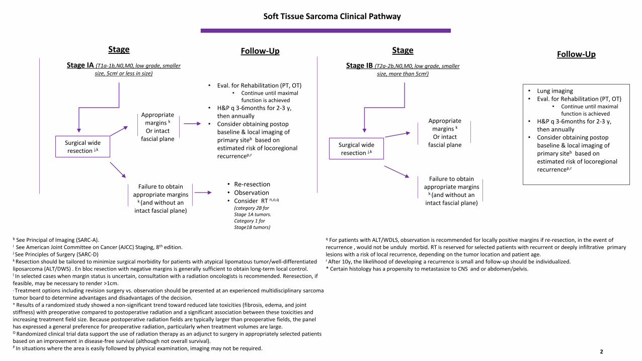
Surgical wide resection j,k
Appropriate margins k
Or intact fascial plane
Failure to obtain appropriate margins
k (and without an intact fascial plane)
• Eval. for Rehabilitation (PT, OT)• Continue until maximal
function is achieved
• H&P q 3-6months for 2-3 y, then annually
• Consider obtaining postop baseline & local imaging of primary siteb based on estimated risk of locoregional recurrencep,r
• Re-resection• Observation• Consider RT n,o,q
(category 2B for Stage 1A tumors. Category 1 for Stage1B tumors)
Stage IA (T1a-1b,N0,M0, low grade, smaller
size, 5cmj or less in size)
2
Follow-Up
b See Principal of Imaging (SARC-A). i See American Joint Committee on Cancer (AJCC) Staging, 8th edition. J See Principles of Surgery (SARC-D) k Resection should be tailored to minimize surgical morbidity for patients with atypical lipomatous tumor/well-differentiated liposarcoma (ALT/DWS) . En bloc resection with negative margins is generally sufficient to obtain long-term local control. l In selected cases when margin status is uncertain, consultation with a radiation oncologists is recommended. Reresection, if feasible, may be necessary to render >1cm. , Treatment options including revision surgery vs. observation should be presented at an experienced multidisciplinary sarcoma tumor board to determine advantages and disadvantages of the decision. n Results of a randomized study showed a non-significant trend toward reduced late toxicities (fibrosis, edema, and joint stiffness) with preoperative compared to postoperative radiation and a significant association between these toxicities and increasing treatment field size. Because postoperative radiation fields are typically larger than preoperative fields, the panelhas expressed a general preference for preoperative radiation, particularly when treatment volumes are large. O Randomized clinical trial data support the use of radiation therapy as an adjunct to surgery in appropriately selected patients based on an improvement in disease-free survival (although not overall survival). P In situations where the area is easily followed by physical examination, imaging may not be required.
q For patients with ALT/WDLS, observation is recommended for locally positive margins if re-resection, in the event of recurrence , would not be unduly morbid. RT is reserved for selected patients with recurrent or deeply infiltrative primarylesions with a risk of local recurrence, depending on the tumor location and patient age. r After 10y, the likelihood of developing a recurrence is small and follow-up should be individualized. * Certain histology has a propensity to metastasize to CNS and or abdomen/pelvis.
Stage IB (T2a-2b,N0,M0, low grade, smaller
size, more than 5cmj)
Appropriate margins k
Or intact fascial plane
Failure to obtain appropriate margins
k (and without an intact fascial plane)
• Lung imaging• Eval. for Rehabilitation (PT, OT)
• Continue until maximal function is achieved
• H&P q 3-6months for 2-3 y, then annually
• Consider obtaining postop baseline & local imaging of primary siteb based on estimated risk of locoregional recurrencep,r
Follow-Up Stage Stage
Surgical wide resection j,k
Primary Treatment
Soft Tissue Sarcoma Clinical Pathway
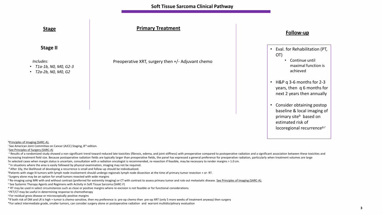
Stage II
Includes: • T1a-1b, N0, M0, G2-3 • T2a-2b, N0, M0, G2
Preoperative XRT, surgery then +/- Adjuvant chemo
Follow-up
3
bPrinciples of Imaging (SARC-A). i See American Joint Committee on Cancer (AJCC) Staging, 8th edition. j See Principles of Surgery (SARC-A) n Results of a randomized study showed a non-significant trend toward reduced late toxicities (fibrosis, edema, and joint stiffness) with preoperative compared to postoperative radiation and a significant association between these toxicities and increasing treatment field size. Because postoperative radiation fields are typically larger than preoperative fields, the panel has expressed a general preference for preoperative radiation, particularly when treatment volumes are largelIn selected cases when margin status is uncertain, consultation with a radiation oncologist is recommended, re-resection if feasible, may be necessary to render margins > 1.0 cm. P In situations where the area is easily followed by physical examination, imaging may not be required.R After 10y, the likelihood of developing a recurrence is small and follow-up should be individualized.
SPatients with stage III tumors with lymph node involvement should undergo regionals lymph node dissection at the time of primary tumor resection + or- RT. t Surgery alone may be an option for small tumors resected with wide marginsU Re-imaging using MRI with and without contrast (preferred for extremity imaging) or CT with contrast to assess primary tumor and rule out metastatic disease. See Principles of Imaging (SARC-A). v See Systemic Therapy Agents and Regimens with Activity in Soft Tissue Sarcoma (SARC-F) w RT may be used in select circumstances such as close or positive margins where re-excision is not feasible or for functional considerations. x PET/CT may be useful in determining response to chemotherapyy For residual gross disease or microscopically positive margins *If both risk of DM and LR is high + tumor is chemo-sensitive, then my preference is: pre-op chemo then pre-op XRT (only 5 more weeks of treatment anyway) then surgery*For select intermediate grade, smaller tumors, can consider surgery alone or postoperative radiation and warrant multidisciplinary evaluation
Stage
• Eval. for Rehabilitation (PT, OT)
• Continue until maximal function is achieved
• H&P q 3-6 months for 2-3 years, then q 6 months for next 2 years then annually
• Consider obtaining postop baseline & local imaging of primary siteb based on estimated risk of locoregional recurrencep,r
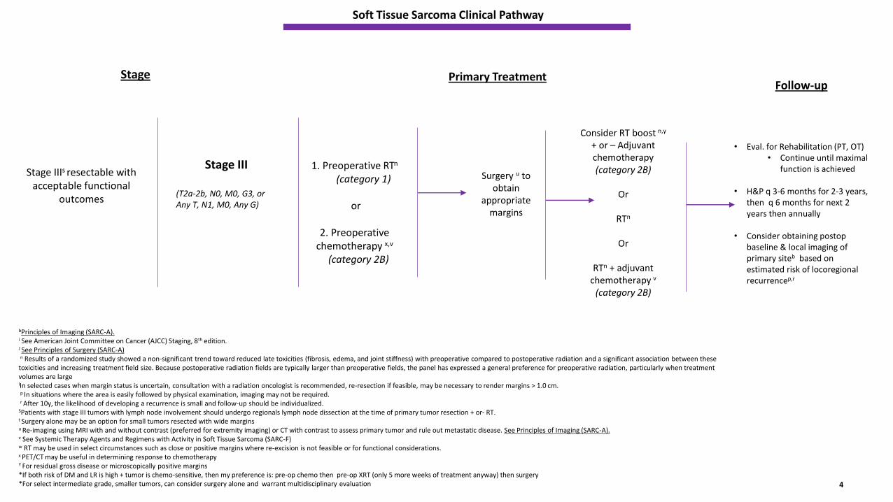
Stage IIIs resectable with acceptable functional
outcomes
Stage
Soft Tissue Sarcoma Clinical Pathway
Stage III
(T2a-2b, N0, M0, G3, or Any T, N1, M0, Any G)
1. Preoperative RTn
(category 1)
or
2. Preoperative chemotherapy x,v
(category 2B)
Surgery u to obtain
appropriate margins
4
Primary Treatment
bPrinciples of Imaging (SARC-A). i See American Joint Committee on Cancer (AJCC) Staging, 8th edition. J See Principles of Surgery (SARC-A) n Results of a randomized study showed a non-significant trend toward reduced late toxicities (fibrosis, edema, and joint stiffness) with preoperative compared to postoperative radiation and a significant association between these toxicities and increasing treatment field size. Because postoperative radiation fields are typically larger than preoperative fields, the panel has expressed a general preference for preoperative radiation, particularly when treatment volumes are largelIn selected cases when margin status is uncertain, consultation with a radiation oncologist is recommended, re-resection if feasible, may be necessary to render margins > 1.0 cm. p In situations where the area is easily followed by physical examination, imaging may not be required.r After 10y, the likelihood of developing a recurrence is small and follow-up should be individualized.
SPatients with stage III tumors with lymph node involvement should undergo regionals lymph node dissection at the time of primary tumor resection + or- RT. t Surgery alone may be an option for small tumors resected with wide marginsu Re-imaging using MRI with and without contrast (preferred for extremity imaging) or CT with contrast to assess primary tumor and rule out metastatic disease. See Principles of Imaging (SARC-A). v See Systemic Therapy Agents and Regimens with Activity in Soft Tissue Sarcoma (SARC-F) w RT may be used in select circumstances such as close or positive margins where re-excision is not feasible or for functional considerations. x PET/CT may be useful in determining response to chemotherapyY For residual gross disease or microscopically positive margins *If both risk of DM and LR is high + tumor is chemo-sensitive, then my preference is: pre-op chemo then pre-op XRT (only 5 more weeks of treatment anyway) then surgery*For select intermediate grade, smaller tumors, can consider surgery alone and warrant multidisciplinary evaluation
Follow-up
• Eval. for Rehabilitation (PT, OT)• Continue until maximal
function is achieved
• H&P q 3-6 months for 2-3 years, then q 6 months for next 2 years then annually
• Consider obtaining postop baseline & local imaging of primary siteb based on estimated risk of locoregional recurrencep,r
Consider RT boost n,y
+ or – Adjuvant chemotherapy (category 2B)
Or
RTn
Or
RTn + adjuvant chemotherapy v
(category 2B)
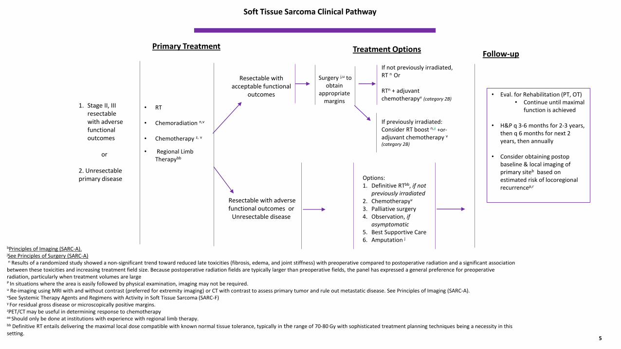
1. Stage II, III resectable with adverse functional outcomes
or
2. Unresectable primary disease
Primary Treatment
Soft Tissue Sarcoma Clinical Pathway
• RT
• Chemoradiation n,v
• Chemotherapy z, v
• Regional Limb Therapybb
Resectable with acceptable functional
outcomes
Resectable with adverse functional outcomes or
Unresectable disease
Follow-up
Options: 1. Definitive RTbb, if not
previously irradiated2. Chemotherapyv
3. Palliative surgery4. Observation, if
asymptomatic 5. Best Supportive Care 6. Amputation j
Treatment Options
bPrinciples of Imaging (SARC-A).jSee Principles of Surgery (SARC-A) n Results of a randomized study showed a non-significant trend toward reduced late toxicities (fibrosis, edema, and joint stiffness) with preoperative compared to postoperative radiation and a significant association
between these toxicities and increasing treatment field size. Because postoperative radiation fields are typically larger than preoperative fields, the panel has expressed a general preference for preoperative radiation, particularly when treatment volumes are large P In situations where the area is easily followed by physical examination, imaging may not be required. u Re-imaging using MRI with and without contrast (preferred for extremity imaging) or CT with contrast to assess primary tumor and rule out metastatic disease. See Principles of Imaging (SARC-A).vSee Systemic Therapy Agents and Regimens with Activity in Soft Tissue Sarcoma (SARC-F) y For residual gross disease or microscopically positive margins. zPET/CT may be useful in determining response to chemotherapyaa Should only be done at institutions with experience with regional limb therapy.bb Definitive RT entails delivering the maximal local dose compatible with known normal tissue tolerance, typically in the range of 70-80 Gy with sophisticated treatment planning techniques being a necessity in this setting.
Surgery j,u to obtain
appropriate margins
If not previously irradiated, RT n Or
RTn + adjuvant chemotherapyv (category 2B)
If previously irradiated: Consider RT boost n,y +or-
adjuvant chemotherapy v
(category 2B)
5
• Eval. for Rehabilitation (PT, OT)• Continue until maximal
function is achieved
• H&P q 3-6 months for 2-3 years, then q 6 months for next 2 years, then annually
• Consider obtaining postop baseline & local imaging of primary siteb based on estimated risk of locoregional recurrencep,r
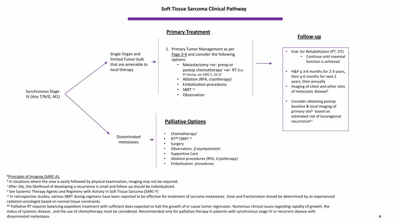
Synchronous Stage IV (Any T/N/G, M1)
Primary Treatment
Soft Tissue Sarcoma Clinical Pathway
Single Organ and limited Tumor bulk that are amenable to local therapy
• Chemotherapyv
• RTdd /SBRT cc
• Surgery• Observation, if asymptomatic• Supportive Care• Ablation procedures (RFA, Cryotherapy) • Embolization procedures
Follow-up
1. Primary Tumor Management as per Page 3-4 and consider the following options:
• Metastectomy +or- preop or postop chemotherapyv +or- RT (For
RT dosing, see SARC E, 2of 2)
• Ablation (RFA, cryotherapy)• Embolization procedures • SBRT cc
• Observation
Palliative Options
Disseminated metastases
bPrinciples of Imaging (SARC-A). P In situations where the area is easily followed by physical examination, imaging may not be required. r After 10y, the likelihood of developing a recurrence is small and follow-up should be individualized. v See Systemic Therapy Agents and Regimens with Activity in Soft Tissue Sarcoma (SARC-F) cc In retrospective studies, various SBRT dosing regimens have been reported to be effective for treatment of sarcoma metastases. Dose and fractionation should be determined by an experienced radiation oncologist based on normal tissue constraints. dd Palliative RT requires balancing expedient treatment with sufficient does expected to halt the growth of or cause tumor regression. Numerous clinical issues regarding rapidity of growth, the status of systemic disease , and the use of chemotherapy must be considered. Recommended only for palliative therapy in patients with synchronous stage IV or recurrent disease with disseminated metastases. 6
• Eval. for Rehabilitation (PT, OT)• Continue until maximal
function is achieved
• H&P q 3-6 months for 2-3 years, then q 6 months for next 2 years, then annually
• Imaging of chest and other sites of metastatic diseaseb
• Consider obtaining postop baseline & local imaging of primary siteb based on estimated risk of locoregional recurrencep,r
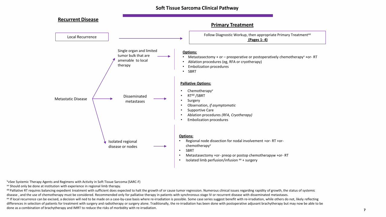
Metastatic Disease
Primary Treatment
Soft Tissue Sarcoma Clinical Pathway
Isolated regional disease or nodes
• Chemotherapyv
• RTdd /SBRT• Surgery• Observation, if asymptomatic• Supportive Care• Ablation procedures (RFA, Cryotherapy) • Embolization procedures
Recurrent Disease
Options: • Regional node dissection for nodal involvement +or- RT +or-
chemotherapyv
• SBRT• Metastasectomy +or- preop or postop chemotherapyw +or- RT• Isolated limb perfusion/infusion aa + surgery
Palliative Options:
Disseminated metastases
Local Recurrence Follow Diagnostic Workup, then appropriate Primary Treatmentee
(Pages 1- 4)
VvSee Systemic Therapy Agents and Regimens with Activity in Soft Tissue Sarcoma (SARC-F) aa Should only be done at institution with experience in regional limb therapy. dd Palliative RT requires balancing expedient treatment with sufficient does expected to halt the growth of or cause tumor regression. Numerous clinical issues regarding rapidity of growth, the status of systemic disease , and the use of chemotherapy must be considered. Recommended only for palliative therapy in patients with synchronous stage IV or recurrent disease with disseminated metastases. ee If local recurrence can be excised, a decision will ned to be made on a case-by-case basis where re-irradiation is possible. Some case series suggest benefit with re-irradiation, while others do not, likely reflecting differences in selection of patients for treatment with surgery and radiotherapy or surgery alone. Traditionally, the re-irradiation has been done with postoperative adjuvant brachytherapy but may now be able to be done as a combination of brachytherapy and IMRT to reduce the risks of morbidity with re-irradiation.
Single organ and limited tumor bulk that are amenable to local therapy
Options:• Metastasectomy + or – preoperative or postoperatively chemotherapyv +or- RT• Ablation procedures (eg, RFA or cryotherapy) • Embolization procedures• SBRT
7
Soft Tissue Sarcoma Clinical Pathway
Retroperitoneal/ Intra-Abdominal
Soft Tissue Sarcoma Clinical Pathway Retroperitoneal/ Intra-Abdominal
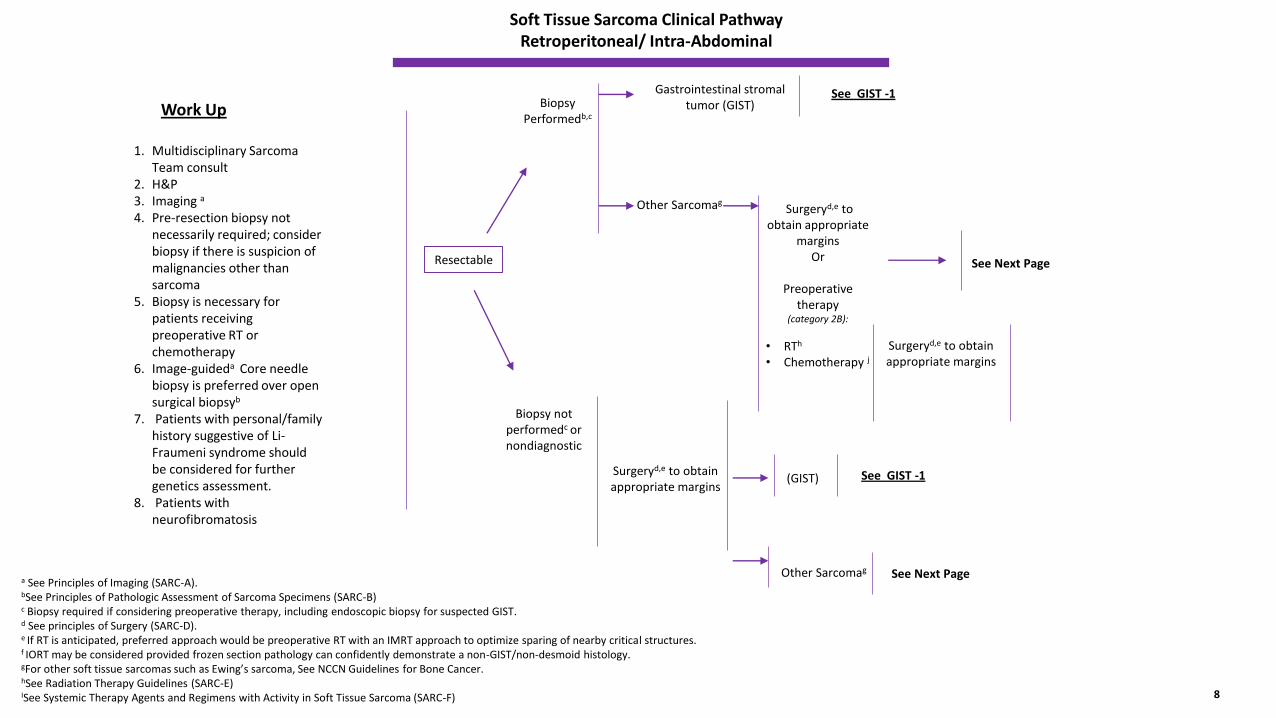
Work Up
a See Principles of Imaging (SARC-A). bSee Principles of Pathologic Assessment of Sarcoma Specimens (SARC-B)c Biopsy required if considering preoperative therapy, including endoscopic biopsy for suspected GIST.d See principles of Surgery (SARC-D). e If RT is anticipated, preferred approach would be preoperative RT with an IMRT approach to optimize sparing of nearby critical structures.f IORT may be considered provided frozen section pathology can confidently demonstrate a non-GIST/non-desmoid histology.gFor other soft tissue sarcomas such as Ewing’s sarcoma, See NCCN Guidelines for Bone Cancer.hSee Radiation Therapy Guidelines (SARC-E)ISee Systemic Therapy Agents and Regimens with Activity in Soft Tissue Sarcoma (SARC-F)
Resectable
1. Multidisciplinary Sarcoma Team consult
2. H&P3. Imaging a
4. Pre-resection biopsy not necessarily required; consider biopsy if there is suspicion of malignancies other than sarcoma
5. Biopsy is necessary for patients receiving preoperative RT or chemotherapy
6. Image-guideda Core needle biopsy is preferred over open surgical biopsyb
7. Patients with personal/family history suggestive of Li-Fraumeni syndrome should be considered for further genetics assessment.
8. Patients with neurofibromatosis
Biopsy not performedc or nondiagnostic
Biopsy Performedb,c
Other Sarcomag
Gastrointestinal stromal tumor (GIST)
Surgeryd,e to obtain appropriate
marginsOr
Preoperative therapy
(category 2B):
• RTh
• Chemotherapy j
Surgeryd,e to obtain appropriate margins
See Next Page
See GIST -1
Surgeryd,e to obtain appropriate margins
(GIST)
Other Sarcomag
See GIST -1
See Next Page
8
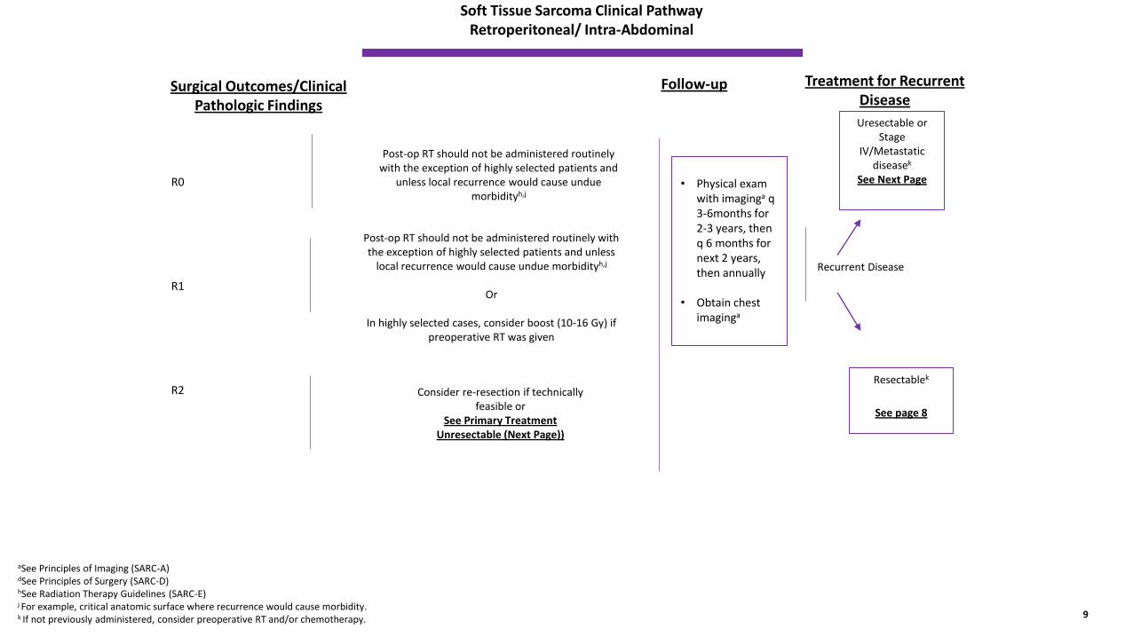
Soft Tissue Sarcoma Clinical Pathway Retroperitoneal/ Intra-Abdominal
Surgical Outcomes/Clinical Pathologic Findings
Consider re-resection if technically feasible or
See Primary Treatment Unresectable (Next Page))
aSee Principles of Imaging (SARC-A) dSee Principles of Surgery (SARC-D) hSee Radiation Therapy Guidelines (SARC-E)j For example, critical anatomic surface where recurrence would cause morbidity.k If not previously administered, consider preoperative RT and/or chemotherapy.
R0
R1
R2
Post-op RT should not be administered routinely with the exception of highly selected patients and
unless local recurrence would cause undue morbidityh,j
Recurrent Disease
Post-op RT should not be administered routinely with the exception of highly selected patients and unless
local recurrence would cause undue morbidityh,j
Or
In highly selected cases, consider boost (10-16 Gy) if preoperative RT was given
Follow-up
Uresectable or Stage
IV/Metastatic diseasek
See Next Page
Resectablek
See page 8
Treatment for Recurrent Disease
9
• Physical exam with imaginga q 3-6months for 2-3 years, then q 6 months for next 2 years, then annually
• Obtain chest imaginga
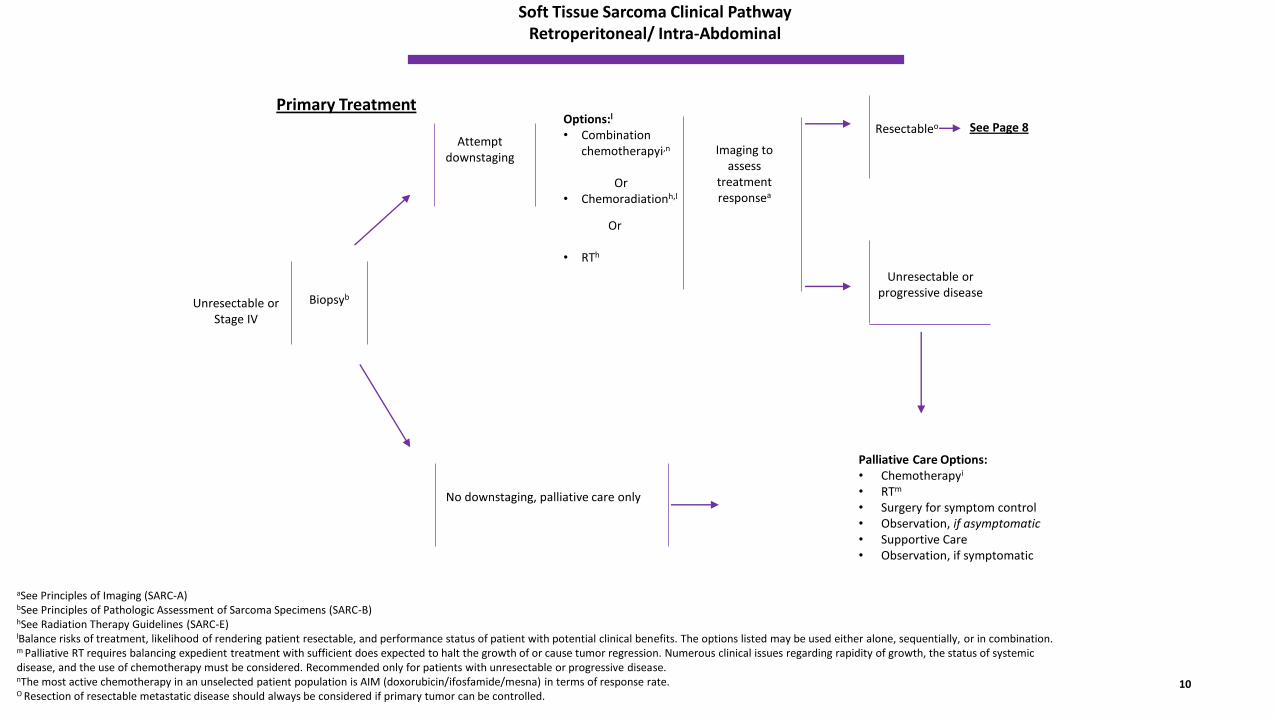
Soft Tissue Sarcoma Clinical Pathway Retroperitoneal/ Intra-Abdominal
Primary Treatment
No downstaging, palliative care only
aSee Principles of Imaging (SARC-A) bSee Principles of Pathologic Assessment of Sarcoma Specimens (SARC-B)hSee Radiation Therapy Guidelines (SARC-E) lBalance risks of treatment, likelihood of rendering patient resectable, and performance status of patient with potential clinical benefits. The options listed may be used either alone, sequentially, or in combination.m Palliative RT requires balancing expedient treatment with sufficient does expected to halt the growth of or cause tumor regression. Numerous clinical issues regarding rapidity of growth, the status of systemic disease, and the use of chemotherapy must be considered. Recommended only for patients with unresectable or progressive disease.nThe most active chemotherapy in an unselected patient population is AIM (doxorubicin/ifosfamide/mesna) in terms of response rate. O Resection of resectable metastatic disease should always be considered if primary tumor can be controlled.
Unresectable or Stage IV
Resectableo
Attempt downstaging
Imaging to assess
treatment responsea
Options:l
• Combination chemotherapyi,n
Or• Chemoradiationh,l
Or
• RTh
Biopsyb
Palliative Care Options: • Chemotherapyi
• RTm
• Surgery for symptom control• Observation, if asymptomatic• Supportive Care• Observation, if symptomatic
Unresectable or progressive disease
See Page 8
10
Soft Tissue Sarcoma Clinical Pathway
Gastrointestinal Stromal Tumors (GIST)
Soft Tissue Sarcoma Clinical Pathway GIST
Work Up At Primary Presentation
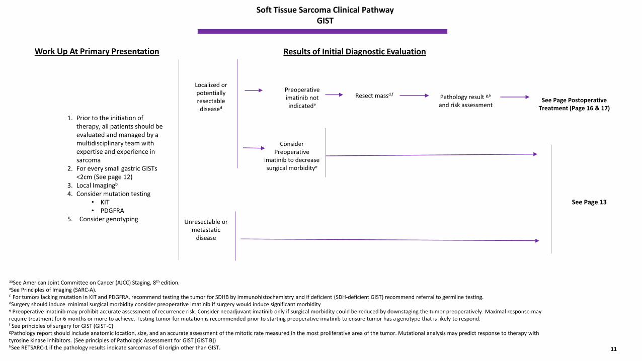
aaSee American Joint Committee on Cancer (AJCC) Staging, 8th edition. aSee Principles of Imaging (SARC-A). C For tumors lacking mutation in KIT and PDGFRA, recommend testing the tumor for SDHB by immunohistochemistry and if deficient (SDH-deficient GIST) recommend referral to germline testing. dSurgery should induce minimal surgical morbidity consider preoperative imatinib if surgery would induce significant morbidity e Preoperative imatinib may prohibit accurate assessment of recurrence risk. Consider neoadjuvant imatinib only if surgical morbidity could be reduced by downstaging the tumor preoperatively. Maximal response may require treatment for 6 months or more to achieve. Testing tumor for mutation is recommended prior to starting preoperative imatinib to ensure tumor has a genotype that is likely to respond. f See principles of surgery for GIST (GIST-C)gPathology report should include anatomic location, size, and an accurate assessment of the mitotic rate measured in the most proliferative area of the tumor. Mutational analysis may predict response to therapy with tyrosine kinase inhibitors. (See principles of Pathologic Assessment for GIST [GIST B])hSee RETSARC-1 if the pathology results indicate sarcomas of GI origin other than GIST.
1. Prior to the initiation of therapy, all patients should be evaluated and managed by a multidisciplinary team with expertise and experience in sarcoma
2. For every small gastric GISTs <2cm (See page 12)
3. Local Imagingb
4. Consider mutation testing • KIT• PDGFRA
5. Consider genotyping Unresectable or metastatic
disease
Localized or potentially resectable diseased
Preoperative imatinib not indicatede
Pathology result g,h
and risk assessment
Consider Preoperative
imatinib to decrease surgical morbiditye
See Page 13
11
Resect massd,f
See Page Postoperative Treatment (Page 16 & 17)
Results of Initial Diagnostic Evaluation
Soft Tissue Sarcoma Clinical Pathway GIST
Results of Initial Diagnostic Evaluation
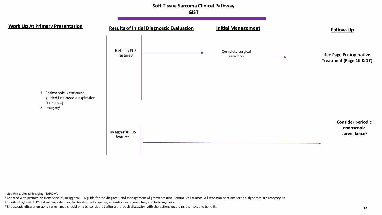
a See Principles of Imaging (SARC-A). i Adapted with permission from Sepe PS, Brugge WR. A guide for the diagnosis and management of gastrointestinal stromal cell tumors. All recommendations for this algorithm are category 2B. j Possible high-risk EUC features include irregular border, cystic spaces, ulceration, echogenic foci, and heterogeneity. k Endoscopic ultrasonography surveillance should only be considered after a thorough discussion with the patient regarding the risks and benefits.
1. Endoscopic Ultrasound-guided fine-needle aspiration (EUS-FNA)
2. Imagingb
No high-risk EUS features
High-risk EUS featuresj
Complete surgical resection
Consider periodic endoscopic
surveillancek
12
See Page Postoperative Treatment (Page 16 & 17)
Work Up At Primary Presentation Initial Management Follow-Up
Soft Tissue Sarcoma Clinical Pathway GIST
Initial Diagnostic Evaluation
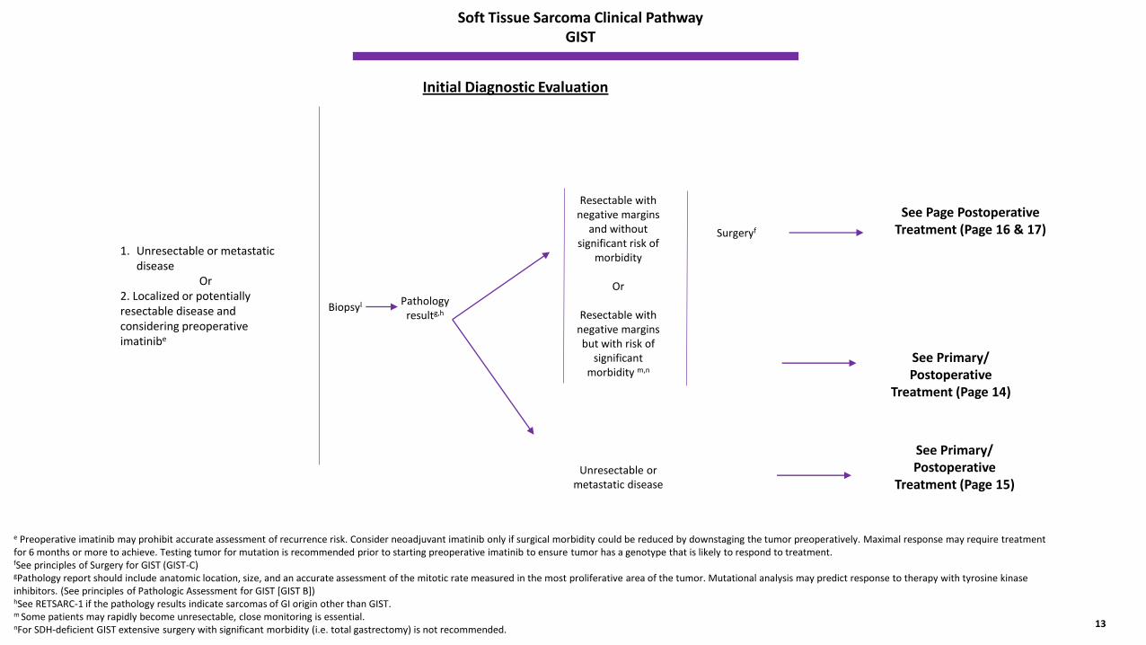
e Preoperative imatinib may prohibit accurate assessment of recurrence risk. Consider neoadjuvant imatinib only if surgical morbidity could be reduced by downstaging the tumor preoperatively. Maximal response may require treatment for 6 months or more to achieve. Testing tumor for mutation is recommended prior to starting preoperative imatinib to ensure tumor has a genotype that is likely to respond to treatment. fSee principles of Surgery for GIST (GIST-C)gPathology report should include anatomic location, size, and an accurate assessment of the mitotic rate measured in the most proliferative area of the tumor. Mutational analysis may predict response to therapy with tyrosine kinase inhibitors. (See principles of Pathologic Assessment for GIST [GIST B])hSee RETSARC-1 if the pathology results indicate sarcomas of GI origin other than GIST. m Some patients may rapidly become unresectable, close monitoring is essential. nFor SDH-deficient GIST extensive surgery with significant morbidity (i.e. total gastrectomy) is not recommended.
1. Unresectable or metastatic disease
Or 2. Localized or potentially resectable disease and considering preoperative imatinibe
Pathology resultg,h
Biopsyl
Resectable with negative margins
and without significant risk of
morbidity
Or
Resectable with negative margins but with risk of
significant morbidity m,n
13
See Page Postoperative Treatment (Page 16 & 17)
Unresectable or metastatic disease
Surgeryf
See Primary/ Postoperative
Treatment (Page 14)
See Primary/ Postoperative
Treatment (Page 15)
Soft Tissue Sarcoma Clinical Pathway GIST
Primary Presentation
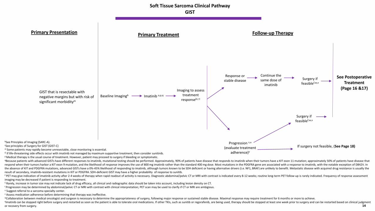
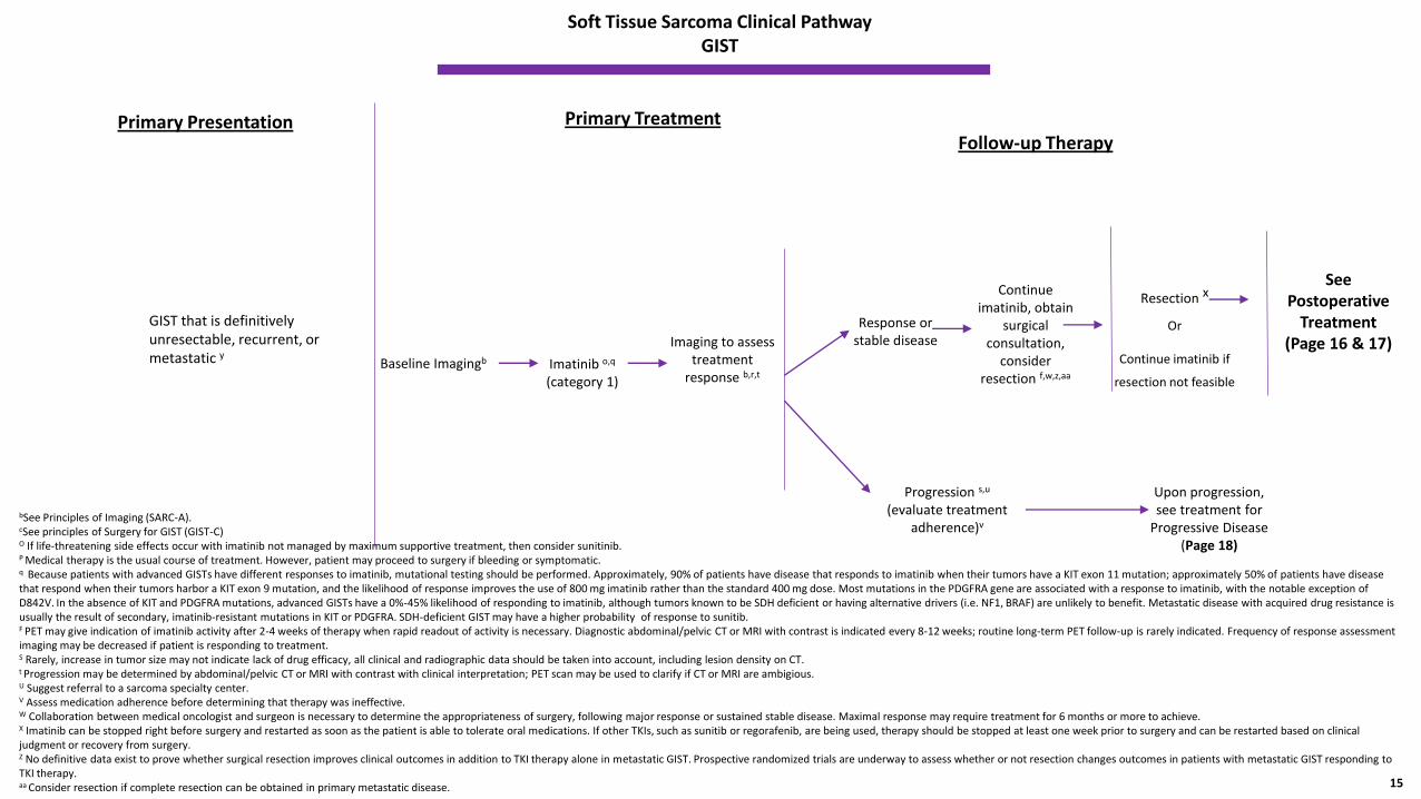
bSee Principles of Imaging (SARC-A). cSee principles of Surgery for GIST (GIST-C)m Some patients may rapidly become unresectable, close monitoring is essential. O If life-threatening side effects occur with imatinib not managed by maximum supportive treatment, then consider sunitinib. p Medical therapy is the usual course of treatment. However, patient may proceed to surgery if bleeding or symptomatic. qBecause patients with advanced GISTs have different responses to imatinib, mutational testing should be performed. Approximately, 90% of patients have disease that responds to imatinib when their tumors have a KIT exon 11 mutation; approximately 50% of patients have disease that respond when their tumors harbor a KIT exon 9 mutation, and the likelihood of response improves the use of 800 mg imatinib rather than the standard 400 mg dose. Most mutations in the PDGFRA gene are associated with a response to imatinib, with the notable exception of D842V. In the absence of KIT and PDGFRA mutations, advanced GISTs have a 0%-45% likelihood of responding to imatinib, although tumors known to be SDH deficient or having alternative drivers (i.e. NF1, BRAF) are unlikely to benefit. Metastatic disease with acquired drug resistance is usually the result of secondary, imatinib-resistant mutations in KIT or PDGFRA. SDH-deficient GIST may have a higher probability of response to sunitib. F PET may give indication of imatinib activity after 2-4 weeks of therapy when rapid readout of activity is necessary. Diagnostic abdominal/pelvic CT or MRI with contrast is indicated every 8-12 weeks; routine long-term PET follow-up is rarely indicated. Frequency of response assessment imaging may be decreased if patient is responding to treatment. S Rarely, increase in tumor size may not indicate lack of drug efficacy, all clinical and radiographic data should be taken into account, including lesion density on CT.t Progression may be determined by abdominal/pelvic CT or MRI with contrast with clinical interpretation; PET scan may be used to clarify if CT or MRI are ambigious.u Suggest referral to a sarcoma specialty center.v Assess medication adherence before determining that therapy was ineffective. wCollaboration between medical oncologist and surgeon is necessary to determine the appropriateness of surgery, following major response or sustained stable disease. Maximal response may require treatment for 6 months or more to achieve. xImatinib can be stopped right before surgery and restarted as soon as the patient is able to tolerate oral medications. If other TKIs, such as sunitib or regorafenib, are being used, therapy should be stopped at least one week prior to surgery and can be restarted based on clinical judgment or recovery from surgery.
GIST that is resectable with negative margins but with risk of significant morbiditym
Imatinib o,p,qBaseline Imagingb
Imaging to assess treatment
responseb,r,t
14
Progression s,u
(evaluate treatment adherence)v
Response or stable disease
See Postoperative Treatment
(Page 16 &17)
Continue the same dose of
imatinib
Surgery if feasiblef,w,x
If surgery not feasible, (See Page 18)
Surgery if feasiblef,w,x
Primary Treatment Follow-up Therapy
Soft Tissue Sarcoma Clinical Pathway GIST
Primary Presentation
bSee Principles of Imaging (SARC-A). cSee principles of Surgery for GIST (GIST-C)O If life-threatening side effects occur with imatinib not managed by maximum supportive treatment, then consider sunitinib. P Medical therapy is the usual course of treatment. However, patient may proceed to surgery if bleeding or symptomatic. q Because patients with advanced GISTs have different responses to imatinib, mutational testing should be performed. Approximately, 90% of patients have disease that responds to imatinib when their tumors have a KIT exon 11 mutation; approximately 50% of patients have disease that respond when their tumors harbor a KIT exon 9 mutation, and the likelihood of response improves the use of 800 mg imatinib rather than the standard 400 mg dose. Most mutations in the PDGFRA gene are associated with a response to imatinib, with the notable exception of D842V. In the absence of KIT and PDGFRA mutations, advanced GISTs have a 0%-45% likelihood of responding to imatinib, although tumors known to be SDH deficient or having alternative drivers (i.e. NF1, BRAF) are unlikely to benefit. Metastatic disease with acquired drug resistance is usually the result of secondary, imatinib-resistant mutations in KIT or PDGFRA. SDH-deficient GIST may have a higher probability of response to sunitib. F PET may give indication of imatinib activity after 2-4 weeks of therapy when rapid readout of activity is necessary. Diagnostic abdominal/pelvic CT or MRI with contrast is indicated every 8-12 weeks; routine long-term PET follow-up is rarely indicated. Frequency of response assessment imaging may be decreased if patient is responding to treatment. S Rarely, increase in tumor size may not indicate lack of drug efficacy, all clinical and radiographic data should be taken into account, including lesion density on CT.t Progression may be determined by abdominal/pelvic CT or MRI with contrast with clinical interpretation; PET scan may be used to clarify if CT or MRI are ambigious.U Suggest referral to a sarcoma specialty center.V Assess medication adherence before determining that therapy was ineffective. W Collaboration between medical oncologist and surgeon is necessary to determine the appropriateness of surgery, following major response or sustained stable disease. Maximal response may require treatment for 6 months or more to achieve. X Imatinib can be stopped right before surgery and restarted as soon as the patient is able to tolerate oral medications. If other TKIs, such as sunitib or regorafenib, are being used, therapy should be stopped at least one week prior to surgery and can be restarted based on clinical judgment or recovery from surgery. Z No definitive data exist to prove whether surgical resection improves clinical outcomes in addition to TKI therapy alone in metastatic GIST. Prospective randomized trials are underway to assess whether or not resection changes outcomes in patients with metastatic GIST responding to TKI therapy. aa Consider resection if complete resection can be obtained in primary metastatic disease.
GIST that is definitively unresectable, recurrent, or metastatic y Imatinib o,q
(category 1)Baseline Imagingb
Imaging to assess treatment
response b,r,t
15
Progression s,u
(evaluate treatment adherence)v
Response or stable disease
See Postoperative
Treatment (Page 16 & 17)
Continue imatinib, obtain
surgical consultation,
consider resection f,w,z,aa
Resection x
Or
Continue imatinib if
resection not feasible
Upon progression, see treatment for
Progressive Disease (Page 18)
Primary Treatment Follow-up Therapy
Soft Tissue Sarcoma Clinical Pathway GIST
Postoperative Outcomes
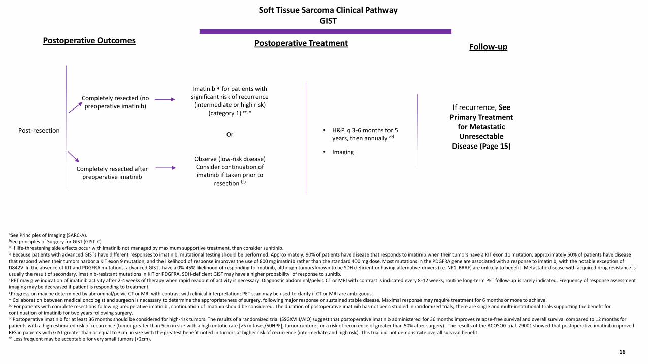
bSee Principles of Imaging (SARC-A). fSee principles of Surgery for GIST (GIST-C)O If life-threatening side effects occur with imatinib not managed by maximum supportive treatment, then consider sunitinib. q Because patients with advanced GISTs have different responses to imatinib, mutational testing should be performed. Approximately, 90% of patients have disease that responds to imatinib when their tumors have a KIT exon 11 mutation; approximately 50% of patients have disease that respond when their tumors harbor a KIT exon 9 mutation, and the likelihood of response improves the use of 800 mg imatinib rather than the standard 400 mg dose. Most mutations in the PDGFRA gene are associated with a response to imatinib, with the notable exception of D842V. In the absence of KIT and PDGFRA mutations, advanced GISTs have a 0%-45% likelihood of responding to imatinib, although tumors known to be SDH deficient or having alternative drivers (i.e. NF1, BRAF) are unlikely to benefit. Metastatic disease with acquired drug resistance is usually the result of secondary, imatinib-resistant mutations in KIT or PDGFRA. SDH-deficient GIST may have a higher probability of response to sunitib. r PET may give indication of imatinib activity after 2-4 weeks of therapy when rapid readout of activity is necessary. Diagnostic abdominal/pelvic CT or MRI with contrast is indicated every 8-12 weeks; routine long-term PET follow-up is rarely indicated. Frequency of response assessment imaging may be decreased if patient is responding to treatment. t Progression may be determined by abdominal/pelvic CT or MRI with contrast with clinical interpretation; PET scan may be used to clarify if CT or MRI are ambiguous.w Collaboration between medical oncologist and surgeon is necessary to determine the appropriateness of surgery, following major response or sustained stable disease. Maximal response may require treatment for 6 months or more to achieve. bb For patients with complete resections following preoperative imatinib , continuation of imatinib should be considered. The duration of postoperative imatinib has not been studied in randomized trials; there are single and multi-institutional trials supporting the benefit for continuation of imatinib for two years following surgery. cc Postoperative imatinib for at least 36 months should be considered for high-risk tumors. The results of a randomized trial (SSGXVIII/AIO) suggest that postoperative imatinib administered for 36 months improves relapse-free survival and overall survival compared to 12 months for patients with a high estimated risk of recurrence (tumor greater than 5cm in size with a high mitotic rate [>5 mitoses/50HPF], tumor rupture , or a risk of recurrence of greater than 50% after surgery) . The results of the ACOSOG trial Z9001 showed that postoperative imatinib improved RFS in patients with GIST greater than or equal to 3cm in size with the greatest benefit noted in tumors at higher risk of recurrence (intermediate and high risk). This trial did not demonstrate overall survival benefit. dd Less frequent may be acceptable for very small tumors (<2cm).
Post-resection
Imatinib q for patients with significant risk of recurrence (intermediate or high risk)
(category 1) cc, o
Or
Observe (low-risk disease) Consider continuation of imatinib if taken prior to
resection bb
Completely resected (no preoperative imatinib)
16
If recurrence, See Primary Treatment
for Metastatic Unresectable
Disease (Page 15)
Postoperative Treatment Follow-up
Completely resected after preoperative imatinib
• H&P q 3-6 months for 5 years, then annually dd
• Imaging
Soft Tissue Sarcoma Clinical Pathway GIST
Postoperative Outcomes
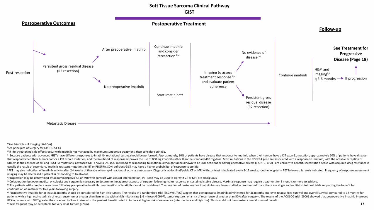
Post-resection
After preoperative Imatinib
No preoperative imatinib
Continue imatinib and consider
reresection f,w
17
Imaging to assess treatment response b,r,t
and evaluate patient adherence
Start Imatinib o,q
See Treatment for Progressive
Disease (Page 18)
No evidence of disease bb
Continue imatinibIf progression
Postoperative Treatment Follow-up
Persistent gross residual disease (R2 resection)
Metastatic Disease
Persistent gross residual disease (R2 resection)
H&P and imagingb,t
q 3-6 months
bSee Principles of Imaging (SARC-A). fSee principles of Surgery for GIST (GIST-C)O If life-threatening side effects occur with imatinib not managed by maximum supportive treatment, then consider sunitinib. q Because patients with advanced GISTs have different responses to imatinib, mutational testing should be performed. Approximately, 90% of patients have disease that responds to imatinib when their tumors have a KIT exon 11 mutation; approximately 50% of patients have disease that respond when their tumors harbor a KIT exon 9 mutation, and the likelihood of response improves the use of 800 mg imatinib rather than the standard 400 mg dose. Most mutations in the PDGFRA gene are associated with a response to imatinib, with the notable exception of D842V. In the absence of KIT and PDGFRA mutations, advanced GISTs have a 0%-45% likelihood of responding to imatinib, although tumors known to be SDH deficient or having alternative drivers (i.e. NF1, BRAF) are unlikely to benefit. Metastatic disease with acquired drug resistance is usually the result of secondary, imatinib-resistant mutations in KIT or PDGFRA. SDH-deficient GIST may have a higher probability of response to sunitib. r PET may give indication of imatinib activity after 2-4 weeks of therapy when rapid readout of activity is necessary. Diagnostic abdominal/pelvic CT or MRI with contrast is indicated every 8-12 weeks; routine long-term PET follow-up is rarely indicated. Frequency of response assessment imaging may be decreased if patient is responding to treatment. t Progression may be determined by abdominal/pelvic CT or MRI with contrast with clinical interpretation; PET scan may be used to clarify if CT or MRI are ambiguous.w Collaboration between medical oncologist and surgeon is necessary to determine the appropriateness of surgery, following major response or sustained stable disease. Maximal response may require treatment for 6 months or more to achieve. bb For patients with complete resections following preoperative imatinib , continuation of imatinib should be considered. The duration of postoperative imatinib has not been studied in randomized trials; there are single and multi-institutional trials supporting the benefit for continuation of imatinib for two years following surgery. cc Postoperative imatinib for at least 36 months should be considered for high-risk tumors. The results of a randomized trial (SSGXVIII/AIO) suggest that postoperative imatinib administered for 36 months improves relapse-free survival and overall survival compared to 12 months for patients with a high estimated risk of recurrence (tumor greater than 5cm in size with a high mitotic rate [>5 mitoses/50HPF], tumor rupture , or a risk of recurrence of greater than 50% after surgery) . The results of the ACOSOG trial Z9001 showed that postoperative imatinib improved RFS in patients with GIST greater than or equal to 3cm in size with the greatest benefit noted in tumors at higher risk of recurrence (intermediate and high risk). This trial did not demonstrate overall survival benefit. dd Less frequent may be acceptable for very small tumors (<2cm).
Soft Tissue Sarcoma Clinical Pathway GIST
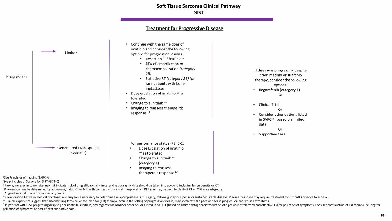
Progression
• Continue with the same does of imatinib and consider the following options for progression lesions:
• Resection f, if feasible w
• RFA of embolization or chemoembolization (category 2B)
• Palliative RT (category 2B) for rare patients with bone metastases
• Dose escalation of imatinib ee as tolerated
• Change to sunitinib ee
• Imaging to reassess therapeutic response b,t
18
If disease is progressing despite prior imatinib or sunitinib
therapy, consider the following options:
• Regorafenib (category 1) Or
• Clinical Trial Or
• Consider other options listed in SARC-F (based on limited data
Or• Supportive Care
Treatment for Progressive Disease
Limited
Generalized (widespread, systemic)
bSee Principles of Imaging (SARC-A). fSee principles of Surgery for GIST (GIST-C)S Rarely, increase in tumor size may not indicate lack of drug efficacy, all clinical and radiographic data should be taken into account, including lesion density on CT.t Progression may be determined by abdominal/pelvic CT or MRI with contrast with clinical interpretation; PET scan may be used to clarify if CT or MRI are ambiguous.U Suggest referral to a sarcoma specialty center.w Collaboration between medical oncologist and surgeon is necessary to determine the appropriateness of surgery, following major response or sustained stable disease. Maximal response may require treatment for 6 months or more to achieve. ee Clinical experience suggest that discontinuing tyrosine kinase inhibitor (TKI) therapy, even in the setting of progressive disease, may accelerate the pace of disease progression and worsen symptoms.ff In patients with GIST progressing despite prior imatinib, sunitinib, and regorafenib consider other options listed in SARC-F (based on limited data) or reintroduction of a previously tolerated and effective TKI for palliation of symptoms. Consider continuation of TKI therapy life-long for palliation of symptoms as part of best supportive care.
For performance status (PS) 0-2:• Dose Escalation of imatinib
ee as tolerated • Change to sunitinib ee
(category 1) • Imaging to reassess
therapeutic response b,t
The following pathway was developed through multidisciplinary efforts with physicians from the Mary Bird Perkins – Our Lady of the Lake Cancer Center. These pathways should be used as a supplemental guide for treatment for physicians at the Mary Bird Perkins – Our Lady of the Lake Cancer Center, and are not intended to replace the independent medical or professional judgment of physicians or other health care providers.
The pathway does not include principles or practices for therapy. Review the following pages within the NCCN Guidelines as an additional resource:
• Principles of Imaging (SARC-A)• Principles of Pathologic Assessment of Sarcoma Specimens (SARC-B)• Principles of Ancillary Techniques Useful in the Diagnosis of Sarcomas (SARC-C)• Principles of Surgery (SARC-D) • Review Radiation Therapy Guidelines for Soft Tissue Sarcoma of Extremity/Trunk/Head-Neck (SARC-E)• Review Systemic Therapy Agents and Regimens with Activity in Soft Tissue Sarcoma Subtypes (Non-specific)
(SARC- F) • Principles of Biopsy for GIST (GIST – A) • Principles of Pathologic Assessment for GIST (GIST-B)• Principles of Surgery (GIST- C)
References
1. Kelly K Curtis , Jonathan B Ashman , Christopher P Beauchamp, Adam J Schwartz, Matthew D Callister, Amylou C Dueck, Leonard L Gunderson and Tom R Fitch. 2011. Neoadjuvant chemoradiation compared to neoadjuvant radiation alone and surgery alone for Stage II and III soft tissue sarcoma of the extremities. Radiation Oncology.
2. Sandro Pasquall, Alessandro Gronchi. 2017. Neoadjuvant chemotherapy in soft tissue sarcomas: latest evidence and clinical implications. Therapeutic Advances in Medical Oncology.
3. Adam Dangoor, Beatrice Seddon, Craig Gerrand, Robert Grimer, Jeremy Whelan and Ian Judson. 2016. UK guidelines for the management of soft tissue sarcomas. Clinical Sarcoma Research.
4. NCCN Guidelines. October 2017 Version. Soft Tissue Sarcoma. 5. A. Lo´pez-Pousa, J. Martin Broto, J. Martinez Trufero, I. Sevilla, C. Valverde, R. Alvarez, J. A. Carrasco Alvarez, J. Cruz Jurado, N. Hindi, X. Garcia del
Muro. 2016. SEOM Clinical Guideline of management of soft-tissue sarcoma .Clinical Guides in Oncology. 6. The ESMO/Europeran Sarcoma Network Working Group. 2014. Soft tissue and visceral sarcomas. ESMO Clinical Practice Guidelines for diagnosis,
treatment and follow-up. ESMO.7. Sylvie Bonvalot, MD, PhD, Antonin Levy, MD, Philippe Terrier, MD, Dimitri Tzanis, MD, PhD, Sara Bellefqih, MD, Axel Le Cesne, MD, and Ce´cile Le
Pe´choux, MD. 2017. Primary Extremity Soft Tissue Sarcomas: Does Local Control Impact Survival?. Annals of Surgical Oncology. 8. Thomas F. Delaney, M.D., Ira J. Spiro, M.D., PH.D., Herman D. Suit, M.D., D. Phil.,Mark C. Gebhardt, M.D.,Franci J. Hornicek, M.D., PH.D., Henry J.
Mankin, M.D., Andrew L. Rosenberg, M.D., Daniel I. Rosenthal, M.D.,Fariba Miryousefi, M.D.,Marcus Ancukiewicz, PH.D., and David C. Harmon, M.D. 2003. Neoadjuvant chemotherapy and Radiotherapy for Large Extremity Soft-tissue Sarcomas. International Journal Radiation Oncology Biology Physics.