Embed Size (px)
Citation preview

Soft-ECC®SFT-2000
Endocervical Curettage Device
Physician and Clinician PrimerVer: July 2012
Histologics LLC20409 Yorba Linda Blvd.
Suite 119Yorba Linda, CA 92886
Phone Toll Free and FAX: (888) 738-9757
Website; www.histologics.comEmail; [email protected]
LLC

Q 1: What is Kylon® and how does it work to obtain a biopsy?
Kylon® is a hooked nylon fabric that can be mounted to the applicator stick, that when pressed on tissue surfaces, allows the hook tip to contact the target tissues, and with pressure and rotation, a trans-epithelial biopsy is obtained (Figure 1). The procedure of applying, pressing, and rotating a Kylon® covered tapered tip device into the endocervical os and rotating it to obtain a curettage specimen is called a Soft-ECC® (Figure 2). The procedure of directing a Kylon® tipped applicator directly on an epithelial lesion is called a “SoftBiopsy®”.
Physician /Clinician Primer : © All Rights Reserved 2010
Question 1 - Figure 1
Question 1 - Figure 2
The Kylon® gently collects tissue samplesfrom the endocervical columnar epithelium.
Collected Tissue
Endocervical
Front End ofSoft-E Device
Stromal Tissue
Endocervical Canal
Lining
Q 2: Why were biopsy tools using this patent pending “hooked” curettage fabric called “Kylon®” developed?
Biopsy of mucosal tissues inside a body cavity, such as the cervix or vagina can be traumatic, physically (with evidence of pain and bleeding) and psychologically (fear of cancer, fear of pain or trauma). If trauma is “perceived” by the clinician or the patient, either or both might be less inclined to perform necessary biopsies or seek care to receive them (health seeking behavior may decrease). Neither is good for the quality of the care rendered or received. A minimally invasive biopsy tool was developed with the intent to take a high quality trans-‐epithelial tissue biopsy while simultaneously collecting most of the specimen inside the fabric. The device is intended to be minimally invasive, keeping the patient care experience in mind. The goal is also to reduce or eliminate the sampling error associated with biopsy procedures by harvesting and retaining the sample optimally. Thus, both the clinician and the patient are inclined to deliver and receive the necessary care to make the diagnosis and guide therapy.
Clinical trials have been performed, and are ongoing to demonstrate the intended goals of using the Kylon fabric to obtain a biopsy.
Physician /Clinician Primer : © All Rights Reserved 2010
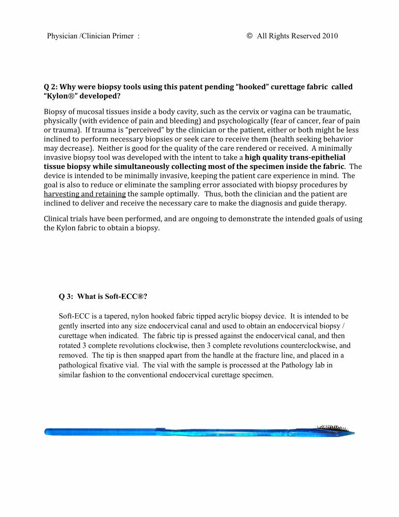
Q 3: What is Soft-ECC®?
Soft-ECC is a tapered, nylon hooked fabric tipped acrylic biopsy device. It is intended to be gently inserted into any size endocervical canal and used to obtain an endocervical biopsy / curettage when indicated. The fabric tip is pressed against the endocervical canal, and then rotated 3 complete revolutions clockwise, then 3 complete revolutions counterclockwise, and removed. The tip is then snapped apart from the handle at the fracture line, and placed in a pathological fixative vial. The vial with the sample is processed at the Pathology lab in similar fashion to the conventional endocervical curettage specimen.

Q 4: How do I use the Soft-ECC® device to obtain an endocervical curettage?
Place the handle of the device in the palm and hold the near portion of the shaft in between the thumb and first two fingers (Fig 1). This will permit a twirling motion during the rotational biopsy. (Fig 2) Gently insert the tapered tip into the endocervical canal and press, wiggle, or slightly agitate it so there is a tight fit, or the KYLON pad has completely entered the endocervical canal (Fig 3 and Fig 4). Make note of the placement of the KYLON pad and gently press the pad laterally against the canal surface (Fig 5) . Twirl or rotate the device two to three complete 360 degree turns clockwise, then 3 complete revolutions counterclockwise (Fig 6).
If the fabric pad on the tip does at least enter partially, a sample can still be obtained, but note that the sample is being removed from the outer endocervical canal.
Alternatively, on could rotationally agitate (like a washing machine effect) 5-8 times while rotating the surface to contact all four quadrants inside the canal.
Once an adequate tissue specimen is obtained, simply snap off the device head (Figure 7) from the shaft of the device at the scored area and place it in the fixative vial provided by the laboratory (Figure 8).
Physician /Clinician Primer : © All Rights Reserved 2010
Fig.
Place the bottom tip of the Soft-EEC in the center of your palm.
Note: Medical gloves must be worn.

Physician /Clinician Primer : © All Rights Reserved 2010
Fig.
Close your hand around the Soft-E C,with your fingers along the ridges, andyour forefinger on the tactile notch.
bottom (tactile) notch
top notch
Note: Medical gloves must be worn.
rotate
Fig
Notches are aligned with KylonTM pad.
Fingers will rotate E C device. Notches serve to keep track of rotations.
rotate
Note: Medical gloves must be worn.

Physician /Clinician Primer : © All Rights Reserved 2010
Fig.
Gently but firmly inserthead of device into cervix.
Slightly agitate until KylonTM pad has completely entered theendocervical canal.
Note: Medical gloves must be worn.
Fig.
KylonTM pad on E C head is fullypositioned inside cervix.
1. Rotate clockwise 360º three complete turns.
2. Rotate counterclockwise three complete turns.
Use notches as guides.
Cervix
Rotate 360º
Note position of notch
Cervical Canal

Physician /Clinician Primer : © All Rights Reserved 2010
Fig.
Press the pad laterally against the canal surface.KylonTM pad contacts cells along interior of canal.
collected by KylonTM.
Cervix
Cervical Canal
Collected
Fig 7
After gently removing Soft-ECC® from cervix, separate head (with Kylon® pad and tissue samples) from body of device by snapping at recessed joint.
Snap at Recess
Note: Medical gloves must be worn.

Q 5: Why is the tip of the Soft-ECC® curette so pointed and how much of the curette should enter the canal to be considered adequate?
The device is designed to enter almost any endocervical canal. The tip is sufficiently rounded as not to puncture tissue surfaces, but the clinician should be careful to enter the canal in alignment with the direction of the endocervical canal, and not force the tool into the canal. If applied with gentle pressure, a good portion of the Kylon® pad will enter into the canal and contact the endocervical epithelial surface. In the majority of cases, at least one third of the Kylon® pad should enter the canal to obtain an adequate sample for analysis. However, the likelihood the tapered tip will successfully enter the canal is higher than other rigid conventional endocervical biopsy tools such as the Kevorkian Curette.
Physician /Clinician Primer : © All Rights Reserved 2010
Fig 8
Place the head of the Soft-ECC (with Kylon® pad and tissue samples) intovial of fixative liquid and seal tightly.
Soft-E C Head

Top Side Angle View
Soft-ECC® Tip in Vial with TissueThe tip was inserted into a slightly stenotic and shortened cervical canal.
You can see that the curette entered 2/3 of the way into thecervical canal and then met resistance, thus the fabric
collected abundant tissue at the curettage surface. The proximal1/3 did not enter the canal, thus does not contain tissue.
The specimen was taken to the lab and combed out of the fabric onto a Telfa-pad to be prepared for processing.
Question 5 Figure 2
Q 6: Do the hooks puncture the tissue when pressed? How does the abrasive fabric compare with other biopsy devices?
No, the tips of the hooks angle away from the tissue surface before pressure is applied. Once pressed, the hooks open to allow the nylon fibers to contact the tissue surface to perform the curettage. With agitational or rotational forces, the hook tips then frictionally abrade the tissue surface, causing it to buckle and fracture. The hooks then recess under the basement membrane lifting the pieces of trans-epithelial tissue into the fabric. The device edge also abrades the tissue as a curette. The rows hooks function as the basket to catch and retain tissue fragments.Other curettes that use a cutting surface, like a forceps, a curette, or a stiff bristle brush are more rigid and are not curved, thus, with pressure, they will puncture or enter the surface with initial placement or pressure. With rotation or lateral motion, the tissue is first sheared, then torn free from the underlying stroma. With a conventional curettage sharp or bristle brush instrument, the tissue commonly falls free from a curette or straight bristle, and may be incompletely collected in the specimen. If the cutting surface is sharp, the tissue will be shaved or dissected in intact fragments. Often, the clinician has to use a soft nylon cytobrush or forceps to retrieve an endocervical curettage specimen specimen, taking additional time. Not so for the Soft-ECC® device that simultaneously collects the specimen inside the hooks of the fabric.
Physician /Clinician Primer : © All Rights Reserved 2010
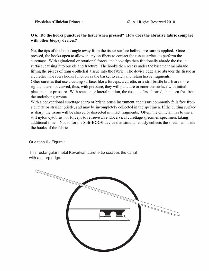
Question 6 - Figure 1
This rectangular metal Kevorkian curette tip scrapes the canal with a sharp edge.

Physician /Clinician Primer : © All Rights Reserved 2010
Q 7: How do I apply the device and how much pressure do I apply when pressing the applicator stick and Kylon® pad on to the tissue surface?
Pressure on a Ilat, irregular, or rounded tissue surface (like exocervix or vagina) is applied perpendicularly, while pressure inside a canal (radially mounted Kylon® pad like the Soft-‐ECC® endocervical canal biopsy device) is applied laterally against the canal epithelium. The amount of pressure needed to expose the hook surface to the epithelium would be equivalent of a light massage, depressing the device into the tissue surface so it is Iirmly applied, but not so hard as to depress the tissue surface more than one or two millimeters.
Q 8: How do I know I have obtained a sufficient sample? What do I do to save the sample for analysis?
After performing the biopsy procedure, gently remove the tip from the endocervix. You will note the “excavated” tissue has recessed between the fiber hooks, and will be combined with some blood tinged mucous. The tip is designed to be released (snapped off) from the handle with moderate lateral traction, decoupled from the device, and dropped in a vial of fixative. If you do not see any tissue or mucous in the fabric, repeat the procedure.
\ Question 8 - Figure 1
After performing the biopsy procedure, gently remove the tip from the endocervix. You will note the “excavated” tissue has recessed between the fiber hooks, and will be combined with some blood tinged mucous.
Collected Tissue

Q-‐9: Is there Evidence that the biopsy taken with Soft-‐ECC® or SoftBiopsy® is trans-‐epithelial (full thickness)?
Kylon Based Trans-‐epithelial Biopsy Sample
Research has shown that the biopsy sample removed with frictional abrasion using the KYLON® hooked fabric is deep enough to remove the full thickness of the epithelium of the cervix into the upper stroma. This includes both the glandular endocervical and squamous exocervical tissue and stromal tissue. Both columnar/glandular and squamous epithelium exist together, often near the canal, at the “transformation zone” of the cervix.
Cervical Stroma(deep connective and vasculartissue)
Biopsy site, epithelium denuded
Surface Epithelium
Transformation Zone
Biopsy: Glands and Stromal Tissue (Intact Architecture)
Endocervical Biopsy
Physician / Clinician Primer : © All Rights Reserved 2010

Q 10: How is the specimen taken off the fabric so it can be processed in the Pathology Lab?
A: The pathologist has several methods available to remove the tissue from the fabric. Tweezers or a scalpel blade can be used to gently scrape off the tissue from the hooks and fabric. Alternatively, a small comb has been developed that can be raked across the hooks repeatedly under the surface of the fixative while still in the vial, thus “combing” the tissue from the fabric into the vial. Once the tissue is released, it resembles conventional curettings one would obtain during endocervical curettage. The specimen can then be processed by the lab in an identical manner that is customary for the lab, including cell block if desired.
Pathologist / Pathology Lab Primer : © All Rights Reserved 2010
Question 10 - Figure 1
Tweezers can be used to gently scrape off the tissue from the hooks and fabric.
Soft-EECTM Head
Filter Paper

Pathologist / Pathology Lab Primer © All Rights Reserved 2010
Question 10 - Figure 2
Alternatively, a scalpel blade can be used to gently scrape off the tissue from the hooks and fabric.
Soft-EECTM Head
Filter Paper
Small Comb
Question 10 - Figure 3
Another viable technique is to comb the tissue out of the fabric.Combing is done under the liquid fixative.
Tissue Sample
Liquid Fixative
All Rights to this Physician/Clinician Primer on Soft-ECC® are reserved by Histologics LLC : ©
It can also be done outside the vial onto a Telfa Pad.