Embed Size (px)
Citation preview
ISSN 0034-7000 REV. ARG. CARDIOL., 51, NO 3, 126-149
T emade actualidad
Estado actual del electrocardiograma de esfuerzo
Current status of the exercise ECG
V. F. FROELICHER, M.D. Associate Professor of Medicine
From the Division of Cardiology, Departmentof Medicine, University of California, San Diego
Presentado en el XVIII Congreso de Cardiología Argentina, el X Congteso Sudamericano de Cardiología y el Simposio !nternacional de Técnicas No Invasivas.
Supported by Specialized Center of Research on Ischemic Heart Disease, NIH Research Grant HL-17682 Awarded by the National Heart, Lung, and Blood Institute awarded to John Ross, Jr. M.D.
There has been a renewed emphasis on the total approach to the analysis of the exercise test. Other objective and subjective rèsults
need to be considered besides just ST-segment depression. ST-segment elevation and U wave inversion are ominous findings that are relatively
insensitive but highly specific. R wave amplitude changes are normal phenomena and failure for them to decrease during exercise probably is
due to submaximal stress, and currently they are not reliable indica~ors of coronary disease
or left ventricular dysfunction. The monitoring of multiple leads or the consideration of spatial changes using the Frank leads and computers should improve the diagnostic value of the
test. Bayes' theorem and the predictive model explain how the test has a different diagnostic impact in different populations. The exercise test not only is a diagnostic tool but has other valuable applications. It probably will remain for some time the most useful office and clinic tool in Cardiology.
Dirección postal: Adress for reprints: Division of Cardiology, Department of Medicine, University of California. San Diego, California, USA.
Hay un énfasis renovado en el método de análisis de la prueba de esfuerzo. Además de la depresión del segmento ST es necesario tener en cuenta otros resultados objetivos y subjetivos.
La elevación del segmento ST y la inversión de la onda U son descubrimientos ominosos, rela- tivamente insensibles pero altamente específicos.
Los cambios en fa amplitud de fa onda R son un fenómeno normal y si no se logra dismi- nuirlos durante el esfuerzo, probablemente se deba a un stress submáximo. Comúnmente no son indicadores confiables de enfermedad coronaria 0 disfunción ventricular izquierda. EI monitoreo de derivaciones múltiples 0 la
consideración de cambios de espacio usando las derivaciones de Frank y computadoras, pueden mejorar el valor diagnóstico de la prue- ba. EI teorema de Bayes y el modelo de pre- dicción explican cómo la prueba tiene un di- ferente valor de diagnóstico en poblaciones diferentes. La prueba ergométrica no es sola- mente un elemento de diagnóstico sino que tiene otras aplicaciones valiosas. Probablemente seguirá siendo por algún tiempo el elemento clínico más útil en Cardiología.
EI esfuerzo es el mejor test dinámico del corazón entre todos los tipos de stress que se han ùsado
127 REVISTA ARGENTINA DE CARDIOLOGIA, MA YO-JUNIO 1983, VOL. 51, NO 3
Although many different types of stresses have been used to bring out ECG changes consistent with myocardial ischemia, exercise is the best dynamic test of the heart since it is man's most common physiological stress. The exercise ECG test is the most practical and useful procedure in clinical practice because of its many applications-
and results. In regard to medical economics, the exercise test is the best way of determining
which patients need or do not need more expensive diagnostic or therapeutic procedures. This chapter will discuss the current applications, methodology and interpretation of exercise ECG testing. .
APPLICATIONS .
To diagnose chest pain and other cardiac find- . ings: The exercise test may be used to evaluate
patients currently symptomatic with chest pain, patients with a past history of worrisome chest pain, or' patients with other findings suggestive, but not diagnostic, of coronary artery disease. Such other suggestive findings include extra sounds, murmurs, abnormal precordial impulses, cardiac enlargement, and
.
resting electrocardiographic abnormalities such as right bundle branch block, nonspecific ST and T wave changes, or non-diagnostic Q waves. The questions posed to the clinician by such patients would be: is this chest pain, previous episode of chest pain, or this finding due to coronary artery disease? The results of exercise testing along with the clinical presentation can h<;lp establish the probability of such
patients having coronary artery disease. The evaluation of the patient with chest pain is
probably' the most common use of exercise testing in clinical practice.
To determine prognosis and severity of disease: Exercise testing may be used to deter- mine the prognosis of patients with coronary artery disease. The results of an exercise test can help to establish the risk of mortality and morbidity after an acute myocardial infarction and in patients with angina pectoris. The severity of coronary artery disease is reflected by the response to exercise testing.1
Exercise-induced hypotension, very limited functional capacity (less than 5 METS), exercise-
para poner de manifiesto cambios en el electro- cardiograma compatibles con isquemia de mio- cardio, ya que es el stress fisiológico más común del hombre. Clínicamente, el ECG de esfuerzo es el procedimiento más práctico y útil, debido a sus múlJ:iples aplicaciones y resultados. En 10
que concierne a economía médica, es el mejor sistema para determinar qué paciente necesita o no form as de diagnóstico 0 procedimientos terapéuticos más costosos. Aquí discutiremos las aplicaciones, metodología e interpretación actuales de la prueba de electrocardiograma de esfuerzo.
APLICACIONES Para el diagnósticadel dolor precordial y otros hallazgos: La prueba ergométrica se puede usar para evaluar pacientes con dolor anginoso, con una historia de dolor de pecho sospechoso, 0
con otros hallazgos sugestivos, pero no diag-
nósticos, de enfermedad coronaria, tales como ruidos agregados, soplos, latidos precordiales
anormales, agrandamiento del corazón, y anor- malidades electrocardiográficas en reposo (blo- queo de rama derecha, cambios no específicos
en el segmento ST y onda T, ondas Q no diag-
nósticas). La pregunta que de be formularse el cIínico ante tales pacientes es si este dolor de pecho 0 estos hallazgos son atribuibles a una el}fermedad coronaria. Los resultados de la
prueba ergométrica junto a la forma de presen- tación clínica pueden ayudar a establecer la probabilidad de que exista enfermedad corona- ria. La evaluación del paciente con dolor de pecho es probablemente la aplicación más común de la prueba ergométrica en la práctica clínica.
Para determinar el pronóstico y gravedad de la enfermedad: Los resultados de una prueba de esfuerzo pueden ayudar a establecer el riesgo de mortalidad y morbilidad después de un infar- to agudo de miocardio y en pacientes con angina de pecho. La severidad de la enfermedad coro- naria se refleja en la respuesta al esfuerzo.1
EI esfuerzo que provoca hipotensión, baja capacidad funcional (inferior a 5 METS), angina,
incompetencia cronotrópica (respuesta de fre- cuencia cardíaca inadecuada), ondas U inverti- das, sobreelevación del segmento ST, segmento
\.'-:;:
ECG DE ESFUERZO / V. F. Froelicher 128
test induced angina, chronotropic incompetence (inadequate heart rate response), inverted U waves, ST-segment elevation, down-sloping ST-segment depression and ST-segment depres-
sion beginning at a low heart rate and in multiple leads have a bad prognosis and are more pre- dictive of severe coronary artery disease and left ventricular dysfunction than a test response of only 0.1 mv of horizontal ST-segment depres-
sion at a good maximal workload. Patients who are able to achieve a normal work load, normal maximal heart rate, and normal maximal systolic blood pressure during treadmill testing
appear to have better ventricular function and less coronary obstruction than those unable to do so. Responses predicting poor prognosis and
severity of disease are summarized in Table 1.
Screening for silent coronary artery disease:
The exercise test may be used to screen asymp- tomatic individuals in order to identify a group at high risk for developing coronary artery disease. It has been demonstrated that indivi- duals with an abnormal ST-segment response have 10 to 15 times the chance of developing
"coronary artery disease than tho~e with a normal response.2 Recent studies have demonstrated a lower sensitivity for ST depression than previous studies. They have suggested that the
Table 1
Responses to exercise reflecting prognosis and severity
of coronary artery disease
1) Systolic hypotension or drop in systolic blood pressure
2) Limited functional capacity (less than 5 METS) 3) Inadequate heart rate response (chronotropic
incompetence or heart rate impairment) 4) ST segment elevation 5) The amount of ST segment depression, and relative
negativity of the ST segment slope, number of leads
it occurs in
6) The lower the heart rate at which ECG abnormalities occur 7) Duration of ECG abnormality 8) Symptoms, particularly true angina pectoris
9) Ventricular dysrhythmias 10) Exercise-induced rise in diastolic pressure
11) Inverted U waves
12) QT prolongation
ST descendente y depresión del segmento ST
que comienza con baja frecuencia cardíaca y
generalmente da mal pronóstico, predice enfer- medad coronaria severa y disfunciór ventricular izquierda, mucho más que uria respuesta de
sólo 0,1 mv de depresión horizontal del seg-
mento ST a una buena carga máxima de trabajo. Los pacientes que son capaces de lograr una
carga normal de trabajo, frecuencia cardíaca máxima normal, y presión sanguínea máxima normal durante la prueba. ergométrica, parecen tener una función ventricular mejor y una me- nor obstrucción coronaria que aquellos que no 10 pueden lograr. Las respuestas que predicen
un mal pronóstico y gravedad de la enfermedad se resumen en la Tabla 1.
Para evidenciar una enfermedad coronaria silenciosa: La prueba de esfuerzo se pu~de
usar para estudiar individuos asintomáticos con el fin de identificar un grupo con alto riesgo
de desarrollar enfermedad coronaria. Se ha demostrado que individuos con un segmento ST anormal tienen de lOa 15 veces más posi- bilidad de desarrollar enfermedad coronaria que
aquellos con respuesta normal. 2
Estudios recientes han demostrado una sen- sibilidad de depresión del segmento ST menor que la señalada en estudios anteriores. Esto su-
Tabla 1
Pronóstico y severidad de la enfermedad arterial coronaria de acuerdo con la respuesta al ejercicio
1) Hipotensión sistólica 0 caída en la presión arterial sistólica
2) Capacidad funcionallimitada (menos de 5 METS) 3) Respuesta inadecuada de la frecuencia cardíaca
(incompetencia cronotrópica 0 deterioro de la frecuencia cardíaca)
4) Elevación del segmento ST 5) Grado de depresión del segmento ST y relativa negatividad
de la pendiente y número de derivaciones en que se presenta 6) La menor Fc en la que las anormalidades ECG aparecen 7) Duración de la anormalidad en el ECG 8) Síntomas, particularmente anginosos 9) Arritmias ventriculares
10) Ascenso de la presión arterial diastólica inducido por el esfuerzo
11) Ondas U invertidas 12) Prolongación del intervalo QT
129 REVISTA ARGENTINA DE CARDIOLOGIA, MAYO-jUNIO 1983. VOL. 51, NO 3
test is only useful in men older than 40 and in populations that have an excess of the standard risk factors.
Some serious complications of ex~rcise testing used in screening asymptomatic indivi- duals are the insurance and occupational pro- blems that abnormal responders can have along with the possibility of making some of them "cardiac cripples". Because of these problems and the high false positive rate seen when exercise testing individuals with a relatively low prevalence of coronary artery disease, it may be appropriate to use exercise testing only for screening individuals in whom sudden
incapacitation could present a danger to public safety. Because of the recent enthusiasm for physical conditioning, it may also be desirable
to exercise test coronary prone middle-aged American males before they embark on a
potentially hazardous exercise program. Factors which identify a population with a higher prevalence of disease make the test more valuable for screening; i.e., male sex, older age, elevated serum cholesterol, high blood pressure, a family history of heart disease in individuals less than 65 years of age, and diabetes mellitus.
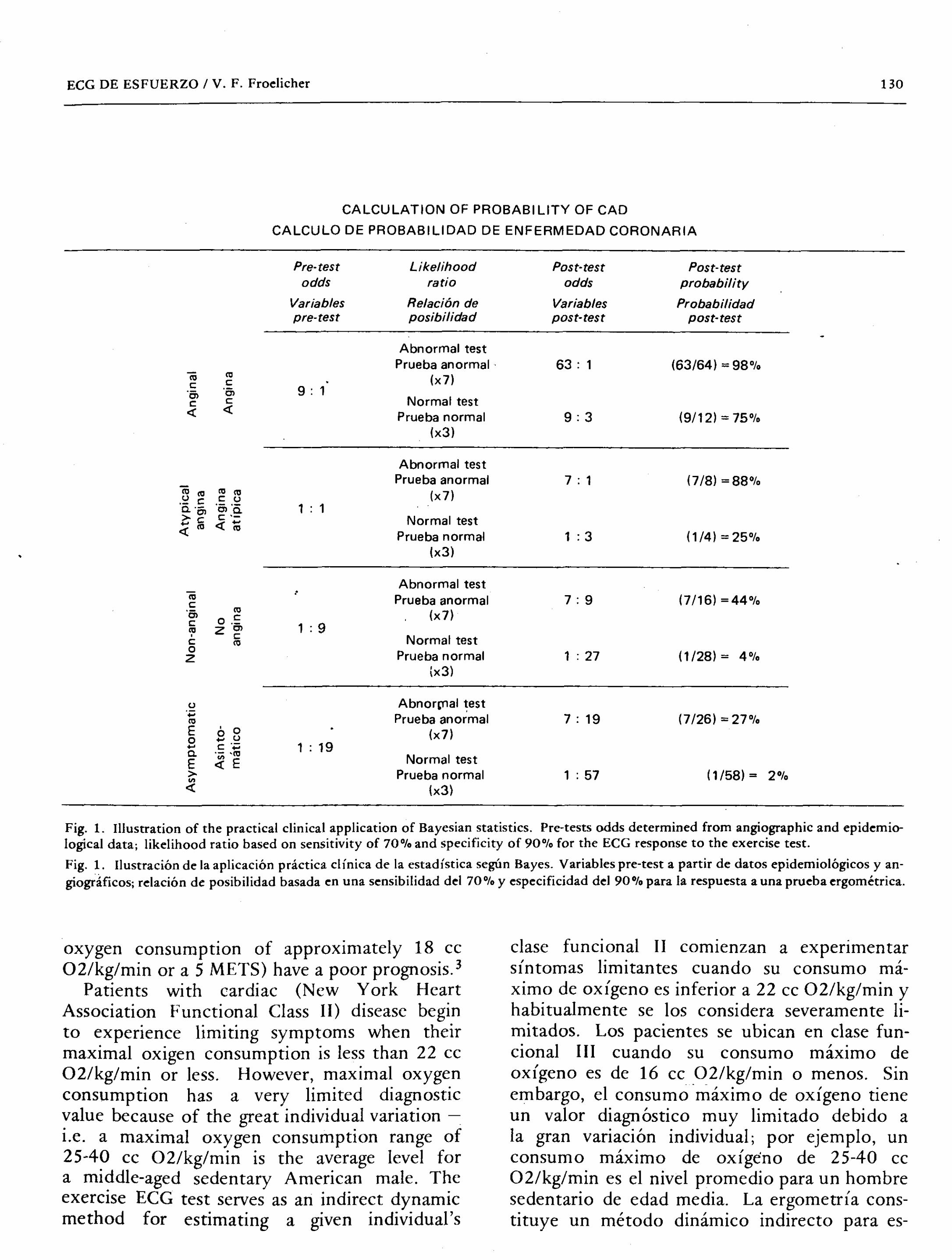
The exercise ECG test has a sensitivity of 60-80% and a specificity of about 90%. By using Bayes' theorem which says that the odds
of a patient having a disease after a test (post
test odds) will be the product of the odds
before the test (pre-test odds) and the odds
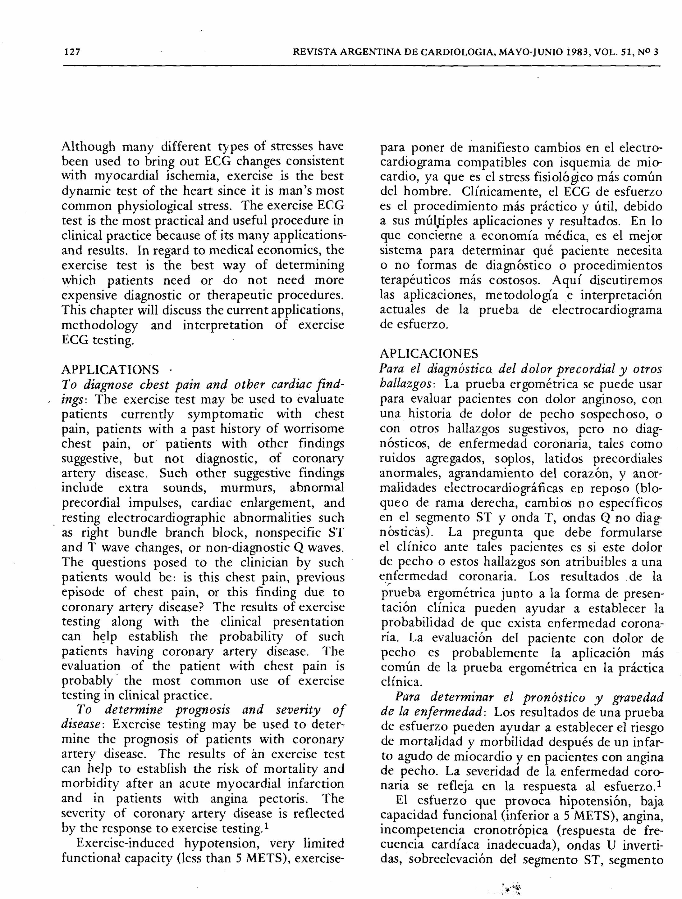
that the test result was a true one (the likelihood ratio). Figure 1 employs Bayes' theorem and demonstrates the usefulness of the exercise ECG test in predicting coronary artery disease
(CAD) in patients with different types of chest pain or asymptomatic.
Evaluation of functional capacity: Exercise tests may be used to evaluate the functional capacity of patients. Maximal oxygen consump- tion is the best non-invasive measurement of functional capacity and may either be estimated
or measured during exercise testing. Maximal . oxygen consumption can more accurately
determine the degree of cardiac impairment than a physician's assessment of functional classification. Patients ur:able to advance beyond the first stage of the Bruce test (an
giere que el esfuerzo es útil solamente en hom- bres mayores de 40 años y en poblaciones con un exceso de factores de riesgo standard.
Algunos de los serios inconvenientes que pue- den tener las personas con una respuesta anor- mal a la prueba de esfuerzo, son el seguro y problemas laborales, junto a la posibilidad de
que algunas de ellas se conviertan en "inválidos
cardíacos". Por esto es más apropiado usar la prueba de esfuerzo en aquellos con una incapa- cidad repentina que puedan constituirse en un peligro para la seguridad pública.
Debido al entusiasmo actual por el ejercicio físico, esta prueba puede ser conveniente. tam- bién en hombres de mediana edad prop ens os a enfermedades coronarias, antes de que se
embarquen en un program a de esfuerzo poten- cialmente riesgoso. Los factores que identifican a una población con un mayor predominio de
enfermedad hacen a la prueba más valiosa, por ejemplo: sexo masculino, mayor edad, coleste- rol elevado, presión sanguínea alta, una historia familiar de enfermedad cardíaca en personas menores de 65 años, y diabetes mellitUs.
El electrocardiograma de esfuerzo tiene una sensibilidad de un 60% a 80% y una especifici-
dad del 90%. Usando el teorema de Bayes, que dice que las variables de un paciente que presen- ta enfermedad coronaria después de una prueba serán el producto de las variables antes de la
prueba (pretest) y las variables reales que la prue- ba provoque (relación de probabilidad).
La Figura 1 emplea el teorema de Bayes y demuestra la eficacia de la ergometría para pre- decir una enfermedad coronaria en pacientes
asintomáticos 0 con distintos tipos de dolores de pecho.
Evaluación de la capacidad funcional: Las
pruebas de esfuerzo pueden utilizarse para eva- luar la capacidad funcional de los pacientes. EI
máximo consumo de oxígeno es la mejor medida no invasiva de la capacidad funcional y puede ser estimada 0 medida durante una prueba de
esfuerzo. Los pacientes que no pueden superar la pri-
'mera etapa del test de Bruce (un consumo de
oxígeno de aproximadamente 18 cc 02/kg/min o 5 MI,TS) tienen un mal pronóstico.3
Los pacientes con enfermedad cardíaca en
ECG DE ESFUERZO / V. F. Froelicher 130
CALCULATION OF PROBABILITY OF CAD
CALCULO DE PROBABILIDAD DE ENFERMEDAD CORONARIA
Pre- test Likelihood Post-test Post-test odds ratio odds probability
Variables Relaciðn de Variables Probabilidad pre-test posibilidad post-test post-test
Abnormal test Prueba anormal 63: 1 (63/64) = 98%
ëij co (x7) c: c:
-5> '5> 9: 1
c: c: Normal test ~ ~
Prueba normal 9:3 (9/12) = 75% (x3)
Abnormal test Prueba anormal 7 : 1 (7/8) =88%
ëij co co co (x7) .~ c c: u
~"5> 'g>:ë- 1 : 1
:i 16 ~ 1;; Normal test
Prueba normal 1 : 3 (1/4) =25% (x3)
Abnormal test ëij Prueba anormal 7:9 (7/16) =44% c: '5> co (x7) c: 0.= 1 : 9 co z'" C: c: Normal test co 0
Prueba normal 1 : 27 (1/28) = 4% Z
(x3)
u AbnortTlal test ".:;
Prueba anormal 7: 19 (7/26) =27% co E '0 (x7) 0 B u
ë. c '';:; 1 : 19 .- 'm Normal test E :; E
> Prueba normal 1 : 57 (1/58) = 2% :; (x3)
Fig. 1. Illustration of the practical clinical application of Bayesian statistics. Pre-tests odds determined from angiographic and epidemio- logical data; likelihood ratio based on sensitivity of 70% and specificity of 90% for the ECG response to the exercise test.
Fig. 1. Ilustración de la aplicación práctica cIínica de la estadística según Bayes- Variables pre-test a partir de datos epidemiológicos y an- giogråficos; relación de posibilidad basada en una sensibilidad del 70% Y especificidad del 90% para la respuesta a una pruebaergométrica.
oxygen consumption of approximately 18 cc
02/kg/min or a 5 METS) have a poor prognosis.3
Patients with cardiac (New York Heart Association Functional Class II) disease begin
to experience limiting symptoms when their maximal oxigen consumption is less than 22 cc
02/kg/min or less. However, maximal oxygen consumption has a very limited diagnostic
value because of the great individual variation -
i_e. a maximal oxygen consumption range of 25-40 cc 02/kg/min is the average level for a middle-aged sedentary American male. The exercise ECG test serves as an indirect dynamic method for estimating a given individual's
clase funcional II comienzan a experimentar síntomas limitantes cuando su consumo má- ximo de oxígeno es inferior a 22 cc 02/kg/min y habitualmente se los considera severamente li-
mitados. Los pacientes se ubican en clase fun- cional III cuando su consumo máximo de
oxígeno es de 16 cc 02/kg/min 0 menos. Sin
embargo, el consumo máximo de oxígeno tiene un valor diagnóstico muy limitado debido a
la gran variación individual; por ejemplo, un consumo máximo de oxígeno de 25-40 cc 02/kg/min es el nivel promedio para un hombre sedentario de edad media. La ergometría cons- tituye un método dinámico indirecto para es-
131 REVISTA ARGENTINA DE CARDlOLOGIA, MAYO-JUNIO 1983, VOL. 51, NO 3
maximal oxygen consumption. Evaluation of treatment: Exercise testing has
been reponed as a means of evaluating patients
who have been treated with rehabilitation programs, various medications, and coronary artery by-pass surgery. However, one problem with this use of exercise testing is that when performing serial tests patients learn to do better without a change in their maximal oxygen consumption. They can have lower heart rates and blood pressures at submaximal work loads, walk longer, and achieve higher work loads without having had any true cardiovascular improvement. This has been called the placebo
effect of exercise testing and is a learned adaptation rather than a true functional change.4 This improvement in performance without cardiovascular change can be somewhat lessened by giving the patient a chance to learn to walk on the treadmill before his ini tial test begins.
Evaluation of dysrhythmias: An exercise test may be used to evaluate patients with dysrhythmias or to induce dysrhythmias. The dysrhythmias that can be evaluated include: premature ventricular contractions, sick sinus
syndrome, atrial fibrillation, and various degrees
of heart block. Whe used in the same population, ambulatory monitoring detects a higher pre- valence of dysrhythmias (particularly serious dysrhythmias) than does exercise testing.
However, the findings in each of these tests have different significance and clinical value.
As part of cardiac rehabilitation: An exercise test can be used to evaluate the safety of participating in an exercise program or for performing other activities. It is essential informula ring an individualized exercise pres- cription based on a person's actual maximal heart rate rather than on an estimated value. After a heart attack or after coronary artery by-pass surgery, a patient's response to an exercise test can be reassuring to both him and those who care about him. Though aerobic exercise training has not been demonstrated to have a beneficial effect on the atherosclerotic process, its beneficial effects on the cardiovascular system and cardiovascular hemodynamics have been demonstrated. Exercise testing is also
timar el consumo máximo de oxígeno de cada individuo.
Evaluación de tratamiento: Mediante la
prueba de esfuerzo se evalúa a los pacientes
que han seguido un program a de rehabilitación, tratamiento médico y cirugía de by-pass aorto- coronario. El problema está en que los pacientes
que realizan pruebas seriadas aprenden a cum- plirlas mejor sin cambios en su consumo máxi- mo de oxígeno. Pueden tener frecuencias car- díacas y presión sanguínea inferiores a una car- ga submáxima de trabajo, caminar más y sopor- tar cargas superiores sin haber registrado ningún
progreso cardiovascular verdadero. A esto se 10 llama el efecto placebo de la prueba de
esfuerzo y es una adaptación aprendida, más que un verdadero cambio funcional. 4
Este progreso en el rendimiento, sin cambio cardiovascular, puede ser disminuido ofrecién- dole aI, paciente la posibilidad de caminar sobre la cinta antes que comience su prueba inicial.
Evaluación de arritmias: La prueba de esfuer- zo puede ser usada para evaluar pacientes con arritmias 0 para inducirlas. Las arritmias que pueden ser evaluadas son: extrasístoles ventricu- lares, síndrome del nódulo sinusal, fibrilación auricular, y varios grados de bloqueo cardíaco. Cuando se usa en la misma población, el moni- toreo ambulatorio detecta mayor prevalencia de ardJmias (particularmente arritmias severas) que la prueba de esfuerzo. Sin embargo los hallazgos
en cada una de estas pruebas tienen una impor- tancia y un valor clínico diferentes.
Como parte de la rehabilitación cardtaca: Me- diante la prueba de esfuerzo se puede evaluar el riesgo de participar en un programa de ejerci- cios 0 de realizar otras actividades. Después de
un ataque cardíaco 0 cirugía de by-pass aorto- coronario, la respuesta del paciente a una prueba de esfuerzo puede tranquilizarlo a él y a quienes cuidan de él. Se han demostrado los efectos beneficiosos del aerobismo sobre el sistema y hemodinamia cardiovasçular, no así sobre el
proceso de aterosclerosis. La prueba de esfuerzo también es valiosa
para demostrarle a un participante su progreso documentado con el fin de alentarlo a que continúe su programa de ejercicios y guiarlo sin peligro hacia actividades más intensas.
ECG DE ESFUERZO IV. F. Froelicher 132
valuable in demonstrating to a participant his
documented progress in order to encourage him to continue his exercise program and to safely advance him to more strenuous activities.
Submitting patients with a recent acute infarction to an exercise test to optimize. their progression through hospitalization and discharge is desirable and may be used to demonstrate the patient's reaction to exercise.5 Since
some of these reactions may be adverse, it is
certainly better to have controlled circumstances when determining the patient's work capacity and limiting factors at the time of discharge. This test provides a safer basis for advising a patient's activity level and return to work as well as demonstrating to his relatives and employer how his capacity for physical performance was affected by his infarction. It may also have a therapeutic effect by making a patient less anxious about daily physical activities. The exercise test can be the first step in cardiac rehabilitation.
The benefit/risk ratio of this procedure can be improved by a number of considerations. Although maximal testing has been reported,' we still feel that until two months after an infarction a heart rate limited test is indicated. Arbitrarily, a heart rate limit of 140 is used
for patients under 40 and 130 for patients over 40. Often conservative clinical judgment must be used instead of these criteria. For this
reason, we believe that the primary physician who knows the patient best take an active role in the performance of tb e exercise ECG test.
Although the use of electrocardiographic monitoring of activities after an acute infarction requires additional time and an interested staff, it would appear to be the ideal way of prescrib- ing a safe level of physical activity. An exercise test prior to discharge is important for giving a
patient guidelines for exercise at home, reassur- ing him of his physical status, and determining his risk of complications. An interesting question is whether the pre-discharge exercise test is
therapeutic. The psychological impact of a good performance on the exercise test is
impressive.
Many patients increase their activity and actually rehabilitate themselves after being
Es conveniente someter a la prueba de esfuer- zo a los pacientes con un infarto agudo reciente para valorar su progreso durante su hospitaliza- ción y determinar el alta, y demostrar las reac- ciones del paciente al esfuerzo.5 Puesto que al- gunas de estas reacciones pueden ser adversas, es
mejor tomar las precauciones debidás al deter- minar la capacidad funcional del paciente y establecer los factores que limitan el alta. Esta prueba ofrece un fundamento más seguro para aconsejar al paciente åcerca de su nivel de actividad y retorno al trabajo, así como para mostrar a los parientes y empleadores en qué medida afectó el infarto su rendimiento físico. También puede eje~cer un efecto terapéutico al calmar la ansiedad del paciente acerca de su actividad diaria. La prueba de esfuerzo puede constituir la primera etapa en la rehabilitación cardíaca.
El promedio de beneficio 0 riesgo de este procedimiento se puede sacar de un número de consideraciones. A pesar de que se ha conside- rado un esfuerzo máximo, creemos que hasta dos meses después de un infarto, debe indicarse un esfuerzo limitado. Arbitrariamente se usa un límite de frecuencia cardíaca que no sobre- pase de 140 en pacientes men ores de 40 años y 130 para mayores de esa edad. A menu do se
toma en cuenta un juicio clínico conservador en lugar dç este criterio. Es por esto que cree- mos que el médico que conoce mejor al pacien- te es quien tiene una participación activa en la realización de la ergometría.
Aunque d uso de la electrocardiografía diná- mica, después de un infarto agudo, requiere ticmpo adicional y personal especializado, parece constituir la manera ideal de prescribir un niveI no riesgoso de actividad física. Una prueba de esfuerzo anterior al alta es impor- tante para dade al paciente las pautas de los ejercicios en la casa, tranquilizándolo sobre su estado físico y determinando los riesgos. Lo in- teresante es saber si esta prueba previa al alta tiene valor terapéutico. El impacto psicoló- gico de un buen rendimiento es muy gran de. Muchos pacicntes incrementan su actividad y se rehabilitan solos luego de haber sido alentados y tranquilizados por Sll respuesta a este test.
Esta prueba presenta nllmerosas aplicaciones.
133 REVISTA ARGENTINA DE CARDIOLOGIA. MAYO-JUNIO 1983. VOL. 51. NO 3
encouraged and reassured by their response
to this test. There are a number of other miscellaneous
applications of exercise testing including the evaluation of exercise-induced bronchospasm, pulmonary disease, congenital heart disease
and peripheral vascular disease. The analysis of the response to exercise
using ancillary techniques such as phonocar- diography, apexcardiography, systolic intervals,
cardiokymography, radioisotope gated imaging studies, Thallium scans, echocardiography, and
computer analysis of ST-segment changes will increase the information gained from exercise testing. In addition, the significance of different patterns of ST-segment change and the leads
that they occur ill requires turther analysis. An increased knowledge of these aspects of exercise testing should improve its diagnostic value.
METHODOLOGY Skin preparation: Proper skin preparation is
essential for the performance of an exercise
test. During exercise, because noise increases with the square of resistance, it is extremely important to lower the skin resistance and thereby improve the signal to noise ratio. It is important to adequately stress the need for proper skin preparation even though it may cause the patient some discomfort or minor skin
irritation. It is worthless and even dangerous
to perform an exercise test with an electro- ca[diographic signal that cannot be continuously monitored and accurately interpreted because
of interference. Computer averaging techniques should not be relied upon to smooth a widely wandering baseline because they can cause distortion. Muscle artifact, which cannot be
completely removed by good skin preparation, can be decreased by having patients relax their arms during testing.
Safety precautions: The safety precautions indicated by the American Heart Association (AHA) are very explicit regarding the require- ments for exercise testing.6 Everything necessary for cardiopulmonary resuscitation must be
available, and regular drills should be performed to make certain that both the personnel and the equipment are ready for a cardiac emergency.
El uso de técnicas auxiliares, tales como fonocardiografía, apexcardiografía, intervalos s1.stólicos, cardiokimografía, estudios radioisotó- picos con talio, ecocardiografía, y el análisis
computado de las variaciones del segmento ST, para anlizar la respuesta al esfuerzo, completa la información obtenida mediante la prueba. Además la importancia de la morfología de cambio del segmento ST y las variaciones que presenta, requieren un análisis más amplio. Un mejor conocimiento de estos aspectos de la prueba de esfuerzo mejorará su valor de diag-
nóstico.
METODOLOGIA Preparación de la piel: Es esencial una adecua- da preparación de la piel para disminuir la resis-
tencia de la misma y obtener una buena señal. Es importante, aun cuando pueda ocasionar al
paciente cierta incomodidad 0 una leve irrita- ción de la piel.
No tiene ningún valor y hasta puede resuItar peligroso realizar una prueba de esfuerzo con una señal electrocardiográfica que no pueda ser monitoreada continuamente e interpretada con precisión, debido a interferencias. Cuando no se puedan eliminar los artificios de origen
muscular mediante una buena preparación de
piel, es posible atenuarlos haciendo que los paci~ntes tengan sus brazos relajados durante la prueba.
Medidas de seguridad: Las medidas de seguri- dad indicadas por la American Heart Association (AHA) son muy explícitas.6 Se debe tener a
mano todo 10 necesario para una resucitación cardiopulmonar y hay que realizar prácticas frecuentes para asegurarse de que tanto el per- sonal como el equipo están listos para una emergencia. Se ha demostrado que esta prueba es un procedimiento seguro, con una muerte y cinco complicaciones no fatales por cada 10.000 pruebas, aproximadamente. Sin embargo se han registrado infartos agudos y muertes. Hay una asociación relativamente frecuente entre la hi- potensión provocada por el esfuerzo y la fibri- l:;l.ción ventricular. Aunque la prueba es desta- cablemente segura, la población sometida a
este procedimiento tiene un alto riesgo de acci- dentes coronarios. El riesgo puede crecer
ECG DE ESFUERZO / V. F. Froelicher 134
., A survey of clinical exercise facilities has shown exercise testing to be a safe procedure with approximately one death and five non-fatal complications per 10.000 tests. However, acute myocardial infarctions and deaths have been reported. There is a relatively frequent association between exercise-in duced hypotension
and ventricular fibrillation. Though the test is
remarkably safe, the population referred for this
procedure usually is at high risk for coronary events. The risk may be 60 times normal when exercise is performed by coronary disease
patients in a stressful environment, such as a
physician's office. The risk of exercise testing
to coronary artery disease patients èannot be disregarded in spite of an excellent safety
record. The treadmill should have front and side
rails for patients to steady themselves, a.nd
some patients may benefit from the helping hand of the tester. Patients should not grasp the front or side rails since this decreases oxygen uptake and work and increases exercise time and muscle artifact. It is helpful to have patients close their fists and extend one finger touching
the rails in order to maintain balance while walking.
Most problems can be avoided by having an experienced physician or exercise physiologist standing next to the patient, measuring blood
pressure, judging skin temperature, and assessing
the patient during, the test. The exercise' technician should operate the recorder and
treadmill, take the appropriate tracings, enter data on a form, and alert the physician of any abnormalities which may have been missed on the monitor scope. If the patient's appearance is worrisome, if blood pressure drops or plateaus, if there are alarming electrocardiographic abnormalities, if chest pain occurs and becomes worse than the patient's usual pain, or if a
patient feels he or she is harming himself in
any way, the test should be stopped even at a "submaximal" heart rate. In most instances, a symptom limited maximal test is preferred, but it is usually advisable to stop if 0.2 mv or more of horizontal or downward sloping
ST-segment depression or 0.1 mv of ST-segment elevation occurs. In some patients estimated
,60 veces cuando el esfuerzo es realizado por pacientes can enfermedad coronaria en un am- biente de stress, tal como el consultorio del
médico. No puede descartarse el riesgo en las pruebas de esfuerzo en pacientes con enferme- dades coronarias aun cuando su seguridad haya sido excelentemente comprobada.
La cinta de esfuerzo de be tener barandas frontales y laterales para que los pacientes se
afirmen y algunos pacientes puedan sostenerse de la mana de la persona que los prueba. Los pacientes no se deben agarrar con fuerza de las
barandas, ya (;}ue esto disminuye el consumo de oxígeno y trabajo e incrementa el tiempo del esfuerzo y la producción de artificio muscu- lar. Es útil hacer al paciente cerrar sus puños y extender un dedo tocando las barandas para mentener el equilibrio mientras camina.
Pueden evitarse much os problemas teniendo a un médico 0 auxiliar experimentado cerca del paciente, para controlar la presión, temperatura de la piel, y asesorando al paciente durante la
prueba. El técnico debe operar el registrador y la
cinta, tomar los registros apropiados, anotar los datos en una planilla e informar al médico cualquier anormalidad que haya pasado desaper- cibida en la pantalla del monitor.
Si el aspecto del paciente es preocupante, si
la presión' sanguínea baja 0 no aumenta, si hay anormalidades electrocardiográficas alarmantes, si da dolor de pecho y es peor que el dolor ha- bitual del paciente 0 si el paciente siente que se
está dañando a sí mismo de alguna manera, se
debe parar la prueba, aun con una frecuencia cardíaca "submáxima". En muchas circuns- tancias se limita la prueba frente al síntoma, pero es aconsejable parar si se observa una de- presión del segmento ST horizontal 0 descen- dente mayor de 0,2 mv 0 si hay un supradesni-
vel del segmento ST de 0,1 mv. En algunos pacientes con alto riesgo, debido
a su historia clínica, es apropiado parar a un nivel submáximo opuesto que no sería raro que se diera una severa depresión del segmen- to ST y/o arritmias sólo después del esfuerzo. Si se qui ere probar la capacidad funcional, es mejor repetir la prueba después, un.a vez que se ha demostrado que se puede llevar
135 REVISTA ARGENTINA DE CARDIOLOGIA, MAYO-JUNIO 1983, VOL. 51, NO 3
to be high risk because of their clinical history, it may be appropriate to stop at a submaximal level since it is not unusual for severe ST-segment depression and/or dysrhythmias to occur only after exercise. If the measurement of functional capacity is desired, it may be better to repeat the test later, once the patient has shown that a submaximal work load can be safely performed.
Exercise testing should be an extension of the physical examination. A physician or exercise physiologist obtains the most infor- mation by being present to talk to, to observe, and to examine the patient in conjunction with the test. In this way, patient safety and an optimal yield of information are assured. In some instances, possibly when screening
asymptomatic men, when performing research studies, or when performing a repeat treadmill test on a patient whose condition is stable, the test can be performed by a technician without an experienced professional present, but one should be within close proximity.
I t is advisable to perform an abbreviated medical history, cardiovascular examination, and 12-lead electrocardiogram prior to an exercise test even if the patient was referred by a physician. In this manner, patients with the recent onset of an infectious disease or worsening of ischemic heart disease should be
identified. Patients with a history of increasing
or "unstable" angina should only be exercise tested in certain circumstance. A cardiac examìnation should identify patients out of those with valvular or congenital heart disease, particularily those with severe aortic stenosis, who should not be exercised.
Recording instruments: Many technological
advances have taken place in electrocardiographic recorders during the past decade partially in response to the specifications set forth by the American Heart Association.7 Some ECG
equipment has a monitor and diagnostic mode, particularly that used in coronary care units. The diagnostic mode follows the AHA diagnostic
instrument specifications with a frequency response from 0.05 hz to 100 hz while the monitor mode has a frequency range of 4 hz to 50 hz. In the monitor mode there is distortion
a cabo un trabajo con carga submáxima. La prueba de esfuerzo puede ser una exten-
sión del examen físico. El médico 0 auxiliar obtiene una mayor información si está presente para hablar, observar y examinar al paciente durante la prueba. De esta manera se reafirma la seguridad del paciente y un óptimo caudal de
información. En algunos casos (hombres asinto- máticos, estudios de investigación, pacientes
con condición estable), la prueba la puede reali- zar el técnico sin la presencia del médico, siem- pre que éste esté cerca.
Es aconsejable realizar una breve historia clínica, un examen cardiovascular y un ECG con 12 derivaciones antes de una prueba ergo- métrica, aun cuando el paciente haya sido de- rivado por otro médico.
Los pacientes con angina inestable 0 progre- siva sólo deben ser estudiados en ciertas cir- cunstancias. Un examen cardíaco de be identifi- car a los pacientes con enfermedades cardíacas valvulares 0 congénitas, particularmente aquellos
con estenosis aórtica severa, los que no pueden ser sometidos a esfuerzo.
Instrumentos de registro: Durante la década pasada han tenido lugar muchos adelantos tec- nológicos en registros electrocardiográficos, par- ticularmente en respuesta alas especificaciones
puestas en marcha por la AHA.7 Ciertos equi- pos.de ECG, particularmente los usados en unidades coronarias, poseen un modo monitor y uno para diagnóstico. El modo diagnóstico sigue las especificaciones para el instrumento de diagnóstico de la AHA, con una respuesta de
frecuencia de 0,05 hz hasta 100 hz, mientras el modo monitor tiene un rango de frecuencia de 4 hz hasta 50 hz.
En el modo monitor hay distorsión del ECG. La misma distorsión se complica con la forma de la onda presentada en el ECG. Si ésta es una onda R alta sin onda S, la distorsión del segmen- to ST puede ser diferente si hay una onda R
seguida de una onda S grande. En general, una respuesta de baja frecuencia inadecuada puede
causar la distorsión del segmento ST y una respuesta de alta frecuencia adecuada puede dis-
minuir enormemente la amplitud de las ondas Q y R Y crear ondas S.
Debe considerarse la respuesta de frecuencia
ECG DE ESFUERZO / V. F. Froelicher 136
of the electrocardiogram. The same distortion is complicated by the presented ECG wave form. If the ECG wave form is a tall R wave without an S wave, the ST-segment distortion can be different than if there is a R wave followed by a large S wave. In general, an inadequate
low-frequency response can cause ST -segment distortion, and an inadequate high-frequency
response can greatly decrease the Q and R wave amplitude and create S waves.
The middle-range frequency response that can be particularly affected by stylus overpressure must be considered. Alteration of the 25 hz to 45 hz frequency response is that most common cause of ST-segment distortion. A simple office test is available for checking the 0.05 to 45 hz
frequency response of a recorder.8 Just record approximately 5 seconds of the decay curve of a 1 cm/mv calibration pulse at the standard
paper speed of 25 mm/sec. The time between the initial upstroke of this calibration pulse
and the point at which the initial signal has
decayed to 3.7 mm should be at least 3.2 seconds in order to meet the 0.05 hz low- frequency end point. In this same recording,'
the presence of a sharp, square-cornered leading edge at the peak of the pulse, reflects the existence of a high-frequency response of at least 45 hz since roundness at that junction becomes visually apparent below this frequency. The monitor mode is available to lessen the effects of electrical interference, motion, and respiration in the ECG and should not be
used for exercise testing. Not all ambulatory electrocardiographic monitoring recorders or telemetry equipment meet diagnostic frequency requirements, and this complicates making thc diagnosis of ischemia from such instruments.
Analog and digital averaging techniques have made it possible to average electrocardiographic signals in order to remove noise.9 There is a
need for consumer protection in these areas since most manufacturers do not specify how their use of such procedures modifies the electrocardiogram. Signal averaging can distort the electrocardiographic signal. These techniques
are attractive since they can produce a pretty tracing in spite of poor skin preparation. However, the old expression used by computer scientists,
de rango medio que puede ser afectada particu- larmente por la sobrepresión del estilo.
La alteración de la respuesta de frecuencia de
25hz a 45hz es la causa más común de la dis-
torsión del segmento ST. Existe una prueba simple para el chequeo de una respuesta de fre- cuencia de 0,05 a 45 hz de un régistrador.8 Al registrar aproximadamente 5 segundos de la
curva descendente de un pulso de calibración de 1 cm/mw en la velocidad del papel estándar de 25 mm/seg, el tiempo entr"e la elevación inicial de este pulso de calibración y el punto en el cual la señal inicial ha descendido a 3,7 mm de be ser de por 10 menos 3,2 seg para alcanzar el punto terminal de baja frecuencia de 0,05 hz. En este mismo registro, la presencia de un redondeamien- to de la onda cuadrada de pulso refleja la exi.s-
tencia de una respuesta de alta frecuencia de, por 10 menos, 45 hz, puesto que un redondea- miento en esa unión se torna visible por debajo de esta frecuencia.
El modo monitor se usa para disminuir los
efectos de interferencia eléctrica y respiratoria en el ECG Y no se debe usar para prueba de
esfuerzo. No todos los registradores de moni- toreo electrocardiográfico continuos 0 equipos de telemetría tienen los elementos necesarios
para frecuencia diagnóstica y esto hace dificul-
toso el diagnóstico de isquemia. Las técn.icas analógicas y digitales de compu-
tación han hecho posible computar signos elec- trocardiográficos para eliminar ruidos.9
Muchos tabricantes no especÜlcan còmo el
uso de tales procedimientos modi fica el elec- trocardiograma. El promedio de señales por computación puede distorsionar la señal elec- trocardiográfica. Estas técnicas son interesantes ya quc pueèen dar un buen trazado a pesar de
una mala preparación de la piel.
La aparición de una nítida señal electrocar- diográfica puede no ser una representación
verdadera de la forma real de la curva y puede
estar confundiendo peligrosamente. Además, los instrumentos que hacen mediciones compu- tadas de segmento ST no son totalmente con- fiablcs, ya que dependen de algoritmos imper- fectos. Por ejemplo, el algoritmo que mide el fin de QRS a 70 u 80 mseg después del pi co de
la onda R, difícilmente puede ser válido,parti-
137 REVISTA ARGENTINA DE CARDIOLOGIA, MAYO-JUNIO 1983, VOL. 51, NO 3
"garbage in, garbage out" applies here. The clean appearing ECG signal produced may not be a true representation of the actual wave form and, in fact, may be dangerously misleading.
Also, the instruments that make computed ST-segment measurements should not be totally relied on since they are dependent upon imperfect algorithms. F or instance, the algorithm that measures QRS end at 70 or 80 msec after the peak of th~ R wave can hardly be valid
particularly with changing heart rate.
Bicycle ergometer and treadmill: Nume- rous modalities have been used to provide the dynamic exercise for exercise testing. Steps,
escalators, and ladder mills have been used.
However, today the bicycle ergometer and the treadmill are the most commonly used dynamic exercise devices. The bicycle ergometer is
usually cheaper, takes up less room, and makes less noise. Upper body motion usually is reduced, but care must be taken that isometric exercise is not being performed by the arms. The work load administered by the simpler bicycle ergo- meters is not well calibrated, and it is very dependent upon pedaling speed. It is too easy for a patient to slow the pedaling speed during exercise testing and decrease the administered work load. More expensive bicycle ergometers keep the administered work Load at a determined level over a wide range of pedaling speeds. This latter type of instrument is particularily needed for supine bicycle exercise testing.
In most studies comparing erect bicycle
exercise to treadmill exercise, maximal heart rate has been similar while maximal oxygen consumption was greater during treadmill exercise. However, these studies were based
mostly on the performance of athletes. Some feel that in cardiac patients, bicycle exercise constitutes a greater stress on the cardiovascular system in terms of the double product at any given oxygen uptake than does treadmill exercise. The clinical importance of this in relation to exercise testing patients with cardiovascular disease is that slightly higher maximal oxygen uptakes are achieved with slightly less hemo- dynamic stress using treadmill exercise. Not for any medical reason, however, the treadmill
cularmente con cambios de frecuencia cardíaca. Bidcleta ergométrica y cinta deslizante: Oi-
versas modalidades fueron adoptadas para pro- veer el ejercicio dinámico para pruebas de es-
fuerzo. Se han usado escalones, escaladores y
escaleras deslizantes. Sin embargo, 10 que más se usa hoy es la bicicleta ergométrica y la cinta deslizante. La bicicleta es habitualmente más
barata, ocupa menos lugar y hace menos ruido. El movimiento de la parte superior del cuerpo es reducido, pero hay que tener cuidado con el esfuerzo isométrico realizado por los brazos.
La carga de trabaJo proporcionada por la bicicle-
ta ergométrica simple no está bien calibrada y depende mucho de la velocidad de pedaleo. Es muy fácil para el paciente reducir la velocidad de pedaleo y disminuir la carga de trabajo a un determinado nivel sobre un amplio promedio de velocidades de pedaleo. Este último tipo de instrumento se necesita particularmente para pruebas de esfuerzo en bicicleta supina.
En la mayor parte de los estudios compara- tivos del esfuerzo en bicicleta vertical con el de la ciI1ta deslizante, la frecuencia cardíaca máxima resultó similar, mientras el consumo máximo de oxígeno fue mayor durante el es-
fuerzo en cinta. Sin embargo, estos estudios se basaron casi siempre en pruebas realizadas por atletas. Algunos piensan que en cardíacos el esfu~rzo en bicicleta es mayor en términos del doble producto que el realizado en cinta desli-
zante. La importancia clínica de esto en rela- ción a someter pacientes con enfermedades cardiovasculares a un esfuerzo, está en que se
consiguen consumos máximos de oxígeno un poco más elevados con un poco menos de es-
fuerzo hemodinámico usando la cinta desli-
zante. En los EE.UU. se usa más La cinta des-
lizante, debido a que los pacientes están más habituados a caminar que a andar en bicicleta. Es más fácil que hagan el esfuerzo muscular necesario para aumentar la demanda de oxí- geno miocárdico caminando que pedaleando.
PROTOCOLOS Se necesita una cierta estandarización de prue- bas de esfuerzo para comparar las pruebas
. entre pacientes y entre pruebas consecutivas del mismo paciente. Oesafortunadamente hay
t,~'~'ft
ECG DE ESFUERZO IV. F. Froelicher 138
is the most commonly used dynamic testing modality in the United States because patients
are more familiar with walking that bicycling. They are more likely to give the muscular effort necessary to adequately increase myocardial oxygen demand by walking rather than by bicycling.
PROTOCOLS Some standardization of exercise testing is
necessary in order to compare tests among patients and between subsequent tests in the same patient. Unfortunately, there are many treadmill protocols to choose. The most com- monly used protocols are progressive; i.e., they are uninterrupted, and the work load is increased in stages. Of note are branching protocols which increase grade and speed depending upon the patient's heart rate response. In this type of protocol, patients of different functional capacity perform for nearly the same time SQ
that differences in endurance are minimized. This type of protocol is usually too complicated for clinical use.
'
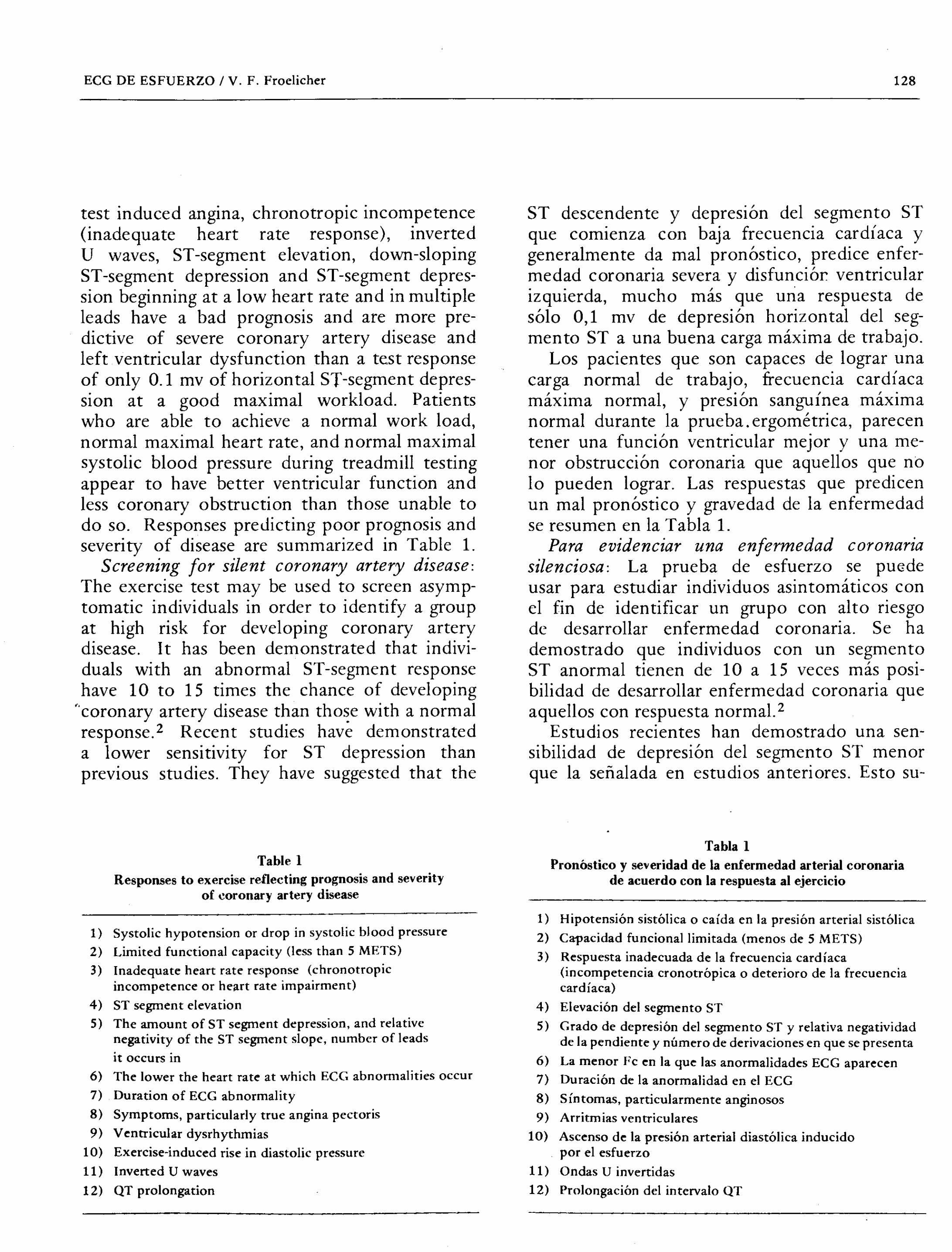
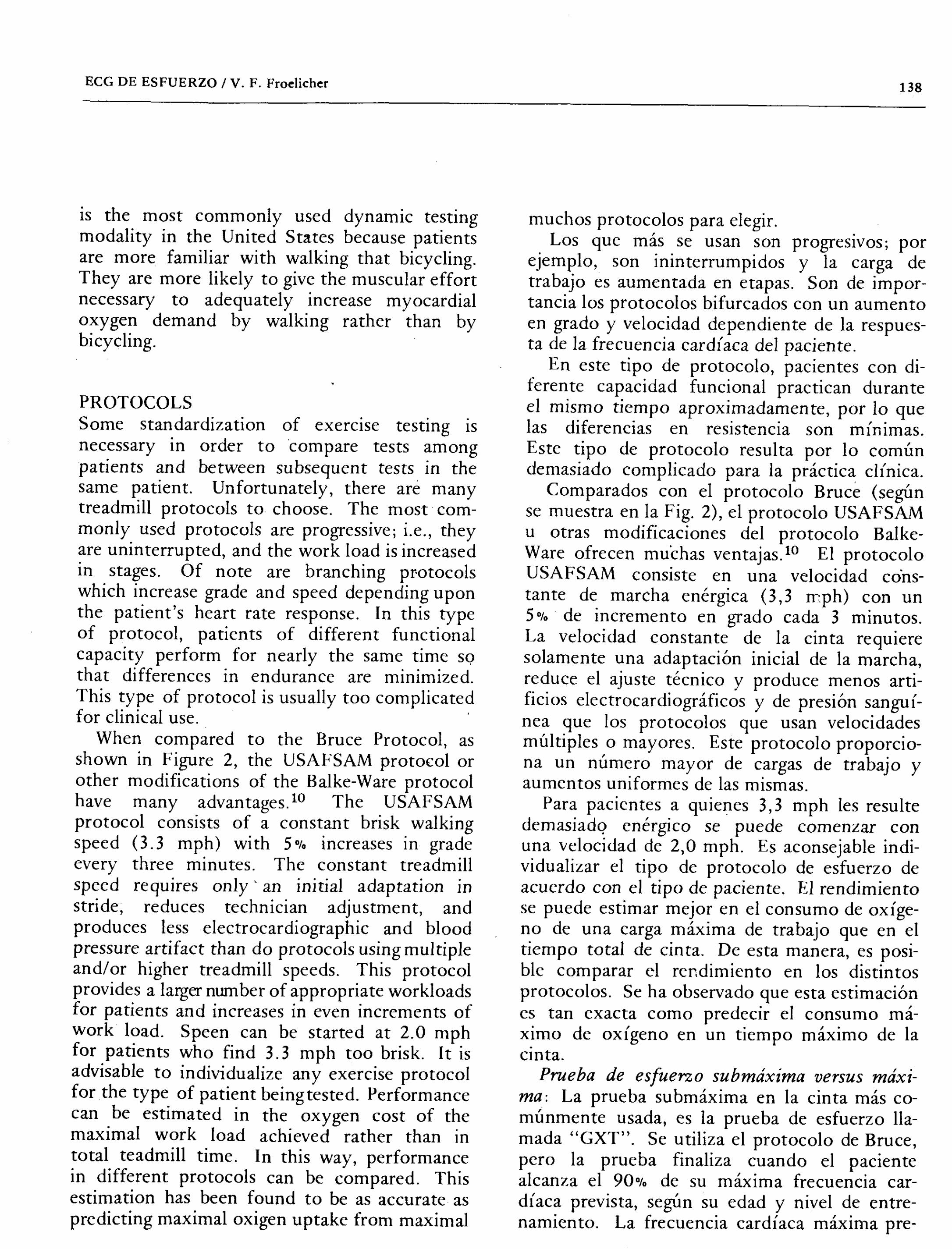
When compared to the Bruce Protocol, as
shown in Figure 2, the USAFSAM protocol or other modifications of the Balke-Ware protocol have many advantages.lO The USAFSAM protocol consists of a constant brisk walking speed (3.3 mph) with 5% increases in grade
every three minutes. The constant treadmill speed requires only' an initial adaptation in stride, reduces technician adjustment, and produces less electrocardiographic and blood pressure artifact than do protocols using multiple and/ or higher treadmill speeds. This protocol provides a larger number of appropriate workloads for patients and increases in even increments of work load. Speen can be started at 2.0 mph for patients who find 3.3 mph too brisk. It is
advisable to individualize any exercise protocol for the type of patient beingtested. Performance can be estimated in the oxygen cost of the maximal work load achieved rather than in total teadmill time. In this way, performance in different protocols can be compared. This estimation has been found to be as accurate as predicting maximal oxigen uptake from maximal
muchos protocolos para elegir. Los que más se usan son progresivos; por
ejemplo, son ininterrumpidos y la carga de trabajo es aumentada en etapas. Son de impor- tancia los protocolos bifurcados con un aumento en grado y velocidad dependiente de la respues- ta de la frecuencia cardíaca del paciente.
En este tipo de protocolo, pacientes con di- ferente capacidad funcional practican durante el mismo tiempo aproximadamente, por 10 que las diferencias en resistencia son mínimas. Este tipo de protocolo resulta por 10 común demasiado complicado para la práctica clínica.
Comparados con el protocolo Bruce (según se muestra en la Fig. 2), el protocolo USAFSAM u otras modificaciones del protocolo Balke- Ware ofrecen mu'chas ventajas.lO EI protocolo USAFSAM consiste en una velocidad co'ns-
tante de marcha enérgica (3,3 rrph) con un 5 % de incremento en grado cada 3 minutos. La velocidad constante de la cinta requiere solamente una adaptación inicial de la marcha, reduce el ajuste técnico y produce menos arti- ficios electrocardiográficos y de presión sanguí- nea que los protocolos que usan velocidades múltipIes 0 mayores. Este protocolo proporcio- na un número mayor de cargas de trabajo y aumentos uniformes de las mismas.
Para pacientes a quienes 3,3 mph les resulte demasiadQ enérgico se puede comenzar con una velocidad de 2,0 mph. Es aconsejable indi- vidualizar el tipo de protocolo de esfuerzo de
acuerdo con el tipo de paciente. El rendimiento se puede estimar mejor en el consumo de oxíge- no de una carga máxima de trabajo que en el tiempo total de cinta. De esta manera, es posi- bIe comparar el rendimiento en los distintos protocolos. Se ha observado que esta estimación es tan exacta como predecir el consumo má- ximo de oxígeno en un tiempo máximo de la
cinta. Prueba de esfuerzo submáxima versus máxi-
ma: La prueba submáxima en la cinta más co- múnmente usada, es la prueba de esfuerzo lla- mada "GXT". Se utiliza el protocolo de Bruce, pero la prueba finaliza cuando el paciente
alcanza el 90% de su máxima frecuencia car- díaca prevista, según su edad y nivel de entre- namiento. La frecuencia cardíaca máxima pre-
139 REVISTA ARGENTINA DE CARDIOLOGIA, MAYO-JUNIO 1983, VOL. 51, NO 3
ESTIMACION DEL CONSUMO DE OXIGENO DE ACUERDO CON EL TIEMPO DE PRUEBA DE LA CINTA DESLIZANTE
5.5/20 20 -
c
E -
C) .::t. -
N
o t,) t,)
70 . '.""" : Bruce
5 0/18 :
"""""" 3.3/25
4.2/16 :
............ 3.3/20
15 -;; Q) E
Velocidad de cinta (mph!% grado
58
o z w ~
X o w C
<( C <( :E X o a: 0- <( <( C ::> w c
52
46
44
2.5/12
12 . 15
... II
C
E -
C)
10 ~ o t,) t,)
~ ~
36 34
29 28 25 24
20 18
14 13
9
5
Balke-Ware modifìcado
3.3/15 USAFSAM - 3,3 mph
2/25 ,..---- I
2/20 I
r----..I 2/15 :
r-----~ 2/10: Balke-Ware modificado
,- - - - -... USAFSAM - 2,0 mph I
2/5 ,
,..---_.J 2/0 I
--_.J
treadmill time. Submaximal versus maximal exercise testing:
The most commonly used submaximal treadmill test is the exercise test, labeled the "GXT". It utilizes the Bruce protocol but the test is
terminated when the patient reaches 90% of his
prediëted maximal heart rate for age and level of training. Predicted maximal heart rate was determined from a study of normal individuals in which athletically trained subjects had a
slightly lower maximal heart rate. Unfortunately, as in other studies, there is a wide spread of maximal heart rate around the regression line declining with age (SO:t 12 beats/min). Thus, the target heart rate is maximal for some, beyond the limits of others, and submaximal for others. This testing procedure has the
3.4/14 . .........
3.3/10
.......... : 3.3/5
3 6 9 MINUTOS
8
5 en I- w
~
2
18
Fig. 2. Three progressive continuous treadmill protOcols with the approximate oxygen cost to perform each stage. The Bruce protocol and modifications of the Balke protocol are the two most commonly used treadmill protocols. Fig. 2. Tres protocolos de cinta deslizante continua con la deuda de oxígeno aproximada para cada etapa. El protocolo de Bruce y las modificaciones del protocolo de Balke son los dos más comúnmente usados. ..
vista se determinó mediante un estudio de
individuos normales en el que atletas entrenados tenían un promedio de frecuencia cardíaca máxima prevista, levemente inferior. Oesgra- ciadamente, como en otros estudios, hay un límite amplio de frecuencia cardíaca máxima alrededor de la línea de regresión, declinando según la edad (OS:t 12 latidos/minuto). Este
procedimiento tiene la ventaja de que el pa- ciente pucde realizar la prueba con zapatos y
ropa de calle; no se encuentra incómodo debido a que generalmentc no es sometido a un esfuerzo
máximo. Sin embargo, no se obtiene una sen- sibilidad máxima y la capacidad funcional no se puede estimar 0 medir con precisión.
Sistemas de derivaciones para pruebas de
esfuerzo: Los tres que se pueden usar son: el
ECG DE ESFUERZO / V. F. Froelicher 140
advantage that patients can be tested in street shoes and clothes and do not usually find the test uncomfortable since most are not stressed to a maximal effort. However, maximal sensitivity is not obtained and functional capacity cannot be accurately estimated or measured.
Lead systems for exercise testing: The three exercise lead systems that can be used include bipolar, the Mason-Likar 12.1ead adaptation, and orthogonal or non-orthogonal three-dimen- sional or VCG leads. Bipolar leads have been favored by many investigators because of the relatively short time required for application, the relative freedom from motion and muscle artifact, and the ease in which the source of noise can be located. When using the Mason- Likar torso mounted lead system the conven- tional ankle and wrist electrodes are replaced by fluid column adhesive electrodes mounted on the torso at the base of the limbs. The advantage of this system is that a reasonable fac- simile of the standard 12-lead electrocardiogram can be obtained. Moving these electrodes on to the chest and abdomen further decreases motion artifact but distorts the electrocardiogram'. Theoretically, the triangular configuration of Wilson's central terminal for the standard precordial leads requires the negative reference to be a combination of three additional electrodes rather than a single electrode used as a negative
reference for bipolar leads. We prefer to alternate between two sets of 3 leads for diagnostic
testing; they are: 1) V4, 5 and 6; and 2) V2, VS
and II. Probably only a single left ventricular lead is needed if the resting ECG is normal. However, ST elevation anteriorly can be missed and is highly specific for a proximal left anterior descending lesion.
Blood pressure measurement: Blood pressure should be taken at least at the mid portion of each exercise stage and with the appearance of chest pain. The following equipment is
preferable to the available automated blood pressure devices: a) a mercury manometer or a damped anaeroid pressure gauge mounted on the treadmill; b) an anesthesiologist's blood pressure. stethoscope with a diaphragm fastened over the brachial artery or a small crystal micro- phone attached over the brachial artery and
bipolar, la adaptación de las 12 derivaciones de
Mason-Likar y la tridimensional ortogonal 0 no ortogonal 0 derivaciones VCG.
Muchos investigadores prefieren las deriva- ciones bipolares debido a su tiempo de aplica-
ción relativamente corto, su relativa libertad de artificios musculares y de movimiento, y a la facilidad con que es posible localizar la causa de '
ruidos exteriores. Usando el sistema de Mason- Likar los electrodos convencionales de ingle y
muñeca son reemplazados por electrodos adhesi-
vos aplicados sobre el torso adyacente a la base de los miembros. La ventaja de este sistema ra- dica en que se puede obtener un facsímil de un electrocardiograma estándar de 12 derivaciones. Moviendo los electrodos hacia el pecho y abdo-
men, disminuyep los artificios de movimiento, pero se distorsiona el electrocardiograma. Tèó- ricamente, la configuración triangular de la central terminal de Wilson para las derivaciones pre cordi ales estándar, requiere que la referencia negativa sea una combinación de 3 electrodos adicionales, más que un solo electrodo usa do
como una referencia negativa para las deriva- ciones bipolares. Preferimos alternar entre 2
grupos de 3 derivaciones para prueba de diag-
nôstico. Ellos son: 1) V4, 5 Y 6: y 2) V2, VS, Y II. Probablemente se necesite una sola deri-
vación ventricular izquierda si el ECG en reposo es norm<j.l. Sin embargo, una sobreelevación ST anterior se puede perder y es altamente es-
pecífica para una obstrucción proximal de la descendente anterior.
Medición de presión sanguinea: Se debe to- mar la presión por 10 menos en la mitad de
cada etapa de esfuerzo y ante la aparición de
dolor de pecho. Se prefiere el siguiente equipo: a) un manó-
metro de mercurio 0 un manómetro aneroide montado sobre la cinta; b) un estetoscopio 0
un pequeño micrófono de cristal con amp li- ficador; c) un aparato de presión en el cual el inflado y desinflado de la almohadilla se accione mediante un manguito inflador estándar.
El brazo del paciente debe permanecer de- recho y libre de la baranda de la cinta cuando se tom a la presión.
La prueba se debe detener si la presión san-
guínea sistólica tiene una caída sostenida, espe-
141 REV 1ST A ARGENTINA DE CARDIOLOGIA, MAYO-JUNIO 1983, VOL. 51, NO 3
amplified through stethophones; c) a pressure device which inflates and then deflates the cuff on pressing a button or a standard blood pressure inflater with a push button bleed-off valve. The patient's arm should be held straight and free of the treadmill siderail when blood pressure is
taken. The test should usually be stopped if the systolic blood pressure has a sustained drop es~ecially if this drop is accompanied by chest
pam. Postexercise: If maximal sensitivity is to be
achieved, the patient should be supine in the postexercise period. According to the Laplace
relationship, the increased supine heart volume increases myocardial oxygen consumption and enhances ST-segment abnormalities. Monitoring should continue for six to eight minutes post- exercise or until changes have stabilized. In the supine position, at the fifth minute of recovery approximately 85% of pacient's with an abnormal exercise test in two large series were abnormal at this time alone or in addition to other times. An abnormal response occurring only in a
recovery period is not unusual. Having a patient do a cool-dcwn walk can delay the time at which ST-segment depression occur or eliminates it altogether. It is advisable to have the patient stop walking immediately after completing exercise, stand still briefly for about ten seconds while ECG data is gathered at near maximal heart rate, and then lay down. Care should be
taken since some people will drop their blood pressure postexercise when blood pools in their legs if they are left standing motionless. A cool- down walk can be advantageous if not perform- ing a diagnostic test.
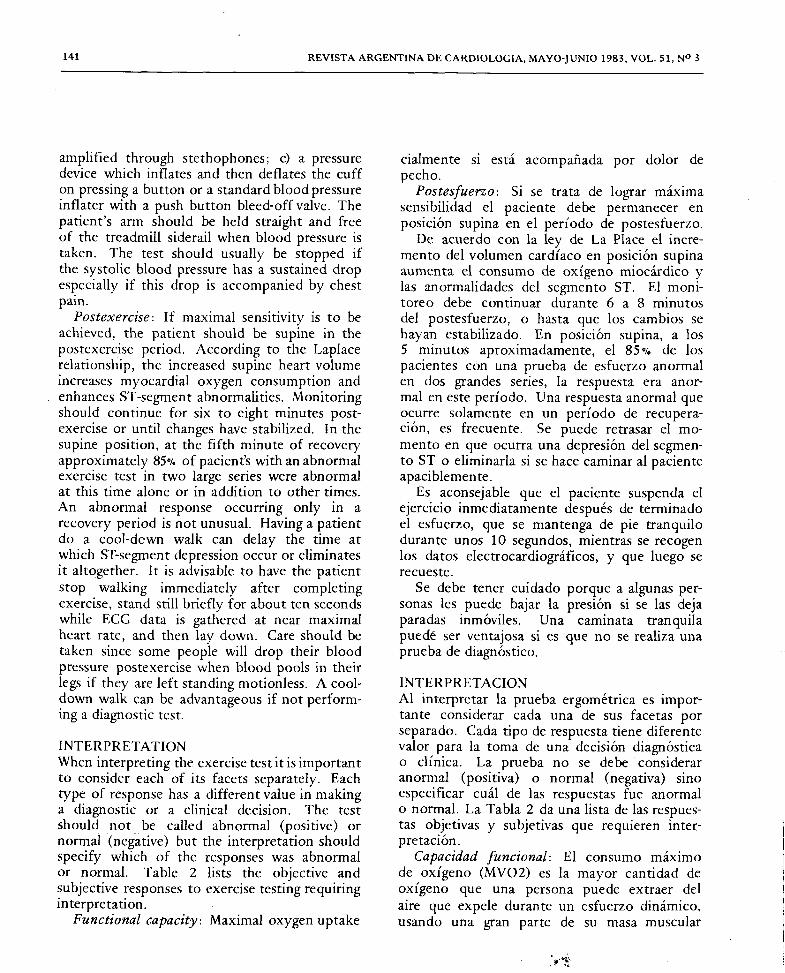
INTERPRET A nON When interpreting the exercise test it is important to consider each of its facets separately. Each
type of response has a different value in making a diagnostic or a clinical decision. The test should not be called abnormal (positive) or normal (negative) but the interpretation should specify which of the responses was abnormal or normal. Table 2 lists the objective and subjective responses to exercise testing requiring
interpretation. Functional capacity: Maximal oxygen uptake
cialmente si está acompañada por dolor de
pecho. Postesfuerzo: Si se trata de lograr máxima
sensibilidad el paciente debe permanecer en posición supina en el período de postesfuerzo.
De acuerdo con la ley de La Place el incre- mento del volumen cardíaco en posición supina
aumenta el consumo de oxígeno miocárdico y las anormalidades del segmento ST. El moni- toreo debe continuar durante 6 a 8 minutos del postesfuerzo, 0 hasta que los cambios se hayan estabilizado. En posición supina, a los 5 minutos aproximadamente, el 85% de los pacientes con una prueba de esfuerzo anormal en dos gran des series, la respuesta era anor- mal en este período. Una respuesta anormal que ocurre solamente en un período de recupera- ción, es frecuente. Se puede retrasar el mo- mento en que ocurra una depresión del segmen- to ST 0 eliminarla si se hace caminar al paciente
apaciblemente. Es aconsejable que el paciente suspenda el
ejercicio inmediatamente después de terminado el esfuerzo, que se mantenga de pie tranquilo durante unos 10 segundos, mientras se recogen los datos electrocardiográficos, y que luego se
recueste. Se debe tener cuidado porque a algunas per-
sonas les puede bajar la presión si se las deja paradas inmóviles. Una caminata tranquila puedé ser ventajosa si es que no se realiza una prueba de diagnóstico.
INTERPRETACION Al interpretar la prueba ergométrica es impor- tante considerar cada una de sus facetas por separado. Cada tipo de respuesta tiene diferente valor para la toma de una decisión diagnóstica o clínica. La prueba no se debe considerar anormal (positiva) 0 normal (negativa) sino especificar cuál de las respuestas fue anormal o normal. La Tabla 2 da una lista de las respues- tas objetivas y subjetivas que requieren inter- pretación.
Capacìdad funcìonal: El consumo máximo de oxígeno (MV02) es la mayor cantidad de
oxígeno que una persona puede extraer del
aire que expele durante un esfuerzo dinámico, usando una gran parte de su masa muscular
:-,.'i'
ECG DE ESFUERZO IV. F. Froelicher 142
Table 2
Exercise-induced electrocardiographic changes possibly
due to ischemia or dysfunction include
1) QRS changes; Transient infarct patterns Changes in R-wave amplitude Changes in S-wave amplitude Changes in Q-wave amplitude Axis changes and left anterior hemiblock QRS prolongation and bundle branch block
2) ST segment; Depression Elevation
Normalization
3) U wave inversion
4) Ventricular ectopy 5) Supraventricular ectopy and blocks
6) P wave changes
Non-ECG exercise test results to be considered include
1) Heart rate
2) Systolic blood pressure
3) Diastolic blood pressure
4) Functional capacity
5) Symptoms 6) Physical findings
7) Patient's appearance
(V02) is the highest amount of oxygen that a person can extract from expired air while performing dynamic exercise using a large part of the total muscle mass. Since maximal oxygen uptake is equal to cardiac output times the
arterial-venous oxygen difference, it is a measure of the functional limits of the cardiovascular
system. Maximal arterial venous oxygen dif- ference is physiologically limited to 15 to 17
volumes percent. Thus, maximal A V-02 difference behaves as a constan t, making maxi- mal oxygen uptake an indirect way of measuring ~aximal cardiac output. Maximal oxygen uptake IS depende~t upon many factors including age, sex, genetic endowments, and past physical activity, but it is the best index of functional capacity and maximal cardiovascular function. The maximal oxygen uptake of the normal sedentar~ individual is approximately 30 cc 02lkg/mm and the minimal level for physical "fitness" is 40 ce 02/kg/min. Aerobic train- ing can increase maximal oxygen uptake by
Tabla 2
Cambios electrocardiográficos por el ejercicio probablemente debidos a isquemia 0 disfunción
1) Cambios de QRS; Patentes transitorias de infarto Cambios en la amplitud de la onda R
Cambios en la amplitud de la onda S
Cambios en la amplitud de la onda Q
Cambios de ejes y hemibloqueo anterior izquierdo Prolongación de QRS y bloqueo de rama
2) Segmento ST; Depresión Elevación N orrnalización
3) Inversión de onda U
4) Extrasístoles ventriculares
5) Extrasístol~s supraventriculares y bloqueo
6) Cambio de onda P
Cambios no relacionados al ECG durante el esfuerzo
1) Frecuencia cardíaca
2) Presión arterial sistólica
3) Presión arterial diastólica
4) Capacidad funcional
5) Síntomas
6) Hallazgos físicos
7) Apariencia del paciente
total. Ya que el consumo máximo de oxígeno es igual al producto del gasto cardíaco por la
diferencia de oxígeno arterio-venoso, es una medida de 'los límites fun cion ales del sistema cardiovascular. El límite máximo fisiológico de la diferencia de oxígeno arterio-venoso es
de 15 ó 17 volúmenes por ciento. Por 10 tanto, la máxima diferencia A V de 02 se comporta como una constante, haciendo que el consumo máximo de oxígeno sea un modo indirecto de
medir el máximo gasto cardíaco. El consumo máximo de oxígeno depende
de muchos factores, tales como: edad, sexo, herencia genética y actividad física pasada, pero es el mejor índice de capacidad funcional y
máxima función cardiovascular. El consumo máximo de oxígeno de un individuo sedentario normal es de aproximadamente 30 cc 02/kg/min y el nivel mínimo de aptitud física es de.40 cc
02/kg/min. Un entreeamiento aeróbico puede
aumentarla en un 25%. Esto depende del nivel inicial de aptitud y de la edad, como así tam-
143 REVISTA ARGENTINA DE CARDIOLOGIA, MAYO-JUNIO 1983, VOL. 51, NO 3
approximately 25 %. This increase is dependent upon the initial level of fitness, and the age of the trainee, as well as the intensity, frequency, and length of training sessions. Trained distance runners can have maximal oxygen uptakes as
high as 60 to 80 cc 02/kg/min but mongrel dogs can exceed 100 cc 02/kg/min.
As technological advances make measuring maximal oxygen consumption and cardiac output practical it may be possible to develop limits or discriminant values for these measure- ments depending upon age, activity status, and
sex. Maximal oxygen consumption and maximal cardiac output should have similar prognostic implications toward cardiac function as do ejection fraction and left ventricular end diastolic pressure (L VEDP) whereas ST-segment changes should reflect the degree of coronary artery obstruction. Until that time, maximal oxygen consumption estimated from the work load achieved during treadmill testing will be clinically valuable.
Heart raÚ and blood pressure response: "Chronotropic incompetence" is defined as a
heart rate response below the 95th percent confidence limits for age and sex. In a follow-up study, patients with chronotropic incompetence had the same incidence of coronary artery disease as did patients with ST-segment depers-
sion. Another term is "heart rate impairment" defined as the percent deviation in measured maximal heart rate from the predicted value. The mechanism of the failure of heart rate to rise normally is poorly understood, but many cf these patiènts have poor left ventricular function, and most have multivessel coronary artery disease. However, the common use of beta-blockers has complicated the interpretation of the heart rate response to exercise.
An inadequate systolic blood pressure rise
can be due to aortic outflow obstruction or left ventricular dysfunction. Serious coronary artery disease has usually been found in patients who develop hypotension along with angina during exercise testing. Bruce defines the term "left ventricular impairment" as the percent deviation in the pressure-rate product at maximal exercise from the predicted value. Coronary artery disease patients have abnormal values but the
bién de la intensidad, frecuencia y duración de las sesiones. Corredores entrenados pueden te- ner consumos máximos de oxígenq tan altos como de 60 a 80 cc 02/kg/min, mientras que perros comunes pueden superar los 100 cc
02/kg/min. El consumo máximo de oxígeno y el gasto
cardíaco máximo deben tener una implicancia pronóstica similar en relación con la función cardíaca, con la fracción de eyección y con la presión ventricular izquierda de fin de diástole (LVEDP), mientras los cambios del segmento ST deben reflejar el grado de obstrucción coronaria. Hasta ese momento el consumo má- ximo de oxígeno estimado mediante la carga de trabajo lograda durante el esfuerzo en la
cinta, será clínicamente valedero. Respuesta de frecuencia cardíaca y presión
sanguínea: Se define como "incompetencia cronotrópica" a una respuesta de la frecuencia cardíaca por debajo del 95 % de los límites, de
acuerdo con edad'y sexo. En un estudio de se-
guimiento, los pacientes con incompetencia cronotrópica presentaban la misma incidencia de enfermedad eoronaria que pacientes con depresión del segmento ST. Otro término es
"deterioro de frecuencia cardíaca", definido
como cl porcentaje de desviación de la frecuen- cia cardíca máxima alcanzada en relación con la frecuencia cardíaca prevista. Es poco conoci- dö el mecanismo de las alteraciones en el aumen- to de la frecuencia cardíaca, pero much os de
est os pacientes tienen una mala función ven- tricular izquierda y la mayor parte tiene enfer- me dad coronaria de múltiples vasos. El uso habitual de betabloqueantes ha complicado la interpretación de la respuesta de la frecuencia cardíaca al esfuerzo.
Un aumento inadecuado de presión sistólica
sanguínea puede deberse a una obstrucción a
la salida del flujo aórtico 0 a una disfunción
ventricular izquierda. Frecuentemente se ha encontrado enfermedad coronaria seria en pa- cientes que presentan hipotensión con angina durante la prueba ergométrica. Bruce define al
término "deterioro ventricular izquierdo" como el porcentaje de desviaClón en el producto en- tre la frecuencia cardíaca y tensión arterial en esfuerzo máximo frente al valor previsto
ECG DE ESFUERZO IV. F. Froelicher 144
CONTIOl
300
ISO
MEAn U,U: fb,.1
'00
[ "
] o
"
40
220
SYSTOLIC 'LOOD 160 'IUSUJI I..H,) [ '40 ] o
110
SUI .AXIMAL IESPONSE (.... minute of .....)
[J [:] '90 ] [ ,:.
[ ::: J_[ "6 ] - '00 II
MAXIMAL (I... ",inUI8) RUPONSE SUPINE IECOVUV IUPONSI
[ 300
1;5 ] 300 [ ':4 ] 110 [ ':. ]
"3 ISO
D.A1J.\ 5}W~'IJ'.I.A5 ÞHDJMJ (oj WffH 'W'" MJIJ 00'" P.E;JC.HrrJ1.E5
40
IS' J-j:::
120
'00
:. J-[ : ]-[ :. ]-[ : ]- ---j : H ~ H ~] -[: ]-[ ~] E [ ::: J-[ :~: ]J ~ ] [ ~:~ J_[ ~~~ J_[ ::~ ] [ ~ J-[
'00
'OOf DIASfOLIC [ I&.OOD 10 -
"EUUIE ,..M,I
60
WORKLOAD 3..3I5"J1, 3.3I1fi U/15"f1
(MPH/GRADE)
"'Oa:eon 20 " .. (cc/K~)
Mln 2 Mln 5 ~f J~ MAX,MAL " -[ -: J-[ -: J-[ ': ]- "
HR-UP 30 29 2' '10'
20 20
-: 24-30 3&-44 "6-50
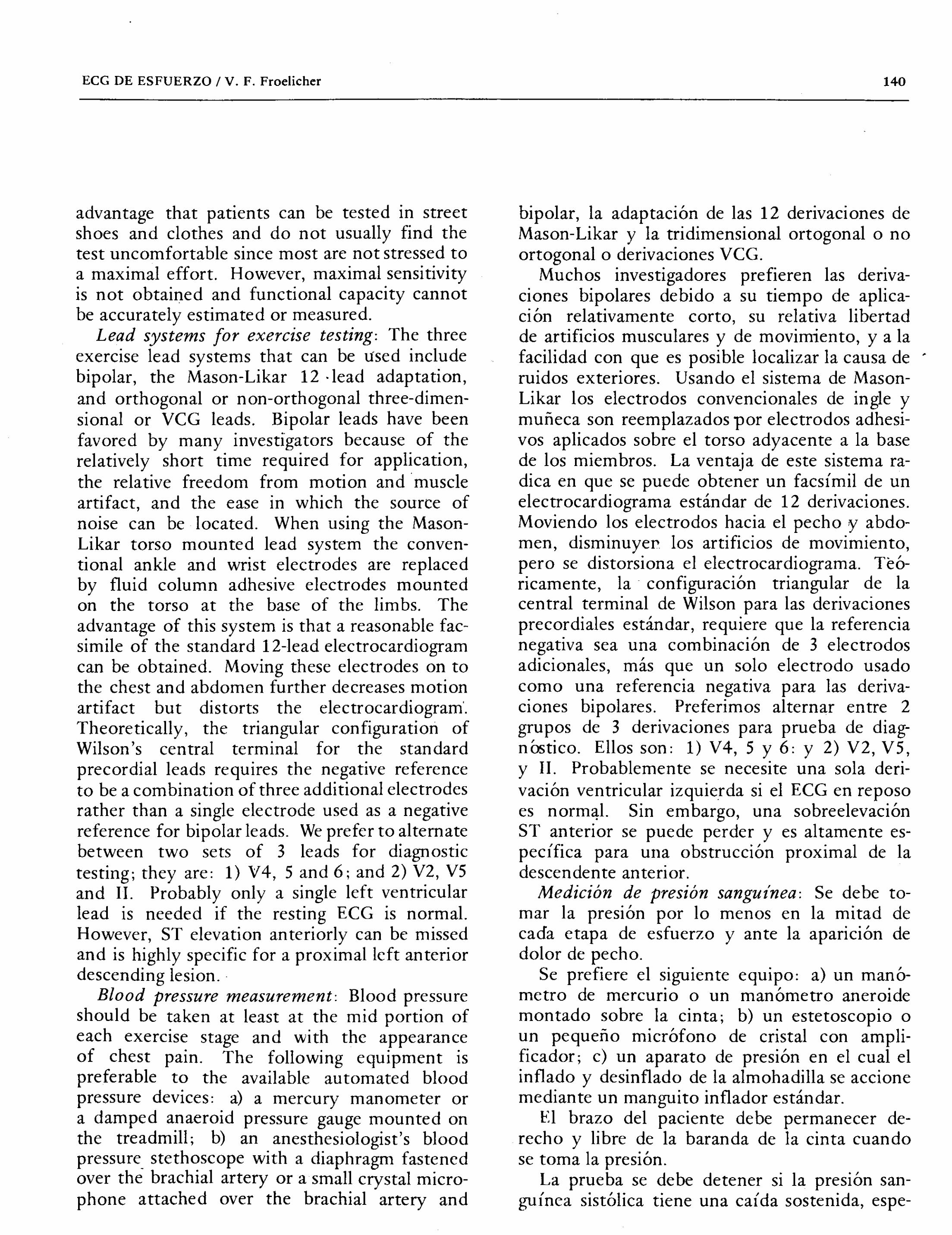
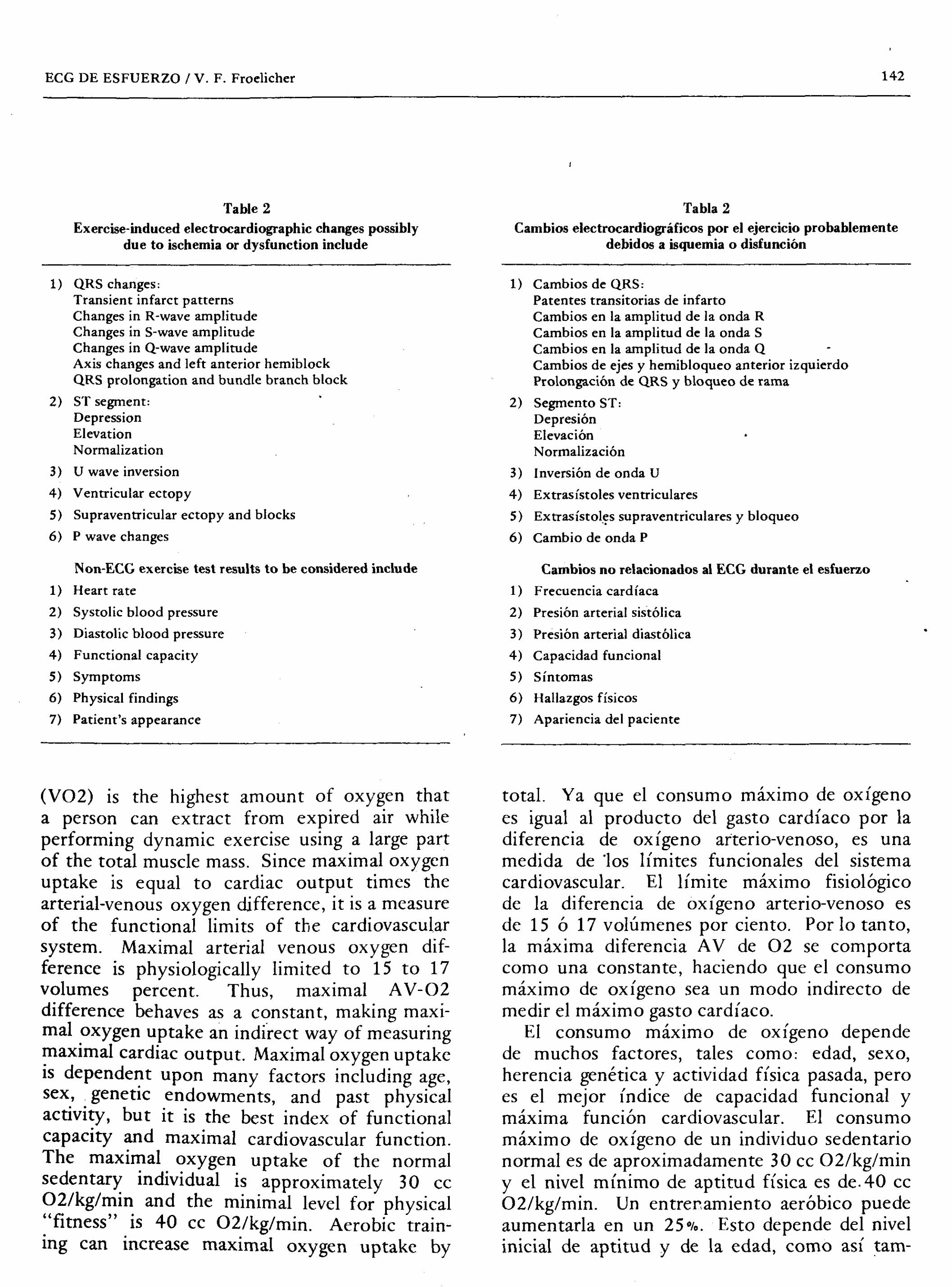
Fig. 3. References of "normal" values for the physiological response to submaximal and maximal treadmill exercise based on testing
over 1,000 apparently healthy men. "Fig. 3. Referencia de los valores "norm.ale.s" para la respuesta iesfuerzo submáximo y máximo en cinta deslizante basado en la prueba
de 1.000 hombres aparentemente sanos.
diagnostic value of this measurement has not been demonstrated. Morris and McHenry have reported the prevalence of exercise-induced hypotension in patients with definite or suspected
coronary artery disease.ll This was defined as
a sustained decrease of 10 mmHg or more in systolic blood pressure and it was found to be a reliable sign of severe coronary artery disease
in men but not in women. Recently, it was suggested that a rise in diastolic blood pressure identifies patients with coronary disease.1:l In normal males diastolic blood pressures vary only slightly during exercise. Figure 3 gives a
10-90% range of values for systolic blood pressures, heart rate, for 1,000 healthy men in response to maximal treadmill testing.13
ST-segment response: Epicardial electrode mapping usually measures ST -segment elevation over areas of severe ischemia and ST-segment depression over areas of lesser ischemia. ST-
para dicho ~sfuerzo. Los pacientes con enfermedad arterial co-
ronaria presentan valores anormales, pero el valor diagnóstico de esta medida no ha sido
demostrado. Morris y Mc Henry han infor- mado sobre la frecuencia de hipotensión pro- vocada por el esfuerzo en pacientes con en- fermedad coronaria definida 0 sopechosa.ll Esto se definió como una disminución soste- nida de 10 mmHg 0 más en la presión sistólica
sanguínea y se ha comprobado que es un signo fidedigno de enfermedad coronaria severa en hombres, pero no en mujeres. Se ha sugerido
recientemente que un aumento en presión diastólica sanguínea puede ayudar a identificar pacientes coronarios.12 En hombres norm ales
la presión diastólica sanguínea varía muy poco durante el esfuerzo. La Fig. 3 muestra una va- riación de val ores dell 0% al 90% para la presión
arterial sistólica y la frecuencia cardíaca para
145 REVISTA ARGENTINA DE CARDIOLOGIA, MAYO-JUNIO 1983, VOL. 51, NO 3
segment depression is thought to be the reciprocal
of the injury effect occurring in the endocardium as viewed from an electrode overlying normal' epicardium. ST-segment elevation seen from the same electrode reflects transmural injury or less frequently epicardial injury. On the surface electrocardiagram, exercise-induced myocardial ischemia can result in one of three ST-segment manifestations: a) ST-segment elevation; b) nor- malization or no change; or c) ST-segment depression.
Studies correlating exercise testing and
coronary angiography in hospital patients have found a 3 to 11% prevalence of ST-segment elevation. This wide range of prevalences is
probably due to the variation in the number of electrocardiographic leads recorded. ST-segment elevation appears to be an insensitive but highly specific indicator of severe myocardial ischemia. The ST -segment vector points through areas of asynergy and usually occurs in an area supplied by vessels with angiographic lesions.14 This is
in contrast to exercise-induced ST-segment depression which does not accurately predict the site of coronary lesions. Inverted U waves
appear to be specific but insensitive markers for myocardial ischemia.
The frequency that cancellation forces result in normalization or no ST-segment changes in the surface electrocardiogram during exercise- induced ischemia is unknown. Patients with three vessel coronary disease would be the ones most likely to have cancellation occur, but they have the highest prevalence of abnormal tests. When exercise testing fails to produce ST-segment depression or elevation in a patient with known coronary artery disease it could be due to two or more severely ischemic myocardial segments causing cancelling ST-segment vectors. This is
important to remember since a patient has been reported who normalized minor ST-segment depression during treadmill testing only to have an acute myocardial infarction ten minutes after the test.15
The classic criteria for abnormal ST-segment depression has been 0.1 nw or more of horizon tal or downward sloping ST-segment depression. The probability and severity of coronary artery disease are directly realted to the amount of
mil individuos sanos en respuesta a la prueba de esfuerzo máxima.13
Respuesta del segmento ST: EI electro do de mapeo epicárdico mide habitualmente la elevacÌón del segmento ST en áreas de isquemia severa y la depresión del mismo en áreas de isquemias más leves. Se piensa que la depresión del segmento ST es la recíproca del efecto del daño en el endocardio, como se ve con un electrodo superyacente al epicardio normal. La elevación del segmento ST que se registra
con el mismo electrodo, refleja un daño trans- mural 0, menos frecuentemente, un daño epi-
cárdico. En el electrocardiogram a de superfi-
cie, la isquemia de miocardio provocada por el esfuerzo puede derivar en alguna de estas mani- festaciones del segmento ST: a) elevación; b) normalización 0 sin cambio; c) depresión.
Estudios que relacionan la prueba ergomé- trica y la angiografía coronaria en pacientes hospitalizados han revelado una frecuencia de
un 3 % a un 1101. de elevación del segmento ST. Este amplio marco de frecuencia se de be proba- blemente a la variación en el número de varia- ciones electrocardiográficas registradas. La ele- vación del segmento ST es un indicador insensi- ble pero altamente específico de severa isquemia de miocardio. EI vector del segmento ST apun- ta hacia áreas de asinergia y comúnmente apa- rece en un área de vasos con lesión angiográfi- ca.1'~ Esto en contraste con la depresión del
segmento ST provocada por el esfuerzo, que no precisa el lugar de las lesiones coronarias. Ondas U invertidas son detectores específicos pero insensibles de isquemia de miocardio.
Se desconoce la frecuencia del fenómeno de cancelación de vectores, que resultan en la nor- malización 0 en la no existencia de cambios del segmento ST, en el ECG de superficie du- rante la isquemia provocada por el esfuerzo.
En pacientes con enfermedad coronaria de
tres vasos, puede ser más factible que haya cancelaci~n, pero también tiene una frecuencia mayor de pruebas anormales. Cuando la prueba de esfuerzo no produce la depresión 0 elevación del segmento ST en un paciente con enfermedad coronaria conocida, esto puede deberse ados 0
más segmentos de isquemia severa de miocardio, causantes del fenómeno de cancelación de vec-
ECG DE ESFUERZO / V. F. Froelicher 146
J-junctional depression and inversely related to the slope of the ST-segment in a V5-type lead.
However, one electrode can show an up sloping
ST-segment depression while adjacent precordial
electrode shows horizontal or downward sloping depression. It is preferable to call tests with an inadequate ST-segment slope but with ST-segment depression a borderline response and place added emphasis on other pàrameters.16 The possible causes of false negative and false positive
responses are listed in Table 3. Exercise-induced ventricular dysrbytbmias:
Premature ventricular contractions oC,cur in
approximately one-third of the asymptomatic men who perform a maximal treadmill test and the prevalence is directly related to age. They most frequently occur at maximal exercise and often are not reproducible on repeat testing. A small subgroup of healthy men (about 2%)
will have serious appearing exercise-induced dysrhythmias.17 This subgroup will have ,three times the normal risk of developing coronary disease but only about 10% of them will do so and only 7 % of those who develop coronary
"disease will have had "ominou~" dysrhythmias.
Coronary disease patients usually have a higher prevalence of severe dysrhythmias, and they usually occur at lower heart rates than in healthy subjects. Dysrhythmias suppressed by exercise do not rule out the presence of coronary disease.
R WAVE CHANGES Recently it was suggested that patients who show an increase or no decrease in R wave amplitude in response to exercise have left ventricular dysfunction. This response has been used to diagnose coronary artery disease. The R wave increase was thought to be due to an increase in left ventricular end diastolic volume secondary to ischemic dysfunction. However, in a simultaneous radionuclide angiographic and ECG exercise study we were unable to demonstrate this. Instead, it appears that R wave increases during submaximal exercise are qllite normal, and that this finding in coronary patients is due to inadequate stress. At the present time, it appears unwise to consider R wave changes in the diagnosis of coronary disease.1S,19 Recently, the Q wave in CM5
tores del segmento ST. Es importante recordar- 10, ya que se ha publicado el caso de un paciente
con una normalización durante el esfuerzo de
una depresión leve del segmentq ST que presen- tab a en condiciones basales, y que hizo un infar- to agudo de miocardio diez minutps después.
El criterio clásico de una depresión anormal del segmento ST ha sido fijado en 0,1 mv 0
más de depresión horizontal 0 descendente del
segmento ST. .
La probabilidad y la gravedad de la enfer- medad coronaria están en relación directa con la magnitud de la depresión del punto J e in- versamente relacionadas con la pendiente del
segmento ST ,en derivación V5. Sin embargo, un electrodo puede mostrar
una curva ascendente del segmento ST mkn- tras que el electrodo precordial adyacente puede
mostrar una depresión horizontal 0 descendente. La Tabla 3 da una lista de las posibles causas
de las respuestas de falso negativo y falso po- sitivo.
Arritmias ventriculares provocadas por el
esfuerzo: Aproximadamente en una tercera par- te de los hombres asintomáticos que realizan una prueba máxima en la cinta, se producen extra- sístoles ventriculares, y la frecuencia está direc- tamente relacionada con la edad. Se presentan más frecuentemente con un máximo esfuerzo y generalmente no se reproducen al repetir la
prueba. Un pequeño subgrupo de hombres sanos (aproximadamente un 2010) presentarán
severas arritmias provocadas por el esfuerzo.17 Este subgrupo puede presentar un riesgo tres veces mayor que el normal de desarrollar enfer- medad coronaria, pero solamente 10 hará un 10% Y solamente un 7% de ell os tendrá arrit- mias "ominosas".
Generalmente los enfermos coronarios presen- tan arritmias severas más frecuentemente, y
comúnmente inferior frecuencia cardíaca que hombres sanos. Las arritmias suprimidas por el
esfuerzo no descartan la existencia de enferme- dad coronaria.
CAMBIOS EN LA ONDA R
Ahora se piensa que los pacientes que muestran un aumento 0 no disminuyen la amplitud de la
onda R, en respuesta al esfuerzo, tienen una
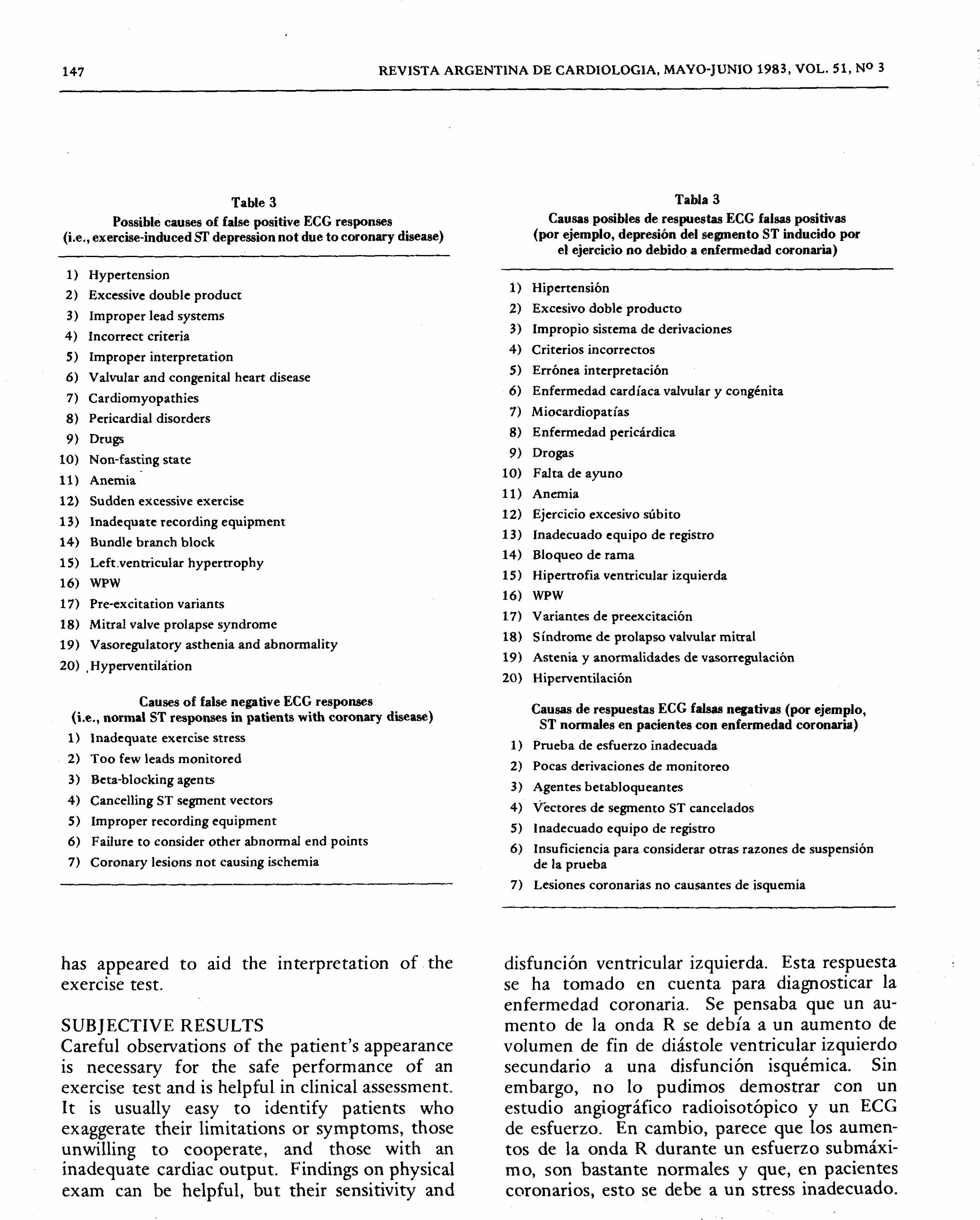
147 REVISTA ARGENTINA DE CARDIOLOGIA, MAYO-JUNIO 1983, VOL. 51, NO 3
Table 3
Possible causes of false positive ECG responses
(i.e., exercise-induced Sf depression not due to coronary disease)
1) Hypertension
2) Excessive double product 3) Improper lead systems
4) Incorrect criteria
5) Improper interpretation
6) Valvular and congenital heart disease
7) Cardiomyopathies
8) Pericardial disorders
9) Drugs
10) Non-fasting state
11) Anemia
12) Sudden excessive exercise
13 ) Inadequate recording equipment
14) Bundle branch block
15) Left.ventricular hypertrophy 16) WPW
17) Pre-excitation variants
18) Mitral valve prolapse syndrome
19) Vasoregulatory asthenia and abnormality
20) ,Hyperventilàtion
Causes of false negative ECG responses
(i.e., normal 8T responses in patients with coronary disease)
1) Inadequate exercise stress
2) Too few leads monitored 3) Beta-blocking agents
4) Cancelling ST segment vectors
5) Improper recording equipment
6) Failure to consider other abnormal end points
7) Coronary lesions not causing ischemia
has appeared to aid the interpretation of the
exercise test.
SUBJECTIVE RESULTS Careful observations of the patient's appearance is necessary for the safe performance of an exercise test and is helpful in clinical assessment. It is usually easy to identify patients who exaggerate their limitations or symptoms, those
unwilling to cooperate, and those with an inadequate cardiac output. Findings on physical
exam can be helpful, but their sensitivity and
Tabla 3
Causas posibles de respuestas ECG falsas positivas
(por ejemplo, depresión del segmento 8T inducido por el ejercicio no debido a enfermedad coronaria)
1) Hipertensión
2) Excesivo doble producto
3) Impropio sistema de derivaciones
4) Criterios incorrectos
5) Errónea interpretación
6) Enfermedad cardíaca valvular y congénita
7) Miocardiopatías
8) Enfermedad pericárdica
9) Drogas
10) Falta de ayuno
11) Anemia
12) Ejercicio excesivo súbito
13) Inadecuado equipo de registro
14) Bloqueo de rama 15) Hiperrrofia ventricular izquierda
16) WPW
17) Variantes de preexcitación
18) Síndrome de prolapso valvular mitral
19) Astenia y anormalidades de vasorregulación
20) Hiperventilación
Causas de respuestas ECG falsas negativas (por ejemplo, 8T normales en pacientes con enfermedad coronaria)
1) Prueba de esfuerzo inadecuada
2) Po cas derivaciones de monitoreo 3) Agentes betabloqueantes
4) Vectores de segmento ST cancelados
5) Inadecuado equipo de registro
6) Insuficiencia para considerar otras razones de suspensión
de la prueba
7) Lesiones coronarias no causantes de isquemia
disfunción ventricular izquierda. Esta respuesta se ha tornado en cuenta para diagnosticar la
enfermedad coronaria. Se pen saba que un au-
menta de la onda R se debía a un aumento de
volumen de fin de diástale ventricular izquierdo
secundario a una disfunción isquémica. Sin
embargo, no 10 pudimos demostrar con un estudio angiográfico radioisotópico y un ECG de esfuerzo. En cambio, parece que los aumen- tos de la onda R durante un esfuerzo submáxi-
mo, son bastante norm ales y que, en pacientes
coronarios, esto se de be a un stress inadecuado.
ECG DE ESFUERZO IV. F. Froelicher 148
specificity have not been demonstrated. Gallop sounds, a mitral regurgitant murmur, or a
precordial bulge could be due to left ventricular dysfunction, but these findings might not always be secondary to coronay artery disease.
Ischemic chest pain induced by the exercise test predicts the presence of coronary artery disease as well as does ST-segment shifts, and when both occur they are even more predictive of coronary disease than either alone.20 This is an important reason for the physician to be
present during the exercise test. The reproduction of atypical chest pain does not help, but if a
physician is there to diagnose classic ischemic
chest pain by questioning the patient then this
symptom finding is important.
REFERENCES (BIBLIOGRAFIA)
1. Froelicher VF: Exercise testing as part of the reasonable
workup before recommending medical or surgical therapy for coronary heart diSease. Circulation (in press).
2. Froelicher VF, Maron D: Using exercise testing and ancil- lary techniques to screen for coronary heart disease: A
review. Progress Cardiovasc Dis (in press).
3. McNeer JF, Margolis JR, Lee KL, Kisslo JA, Peter RH, Kong Y, Behar VS, Wallace AG, McCants CB, Rosati RA: The role of the exercise test in the evaluation of patients for ischemic heart disease. Circulation 57: 64, 1978.
4. Froelicher VF, Brammell H, Davis G, Noguera I, Stewart A, Lancaster MC: A comparison of the reproducibility and physiologic response to three maximal treadmill exercise protocols. Chest 65: 512, 1974.
5. Niederberger M: Values and limitations of exercise testing after myocardial infarction. Verlag Bruder Hollinek. Wien, Vienna, Switzerland, 1977.
Actualmente no parece acertado tener en cuenta cambios de la onda R en el diagnóstico de enfer- medad coronaria.18, 19
Recientemente se ha descubierto que la onda Q en CM5 ayuda a la interpretación de
la prueba de esfuerzo.
RESULTADOS SUBJETIVOS Un examen cuidadoso del aspecto del paciente es necesario para la realización de la prueba con seguridad y resulta dè gran ayuda para la evaluación clínica. Por 10 común es fácil identi- ficar a los pacientes que exageran sus limitacio- nes 0 síntomas, a Quienes no quieren cooperar y aquellos con un gasto cardíaco inadecuado. Los hallazgos que surjan de un examen físico pueden ser útiles, pero su sensibilidad y especi- ficidad no han sido demostradas.
.
Puede ser que ruidos de gal ope, un soplo mitral regurgitativo, 0 una expansión precordial, Sf deban a una disfunción ventricular izquierda,
pero esto no siempre puede ser secundario a
una enfermedad coronaria. El dolor de pecho de carácter isquémico pro-
vocado por el esfuerzo 0 los cambios del seg-
mento ST, denotan la presencia de enfermedad coronaria, y más aún si se presentan ambos.2o Son una razón para que el médico esté presente durante la prueba.
.
La repr<;>ducción de un dolor de pecho atípico
no ayuda, pero la habilidad del médico para interrogar permitirá concluir la característica isquémica del dolor.
6. American Heart Association Committee Report: Standards for adult exercise testing laboratories. Circulation 59: 421A, 1979.
7. American Heart Association: The Exercise. Standard Book (70-041A). Dallas, Texas, 1979.
8. Lategola MT, Busby DE, Lyne PJ: ST-segment distortions by high-side frequency filtration in direct-writing ECG recorders. Aviation Space Environmental Medicine 48: 264, 1977.
9. Bhargava V, Watanabe K, Froelicher VF: Progress in computer analysis of the exercise electrocardiogram. Am J Cardiol47: 1143, 1981.
10. Wolthuis R, Froelicher V, Fischer J: A new practical clinical treadmill protocol. American Journal of Cardiol 39: 697, 1977.
11. Morris SN, Phillips J, Jordan 1. McHenry PL: The incidence and significance of decreases in systolic BP during treadmill
149 REVISTA ARGENTINA DE CARDlOLOGIA, MAYO-JUNIO 1983, VOL. 51, NO 3
testing. Am J Cardiol41: 221-226, 1978.
12. Sheps DS, Ernst JC, Briese FW, Myerburg RJ: Exercise- induced increase in diastolic pressure: Indicator of severe
coronary artery disease. Am Journ of Cardiol 43: 708, 1979.
13. Wolthuis RA, Froelicher VF, Fischer J, Triebwasser J: The response of healthy men to treadmill exercise. Circulation 55: 153,1977.
14. Simoons ML, Brand M, Hugenholt:t PG: Quantitative analy- sis of exercise ECGs and left ventricular angiograms in
patients with abnormal QRS complexes at rest. Circulation 55: 55, 1977.
15. Sweet RL, Sheffield LT: Myocardial infarction after exer- cise induced ECG changes in a patient with variant angina
pectoris. American Journ of Cardiol 33: 813, 1974. 16. Chaitman BR, Hansen J: Comparative sensitivity and
specificity of exercise ECG lead systems. Am J Cardiol47:
1335,1981. 17. Froelicher VF, Thompson AJ, Longo MR, Triebwasser JH,
Lancaster MC: Value of exercise testing for screening asymptomatic men for latent coronary artery disease.
Progress in Cardiovasc Dis 18: 265, 1976. 18. Battler A, FroeliCher V, Slutsky R, Ashburn W: Relation-
ship of QRS amplitude changes during exercise to left ventricular function, volumes, and the diagnosis of coro- nary artery disease. Circulation 60: 1004,1979.
19. Froelicher VF, Wolthuis R, Fischer J, Uhl G: Variations in normal electrocardiographiC response to treadmill testing.
Am J Cardiol47: 1161, 1981. 20. Weiner DA, McCabe C, Hueter DC, Ryan TJ, Hood WB:
The prediCtive value of anginal chest pain as an indicator of coronary disease during exercise testing. Am Heart Journal 96: 458, 1978.