Embed Size (px)
Citation preview
BIOCHEMICAL AND BIOPHYSICAL RESEARCH COMMUNICATIONS 238, 297–300 (1997)ARTICLE NO. RC977286
SNAP-25 Is Phosphorylated by Glucose and GLP-1in RIN 1046-38 Cells
Jie Zhou and Josephine M. EganNIA/GRC/NIH, Diabetes Section, 4940 Eastern Avenue, Baltimore, Maryland 21224
Received August 8, 1997
target membrane interface, resulting in the fusion ofWe investigated the possibility that tyrosine phos- both the vesicular and plasma membranes followed by
phorylation might play a role in insulin secretion in exocytosis (6). Under certain circumstances, exocytosisthe insulinoma cell line, RIN 1046-38 cells. At least 4 from vesicles is calcium-dependent (7). Posttransla-proteins of 18, 25, 35, and 46 kDa size were found to tional modification of proteins may also alter proteinbe tyrosine phosphorylated in the presence of glu- function. Indeed, the state of phosphorylation of SNAP-cose and an insulin secretagogue, glucagon-like pep-
25 appears to regulate catecholamine release (8), andtide-1 (GLP-1). The addition of glucose and GLP-1 topossibly other hormones.cells that were exposed to the tyrosine kinase inhibi-
In acinar cells of the pancreas, it has been showntor genistein resulted in a decrease in the extent ofthat tyrosine phosphorylation events are involved inphosphorylation of the 18, 25, and 35 kDa proteinsthe regulation of amylase release by cholecystokininand a concomitant reduction in insulin secretion,(9, 10). The nature of these signalling proteins are un-whereas treatment with vanadate, a tyrosine phos-known but may involve phosphorylation of eitherphatase inhibitor, led to enhanced responses. Immu-VAMP or the plasma membrane proteins involved innoprecipitation of cellular proteins with an anti-fusion and/or exocytosis of amylase. Both endocrinephosphotyrosine antibody followed by immunoblot-
ting with a specific monoclonal antibody to SNAP-25 and exocrine cells of the pancreas are derived from a(synaptosome-associated protein of 25 kDa) revealed common progenitor cell (11, 12) and both containthat the 25 kDa protein is SNAP-25. These results LDCVs for the regulated release of insulin and amy-suggest that tyrosine phosphorylation of SNAP-25 lase, respectively. These observations led to the postu-may be involved in the regulation of insulin secre- late that tyrosine phosphorylation events might be in-tion in RIN 1046-38 cells. q 1997 Academic Press volved in insulin release. The aim of this study was
to explore the role of tyrosine phosphorylation in theregulation of insulin release by glucose and the insulinsecretagogue, GLP-1, in an insulinoma cell line, RIN
Insulin is stored in beta cells in large dense-core se- 1046-38.cretory vesicles (LDCVs) that accumulate in proximityof release sites and fuse with the plasma membrane in
MATERIALS AND METHODSresponse to secretagogues (1). The molecules involvedin neuronal synaptic vesicle exocytosis have been char-
Materials. All chemicals used were of the highest purity avail-acterized (2); however, little is known about exocytosisable. Medium 199 (M199) was from the NIH Media Unit. Fetal bovine
in endocrine cells and it is just lately that some infor- serum (FBS) and trypsin/EDTA solution were purchased from Gibcomation has become forthcoming (3, 4). It appears that BRL (Grand Island, NY). GLP-1, glucagon and insulin were pur-
chased from Bachem (Torrence, CA). Monoclonal and polyclonal anti-beta cells and neurons share similar molecular mecha-phosphotyrosine antibodies were purchased from Upstate Biotech-nisms for exocytosis (4). At the point of synapsis, vesi-nology, Inc. (Lake Placid, NY) and protein G Plus/protein A agarosecle-associated membrane protein (VAMP) of the vesicle beads from Oncogene Science, Inc. (Uniondale, NY). Monoclonal anti-
membrane interacts with two presynaptic plasma body (Clone #SP12) against SNAP-25 was from Serotec Co. (Oxford,membrane proteins; the synaptic-associated protein of England). Enhanced chemiluminescent (ECL) detection system,
horseradish peroxidase conjugated donkey anti-rabbit IgG, sheep25 kDa (SNAP-25) and syntaxin (5). Together theseanti-mouse IgG and 125-I-insulin kit were from Amersham Co. (Ar-three molecules form a complex which functions as alington Heights, IL). Polyvinylidene difluoride (PVDF) membraneSNAP receptor, also known as SNARE. SNAPs (soluble and precast gradient 4-20% polyacrylamide gels were from Novex
NSF attachment proteins) and NSF (N-ethylmalei- (San Diego, CA). Tween-20, Triton X-100, phenylmethylsulfonyl flu-oride (PMSF), genistein and Na-orthovanadate were from Sigmamide-sensitive factor) bind to the SNARE at the vesicle/
0006-291X/97 $25.00Copyright q 1997 by Academic PressAll rights of reproduction in any form reserved.
297
AID BBRC 7286 / 6938$$$241 08-29-97 08:01:47 bbrcg AP: BBRC

Vol. 238, No. 2, 1997 BIOCHEMICAL AND BIOPHYSICAL RESEARCH COMMUNICATIONS
Chemical Co. (St. Louis, MO). Cells were from American Type Cul-ture Collection (Rockville, MD). Benzamidine, leupeptin and aproti-nin were obtained from ICN (Costa Mesa, CA). Protein assay reagentwas from BIO-RAD Laboratories (Hercules, CA).
Cell culture and treatment. RIN cells were propagated in cultureconditions, as previously described (13). In brief, cells were grown toapproximately 80% confluency in M199, 5% heat-inactivated FBS,100 IU/ml penicillin, 100 mg/ml streptomycin, 2 mM glutamine, and2 mM glucose at 377C with 95% air-5% CO2. They were then washedtwice with Kreb-Ringer’s balanced salt solution (KRB) without glu-cose and were preincubated in KRB for 2 h at 377C. Then the culturedishes were transferred to an aluminium cooling plate (Pharmacia)connected to a thermostatic water circulator (Lauda). After a 5-minincubation at 377C, the phosphorylation reaction was carried out byadding 20 mM glucose and/or 10 nM GLP-1. In some cases, the cellswere treated with the tyrosine kinase inhibitor genistein or the tyro-sine phosphatase inhibitor vanadate for 30 min prior to addition ofglucose and GLP-1. The incubation medium was collected after 2min, and the cells rapidly washed before their immersion in liquidnitrogen. The collected media were centrifuged (2 min, 2,000 xg,47C) and the supernatants stored at 0207C until further analysis forinsulin content by RIA.
Immunoprecipitation, SDS-PAGE, and immunoblotting. The fro-zen cells were scraped and lysed in the immune precipitation buffercontaining 25 mM Tris-HCl (pH 8.0), 137 mM NaCl, 1% Triton X-100, 0.5% deoxycholate, 0.1% SDS, 0.2 mM PMSF, 1 mM Na-ortho-vanadate, 1 mM benzamidine, 10 mg/ml leupeptin and 20 mg/ml apro-tinin. Insoluble material was removed by centrifugation at 15,000xg for 15 min, and the clarified lysates were incubated with a mono-clonal anti-phosphotyrosine antibody or monoclonal anti-SNAP-25antibody at 47C overnight with constant agitation. Protein G Plus/protein A agarose beads were added and 4 h later the immunocom-plexes were washed three times with the immune precipitation bufferand once with washing buffer (25 mM Hepes [pH7.4], 0.1% TritonX-100 and 1 mM Na-orthovanadate). The samples were denaturedin Laemlli sample buffer (14) in the presence of 10% 2-mercaptoetha-nol by heating at 1007C for 5 min, and then resolved by electrophore-sis on 4-20% or 12% precasted gradient polyacrylamide gel. Afterelectrophoresis, proteins were electrotransferred to PVDF mem-brane. The PVDF membranes were blocked with 5% (w/v) non-fatdry milk in TBST buffer (20 mM Tris-HCl [pH 7.5], 137 mM NaCland 0.1% Tween 20) for 1 h at room temperature, and then incubatedwith antibody to SNAP-25 (1: 5,000 dilution) or polyclonal anti-phos- FIG. 1. Protein tyrosine phosphorylation in RIN 1046-38 cells inphotyrosine (1:1,500) for 1 h at room temperature. PVDF membranes response to glucose and GLP-1. (A) RIN cell lysates were immunopre-were washed three times with TBST and incubated with horseradish cipitated with a monoclonal antibody to phosphotyrosine, and theperoxidase-conjugated anti-mouse or anti-rabbit secondary antisera immunoprecipitated complexes were subjected to SDS-PAGE andfor 1 h at room temperature. After a series of washes in TBST, transferred to PVDF membrane. The blot was probed with polyclonalthe blots were developed using the ECl chemiluminescent detection anti-phosphotyrosine antibody and detected using a chemilumines-system. Autoradiographs were quantified by using Image-Quant soft- cent technique. Cells were treated for 2 min in the following manner:ware (version 3.3) on a Molecular Dynamics laser densitometer. Ali- (1) no treatment, (2) glucose alone (20 mM) (3) GLP-1 alone (20 nM),quots (20 ml) of clarified cell lysates were used to determine protein and (4) glucose / GLP-1. (B) densitometric quantification of selectedconcentration which was estimated by the Bradford method with bands from the gel in A.bovine gamma globulin as standard.
Statistical analysis. All data are expressed as mean { SEM. Aosine antibody (Fig. 1). An increase in phosphorylationvalue of p õ 0.05 by non-paired Student’s t-test was considered sig-
nificant. of proteins at 18 (1.5-fold), 25 (1.65-fold), 35 (1.3-fold),and 46 (2.0-fold) kDa was observed when compared tountreated cells. Likewise, GLP-1 caused an increase in
RESULTS AND DISCUSSION the phosphorylation of the same proteins but with adifferent pattern. While GLP-1 treatment lead to a 2.5-fold increase in phosphorylation of the 18 and 25 kDaTo determine if glucose is able to enhance tyrosine
phosphorylation of cellular proteins in RIN cells, we proteins phosphorylation of the 35 and 46 kDa proteinswas increased 1.3-fold only. Both glucose and GLP-1incubated lysates from glucose-treated cells with a
monoclonal anti-phosphotyrosine antibody followed by had additive effects on the 4 proteins of interest.Next, we analysed the antiphosphotyrosine immuno-Western blot analysis with polyclonal anti-phosphotyr-
298
AID BBRC 7286 / 6938$$$242 08-29-97 08:01:47 bbrcg AP: BBRC

Vol. 238, No. 2, 1997 BIOCHEMICAL AND BIOPHYSICAL RESEARCH COMMUNICATIONS
(Fig. 4 A,B). The 18 and 35 kDa proteins were immuno-precipitated also by the anti-SNAP-25 antibody. Thesematch the size of VAMP (18 kDa) and syntaxin (35kDa) (15,16) and probably indicates that phosphoryla-tion activates SNAP-25 and allows its association withthe other two SNARE proteins, syntaxin and VAMP.This complex formation as has already been describedin neurons and beta cells (17, 18).
In this report, we have demonstrated that; 1) SNAP-25 is phosphorylated on tyrosine; 2) tyrosine phosphor-ylation events occur in response to glucose and GLP-1; and, 3) an increase in tyrosine phosphorylation byvanadate and a decrease by genistein altered insulin
FIG. 2. Effects of insulin-secreting agents on tyrosine phosphory-lation of SNAP-25. (A) RIN 1046-38 cells were treated in the followingmanner: (1) no treatment, (2) glucose alone (20 mM), (3) GLP-1 alone(20 nM), and (4) glucose / GLP-1 for 2 min. Cells were lysed andthe phosphoproteins were immunoprecipitated with a monoclonalanti-phosphotyrosine antibody. The immunoprecipitates were sepa-rated by SDS-PAGE and the proteins were electrotransfered to PVDFmembrane. The membrane was immunoblotted with a monoclonalanti-SNAP-25 antibody. (B) Alternatively the PVDF membrane wasincubated with an anti-phosphotyrosine antibody in the absence (1)or presence of (2) 1000-fold excess phosphorylated tyrosine.
precipitate with an anti-SNAP-25 antibody (Fig. 2A).The fact that a 25 kDa phosphoprotein co-migratedwith SNAP-25 may indicate that SNAP-25 is phosphor-ylated. To verify the specificity of the tyrosine phos-phorylation PVDF membrane was reprobed with anti-phosphotyrosine antibody in the presence of 1,000-foldexcess phosphorylated tyrosine. The intensity of thesignal associated with the 18 kDa protein markedlydecreased while that of the 46 kDa band was not af-fected. Furthermore, the bands at 25 and 35 kDa aswell as other lesser bands were abolished (Fig. 2B).
To further confirm that tyrosine phosphorylationevents occurred following cell treatment with glucoseand/or GLP-1, the effect of a chemical inhibitor of tyro-sine kinase was assessed. Genistein at 300 and 500mM(but not 100 mM) inhibited phosphorylation of all thephosphorylated proteins in a concentration-dependentmanner (Fig. 3A). These concentrations of genisteinalso decreased glucose- and GLP-1-mediated insulinsecretion (Fig. 3B). However, even though genistein at300 mM totally abolished insulin secretion in response
FIG. 3. Effects of genistein on SNAP-25 tyrosine phosphorylationto secretagogues, the proteins of interest were stilland insulin secretion in RIN 1046-38 cells. (A) Cells were pre-incu-
phosphorylated to a greater degree than non-stimu- bated with genistein for 30 min prior to the addition of the secreta-lated cells. The implication is that other proteins in- gogues. Two min later, the media were collected for the measurement
of insulin content (B). (A) Cells were lysed and a monoclonal anti-volved in insulin secretion are also not being activatedSNAP-25 antibody was added to the clarified cell lysates. The immu-in the presence of genistein. Vanadate (200 mM), a tyro-noprecipitates were separated by SDS-PAGE and the proteins elec-sine phosphatase inhibitor, potentiated glucose- and trotransferred to PVDF membrane which was immunoblotted with
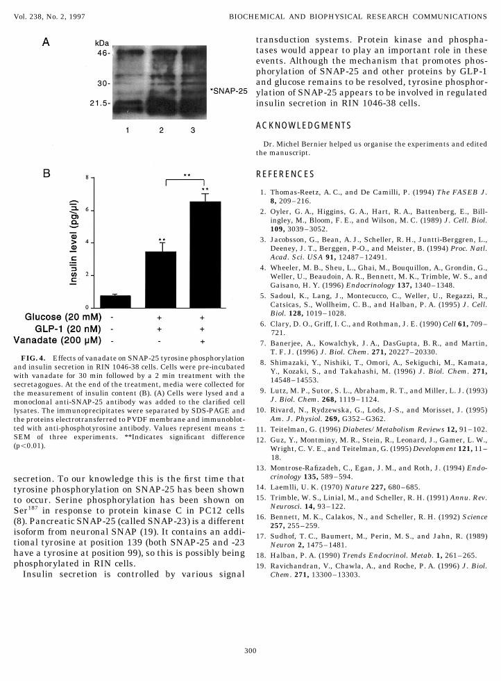
GLP-1-mediated tyrosine phosphorylation and potenti- antiphosphotyrosine antibody. Values represent means { SEM ofthree experiments. *Indicates significant difference (põ0.05).ated insulin secretion in response to secretagogues
299
AID BBRC 7286 / 6938$$$242 08-29-97 08:01:47 bbrcg AP: BBRC
Vol. 238, No. 2, 1997 BIOCHEMICAL AND BIOPHYSICAL RESEARCH COMMUNICATIONS
transduction systems. Protein kinase and phospha-tases would appear to play an important role in theseevents. Although the mechanism that promotes phos-phorylation of SNAP-25 and other proteins by GLP-1and glucose remains to be resolved, tyrosine phosphor-ylation of SNAP-25 appears to be involved in regulatedinsulin secretion in RIN 1046-38 cells.
ACKNOWLEDGMENTS
Dr. Michel Bernier helped us organise the experiments and editedthe manuscript.
REFERENCES
1. Thomas-Reetz, A. C., and De Camilli, P. (1994) The FASEB J.8, 209–216.
2. Oyler, G. A., Higgins, G. A., Hart, R. A., Battenberg, E., Bill-ingley, M., Bloom, F. E., and Wilson, M. C. (1989) J. Cell. Biol.109, 3039–3052.
3. Jacobsson, G., Bean, A. J., Scheller, R. H., Juntti-Berggren, L.,Deeney, J. T., Berggen, P-O., and Meister, B. (1994) Proc. Natl.Acad. Sci. USA 91, 12487–12491.
4. Wheeler, M. B., Sheu, L., Ghai, M., Bouquillon, A., Grondin, G.,Weller, U., Beaudoin, A. R., Bennett, M. K., Trimble, W. S., andGaisano, H. Y. (1996) Endocrinology 137, 1340–1348.
5. Sadoul, K., Lang, J., Montecucco, C., Weller, U., Regazzi, R.,Catsicas, S., Wollheim, C. B., and Halban, P. A. (1995) J. Cell.Biol. 128, 1019–1028.
6. Clary, D. O., Griff, I. C., and Rothman, J. E. (1990) Cell 61, 709–721.
7. Banerjee, A., Kowalchyk, J. A., DasGupta, B. R., and Martin,T. F. J. (1996) J. Biol. Chem. 271, 20227–20330.
FIG. 4. Effects of vanadate on SNAP-25 tyrosine phosphorylation 8. Shimazaki, Y., Nishiki, T., Omori, A., Sekiguchi, M., Kamata,and insulin secretion in RIN 1046-38 cells. Cells were pre-incubated Y., Kozaki, S., and Takahashi, M. (1996) J. Biol. Chem. 271,with vanadate for 30 min followed by a 2 min treatment with the 14548–14553.secretagogues. At the end of the treatment, media were collected for9. Lutz, M. P., Sutor, S. L., Abraham, R. T., and Miller, L. J. (1993)the measurement of insulin content (B). (A) Cells were lysed and a
J. Biol. Chem. 268, 1119–1124.monoclonal anti-SNAP-25 antibody was added to the clarified cell10. Rivard, N., Rydzewska, G., Lods, J-S., and Morisset, J. (1995)lysates. The immunoprecipitates were separated by SDS-PAGE and
Am. J. Physiol. 269, G352–G362.the proteins electrotransferred to PVDF membrane and immunoblot-ted with anti-phosphotyrosine antibody. Values represent means { 11. Teitelman, G. (1996) Diabetes/Metabolism Reviews 12, 91–102.SEM of three experiments. **Indicates significant difference 12. Guz, Y., Montminy, M. R., Stein, R., Leonard, J., Gamer, L. W.,(põ0.01). Wright, C. V. E., and Teitelman, G. (1995) Development 121, 11–
18.13. Montrose-Rafizadeh, C., Egan, J. M., and Roth, J. (1994) Endo-
crinology 135, 589–594.secretion. To our knowledge this is the first time that14. Laemlli, U. K. (1970) Nature 227, 680–685.tyrosine phosphorylation on SNAP-25 has been shown15. Trimble, W. S., Linial, M., and Scheller, R. H. (1991) Annu. Rev.to occur. Serine phosphorylation has been shown on
Neurosci. 14, 93–122.Ser187 in response to protein kinase C in PC12 cells16. Bennett, M. K., Calakos, N., and Scheller, R. H. (1992) Science(8). Pancreatic SNAP-25 (called SNAP-23) is a different 257, 255–259.
isoform from neuronal SNAP (19). It contains an addi- 17. Sudhof, T. C., Baumert, M., Perin, M. S., and Jahn, R. (1989)tional tyrosine at position 139 (both SNAP-25 and -23 Neuron 2, 1475–1481.have a tyrosine at position 99), so this is possibly being 18. Halban, P. A. (1990) Trends Endocrinol. Metab. 1, 261–265.phosphorylated in RIN cells. 19. Ravichandran, V., Chawla, A., and Roche, P. A. (1996) J. Biol.
Chem. 271, 13300–13303.Insulin secretion is controlled by various signal
300
AID BBRC 7286 / 6938$$$242 08-29-97 08:01:47 bbrcg AP: BBRC