Embed Size (px)
Citation preview

Review
Spinal muscular atrophy: disease & clinical manifestationsSpinal muscular atrophy (SMA) is an inherited autosomal recessive neurodegenerative disease. It is the leading genetic cause of infantile mortality worldwide with a disease prevalence of approxi-mately 1:6000–1:10,000 and a carrier frequency of approximately 1:35–1:40 [1,2]. SMA preva-lence is static throughout all ethnic groups, with few exceptions, although an isolated population from South Dakota, the Hutterites, possesses a dramatically higher carrier frequency approach-ing 1:8 [3]. SMA is characterized by the degen-eration of motor neurons within the anterior horn of the spinal cord leading to skeletal muscle weakness and atrophy [4]. Muscle weakness and atrophy is symmetrical and progressive, often impacting the legs more so than the arms, even-tually leading to a decline in intercostal activity. Respiratory failure and complications account for the majority of premature deaths in SMA patients [4].
Clinically, SMA disease severity is broad and for classification purposes, patients are catego-rized based upon the severity, the age of onset, and achieving (or failing to achieve) physical milestones [5]. Type 0 is extremely severe and initiates during prenatal development and results in death within weeks. Type I, or Werdnig–Hoffman disease, is a severe form characterized by an infantile onset ranging from birth to 6 months and accounts for approximately 50% of all newly diagnosed cases of SMA [6]. The natu-ral history of all forms of SMA has significantly changed due to supportive measures, including
respiratory and physical therapy, however, in the absences of these advances, there is a 32% sur-vival probability at 2 years of age and at no time can type I patients sit upright without support [7]. Type II onset occurs between 6–18 months and initiates with proximal limb weakness, with progressive weakness and respiratory complica-tions, joint contractures and scoliosis appearing in childhood [7]. At some point during child-hood, type II patients can sit upright with-out assistance. Approximately 70% of type II patients live to adulthood. Type III SMA pre-sents past 1 year (>1 year for type IIIa; >3 years for IIIb) and individuals can initially stand/walk without assistance and have a normal lifespan, although many become wheelchair-bound dur-ing adolescence [4]. Type IV patients develop proximal leg weakness in adulthood and have a normal lifespan [4]. Even within each disease cat-egory, further delineation exists and it could be argued that there is a continuous disease severity spectrum, however, in the age of molecular diag-nostics and clinical trial enrollment, this system retains a great deal of utility.
Molecular genetics: SMN1 & SMN2SMN1 is the SMA-determining gene and is located on chromosome 5q13 [8]. While a gen-eralized region of chromosome 5 had been iden-tified several years earlier as the SMA locus, it was not until 1995 that the SMA-determining gene was conclusively identified as SMN1 [9–11]. However, the genetics and the identification were clearly complicated by the presence of a nearly identical gene also located on chromosome 5q.
SMN-inducing compounds for the treatment of spinal muscular atrophy
Spinal muscular atrophy (SMA) is a leading genetic cause of infant mortality. A neurodegenerative disease, it is caused by loss of SMN1, although low, but essential, levels of SMN protein are produced by the nearly identical gene SMN2. While no effective treatment or therapy currently exists, a new wave of therapeutics has rapidly progressed from cell-based and preclinical animal models to the point where clinical trials have initiated for SMA‑specific compounds. There are several reasons why SMA has moved relatively rapidly towards novel therapeutics, including: SMA is monogenic; the molecular understanding of SMN gene regulation has been building for nearly 20 years; and all SMA patients retain one or more copies of SMN2 that produces low levels of full-length, fully functional SMN protein. This review primarily focuses upon the biology behind the disease and examines SMN1- and SMN2-targeted therapeutics.
Monique A Lorson1 & Christian L Lorson*2,3
1Department of Veterinary Pathobiology, Bond Life Sciences Center, Room 440C, University of Missouri, MO 65211 USA
2Department of Veterinary Pathobiology, Bond Life Sciences Center, Room 471G, University of Missouri, Columbia, MO 65211, USA 3Department of Molecular Microbiology & Immunology, University of Missouri, MO, USA *Author for correspondence: Tel.: +1 573 884 2219 Fax: +1 573 884 9395 E-mail: [email protected]
2067ISSN 1756-891910.4155/FMC.12.131 © 2012 Future Science Ltd Future Med. Chem. (2012) 4(16), 2067–2084
For reprint orders, please contact [email protected]

Key Terms
Spinal muscular atrophy: Pediatric neurodegenerative disease that is a leading genetic cause of infantile death worldwide.
SMN1: SMA-determining gene. Mutations or deletions of this gene give rise to spinal muscular atrophy.
Alternative splicing: Gene regulation process in which exons are joined together in a manner other than a linear progression. In SMN2, the majority of transcripts lack exon 7.
This gene, called SMN2, is positioned within an approximately 500 Kb duplication that lies in a head-to-head orientation [8]. The duplicated region that contains SMN2 is centromeric to SMN1 and additional genes such as p44, SERF and NAIP are also present in each duplicated region [8,12]. Most SMA cases arise from dele-tions including SMN1 exons 7 and 8, however, larger deletions were also detected that encom-passed adjoining genes, such as NAIP [12]. While there was initially some concern that other genes within this region contributed to SMA develop-ment, the identification of SMN intragenic muta-tions including frameshifts and point mutations further confirmed the role of SMN1 [13].
The nucleotide sequences of SMN1 and SMN2 are 99% identical and the amino acid sequences are 100% identical for the overlap-ping coding elements. There are a handful of sequence variations in the promoter regions as well as small variations within the genes, mostly within intronic elements [8,14,15]. Seemingly paradoxically, mutation or loss of SMN2 has no clinical consequence provided SMN1 is intact, whereas loss of SMN1 results in SMA. The key to understanding this complex genetic question resides in a single non-polymorphic nucleotide difference at the 5´ end of exon 7 (840C>T). SMN1 transcripts are predominately full-length, spanning exons 1–8, whereas the majority of SMN2-derived transcripts are alternatively spliced producing an isoform that lacks the typical final coding exon (exon 7) [16,17]. Exon-skipped products then terminate early in exon 8 after encoding only four amino acids. Insight into the importance of the 840C>T transition was initially identified by ‘hybrid’ alleles in SMA patients [18]. Likely due to the relatively large genomic duplication and inversion of the SMN locus, the SMN genes appear to be prone to recombination and partial recombination events that can produce small arrays of SMN1 or SMN2 genes on a single chromosome or can result in a single ‘hybrid’ SMN gene that is partially derived from SMN1 and SMN2. Through the analysis of hybrid genes as well as synthetic mini-genes, it was determined that the 840C>T dictated the exon 7 alternative splicing event: an SMN1-derived ‘C’ resulted in a hybrid gene that exhib-ited a SMN1-like expression profile, whereas a SMN2-derived ‘T’ resulted in a hybrid gene that exhibited a SMN2-like expression profile [15,19].
Importantly, approximately 10%, of SMN2-derived transcripts encodes fully functional, full-length SMN protein. It is important to stress that
SMA does not arise from the complete absence of SMN, consistent with observations that a SMN null state appears to be lethal in all organ-isms [20]. Rather, SMA develops from a severe reduction of SMN. The low levels of full-length SMN produced by SMN2 are sufficient to pre-vent embryonic lethality, but are insufficient to prevent SMA development [21]. Additionally, murine models suggest that the exon 7-skipped protein product, SMND7, retains a small degree of functionality compared to full-length SMN [22]. One or more copies of SMN2 are retained by all SMA patients and, to date, SMN2 is clearly the most important genetic modifier of disease sever-ity [23–26]. Numerous patient studies have been performed, allowing a relatively straightforward conclusion to be drawn: SMN2 dosage inversely correlates with disease severity. In general, type I patients have two copies; type II patients have 2–3 copies; and type III patients have 3–4 cop-ies [27–29]. Additionally, healthy individuals have been identified that are homozygously deleted for SMN1, but retain 5 or more copies of SMN2 [30]. Genetically, these SMA-carriers could be considered as SMA patients as they lack SMN1, however, the high number of SMN2 copies has provided sufficient SMN protein to fully protect from disease development. Clearly, overlap exists for each of these groups and much like the con-tinuous clinical spectrum, SMN2 dosage covers a similarly broad range as well. At the molecular level, the overwhelming majority of patients are screened by genetic means that are designed to assess the presence or absence of SMN1/SMN2 genes [31,32]. These tests, however, are typically based upon the 840C>T difference and do not discriminate between SMN2 alleles that produce high or low levels of SMN or from those that may be completely dysfunctional. Therefore, while gene dosage is an incredibly important compo-nent of an individual’s diagnosis, this cannot be the sole determinant and must be merged with the clinical manifestations. Molecular diagnostics, however, are playing an increasingly important role in SMA clinical trials and the stratification of patients with a particular SMN2 genotype will likely become increasingly more important as SMA-specific compounds are examined in clinical trials.
SMN protein function: general activity versus motor neuron activitySMN is a multifaceted 38 kDa protein that is ubiquitously expressed throughout develop-ment [33]. Early studies suggested that SMN
Review | Lorson & Lorson
Future Med. Chem. (2012) 4(16)2068 future science group

performed an essential function for all cells since genetic ablation of the murine Smn resulted in pre-implantation lethality [34]. Insight into SMN function initially came through yeast two-hybrid studies and SMN’s hallmark nuclear staining pattern [35]. Within many cell types, SMN localizes into discrete nuclear foci termed ‘gemini of coiled bodies’ or ‘gems’ [35,36]. While coiled bodies and gems are enriched for factors involved in various aspects of gene expression, including transcription and RNA processing components, these nuclear structures are not active sites of transcription or splicing. However, this localization pattern suggested a role in a variety of RNA-associated activities. Detailed biochemical studies have subsequently revealed that SMN often functions with a cohort of pro-teins collectively referred to as Gemins [37]. The core complex consists of SMN, Gemins 2–8 and unrip, and can be found in all tissues and cell types. The SMN/Gemin complex is integral to the assembly of small nuclear ribonuclear proteins (snRNPs) within the cytoplasm and their subsequent transport into the nucleus [37]. snRNPs are composed of a single snRNA and a heptameric ring structure composed of Sm proteins. SMN’s role in this activity is with-out question, both biochemical studies as well as structural data demonstrate the relevance of the SMN/Gemin complex in snRNP assembly. Perhaps one of the most intriguing questions in the SMA field has centered upon how a dysregu-lation of an essential, general cellular activity, snRNP assembly, could account for the motor neuron-specific vulnerability observed in SMA [38]. While general snRNP assembly defects have been observed in SMA mice, these defects were observed in a pathway referred to as the major spliceosome or at a relatively late stage of disease [39,40]. In contrast, an alternative class of introns comprising <1% of all introns referred to as U12-dependent or minor introns has been the target of recent speculation [41–44]. The connection between SMA and U12-dependent introns stems from the observation that U12 introns are not uniformly distributed throughout the genome [45,46]. Rather, U12 introns are enriched within voltage-gated ion channels and other genes likely to be involved in neuronal function [46]. The dis-covery of U12 genes that are aberrantly spliced in SMA contexts would provide mechanistic validity to the role of snRNP assembly in SMA development as well as providing novel drug targets beyond SMN. It is likely, however, too simplistic to assume that correcting one or two
improperly spliced mRNAs could completely correct the entirety of the SMA phenotype and a more nuanced balance of U12 introns may be difficult to achieve pharmacologically.
An alternative, non-snRNP function has also been proposed that could account for the motor neuron specific loss observed in SMA [47]. SMN largely exists in snRNP-free protein/RNA com-plexes within axons and it has been hypothesized that SMN serves as an mRNA chaperone for a subset of mRNAs as they are transported dis-tally to the growth cones [48–50]. SMN has been shown to interact and/or co-localize with several factors involved in RNA transport, including hnRNP-R, hnRNP-Q, HuD, COP1 and with actin and candidate plasticity-related gene 15 mRNA [51–55]. In cultured neurons from SMA mice, the SMN-neuronal complexes are dis-rupted and the cargoes are poorly localized to the developing termini. The current thought is that localized translation of factors, such as b-actin, would be disrupted leading to an alteration in actin dynamics and subsequent cytoskeleton growth and development at the growth cone, however genetic ablation of b-actin does not result in lower motor neuron defects [56]. While the biochemistry behind the SMN-neuronal function is still developing, research in other fronts has supported a role for SMN in some type of cytoskeletal activity. For example, two recent reports have demonstrated that com-pounds Fasudil and Y-27632 that modulate actin dynamics by inhibition of the RhoA/ROCK pathway can significantly extend survival in an intermediate mouse model of SMA [57,58]. The protective affect was independent of SMN as SMN levels remained stable and low. Plastin 3 has also been postulated to function as a disease modifier as its overexpression was detected in discordant siblings and shown to correlate with decreased disease severity in female siblings [59]. Plastin 3 was also capable of compensating for SMN-deficient phenotypes in zebrafish and cultured neurons [59].
While there are two primary schools of thought regarding the SMN-specific function that leads to motor neuron loss, the reality is that neither has been conclusively proven or disproven and the picture remains complex. SMN interacts with an ever-expanding list of proteins, totaling over 60 to date [60]. While SMN missense mutations typically fail to bind to each of the protein substrates, it is unclear whether these factors contribute to SMA devel-opment. The recent analysis of b-actin null mice
SMN-inducing compounds for the treatment of spinal muscular atrophy | Review
www.future-science.com 2069future science group

was hypothesized to result in neuronal defects, however, the b-actin null mice appeared to be surprisingly normal regarding axonal regenera-tion and neuronal function, although a more detailed analysis in a CNS-restricted ablation revealed abnormalities within the hippocampus and cerebellum [56,61].
Regulation of SMN pre-mRNA splicing: to skip or not to skipEukaryotic pre-mRNA splicing is an intricate balance between cis and trans factors that co-ordinate the identification and proper excision of
intronic sequences (FiguRe 1). SMN exon 7 is 54 nucleotides and includes the stop codon for full-length SMN [8]. Although SMN1 and SMN2 are nearly identical, the pre-mRNA splicing pat-terns are dramatically different and underscore the importance of cis- and trans-factors that co-ordinate the complex regulatory environment surrounding SMN exon 7 (FiguRe 2) [16,17]. In the context of SMN1, exon 7 is constitutively included in the majority of transcripts. While SMN1 exon 7 is flanked by relatively weak splice signals including the poly-pyrimidine tract, and the 3 and 5 splice sites, the presence of several
AAG GU AG GUYYYY
AG GU AG GUA YYYY
U1snRNP
U1snRNPU2
snRNPU2AF65
35
ESS
ESS
ISSESS
AG GU AG GUA YYYY
U2snRNP
U2AF6535
U1snRNP
U1snRNP
ESSISS
ESS
AG GU AG GUA YYYY
U2snRNP
U2AF6535
U1snRNP
U1snRNP
SR SRSR SR SRSR
ESS ESS
AG GU AG GUA YYYY
U2snRNP
U2AF6535
U1snRNP
U1snRNPSRSR
SR SRSRhnRNP
hnRNPhnRNP
SR
A
B
C
D
E
Figure 1. Eukaryotic pre-mRNA splicing factors. (A) Typical conserved cis and trans (B) splicing signals found at the intron/exon junction. (C) Auxiliary regulatory cis elements, including repressors (red) such as ESS and ISS or positive factors, such as ESE. (D) SR proteins binding to ESEs. (E) Negatively regulating factors, such as hnRNP proteins binding their cognate sites, thereby inhibiting SR protein binding and exon inclusion. ESE: Exonic splice enhancers; ESS: Exonic splice silencers; ISS: Intronic splice silencers; SR: Ser-/Arg-rich.
Review | Lorson & Lorson
Future Med. Chem. (2012) 4(16)2070 future science group
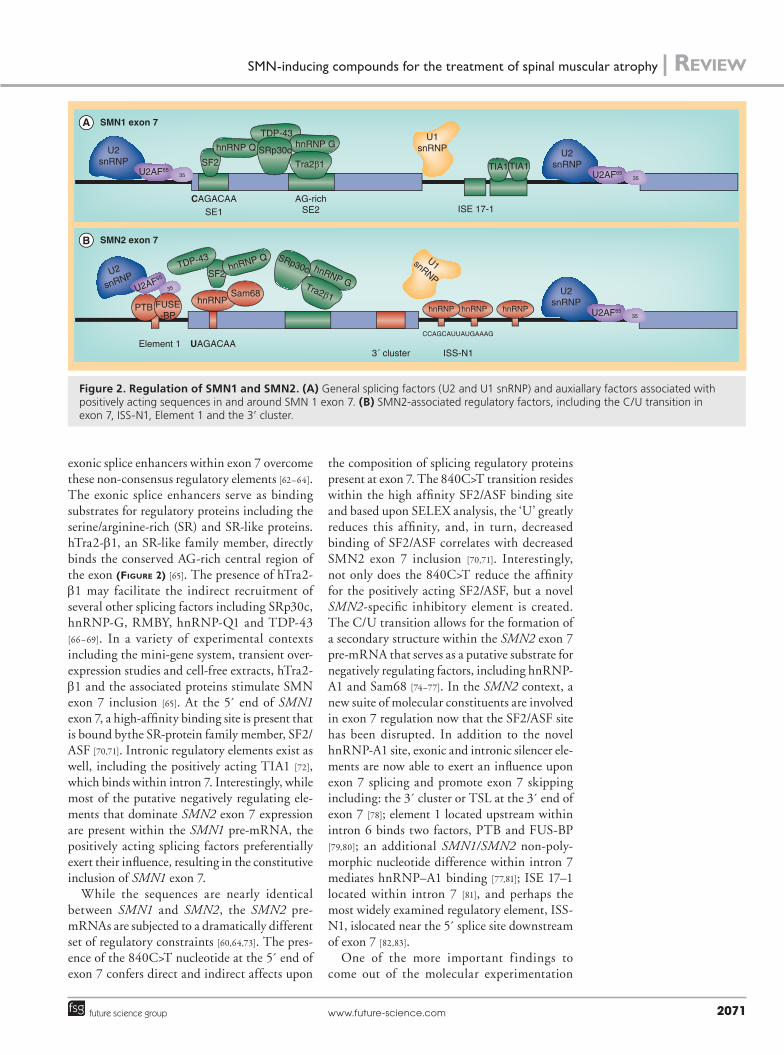
exonic splice enhancers within exon 7 overcome these non-consensus regulatory elements [62–64]. The exonic splice enhancers serve as binding substrates for regulatory proteins including the serine/arginine-rich (SR) and SR-like proteins. hTra2-b1, an SR-like family member, directly binds the conserved AG-rich central region of the exon (FiguRe 2) [65]. The presence of hTra2-b1 may facilitate the indirect recruitment of several other splicing factors including SRp30c, hnRNP-G, RMBY, hnRNP-Q1 and TDP-43 [66–69]. In a variety of experimental contexts including the mini-gene system, transient over-expression studies and cell-free extracts, hTra2-b1 and the associated proteins stimulate SMN exon 7 inclusion [65]. At the 5´ end of SMN1 exon 7, a high-affinity binding site is present that is bound bythe SR-protein family member, SF2/ASF [70,71]. Intronic regulatory elements exist as well, including the positively acting TIA1 [72], which binds within intron 7. Interestingly, while most of the putative negatively regulating ele-ments that dominate SMN2 exon 7 expression are present within the SMN1 pre-mRNA, the positively acting splicing factors preferentially exert their influence, resulting in the constitutive inclusion of SMN1 exon 7.
While the sequences are nearly identical between SMN1 and SMN2, the SMN2 pre-mRNAs are subjected to a dramatically different set of regulatory constraints [60,64,73]. The pres-ence of the 840C>T nucleotide at the 5 end of exon 7 confers direct and indirect affects upon
the composition of splicing regulatory proteins present at exon 7. The 840C>T transition resides within the high affinity SF2/ASF binding site and based upon SELEX analysis, the ‘U’ greatly reduces this affinity, and, in turn, decreased binding of SF2/ASF correlates with decreased SMN2 exon 7 inclusion [70,71]. Interestingly, not only does the 840C>T reduce the affinity for the positively acting SF2/ASF, but a novel SMN2-specific inhibitory element is created. The C/U transition allows for the formation of a secondary structure within the SMN2 exon 7 pre-mRNA that serves as a putative substrate for negatively regulating factors, including hnRNP-A1 and Sam68 [74–77]. In the SMN2 context, a new suite of molecular constituents are involved in exon 7 regulation now that the SF2/ASF site has been disrupted. In addition to the novel hnRNP-A1 site, exonic and intronic silencer ele-ments are now able to exert an influence upon exon 7 splicing and promote exon 7 skipping including: the 3´ cluster or TSL at the 3´ end of exon 7 [78]; element 1 located upstream within intron 6 binds two factors, PTB and FUS-BP [79,80]; an additional SMN1/SMN2 non-poly-morphic nucleotide difference within intron 7 mediates hnRNP–A1 binding [77,81]; ISE 17–1 located within intron 7 [81], and perhaps the most widely examined regulatory element, ISS-N1, islocated near the 5 splice site downstream of exon 7 [82,83].
One of the more important findings to come out of the molecular experimentation
U1snRNP U2
snRNPU2AF65
35
ISE 17-1SE2SE1AG-richCAGACAA
U2snRNP
U2AF6535
SF2
hnRNP Q
TDP-43hnRNP G
Tra2β1 TIA1TIA1
SRp30c
SMN1 exon 7A
U1snRNP
U2snRNP
U2AF6535
3´ clusterUAGACAA
SF2hnRNP Q
TDP-43hnRNP GTra2β1
SRp30c
ISS-N1
CCAGCAUUAUGAAAG
hnRNP hnRNPhnRNPhnRNP
PTB
Sam68
Element 1
U2
snRNPU2AF65
35
FUSE-BP
SMN2 exon 7B
Figure 2. Regulation of SMN1 and SMN2. (A) General splicing factors (U2 and U1 snRNP) and auxiallary factors associated with positively acting sequences in and around SMN 1 exon 7. (B) SMN2-associated regulatory factors, including the C/U transition in exon 7, ISS-N1, Element 1 and the 3’ cluster.
SMN-inducing compounds for the treatment of spinal muscular atrophy | Review
www.future-science.com 2071future science group

surrounding SMN exon 7 splicing was that SMN2 exon 7 is not irreversibly damaged. In fact, subtle mutations in the poly-pyrimidine tract upstream of exon 7 completely reverse the splicing ratio such that SMN2 produces exclu-sively full-length transcripts [63]. Similarly, muta-tions within exon 7 that modulate the inhibitor and enhancer binding sites can overcome the C/U-mediated splicing patterns [78,83–87], while overexpression of modified U1 snRNAs that specifically recognize the non-canonical splice site downstream of exon 7 largely correct the exon skipping phenotype [87]. Several reports have also demonstrated that by reducing splice site recognition at the exon 8 junction, the bal-ance is shifted to the exon 7 splice site leading to an increased incorporation of SMN2 exon 7 [62,88–90]. Naturally occurring mutations also highlight the malleable nature of exon 7 splic-ing. For instance, discordant phenotypes that did not match the predicted disease severity based upon the SMN2 copy numbers revealed a novel mutation within SMN2 exon 7, 859G>C. This mutation was predicted to create a high affinity exonic splice enhancer for SF2/ASFand consistent with the in silico-based predications, the 859G>C mutation resulted in increased inclusion of SMN2 exon 7 – even in the presence of the 840C>T transition [91]. These important observations demonstrated that SMN2 exon 7 is not irreparably damaged and provided a bio-logical foundation for strategies to modulate SMN2 exon 7 pre-mRNA splicing as a potential therapeutic for SMA.
Interestingly, a feedback mechanism may exist that contributes to the motor neuron spe-cific sensitivity observed in SMN deficient con-texts [92,93]. In in vitro assays, SMN reduction increased SMN2 exon 7 inclusion, suggesting that a reduction in SMN was further exacerbated by SMN-induced aberrant splicing of SMN2 exon 7 [92,93]. Consistent with this, RT-PCR analysis of SMN transcripts isolated from laser captured SMA motor neurons from SMA mice demonstrated that SMN2 exon 7 splicing was significantly more impacted by reduced SMN than non-motor n euron tissue [93].
SMN induction: modes of action for SMN therapeutic strategies�� Gene replacement: viral vectors
Since SMA is monogenic and the SMN cDNA is relatively small, SMA is well suited for a viral-based replacement of SMN. Early work with pseudotyped lentivirus vectors and purported
retrograde transport was capable of modest extensions in survival in severe SMA mice following intramuscular injections in SMA mice [94]. However, several hurdles still existed including achieving in vivo tropism for the appropriate tissues and being able to deliver a sufficiently high titer to the CNS. A breakthrough came when self-complementary adeno-associated virus vectors were used to delivery SMN to SMA mice. Intravenous (iv.) delivery of scAAV9–SMN at P1 resulted in a dramatic extension in survival and a signifi-cant rescue of the SMA phenotype [95–100]. Importantly, motor neuron transduction with the scAAV9 vector was high and an extension in survival, albeit reduced to an average of 22 days, could still be achieved if the vector was delivered at P5 [97]. Motor neuron transduc-tion dropped dramatically at the later delivery time point, suggesting that the blood–brain barrier had blocked entry into the CNS or that disease progression has advanced to a stage that is no longer correctable [97]. In a separate report, delivery of AAV8–SMN or scAAV8–SMN via intracerebroventricular (icv.) injection also significantly extended sur-vival; however, instead of the 250 plus days of survival observed in the scAAV9–SMN treated animals, these mice lived on average approximately 150 days [98]. Several possible explanations could account for the differences in life span for scAAV8 and scAAV9 treated animals. Recent work from several labora-tories has demonstrated that severe SMA mice present a multi-organ system pathology including cardiac and vascular tissue and the pancreas [101–105]. scAAV9 has an exceptionally broad tropism, while scAAV8 tropism is more restricted and poorly transduces cardiac tissue [106]. This requirement for SMN within the CNS and the periphery may explain why high motor neuron expression of SMN following scAAV8–SMN results in a decreased degree of rescue compared to scAAV9–SMN delivery. In a head-to-head comparison of icv. versus iv. administration of scAAV9–SMN, icv. admin-istration was shown to produce a greater degree of rescue in SMA [100,107]. Similar conclusions were also drawn regarding icv. administration in an even more severe model of disease that typically dies at approximately P5 [107]. While the extension in survival did not achieve the 150–250 days seen with the less severe model, this work demonstrated that delivery of the scAAV9-SMN vector can significantly extend
Key Term
Adeno-associated virus: Dependo-parvovirus that is frequently a vector of choice for gene therapy based upon its broad tropism, low immunogenicity, and their ability to infect quiescent cells.
Review | Lorson & Lorson
Future Med. Chem. (2012) 4(16)2072 future science group

survival even in severely symptomatic SMA animals.
Results using various mouse models indicated that CNS uptake following an iv. administra-tion scAAV9–SMN was robust and provided the injection was performed almost immediately after birth, however, CNS penetration dropped off dramatically if the injection was given one week later. In contrast, adult cat and non-human primate studies have shown that scAAV9 can still enter the CNS following a single IV injec-tion and, more importantly, efficient motor neuron uptake can still be achieved [108–110]. In terms of translating iv. delivery to SMA patients, current vector production technologies cannot accommodate a large scale clinical trial for older children or adults. However, sufficient virus could be generated for a clinical trial that uti-lized a more focused CNS-specific delivery para-digm. While neutralizing antibodies are clearly an issue that could hinder the advancement of this type of strategy [110], a SMA clinical trial is likely within the next 1–2 years.
�� Targeting SMN2: promoter activationAll SMA patients retain one or more copies of SMN2 and since SMN2 has the potential to encode fully functional SMN protein, this copy gene has been an invaluable therapeutic target. While total expression levels between SMN1 and SMN2 are similar, there has been significant interest in further stimulating the basal level of SMN2 transcription: even though SMN2 pre-mRNAs are preferentially producing SMND7, a global increase in transcription would boost full-length and SMND7 mRNA. While early reports suggested overexpression of SMND7 protein functioned in a dominant-negative manner, the generation of the SMND7 mouse demonstrated that SMND7 was neither toxic nor a dominant-negative [22]. These animals are homozygous null for murine Smn, possess two human SMN2 genes and contain an additional transgene that overexpresses the cDNA for the SMND7 isoform [22]. The addition of the SMND7 cDNA extends survival to approximately 14 days, as compared with approximately 5 days for the Smn-/-;SMN2 SMA mice. This conclusively demonstrated that the SMND7 product was not detrimental and the upregulating the SMN2 promoter could be a viable means of elevating SMN protein.
Multiple layers of regulation exist to tightly control eukaryotic gene expression. A global mechanism to regulate gene expression is through the compaction and relaxation of
DNAaccomplished in part by histone acety-lases (HATs) which acetylate lysine residues found on histones, a primary building block for chromatin. Acetylation relaxes the DNA allowing for transcription while the activity of histone deacetylases (HDACs) promotes DNA chromatin compression and gene repres-sion. Pharmacological manipulation of this system can be accomplished with a class of compounds referred to as HDAC inhibitors (HDACi) [111,112]. Within the context of SMA, repurposed compounds have shown promise in SMA reporter assays, SMA cells and in SMA mice. Sodium butyrate [113], valproic acid (VPA) [114–117], phenylbutyrate [118], trichostatin A (TSA) [119,120], LBH589 [121], and suberoylani-lide hydroxamic acid (SAHA) [122,123] have been shown to increase SMN protein levels from the human SMN2 gene and in some instances from the murine Smn gene. Translating cell-based success into a phenotypic improvement in SMA mice has been challenging, in part, due to the extreme severity of the SMA models. However, TSA and SAHA were able to provide signifi-cant extensions in survival as well as lessening overall disease severity in SMA mice [119,120,123]. LBH589 elevated SMN protein by stimulat-ing exon 7 inclusion and total SMN2 promoter expression, as well as increasing hTra2-b1 levels [121]. TSA plus a rigorous regimen of supportive and dietary care was further capable of extend-ing survival by ~170% [120]. In addition to their SMN-inducing activity, many HDACis appear to confer some degree of general neuroprotection independent of SMN, potentially through the suppression of atrogene pathways [124].
Perhaps one of the greatest challenges going forward for these types of compounds relates to specificity, or more to the point, the lack of specificity. In all likelihood, SMA patients will need to be on medication permanently and the pharmacological induction of a tangible per-centage of the genome is likely to lead to off-target affects and toxicity. A recent inducible SMA mouse model of disease further suggests that elevated SMN levels will be required into adulthood as well [125]. While the investiga-tions into the molecular regulation behind the SMN promoter structure(s) has provided some insight [126], additional work is needed to bet-ter understand the regulatory environment as well as to develop more specific HDACi that could be used for long-term administration. Since VPA and phenybutyrate are US FDA-approved compounds, it was possible to move
SMN-inducing compounds for the treatment of spinal muscular atrophy | Review
www.future-science.com 2073future science group
these compounds into clinical trials for SMA type II and III patients, however, relatively lit-tle effect was observed [127–129]. Similar to the results seen with VPA and phenybutyrate, which performed well in cell-based assays, hydroxyurea was shown to increase SMN full-length RNA levels in SMA primary lymphoblast cultures but when analyzed in a placebo-controlled, double-blind trial, HU did not alter the SMA phenotype or SMN expression [130,131].
Prolactin (PRL), a blood–brain barrier per-meable compound, has recently been shown to increase SMN expression dramatically in SMA cellular models and within the CNS of SMA mice [132]. While a significant extension in survival was observed in prolactin treated ani-mals (~70%), this extension was not as robust as the SMN levels within the CNS might have predicted. One possible interpretation of these results is that while the receptor for PRL is widely expressed throughout the CNS, it is not widely present within the periphery. The rela-tively early mortality of PRL-treated animals did not appear to be due to NMJ-associated defects, but perhaps related to peripheral organ defects. PRL acts through the STAT5 pathway and was initially suggested as a potential target due to the functional overlap of three small molecules that increased SMN2 expression: sodium val-proate, TSA and aclarubicin. STAT5 activa-tion in SMA-like mouse embryonic fibroblasts and SMN2-expressing NSC34 cells increased SMN while a constitutively activated STAT5 mutant increased SMN in SMA patient lym-phocytes [133]. Going forward, delivery of the FDA-approved recombinant PRL will likely be examined in SMA patients. Its activity and the absence of significant non-CNS activity will be of particular interest in the future.
During the first high-throughput screen for SMA-specific compounds, two parallel screens were run in an attempt to capture compounds with two distinct modes-of-action: SMN2 promoter activation and SMN2 exon 7 splic-ing. The SMN2 promoter assay identified a quinazoline structure that exhibited cell-based activity and led to a medicinal chemistry pro-gram that produced several compounds with drug-like properties [134,135]. The lead com-pound, 2,4-diaminoquinazoline, was shown to function through a distinct mechanism: inhibition of DcpS [136], which is an enzyme involved in 5´ cap-mediated degradation of mRNAs. Oral administration of the lead com-pound (RG3039) resulted in a dose-dependent
increase of SMN in SMA mice and extended survival by ~20–30% [137]. A Phase I study per-formed by Repligen Corporation in 32 healthy individuals analyzing dosing, safety and toler-ance has recently concluded and indicated a good safety profile for RG3039.
Targeting SMN2: modulating exon 7 inclusion with nucleic acid-based therapeuticsWhile early work demonstrated that exon 7 splic-ing could be modulated by the over-expression of transacting factors or the genetic removal of splicing inhibitors surrounding exon 7, there was little belief that these insights could translate into potential therapeutic strategies. However, this work laid the foundation for a new wave of research that leveraged the power of antisense oligonucleotides (ASOs) (FiguRe 3). ASOs are relatively short stretches of nucleic acid that recognize a target sequence with a high degree of specificity. In most instances, the molecu-lar backbone has been chemically modified to reduce nuclease sensitivity, thereby extending the half-life for ASO compounds. While ASOs have often been used to knock-down expression of target mRNAs through siRNA pathways, ASOs within the context of SMA are designed as splice site-switching ASOs as a means to alter SMN2 exon 7 pre-mRNA splicing. The most straightforward strategy is to target an inhibitor of SMN2 exon 7 inclusion, such as the intronic splice silencer (ISS) elements surrounding exon 7, including ISS-N1 and E1 (FiguRe 3). ISS-N1 has been targeted by ASOs composed of several different backbone chemistries and of varying lengths [79,138–141], however, two compounds have demonstrated the greatest degree of efficacy in SMA mouse models. ISIS Pharmaceuticals has developed a backbone technology that uti-lizes a 2´-O-2-methyoxyethyl-modified back-bone in their ASO-10–27 (ISIS SMNRx). In an asymptomatic SMN2-transgenic model, ASO-10–27 fully reversed the SMN2 splicing pattern when administered via an icv. injection [142], whereas subsequent work in an SMA mouse model resulted in a significant extension in sur-vival from approximately 14 days to approxi-mately 26 days following a single icv. injection [143]. Somewhat unexpectedly, systemic admin-istration of the same ASO resulted in nearly a 25-fold increase in survival, demonstrating the importance of peripheral tissues in severe SMA mouse models [144]. SMNRx has entered an open label Phase I clinical study that is designed to
Key Term
Antisense oligonucleotide: Short stretch of nucleic acid that is complementary to a specific sequence. By blocking regulatory elements, antisense oligonucleotides can alter pre-mRNA splicing patterns.
Review | Lorson & Lorson
Future Med. Chem. (2012) 4(16)2074 future science group

examine dosing following a single intrathecal injection.
An ASO with a different chemical backbone called Morpholinos has been used to target ISS-N1 with similar success [145]. Using a slightly longer ASO, which spanned the sequences -10 to -29 (relative to the 3´ end of exon 7), a single high-concentration dose administered via icv. injection at P0 conferred a high degree of protection from SMA and extended survival to an average of 112 days. In contrast to the ISIS SMNRx ASO, no significant additional survival benefit was gained when ICV delivery was augmented with periph-eral administration. It has become clear that while the murine blood–brain barrier in early neonatal pups allows compounds and ASOs to pass this will likely not be the case for larger animals and human clinical trials. However, without know-ing the therapeutic time window or the pharma-cokinetics of each compound, it is not possible to directly compare the two delivery paradigms or compounds. Therefore, it will be particularly interesting to determine whether a CNS-directed strategy versus a CNS/peripheral strategy will pre-vail in larger animals and in clinical trials. To this end, a swine model of SMA is under development
and will be exceptionally useful as a means to examine the dosing and delivery strategies for novel molecular entities such as ASO and viral vectors [146,147].
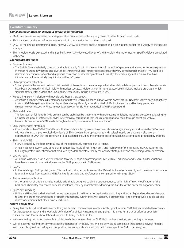
Additional nucleic acid-based targets and strate-gies have been developed that redirect SMN2 splic-ing, including ASOs targeting element 1(E1) or the intron 7/exon 8 junction; bifunctional RNAs; peptide–nucleic acid ESSENCE compounds; and trans-splicing RNAs [62,71,88–90,141,148–154]. The bifunctional RNAs are comprised of two domains: an antisense domain and a separate region that serves as a recruiting and binding substrate for splicing factors, such as hTra2-b1 or SF2/ASF. Bifunctional RNAs and ESSENCE compounds have shown enhanced activation of SMN2 exon 7 splicing medi-ated by the recruiting platform over and above the activity conferred by the antisense domain, how-ever, there is not a dramatic extension of survival using these molecules that is likely attributed to chemistry or stability differences. Similarly, trans-splicing RNAs, which redirect SMN2 splicing from the endogenous pre-mRNA molecule to a vector-derived RNA in trans via a site-specific antisense domain, have shown promise in cell culture and modest activity in a severe mouse model of SMA.
Ex6 Ex7 Ex8Ex6 Ex7 Ex8
Ex7 Ex8
SMN1 Ex7
Ex6
Ex6 Ex7 Ex8
hnRNP
Ex6 Ex7 Ex8
hnRNP
A
B
C
hnRNP
Ex6 Ex7 Ex8
hnRNP
Ex6 Ex7 Ex8
Ex7 Ex8Ex6
SMN1 Ex7
A
B
C
Figure 3. Nucleic acid-based strategies to modulate SMN2 splicing. (A) Antisense oligonucleotides that target E1, ISS-N1 and the intron 7/exon 8 juncture have been shown to stimulate exon 7 inclusion. (B) Bifunctional RNAs and similar derivatives targeting E1, exon 7, ISS-N1 stimulate exon 7 inclusion by recruiting SR proteins (green), whereas bifunctional RNAs targeting the intron 7/exon 8 junction recruit hnRNP proteins as a means to favor exon 7 inclusion. (C) Trans-splicing RNAs re-direct SMN2 splicing from the cis-SMN2 splicing to a ‘corrective’ SMN1 exon 7 that is supplied via vector.
SMN-inducing compounds for the treatment of spinal muscular atrophy | Review
www.future-science.com 2075future science group

Small molecules targeting SMN2 splicingThe novel molecular entities, including the nucleic acid-based therapeutics, have elicited a consider-able amount of excitement based upon the suc-cessful studies in SMA animal models, however, more traditional small molecules have been iden-tified that modulate SMN2 exon 7 splicing. One of the first compounds to be examined in SMA models was aclarubicin that demonstrated activ-ity in cell-based assays [155]. Salbutamol has also been shown to increase the relative ratio of full-length:D7 SMN transcript in cell-based models [156,157]. As salbutamol is an approved compound, clinical trials were initiated based upon the posi-tive SMN induction, demonstrating a high degree of tolerability yet only modest improvement in motor function [156,158].
In a cell-free screen designed to identify com-pounds that increased SMN2 exon 7 splicing, a tetracycline derivative, PTK-SMA1, was iden-tified that increased in vitro SMN2 splicing as well as increasing exon 7 inclusion in mild SMA mouse model following intraperitoneal or iv. administration [159]. The beta-lactam antibiotic ceftriaxone has been evaluated in SMA mice and was shown to modestly, but significantly extend survival and decrease the severity of disease [160]. RNA transcripts were not examined in this report although total SMN protein levels were increased slightly.Ceftriaxone also exhibits a general in vitro and in vivo neuroprotective activity as it upregu-lates glutamate transporter expression, and in the G93A SOD1 amyotrophic lateral sclerosis mouse model it extends survival modestly and delays onset of disease [161].
In large part, the high-throughput screening vectors that have been utilized captured either SMN2 promoter activity or SMN2 exon 7 splic-ing. Recently, a new reporter was developed that incorporated the SMN2 promoter, the SMN cDNA-spanning exons 1–6 and the genomic cas-sette comprised exons 6–8. In this system, multi-ple SMN-inducing mechanisms could be screened simultaneously, such as promoter activation, exon 7 splicing, or RNA stability [162]. A series of novel compounds has been identified that appear to function through different mechanistic path-ways including increasing SMN2 exon 7 inclu-sion. SMN exon 7 inclusion was increased not just in the reporter system but from the endogenous SMN2 gene within SMA patient fibroblasts [163]. Further confirmation of these new compounds is required to demonstrate in vivo activity and in disease relevant tissues.
SMN stabilizationBased upon an image-based cell screen for com-pounds that elevated SMN levels [164], GSK-3 inhibitors were shown to be a druggable target as inhibition of this pathway increased the intra-cellular pool of SMN by stabilizing the protein [165]. A potent inhibitor of GSK-3 called BIP-135 was able to extend survival modestly from 12.8 to 14.7 days in a severe SMA mouse model [165]. Recent work has also focused upon the protea-some as a means to stabilize the low intracellular levels of SMN and to some extent, the exon-skipped SMND7 protein [166]. Bortezomib is a relatively specific proteosomal inhibitor as it selectively blocks chymotrypsin cleavage and has been used in the clinic. Bortezomib was shown to increase SMN and while it failed to signifi-cantly extend survival in SMA mice, there was a mild synergistic effect when Bortezomib was combined with TSA [167].
Aminoglycosides have been shown to stabilize SMN protein presumably by inducing a transla-tional read-through of the SMND7 protein [168]. The extension of the C-terminus either by trans-lational read-through or by the synthetic addi-tion of random amino acids stretches of varying lengths has demonstrated that extension of the C-terminus confers a greater degree of stabil-ity to the SMND7 protein [168–171]. Consistent with these results, several FDA-approved com-pounds were shown to increase SMN levels in SMA fibroblasts and induced pluripotent stem cell-derived neuronal cultures [170–174]. A novel aminoglycoside, TC007, was delivered via ICV injections and was capable of extending survival from 12.6 to 16.0 days and decreasing disease severity in severe SMA mice [172]. Current FDA-approved compounds have several undesirable side effects, including ototoxicity and nephro-toxicity and it is currently unclear whether this approach will suffice as a stand alone therapy or it could be incorporated into a multidrug regimen.
SMN-independent therapeutic strategiesOne of the more promising SMN-independent pathways identified is based upon the delivery of a compound that inhibits ROCK [58]. This compound, Y-27632, alters actin dynamics and without elevating Smn levels, dramatically extends survival in a relatively severe model of SMA called Smn2B/- [58]. One of the hallmarks of SMA involves significant neuromuscular junc-tion pathology (FiguRe 4) [175–178]. Interestingly, the cellular phenotype including motor neuron
Review | Lorson & Lorson
Future Med. Chem. (2012) 4(16)2076 future science group

and muscle pathology was significantly improved, however, the animals failed to gain weight above their untreated littermates. Similarly, another ROCK inhibitor, fasudil, which is FDA approved, significantly extended survival beyond 300 days and largely rescued the NMJ and muscle phe-notypes in Smn2B-/- mice [57]. Not only does this work provide a potential new therapeutic direc-tion for SMA, but it may also shed light upon the biology behind the disease as SMN’s role in actin dynamics may provide clues to the motor neuron sensitivity observed in SMA.
Other approaches have produced mixed or modest results in SMA mice, such as inhibi-tion of the myostatin pathway or overexpress-ing IGF-1 [179–183]. One of the likely complica-tions to SMN-independent strategies is that the most commonly used mouse model, the SMND7 mouse, represents a very severe form of SMA. Clearly, any compound that exhibits activity and efficacy in this model would become immedi-ately worthy of further analysis, however, it is equally important to not immediately disregard a compound that appears to function effectively in many parameters such as increased motor function or decreased muscle pathology yet fails to extend survival in the SMND7 mouse. The recent development of less severe models may lead to the identification of compounds and mol-ecules that function through different pathways that could still yield considerable benefit to SMA patients.
Future perspectiveFor several decades, SMA research has been pushing forward to the point of generat-ing SMA-specif ic compounds. A signif i-cant grassroots movement for SMA research pioneered by Families of SMA, FightSMA, Muscular Dystrophy Association and the SMA Foundation has stimulated governmental fund-ing including the SMA Project and the Network for Excellence in Neuroscience Clinical Trials. The strong research portfolio has also led to broad interest from pharmaceutical compa-nies including: Isis Pharmaceuticals, Genzyme Corporation, Roche, Repligen Corporation, Paratek, Trophos, Biogen Idec and Novartis. Currently, Repligen Coproration, Trophos and Isis Pharmaceuticals have initiated clinical trials for SMA.
It will be important going forward to recall that clinical trials are not merely a confirma-tion of the preclinical work, but a completely independent series of experiments. Carefully designed clinical trials will be essential to mov-ing compounds towards regulatory approval. The identification of the appropriate patient populations that can most clearly confirm the efficacy of a specific compound and its mode of action will be a complex process. It is possible that an effective compound for type I patients may not be the most effective compound for type III patients. A combinatorial approach may provide a SMN increase that addresses the
40 µmControl
NerveAChR
SMA
A B
Figure 4. Spinal muscular atrophy pathology manifests in select neuromuscular junctions. (A) Serratus posterior inferior muscles of control and (B) YFP-SMND7 (right) mice at post-natal day 14 were immunostained for nerve terminals with anti-synaptophysin antibody (in green) and motor end plates with a-bungarotoxin (in red). Unpublished images were generously provided by KKY Ling and CP Ko (Section of Neurobiology, University of Southern California, Los Angeles, CA, USA).
SMN-inducing compounds for the treatment of spinal muscular atrophy | Review
www.future-science.com 2077future science group
Executive summary
Spinal muscular atrophy: disease & clinical manifestations
�� SMA is an autosomal recessive neurodegenerative disease that is the leading cause of infantile death worldwide.
�� SMA is caused by the loss of motor neurons within the anterior horn of the spinal cord.
�� SMN1 is the disease-determining gene, however, SMN2 is a critical disease modifier and is an excellent target for a variety of therapeutic strategies.
�� SMN is ubiquitously expressed and it is still unknown why decreased levels of SMN result in the motor neuron-specific defects associated with SMA.
Therapeutic strategies
�� Gene replacement- The SMN cDNA is relatively compact and able to easily fit within the confines of the scAAV genome and allows for robust expression
in motor neurons in wildtype and SMA mice. Intravenous and intracerebroventricular delivery demonstrates that scAAV9 lead to a dramatic extension in survival and a general correction of disease symptoms. Currently, the early stages of a clinical trial have initiated and a Phase I study may initiate within 1–2 years.
�� SMN2 promoter activation- Suberoylanilide hydroxamic acid and trichostatin A have shown promise in preclinical models, while valproic acid and phenylbuturate
have been examined in clinical trials with modest success. Additional non-histone deacetylase inhibitors include prolacatin which significantly elevates SMN in the CNS and increases SMA mouse survival by ~40%.
�� Modulating exon 7 inclusion with nucleic acid-based therapeutics- Antisense oligonucleotides directed against negatively regulating splice signals within SMN2 pre-mRNA have shown excellent activity
in vivo. ISS-N1-targeting antisense oligonucleotides significantly extend survival of SMA mice and can effectively penetrate disease-relevant tissues. A Phase I study is underway for Isis Pharmaceutical’s SMNRx compound.
�� SMN stabilization- The low level of full-length SMN protein can be stabilized by treatment with proteasome inhibitors, including bortezomib, leading to
an increased pool of intracellular SMN. Alternatively, compounds that induce a translational read-through event on SMND7 transcripts can increase SMN levels through a distinct post-transcriptional mechanism.
�� SMN-independent strategies- Compounds such as Y-27632 and fasudil that modulate actin dynamics have been shown to significantly extend survival of SMA mice
without altering the pathologically low levels of SMN protein. Neuroprotectants and skeletal muscle enhancement also present opportunities in SMA that are continuing to be explored, including the ongoing trial of olesoxmine, a compound produced by Trophos.
�� SMN1 and SMN2 - SMA is caused by the homozygous loss of the ubiquitously expressed SMN1 gene.
- A nearly identical SMN1-copy gene that produces low levels of full-length SMN and high levels of the truncated SMND7 isoform. The full-length protein is identical to that produced by SMN1, therefore, many therapeutic strategies involve modulating SMN2 expression.
�� scAAV9–SMN- An adeno-associated virus vector with the serotype-9 capsid expressing the SMN cDNA. This vector and several similar variations
have been shown to dramatically rescue the SMA phenotype in SMA mice.
�� Exon 7- In the full-length SMN protein, exon 7 is the final coding exon, however, the SMND7 isoform lacks exon 7, and therefore incorporates
four amino acids from exon 8. SMND7 is highly unstable and dysfunctional compared to full-length SMN.
�� Antisense oligonucleotide- A short stretch of single-stranded nucleic acid that is designed to bind a target sequence with high affinity. Modification of the
backbone chemistry can confer nuclease resistance, thereby dramatically extending the half-life of the antisense oligonucleotide.
�� Splice-site switching- Unlike a siRNA that is designed to knock-down a specific mRNA target, splice-site switching antisense oligonucleotides are designed
to alter the pre-mRNA processing of specific transcripts. Within the SMA context, a primary goal is to competitively disable splicing repressor elements that block exon 7 inclusion.
Future perspective
�� Rarely has the first clinical trial become the gold standard for any disease entity. At this point in time, SMA lacks a validated benchmark for therapeutic efficacy and a workable definition of a clinically meaningful end point. This is not for a lack of effort as countless researchers and families have labored for years to bring the field so far.
�� We are entering uncharted waters but this is clearly the moment that the SMA field has been waiting and hoping to witness.
�� Will all patients respond similarly to a specific therapeutic? Probably not. Will delivery issues complicate therapeutic analysis? Perhaps. Will the evolving natural history and supportive care complicate an already broad clinical spectrum? Most certainly, yes.
Review | Lorson & Lorson
Future Med. Chem. (2012) 4(16)2078 future science group

References1 Pearn J. Classification of spinal muscular
atrophies. Lancet 1(8174), 919–922 (1980).
2 Zerres K, Wirth B, Rudnik-Schoneborn S. Spinal muscular atrophy – clinical and genetic correlations. Neuromuscul. Disord. 7(3), 202–207 (1997).
3 Chong JX, Oktay AA, Dai Z, Swoboda KJ, Prior TW, Ober C. A common spinal muscular atrophy deletion mutation is present on a single founder haplotype in the US Hutterites. Eur. J. Hum. Genet. 19(10), 1045–1051 (2011).
4 Lewelt A, Newcomb TM, Swoboda KJ. New therapeutic approaches to spinal muscular atrophy. Curr. Neurol. Neurosci. Rep. 12(1), 42–53 (2012).
5 Munsat TL, Davies KE. International SMA consortium meeting. (26–28 June 1992, Bonn, Germany). Neuromuscul. Disord. 2(5–6), 423–428 (1992).
6 Meldrum C, Scott C, Swoboda KJ. Spinal muscular atrophy genetic counseling access and genetic knowledge: parents’ perspectives. J. Child Neurol. 22(8), 1019–1026 (2007).
7 Zerres K, Rudnik-Schoneborn S. Natural history in proximal spinal muscular atrophy. Clinical analysis of 445 patients and suggestions for a modification of existing classifications. Arch. Neurol. 52(5), 518–523 (1995).
8 Lefebvre S, Burglen L, Reboullet S et al. Identification and characterization of a spinal muscular atrophy-determining gene. Cell 80(1), 155–165 (1995).
9 Munsat TL, Skerry L, Korf B et al. Phenotypic heterogeneity of spinal muscular atrophy mapping to chromosome 5q11.2–13.3 (SMA 5q). Neurology 40(12), 1831–1836 (1990).
10 Brzustowicz LM, Lehner T, Castilla LH et al. Genetic mapping of chronic childhood-onset spinal muscular atrophy to chromosome 5q11.2–13.3. Nature 344(6266), 540–541 (1990).
11 Melki J, Abdelhak S, Sheth P et al. Gene for chronic proximal spinal muscular atrophies maps to chromosome 5q. Nature 344(6268), 767–768 (1990).
12 Roy N, Mahadevan MS, Mclean M et al. The gene for neuronal apoptosis inhibitory protein is partially deleted in individuals with spinal muscular atrophy. Cell 80(1), 167–178 (1995).
13 Wirth B. An update of the mutation spectrum of the survival motor neuron gene (SMN1) in autosomal recessive spinal muscular atrophy (SMA). Human Mutation 15(3), 228–237 (2000).
14 Burglen L, Lefebvre S, Clermont O et al. Structure and organization of the human survival motor neurone (SMN) gene. Genomics 32(3), 479–482 (1996).
15 Monani UR, Lorson CL, Parsons DW et al. A single nucleotide difference that alters splicing patterns distinguishes the SMA gene SMN1 from the copy gene SMN2. Hum. Mol. Genet. 8(7), 1177–1183 (1999).
16 Novelli G, Calza L, Amicucci P et al. Expression study of survival motor neuron gene in human fetal tissues. Biochem. Mol. Med. 61(1), 102–106 (1997).
17 Gennarelli M, Lucarelli M, Capon F et al. Survival motor neuron gene transcript analysis in muscles from spinal muscular atrophy patients. Biochem. Biophys. Res. Commun. 213(1), 342–348 (1995).
18 Hahnen E, Schonling J, Rudnik-Schoneborn S, Zerres K, Wirth B. Hybrid survival motor neuron genes in patients with autosomal recessive spinal muscular atrophy: new insights into molecular mechanisms responsible for the disease. Am. J. Hum. Genet. 59(5), 1057–1065 (1996).
19 Lorson CL, Hahnen E, Androphy EJ, Wirth B. A single nucleotide in the SMN gene regulates splicing and is responsible for spinal muscular atrophy. Proc. Natl Acad. Sci. USA 96(11), 6307–6311 (1999).
20 Monani UR. Spinal muscular atrophy: a deficiency in a ubiquitous protein; a motor
neuron-specific disease. Neuron 48(6), 885–896 (2005).
21 Monani UR, Sendtner M, Coovert DD et al. The human centromeric survival motor neuron gene (SMN2) rescues embryonic lethality in Smn(-/-) mice and results in a mouse with spinal muscular atrophy. Hum. Mol. Genet. 9(3), 333–339 (2000).
22 Le TT, Pham LT, Butchbach ME et al. SMNDelta7, the major product of the centromeric survival motor neuron (SMN2) gene, extends survival in mice with spinal muscular atrophy and associates with full-length SMN. Hum. Mol. Genet. 14(6), 845–857 (2005).
23 Campbell L, Potter A, Ignatius J, Dubowitz V, Davies K. Genomic variation and gene conversion in spinal muscular atrophy: implications for disease process and clinical phenotype. Am. J. Hum. Genet. 61(1), 40–50 (1997).
24 Mcandrew PE, Parsons DW, Simard LR et al. Identification of proximal spinal muscular atrophy carriers and patients by analysis of SMNT and SMNC gene copy number. Am. J. Hum. Genet. 60(6), 1411–1422 (1997).
25 Wirth B, Herz M, Wetter A et al. Quantitative analysis of survival motor neuron copies: identification of subtle SMN1 mutations in patients with spinal muscular atrophy, genotype-phenotype correlation, and implications for genetic counseling. Am. J. Hum. Genet. 64(5), 1340–1356 (1999).
26 Mailman MD, Heinz JW, Papp AC et al. Molecular analysis of spinal muscular atrophy and modification of the phenotype by SMN2. Genetics in Medicine 4(1), 20–26 (2002).
27 Feldkotter M, Schwarzer V, Wirth R, Wienker TF, Wirth B. Quantitative analyses of SMN1 and SMN2 based on real-time lightCycler PCR: fast and highly reliable carrier testing and prediction of severity of spinal muscular atrophy. Am. J. Hum. Genet. 70(2), 358–368 (2002).
28 Parano E, Pavone L, Falsaperla R, Trifiletti R, Wang C. Molecular basis of phenotypic
molecular basis of this disease and an alterna-tive compound could focus upon creating the appropriate cellular environment that fosters motor neuron protection and survival. SMA is a truly devastating disease and while the natural histories have changed significantly due in large part to herculean supportive care efforts, the future on a new wave of rationally designed SMA therapeutics is about to unfold and it is clear that there is a growing sense of promise.
Financial & competing interests disclosureThis work was supported by grants from FightSMA and the Gwendolyn Strong Foundation (MAL) and the NIH (MAL; CLL) (R21NS078299; R56NS041584). The authors have no other relevant affiliations or financial involvement with any organization or entity with a finan-cial interest in or financial conflict with the subject matter or materials discussed in the manuscript apart from those disclosed.
No writing assistance was u tilized in the production of this manuscript.
SMN-inducing compounds for the treatment of spinal muscular atrophy | Review
www.future-science.com 2079future science group
heterogeneity in siblings with spinal muscular atrophy. Ann. Neurol. 40(2), 247–251 (1996).
29 Petit F, Cuisset JM, Rouaix-Emery N et al. Insights into genotype-phenotype correlations in spinal muscular atrophy: a retrospective study of 103 patients. Muscle Nerve 43(1), 26–30 (2011).
30 Jedrzejowska M, Borkowska J, Zimowski J et al. Unaffected patients with a homozygous absence of the SMN1 gene. Eur. J. Hum. Genet. 16(8), 930–934 (2008).
31 Prior TW. Spinal muscular atrophy: a time for screening. Curr. Opin. Ped. 22(6), 696–702 (2010).
32 Prior TW. Spinal muscular atrophy: newborn and carrier screening. Obstet. Gynecol. Clin. North Am. 37(1), 23–36, (2010).
33 Gabanella F, Carissimi C, Usiello A, Pellizzoni L. The activity of the spinal muscular atrophy protein is regulated during development and cellular differentiation. Hum. Mol. Genet. 14(23), 3629–3642 (2005).
34 Schrank B, Gotz R, Gunnersen JM et al. Inactivation of the survival motor neuron gene, a candidate gene for human spinal muscular atrophy, leads to massive cell death in early mouse embryos. Proc. Natl Acad. Sci. USA 94(18), 9920–9925 (1997).
35 Liu Q, Dreyfuss G. A novel nuclear structure containing the survival of motor neurons protein. EMBO J. 15(14), 3555–3565 (1996).
36 Coovert DD, Le TT, Mcandrew PE et al. The survival motor neuron protein in spinal muscular atrophy. Hum. Mol. Genet. 6(8), 1205–1214 (1997).
37 Pellizzoni L. Chaperoning ribonucleoprotein biogenesis in health and disease. EMBO Reports 8(4), 340–345 (2007).
38 Burghes AH, Beattie CE. Spinal muscular atrophy: why do low levels of survival motor neuron protein make motor neurons sick? Nat. Rev. Neurosci. 10(8), 597–609 (2009).
39 Zhang Z, Lotti F, Dittmar K et al. SMN deficiency causes tissue-specific perturbations in the repertoire of snRNAs and widespread defects in splicing. Cell 133(4), 585–600 (2008).
40 Baumer D, Lee S, Nicholson G et al. Alternative splicing events are a late feature of pathology in a mouse model of spinal muscular atrophy. PLoS Genetics 5(12), e1000773 (2009).
41 Patel AA, Steitz JA. Splicing double: insights from the second spliceosome. Nat. Rev. Mol. Cell Biol. 4(12), 960–970 (2003).
42 Will CL, Schneider C, Macmillan AM et al. A novel U2 and U11/U12 snRNP protein that associates with the pre-mRNA
branch site. EMBO J. 20(16), 4536–4546 (2001).
43 Gabanella F, Butchbach ME, Saieva L, Carissimi C, Burghes AH, Pellizzoni L. Ribonucleoprotein assembly defects correlate with spinal muscular atrophy severity and preferentially affect a subset of spliceosomal snRNPs. PLoS ONE 2(9), e921 (2007).
44 Boulisfane N, Choleza M, Rage F, Neel H, Soret J, Bordonne R. Impaired minor tri-snRNP assembly generates differential splicing defects of U12-type introns in lymphoblasts derived from a type I SMA patient. Hum. Mol. Genet. 20(4), 641–648 (2011).
45 Wu Q, Krainer AR. Splicing of a divergent subclass of AT-AC introns requires the major spliceosomal snRNAs. RNA 3(6), 586–601 (1997).
46 Wu Q, Krainer AR. AT-AC pre-mRNA splicing mechanisms and conservation of minor introns in voltage-gated ion channel genes. Mol. Cell. Biol. 19(5), 3225–3236 (1999).
47 Rossoll W, Bassell GJ. Spinal muscular atrophy and a model for survival of motor neuron protein function in axonal ribonucleoprotein complexes. Results Probl. Cell Differ. 48, 289–326 (2009).
48 Rossoll W, Jablonka S, Andreassi C et al. Smn, the spinal muscular atrophy-determining gene product, modulates axon growth and localization of beta-actin mRNA in growth cones of motoneurons. J. Cell Biol. 163(4), 801–812 (2003).
49 Rossoll W, Kroning AK, Ohndorf UM, Steegborn C, Jablonka S, Sendtner M. Specific interaction of Smn, the spinal muscular atrophy determining gene product, with hnRNP-R and gry-rbp/hnRNP-Q: a role for Smn in RNA processing in motor axons? Hum. Mol. Genet.11(1), 93–105 (2002).
50 Jablonka S, Wiese S, Sendtner M. Axonal defects in mouse models of motoneuron disease. J. Neurobiol. 58(2), 272–286 (2004).
51 Fallini C, Zhang H, Su Y et al. The survival of motor neuron (SMN) protein interacts with the mRNA-binding protein HuD and regulates localization of poly(A) mRNA in primary motor neuron axons. J. Neurosci. 31(10), 3914–3925 (2011).
52 Akten B, Kye MJ, Hao Le T et al. Interaction of survival of motor neuron (SMN) and HuD proteins with mRNA cpg15 rescues motor neuron axonal deficits. Proc. Natl Acad. Sci. USA 108(25), 10337–10342 (2011).
53 Hubers L, Valderrama-Carvajal H, Laframboise J, Timbers J, Sanchez G, Cote J. HuD interacts with survival motor neuron
protein and can rescue spinal muscular atrophy-like neuronal defects. Hum. Mol. Genet. 20(3), 553–579 (2011).
54 Glinka M, Herrmann T, Funk N et al. The heterogeneous nuclear ribonucleoprotein-R is necessary for axonal beta-actin mRNA translocation in spinal motor neurons. Hum. Mol. Genet. 19(10), 1951–1966 (2010).
55 Peter CJ, Evans M, Thayanithy V et al. The COPI vesicle complex binds and moves with survival motor neuron within axons. Hum. Mol. Genet. 20(9), 1701–1711 (2011).
56 Cheever TR, Olson EA, Ervasti JM. Axonal regeneration and neuronal function are preserved in motor neurons lacking ss-actin in vivo. PLoS ONE 6(3), e17768 (2011).
57 Bowerman M, Murray LM, Boyer JG, Anderson CL, Kothary R. Fasudil improves survival and promotes skeletal muscle development in a mouse model of spinal muscular atrophy. BMC Medicine 10, 24 (2012).
58 Bowerman M, Beauvais A, Anderson CL, Kothary R. Rho-kinase inactivation prolongs survival of an intermediate SMA mouse model. Hum. Mol. Genet. 19(8), 1468–1478 (2010).
59 Oprea GE, Krober S, Mcwhorter ML et al. Plastin 3 is a protective modifier of autosomal recessive spinal muscular atrophy. Science 320(5875), 524–527 (2008).
60 Coady TH, Lorson CL. SMN in spinal muscular atrophy and snRNP biogenesis. Wiley Interdiscip. Rev. RNA 2(4), 546–564 (2011).
61 Cheever TR, Li B, Ervasti JM. Restricted morphological and behavioral abnormalities following ablation of beta-actin in the brain. PLoS ONE 7(3), e32970 (2012).
62 Lim SR, Hertel KJ. Modulation of survival motor neuron pre-mRNA splicing by inhibition of alternative 3´ splice site pairing. J. Biol. Chem. 276(48), 45476–45483 (2001).
63 Lorson CL, Androphy EJ. An exonic enhancer is required for inclusion of an essential exon in the SMA-determining gene SMN. Hum. Mol. Genet.9(2), 259–265 (2000).
64 Singh RN. Evolving concepts on human SMN pre-mRNA splicing. RNA biology 4(1), 7–10 (2007).
65 Hofmann Y, Lorson CL, Stamm S, Androphy EJ, Wirth B. Htra2-beta 1 stimulates an exonic splicing enhancer and can restore full-length SMN expression to survival motor neuron 2 (SMN2). Proc. Natl Acad. Sci. USA 97(17), 9618–9623 (2000).
66 Chen HH, Chang JG, Lu RM, Peng TY, Tarn WY. The RNA binding protein hnRNP Q
Review | Lorson & Lorson
Future Med. Chem. (2012) 4(16)2080 future science group

modulates the utilization of exon 7 in the survival motor neuron 2 (SMN2) gene. Mol. Cell. Biol. 28(22), 6929–6938 (2008).
67 Young PJ, Didonato CJ, Hu D, Kothary R, Androphy EJ, Lorson CL. SRp30c-dependent stimulation of survival motor neuron (SMN) exon 7 inclusion is facilitated by a direct interaction with hTra2 beta 1. Hum. Mol. Genet. 11(5), 577–587 (2002).
68 Hofmann Y, Wirth B. hnRNP-G promotes exon 7 inclusion of survival motor neuron (SMN) via direct interaction with Htra2-beta1. Hum. Mol. Genet. 11(17), 2037–2049 (2002).
69 Bose JK, Wang IF, Hung L, Tarn WY, Shen CK. TDP-43 overexpression enhances exon 7 inclusion during the survival of motor neuron pre-mRNA splicing. J. Biol. Chem. 283(43), 28852–28859 (2008).
70 Cartegni L, Krainer AR. Disruption of an SF2/ASF-dependent exonic splicing enhancer in SMN2 causes spinal muscular atrophy in the absence of SMN1. Nat. Genet. 30(4), 377–384 (2002).
71 Cartegni L, Krainer AR. Correction of disease-associated exon skipping by synthetic exon-specific activators. Nat. Struct. Biol. 10(2), 120–125 (2003).
72 Singh NN, Seo J, Ottesen EW, Shishimorova M, Bhattacharya D, Singh RN. TIA1 prevents skipping of a critical exon associated with spinal muscular atrophy. Mol. Cell. Biol. 31(5), 935–954 (2011).
73 Bebee TW, Gladman JT, Chandler DS. Splicing regulation of the survival motor neuron genes and implications for treatment of spinal muscular atrophy. Front. Biosci. 15, 1191–1204 (2010).
74 Pedrotti S, Bielli P, Paronetto MP et al. The splicing regulator Sam68 binds to a novel exonic splicing silencer and functions in SMN2 alternative splicing in spinal muscular atrophy. EMBO J. 29(7), 1235–1247 (2010).
75 Kashima T, Manley JL. A negative element in SMN2 exon 7 inhibits splicing in spinal muscular atrophy. Nat. Genet. 34(4), 460–463 (2003).
76 Kashima T, Rao N, David CJ, Manley JL. hnRNP A1 functions with specificity in repression of SMN2 exon 7 splicing. Hum. Mol. Genet. 16(24), 3149–3159 (2007).
77 Kashima T, Rao N, Manley JL. An intronic element contributes to splicing repression in spinal muscular atrophy. Proc. Natl Acad. Sci. USA 104(9), 3426–3431 (2007).
78 Singh NN, Androphy EJ, Singh RN. In vivo selection reveals combinatorial controls that define a critical exon in the spinal muscular
atrophy genes. RNA 10(8), 1291–1305 (2004).
79 Baughan TD, Dickson A, Osman EY, Lorson CL. Delivery of bifunctional RNAs that target an intronic repressor and increase SMN levels in an animal model of spinal muscular atrophy. Hum. Mol. Genet. 18(9), 1600–1611 (2009).
80 Miyajima H, Miyaso H, Okumura M, Kurisu J, Imaizumi K. Identification of a cis-acting element for the regulation of SMN exon 7 splicing. J. Biol. Chem. 277(26), 23271–23277 (2002).
81 Gladman JT, Chandler DS. Intron 7 conserved sequence elements regulate the splicing of the SMN genes. Hum. Genet. 126(6), 833–841 (2009).
82 Hua Y, Vickers TA, Okunola HL, Bennett CF, Krainer AR. Antisense masking of an hnRNP A1/A2 intronic splicing silencer corrects SMN2 splicing in transgenic mice. Am. J. Hum. Genet. 82(4), 834–848 (2008).
83 Singh NK, Singh NN, Androphy EJ, Singh RN. Splicing of a critical exon of human survival motor neuron is regulated by a unique silencer element located in the last intron. Mol. Cell. Biol. 26(4), 1333–1346 (2006).
84 Cartegni L, Hastings ML, Calarco JA, De Stanchina E, Krainer AR. Determinants of exon 7 splicing in the spinal muscular atrophy genes, SMN1 and SMN2. Am. J. Hum. Genet. 78(1), 63–77 (2006).
85 Baughan T, Shababi M, Coady TH, Dickson AM, Tullis GE, Lorson CL. Stimulating full-length SMN2 expression by delivering bifunctional RNAs via a viral vector. Mol. Ther. 14(1), 54–62 (2006).
86 Singh NN, Androphy EJ, Singh RN. An extended inhibitory context causes skipping of exon 7 of SMN2 in spinal muscular atrophy. Biochem. Biophys. Res. Commun 315(2), 381–388 (2004).
87 Fernandez Alanis E, Pinotti M, Dal Mas A et al. An exon-specific U1 small nuclear RNA (snRNA) strategy to correct splicing defects. Hum. Mol. Genet. 21(11), 2389–2398 (2012).
88 Geib T, Hertel KJ. Restoration of full-length SMN promoted by adenoviral vectors expressing RNA antisense oligonucleotides embedded in U7 snRNAs. PLoS ONE 4(12), e8204 (2009).
89 Madocsai C, Lim SR, Geib T, Lam BJ, Hertel KJ. Correction of SMN2 pre-mRNA splicing by antisense U7 small nuclear RNAs. Mol. Ther. 12(6), 1013–1022 (2005).
90 Dickson A, Osman E, Lorson C. A negatively-acting bifunctional RNA increases survival motor neuron in vitro and in vivo. Hum. Gene Ther (2008).
91 Prior TW, Krainer AR, Hua Y et al. A positive modifier of spinal muscular atrophy in the SMN2 gene. Am. J. Hum. Genet. 85(3), 408–413 (2009).
92 Jodelka FM, Ebert AD, Duelli DM, Hastings ML. A feedback loop regulates splicing of the spinal muscular atrophy-modifying gene, SMN2. Hum. Mol. Genet. 19(24), 4906–4917 (2010).
93 Ruggiu M, Mcgovern VL, Lotti F et al. A role for SMN exon 7 splicing in the selective vulnerability of motor neurons in spinal muscular atrophy. Mol. Cell. Biol. 32(1), 126–138 (2012).
94 Azzouz M, Le T, Ralph GS et al. Lentivector-mediated SMN replacement in a mouse model of spinal muscular atrophy. J. Clin. Invest. 114(12), 1726–1731 (2004).
95 Dominguez E, Marais T, Chatauret N et al. Intravenous scAAV9 delivery of a codon-optimized SMN1 sequence rescues SMA mice. Hum. Mol. Genet. 20(4), 681–693 (2011).
96 Valori CF, Ning K, Wyles M et al. Systemic delivery of scAAV9 expressing SMN prolongs survival in a model of spinal muscular atrophy. Sci. Transl. Med. 2(35), 35ra42 (2010).
97 Foust KD, Wang X, Mcgovern VL et al. Rescue of the spinal muscular atrophy phenotype in a mouse model by early postnatal delivery of SMN. Nat. Biotechnol. 28(3), 271–274 (2010).
98 Passini MA, Bu J, Roskelley EM et al. CNS-targeted gene therapy improves survival and motor function in a mouse model of spinal muscular atrophy. J. Clin. Invest. 120(4), 1253–1264 (2010).
99 Glascock JJ, Osman EY, Wetz MJ, Krogman MM, Shababi M, Lorson CL. Decreasing disease severity in symptomatic, Smn(-/-); SMN2(+/+), spinal muscular atrophy mice following scAAV9-SMN delivery. Hum. Gene Ther. 23(3), 330–335 (2012).
100 Glascock JJ, Shababi M, Wetz MJ, Krogman MM, Lorson CL. Direct central nervous system delivery provides enhanced protection following vector mediated gene replacement in a severe model of spinal muscular atrophy. Biochem. Biophys. Res. Commun. 417(1), 376–381 (2012).
101 Somers E, Stencel Z, Wishart TM, Gillingwater TH, Parson SH. Density, calibre and ramification of muscle capillaries are altered in a mouse model of severe spinal muscular atrophy. Neuromuscul. Disord. 22(5), 435–442 (2012).
102 Bevan AK, Hutchinson KR, Foust KD et al. Early heart failure in the SMN(Delta)7 model of spinal muscular atrophy and correction by
SMN-inducing compounds for the treatment of spinal muscular atrophy | Review
www.future-science.com 2081future science group

postnatal scAAV9-SMN delivery. Hum. Mol. Genet. 19(20), 3895–3905 (2010).
103 Heier CR, Satta R, Lutz C, Didonato CJ. Arrhythmia and cardiac defects are a feature of spinal muscular atrophy model mice. Hum. Mol. Genet. 19(20), 3906–3918 (2010).
104 Shababi M, Habibi J, Ma L, Glascock JJ, Sowers JR, Lorson CL. Partial restoration of cardio-vascular defects in a rescued severe model of spinal muscular atrophy. J. Mol. Cell. Cardiol. 52(5), 1074–1082 (2012).
105 Shababi M, Habibi J, Yang HT, Vale SM, Sewell WA, Lorson CL. Cardiac defects contribute to the pathology of spinal muscular atrophy models. Hum. Mol. Genet. 19(20), 4059–4071 (2010).
106 Inagaki K, Fuess S, Storm TA et al. Robust systemic transduction with AAV9 vectors in mice: efficient global cardiac gene transfer superior to that of AAV8. Mol. Ther. 14(1), 45–53 (2006).
107 Glascock J, Osman EY, Wetz MJ, Krogman MM, Shababi M, Lorson C. Decreasing disease severity in symptomatic spinal muscular atrophy mice following scAAV9-SMN delivery. Hum. Gene Ther. 23(3), 330–335 (2011).
108 Duque S, Joussemet B, Riviere C et al. Intravenous administration of self-complementary AAV9 enables transgene delivery to adult motor neurons. Mol. Ther. 17(7), 1187–1196 (2009).
109 Samaranch L, Salegio EA, San Sebastian W et al. Adeno-associated virus serotype 9 transduction in the central nervous system of nonhuman primates. Hum. Gene Ther. 23(4), 382–389 (2012).
110 Asokan A, Schaffer DV, Samulski RJ. The AAV vector toolkit: poised at the clinical crossroads. Mol. Ther. 20(4), 699–708 (2012).
111 Minucci S, Pelicci PG. Histone deacetylase inhibitors and the promise of epigenetic (and more) treatments for cancer. Nat. Rev. Cancer 6(1), 38–51 (2006).
112 Arrowsmith CH, Bountra C, Fish PV, Lee K, Schapira M. Epigenetic protein families: a new frontier for drug discovery. Nat. Rev. Drug Discov. 11(5), 384–400 (2012).
113 Chang JG, Hsieh-Li HM, Jong YJ, Wang NM, Tsai CH, Li H. Treatment of spinal muscular atrophy by sodium butyrate. Proc. Natl Acad. Sci. USA 98(17), 9808–9813 (2001).
114 Brichta L, Hofmann Y, Hahnen E et al. Valproic acid increases the SMN2 protein level: a well-known drug as a potential therapy for spinal muscular atrophy. Hum. Mol. Genet. 12(19), 2481–2489 (2003).
115 Brichta L, Holker I, Haug K, Klockgether T, Wirth B. In vivo activation of SMN in spinal muscular atrophy carriers and patients treated with valproate. Ann. Neurol. 59(6), 970–975 (2006).
116 Piepers S, Cobben JM, Sodaar P et al. Quantification of SMN protein in leucocytes from spinal muscular atrophy patients: effects of treatment with valproic acid. J. Neurol. Neurosurg. Psych. 82(8), 850–852 (2011).
117 Van Bergeijk J, Haastert K, Grothe C, Claus P. Valproic acid promotes neurite outgrowth in PC12 cells independent from regulation of the survival of motoneuron protein. Chem. Biol. Drug Design 67(3), 244–247 (2006).
118 Andreassi C, Angelozzi C, Tiziano FD et al. Phenylbutyrate increases SMN expression in vitro: relevance for treatment of spinal muscular atrophy. Eur. J. Hum. Genet. 12(1), 59–65 (2004).
119 Avila AM, Burnett BG, Taye AA et al. Trichostatin A increases SMN expression and survival in a mouse model of spinal muscular atrophy. J. Clin. Invest. 117(3), 659–671 (2007).
120 Narver HL, Kong L, Burnett BG et al. Sustained improvement of spinal muscular atrophy mice treated with trichostatin A plus nutrition. Ann. Neurol. 64(4), 465–470 (2008).
121 Garbes L, Riessland M, Holker I et al. LBH589 induces up to 10-fold SMN protein levels by several independent mechanisms and is effective even in cells from SMA patients non-responsive to valproate. Hum. Mol. Genet. 18(19), 3645–3658 (2009).
122 Hahnen E, Eyupoglu IY, Brichta L et al. In vitro and ex vivo evaluation of second-generation histone deacetylase inhibitors for the treatment of spinal muscular atrophy. J. Neurochem. 98(1), 193–202 (2006).
123 Riessland M, Ackermann B, Forster A et al. SAHA ameliorates the SMA phenotype in two mouse models for spinal muscular atrophy. Hum. Mol. Genet. 19(8), 1492–1506 (2010).
124 Bricceno KV, Sampognaro PJ, Van Meerbeke JP, Sumner CJ, Fischbeck KH, Burnett BG. Histone deacetylase inhibition suppresses myogenin-dependent atrogene activation in spinal muscular atrophy mice. Hum. Mol. Genet. 20(20), 4448–4459 (2012).
125 Le TT, Mcgovern VL, Alwine IE et al. Temporal requirement for high SMN expression in SMA mice. Hum. Mol. Genet. 20(18), 3578–3591 (2011).
126 Kernochan LE, Russo ML, Woodling NS et al. The role of histone acetylation in SMN gene expression. Hum. Mol. Genet. 14(9), 1171–1182 (2005).
127 Mercuri E, Bertini E, Messina S et al. Randomized, double-blind, placebo-controlled trial of phenylbutyrate in spinal muscular atrophy. Neurology 68(1), 51–55 (2007).
128 Swoboda KJ, Scott CB, Reyna SP et al. Phase II open label study of valproic acid in spinal muscular atrophy. PLoS ONE 4(5), e5268 (2009).
129 Kissel JT, Scott CB, Reyna SP et al. SMA Carnival Trial Part II: a prospective, single-armed trial of l-carnitine and valproic acid in ambulatory children with spinal muscular atrophy. PLoS ONE 6(7), e21296 (2011).
130 Liang WC, Yuo CY, Chang JG et al. The effect of hydroxyurea in spinal muscular atrophy cells and patients. J. Neurol. Sci. 268(1–2), 87–94 (2008).
131 Chen TH, Chang JG, Yang YH et al. Randomized, double-blind, placebo-controlled trial of hydroxyurea in spinal muscular atrophy. Neurology 75(24), 2190–2197 (2010).
132 Farooq F, Molina FA, Hadwen J et al. Prolactin increases SMN expression and survival in a mouse model of severe spinal muscular atrophy via the STAT5 pathway. J. Clin. Invest. 121(8), 3042–3050 (2011).
133 Ting CH, Lin CW, Wen SL, Hsieh-Li HM, Li H. Stat5 constitutive activation rescues defects in spinal muscular atrophy. Hum. Mol. Genet.16(5), 499–514 (2007).
134 Jarecki J, Chen X, Bernardino A et al. Diverse small-molecule modulators of SMN expression found by high-throughput compound screening: early leads towards a therapeutic for spinal muscular atrophy. Hum. Mol. Genet.14(14), 2003–2018 (2005).
135 Thurmond J, Butchbach ME, Palomo M et al. Synthesis and biological evaluation of novel 2,4-diaminoquinazoline derivatives as SMN2 promoter activators for the potential treatment of spinal muscular atrophy. J. Med. Chem. 51(3), 449–469 (2008).
136 Singh J, Salcius M, Liu SW et al. DcpS as a therapeutic target for spinal muscular atrophy. ACS Chem. Biol. 3(11), 711–722 (2008).
137 Butchbach ME, Singh J, Thornorsteinsdottir M et al. Effects of 2,4-diaminoquinazoline derivatives on SMN expression and phenotype in a mouse model for spinal muscular atrophy. Hum. Mol. Genet. 19(3), 454–467 (2010).
138 Singh NN, Hollinger K, Bhattacharya D, Singh RN. An antisense microwalk reveals critical role of an intronic position linked to a unique long-distance interaction in pre-mRNA splicing. RNA 16(6), 1167–1181 (2010).
Review | Lorson & Lorson
Future Med. Chem. (2012) 4(16)2082 future science group

139 Williams JH, Schray RC, Patterson CA, Ayitey SO, Tallent MK, Lutz GJ. Oligonucleotide-mediated survival of motor neuron protein expression in CNS improves phenotype in a mouse model of spinal muscular atrophy. J. Neurosci. 29(24), 7633–7638 (2009).
140 Singh NN, Shishimorova M, Cao LC, Gangwani L, Singh RN. A short antisense oligonucleotide masking a unique intronic motif prevents skipping of a critical exon in spinal muscular atrophy. RNA Biol. 6(3), 341–350 (2009).
141 Osman EY, Yen PF, Lorson CL. Bifunctional RNAs Targeting the Intronic Splicing Silencer N1 Increase SMN Levels and Reduce Disease Severity in an Animal Model of Spinal Muscular Atrophy. Mol. Ther. 20(1), 119–126 (2012).
142 Hua Y, Sahashi K, Hung G et al. Antisense correction of SMN2 splicing in the CNS rescues necrosis in a type III SMA mouse model. Genes Develop. 24(15), 1634–1644 (2010).
143 Passini MA, Bu J, Richards AM et al. Antisense oligonucleotides delivered to the mouse CNS ameliorate symptoms of severe spinal muscular atrophy. Sci. Transl. Med. 3(72), 72ra18 (2011).
144 Hua Y, Sahashi K, Rigo F et al. Peripheral SMN restoration is essential for long-term rescue of a severe spinal muscular atrophy mouse model. Nature 478(7367), 123–126 (2011).
145 Porensky PN, Mitrpant C, Mcgovern VL et al. A single administration of morpholino antisense oligomer rescues spinal muscular atrophy in mouse. Hum. Mol. Genet. 21(7), 1625–1638 (2012).
146 Lorson MA, Spate LD, Prather RS, Lorson CL. Identification and characterization of the porcine (Sus scrofa) survival motor neuron (SMN1) gene: an animal model for therapeutic studies. Dev. Dyn. 237(8), 2268–2278 (2008).
147 Lorson MA, Spate LD, Samuel MS et al. Disruption of the survival motor neuron (SMN) gene in pigs using ssDNA. Transgenic Res. 20(6), 1293–1304 (2011).
148 Marquis J, Meyer K, Angehrn L, Kampfer SS, Rothen-Rutishauser B, Schumperli D. Spinal muscular atrophy: SMN2 pre-mRNA splicing corrected by a U7 snRNA derivative carrying a splicing enhancer sequence. Mol. Ther. 15(8), 1479–1486 (2007).
149 Meyer K, Marquis J, Trub J et al. Rescue of a severe mouse model for spinal muscular atrophy by U7 snRNA-mediated splicing modulation. Hum. Mol. Genet. 18(3), 546–555 (2009).
150 Coady TH, Baughan TD, Shababi M, Passini MA, Lorson CL. Development of a single vector system that enhances trans-splicing of SMN2 transcripts. PLoS ONE 3(10), e3468 (2008).
151 Coady TH, Lorson CL. Trans-splicing-mediated improvement in a severe mouse model of spinal muscular atrophy. J. Neurosci. 30(1), 126–130 (2010).
152 Coady TH, Shababi M, Tullis GE, Lorson CL. Restoration of SMN function: delivery of a trans-splicing RNA re-directs SMN2 pre-mRNA splicing. Mol. Ther. 15(8), 1471–1478 (2007).
153 Skordis LA, Dunckley MG, Yue B, Eperon IC, Muntoni F. Bifunctional antisense oligonucleotides provide a trans-acting splicing enhancer that stimulates SMN2 gene expression in patient fibroblasts. Proc. Natl Acad. Sci. USA 100(7), 4114–4119 (2003).
154 Shababi M, Lorson CL. Optimization of SMN trans-splicing through the analysis of SMN introns. J. Mol. Neurosci. 46(3), 459–469 (2012).
155 Andreassi C, Jarecki J, Zhou J et al. Aclarubicin treatment restores SMN levels to cells derived from type I spinal muscular atrophy patients. Hum. Mol. Genet.10(24), 2841–2849 (2001).
156 Angelozzi C, Borgo F, Tiziano FD, Martella A, Neri G, Brahe C. Salbutamol increases SMN mRNA and protein levels in spinal muscular atrophy cells. J. Med. Genet. 45(1), 29–31 (2008).
157 Tiziano FD, Lomastro R, Pinto AM et al. Salbutamol increases survival motor neuron (SMN) transcript levels in leucocytes of spinal muscular atrophy (SMA) patients: relevance for clinical trial design. J. Med. Genet. 47(12), 856–858 (2010).
158 Pane M, Staccioli S, Messina S et al. Daily salbutamol in young patients with SMA type II. Neuromuscul. Disord. 18(7), 536–540 (2008).
159 Hastings ML, Berniac J, Liu YH et al. Tetracyclines that promote SMN2 exon 7 splicing as therapeutics for spinal muscular atrophy. Sci. Transl. Med. 1(5), 5ra12 (2009).
160 Nizzardo M, Nardini M, Ronchi D et al. Beta-lactam antibiotic offers neuroprotection in a spinal muscular atrophy model by multiple mechanisms. Exp. Neurol. 229(2), 214–225 (2011).
161 Rothstein JD, Patel S, Regan MR et al. Beta-lactam antibiotics offer neuroprotection by increasing glutamate transporter expression. Nature 433(7021), 73–77 (2005).
162 Xiao J, Marugan JJ, Zheng W et al. Discovery, synthesis, and biological evaluation of novel SMN protein modulators. J. Med. Chem. 54(18), 6215–6233 (2011).
163 Cherry JJ, Evans MC, Ni J, Cuny GD, Glicksman MA, Androphy EJ. Identification of novel compounds that increase SMN protein levels using an improved SMN2 reporter cell assay. J. Biomol. Screening 17(4), 481–495 (2012).
164 Makhortova NR, Hayhurst M, Cerqueira A et al. A screen for regulators of survival of motor neuron protein levels. Nat. Chem. Biol. 7(8), 544–552 (2011).
165 Chen PC, Gaisina IN, El-Khodor BF et al. Identification of a Maleimide-based glycogen synthase kinase-3 (GSK-3) inhibitor, BIP-135, that prolongs the median survival time of Delta7 SMA KO Mouse model of spinal muscular atrophy. ACS Chem. Neurosci. 3(1), 5–11 (2012).
166 Burnett BG, Munoz E, Tandon A, Kwon DY, Sumner CJ, Fischbeck KH. Regulation of SMN protein stability. Mol. Cell. Biol. 29(5), 1107–1115 (2009).
167 Kwon DY, Motley WW, Fischbeck KH, Burnett BG. Increasing expression and decreasing degradation of SMN ameliorate the spinal muscular atrophy phenotype in mice. Hum. Mol. Genet. 20(18), 3667–3677 (2011).
168 Mattis VB, Bowerman M, Kothary R, Lorson CL. A SMNDelta7 read-through product confers functionality to the SMNDelta7 protein. Neurosci. Lett. 442(1), 54–58 (2008).
169 Hua Y, Zhou J. Modulation of SMN nuclear foci and cytoplasmic localization by its C-terminus. Cell. Mol. Life Sci. 61(19–20), 2658–2663 (2004).
170 Heier CR, Didonato CJ. Translational readthrough by the aminoglycoside geneticin (G418) modulates SMN stability in vitro and improves motor function in SMA mice in vivo. Hum. Mol. Genet. 18(7), 1310–1322 (2009).
171 Wolstencroft EC, Mattis V, Bajer AA, Young PJ, Lorson CL. A non-sequence-specific requirement for SMN protein activity: the role of aminoglycosides in inducing elevated SMN protein levels. Hum. Mol. Genet. 14(9), 1199–1210 (2005).
172 Mattis VB, Ebert AD, Fosso MY, Chang CW, Lorson CL. Delivery of a read-through inducing compound, TC007, lessens the severity of a spinal muscular atrophy animal model. Hum. Mol. Genet. 18(20), 3906–3913 (2009).
173 Mattis VB, Fosso MY, Chang CW, Lorson CL. Subcutaneous administration of TC007 reduces disease severity in an animal model of SMA. BMC Neurosci. 10, 142 (2009).
174 Mattis VB, Rai R, Wang J, Chang CW, Coady T, Lorson CL. Novel aminoglycosides increase SMN levels in spinal muscular atrophy fibroblasts. Hum. Genet. 120(4), 589–601 (2006).
SMN-inducing compounds for the treatment of spinal muscular atrophy | Review
www.future-science.com 2083future science group

175 Cifuentes-Diaz C, Nicole S, Velasco ME et al. Neurofilament accumulation at the motor endplate and lack of axonal sprouting in a spinal muscular atrophy mouse model. Hum. Mol. Genet. 11(12), 1439–1447 (2002).
176 Kariya S, Park GH, Maeno-Hikichi Y et al. Reduced SMN protein impairs maturation of the neuromuscular junctions in mouse models of spinal muscular atrophy. Hum. Mol. Genet. 17(16), 2552–2569 (2008).
177 Ling KK, Gibbs RM, Feng Z, Ko CP. Severe neuromuscular denervation of clinically relevant muscles in a mouse model of spinal muscular atrophy. Hum. Mol. Genet. 21(1), 185–195 (2012).
178 Murray LM, Comley LH, Thomson D, Parkinson N, Talbot K, Gillingwater TH. Selective vulnerability of motor neurons and dissociation of pre- and post-synaptic pathology at the neuromuscular junction in mouse models of spinal muscular atrophy. Hum. Mol. Genet. 17(7), 949–962 (2008).
179 Rose FF Jr, Mattis VB, Rindt H, Lorson CL. Delivery of recombinant follistatin lessens disease severity in a mouse model of spinal muscular atrophy. Hum. Mol. Genet. 18(6), 997–1005 (2009).
180 Shababi M, Glascock J, Lorson CL. Combination of SMN trans-splicing and a neurotrophic factor increases the life span and body mass in a severe model of spinal
muscular atrophy. Hum. Gene Ther. 22(2), 121–125 (2010).
181 Tsai LK, Chen YC, Cheng WC et al. IGF-1 delivery to CNS attenuates motor neuron cell death but does not improve motor function in type III SMA mice. Neurobiol. Dis. 45(1), 272–279 (2012).
182 Bosch-Marce M, Wee CD, Martinez TL et al. Increased IGF-1 in muscle modulates the phenotype of severe SMA mice. Hum. Mol. Genet. 20(9), 1844–1853 (2011).
183 Sumner CJ, Wee CD, Warsing LC et al. Inhibition of myostatin does not ameliorate disease features of severe spinal muscular atrophy mice. Hum. Mol. Genet. 18(17), 3145–3152 (2009).
Review | Lorson & Lorson
Future Med. Chem. (2012) 4(16)2084 future science group