Embed Size (px)
Citation preview
98 Summer 2013 • Volume 29 • Number 2
Abstract
Restoration design has entered a new technological age. However,
observation remains key to replicating nature and working within the
artistic parameters of smile design. Parameters include tooth shape,
proportion, and anatomy; as well as color, value, and translucency.
This article, the second in a two-part series (Part 1 appeared in the
Spring 2013 issue of jCD), addresses tooth anatomy, morphology,
and the various laboratory applications for digital design.
Key Words: smile design, digital dentistry, tooth anatomy, dental treatment planning, CAD/CAM dental restorations
Smile AnalysisConverting Digital Designs to the Final Smile: Part 2
Lee Culp, CDT, AAACDEdward A. McLaren, DDS, MDCLida C. Swann, DDS
99 Journal of Cosmetic Dentistry
Culp/McLaren/Swann
The way anterior and posterior teeth have been analyzed and characterized for the last 50 years has not been effective, as some of those methods have correlated the shape and morphology of the teeth to the shape and proportion of the head.
100 Summer 2013 • Volume 29 • Number 2
IntroductionThe fabrication of restorations has entered a new tech-nological age, moving from two-dimensional to three-dimensional (3-D). Restoration design—whether it is a framework, full-mouth rehabilitation, or all-ceram-ic—now can be completed on a computer.1 This ar-ticle, the second in a two-part series (the first part of which appeared in the Spring 2013 issue of jCD), ad-dresses tooth anatomy, morphology, and the various laboratory applications for digital design.
Teeth are actually very difficult to recreate. The way anterior and posterior teeth have been analyzed and characterized for the last 50 years has not been effec-tive, as some of those methods correlated the shape and morphology of the teeth to the shape and propor-tion of the head. However, individuals with a square head do not necessarily have square teeth; rounder-faced individuals do not necessarily have round teeth, etc. There are no specific gender or ethnic differences between teeth.2
The American Academy of Cosmetic Dentistry has published guidelines3 directing the artistic parameters of smile design, with the goal of esthetically replicating nature. Observation is fundamental to this endeavor, as is a true understanding of patient expectations.
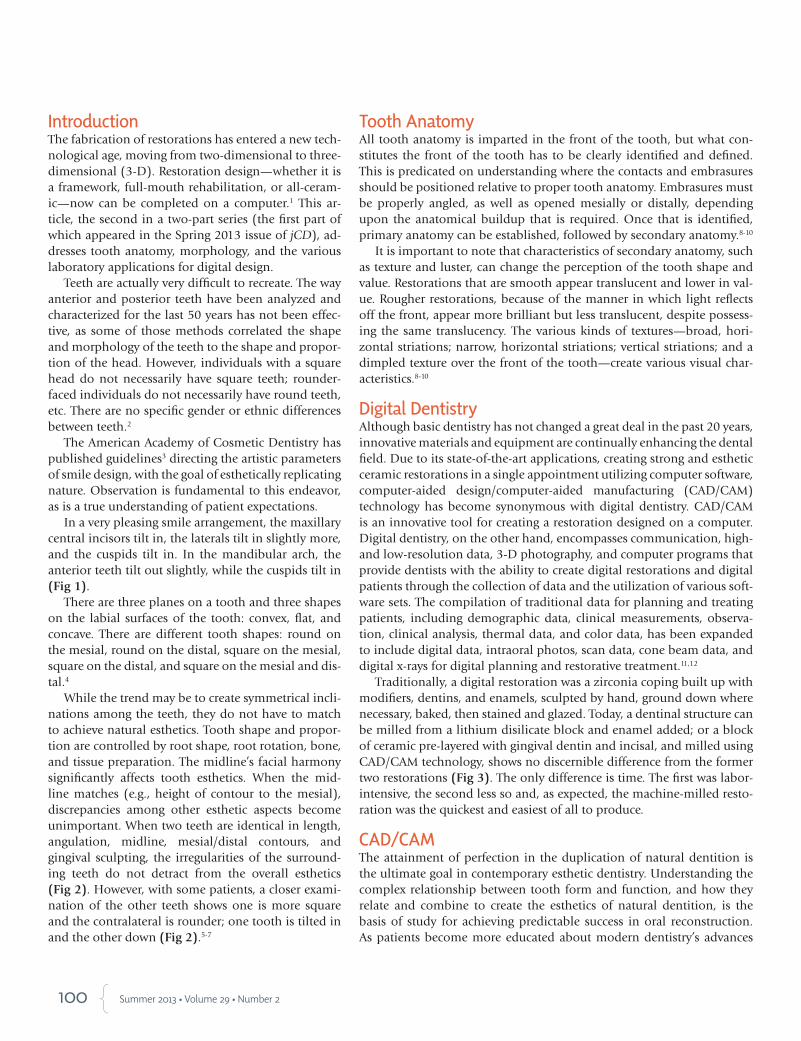
In a very pleasing smile arrangement, the maxillary central incisors tilt in, the laterals tilt in slightly more, and the cuspids tilt in. In the mandibular arch, the anterior teeth tilt out slightly, while the cuspids tilt in (Fig 1).
There are three planes on a tooth and three shapes on the labial surfaces of the tooth: convex, flat, and concave. There are different tooth shapes: round on the mesial, round on the distal, square on the mesial, square on the distal, and square on the mesial and dis-tal.4
While the trend may be to create symmetrical incli-nations among the teeth, they do not have to match to achieve natural esthetics. Tooth shape and propor-tion are controlled by root shape, root rotation, bone, and tissue preparation. The midline’s facial harmony significantly affects tooth esthetics. When the mid-line matches (e.g., height of contour to the mesial), discrepancies among other esthetic aspects become unimportant. When two teeth are identical in length, angulation, midline, mesial/distal contours, and gingival sculpting, the irregularities of the surround-ing teeth do not detract from the overall esthetics (Fig 2). However, with some patients, a closer exami-nation of the other teeth shows one is more square and the contralateral is rounder; one tooth is tilted in and the other down (Fig 2).5-7
Tooth AnatomyAll tooth anatomy is imparted in the front of the tooth, but what con-stitutes the front of the tooth has to be clearly identified and defined. This is predicated on understanding where the contacts and embrasures should be positioned relative to proper tooth anatomy. Embrasures must be properly angled, as well as opened mesially or distally, depending upon the anatomical buildup that is required. Once that is identified, primary anatomy can be established, followed by secondary anatomy.8-10
It is important to note that characteristics of secondary anatomy, such as texture and luster, can change the perception of the tooth shape and value. Restorations that are smooth appear translucent and lower in val-ue. Rougher restorations, because of the manner in which light reflects off the front, appear more brilliant but less translucent, despite possess-ing the same translucency. The various kinds of textures—broad, hori-zontal striations; narrow, horizontal striations; vertical striations; and a dimpled texture over the front of the tooth—create various visual char-acteristics.8-10
Digital DentistryAlthough basic dentistry has not changed a great deal in the past 20 years, innovative materials and equipment are continually enhancing the dental field. Due to its state-of-the-art applications, creating strong and esthetic ceramic restorations in a single appointment utilizing computer software, computer-aided design/computer-aided manufacturing (CAD/CAM) technology has become synonymous with digital dentistry. CAD/CAM is an innovative tool for creating a restoration designed on a computer. Digital dentistry, on the other hand, encompasses communication, high- and low-resolution data, 3-D photography, and computer programs that provide dentists with the ability to create digital restorations and digital patients through the collection of data and the utilization of various soft-ware sets. The compilation of traditional data for planning and treating patients, including demographic data, clinical measurements, observa-tion, clinical analysis, thermal data, and color data, has been expanded to include digital data, intraoral photos, scan data, cone beam data, and digital x-rays for digital planning and restorative treatment.11,12
Traditionally, a digital restoration was a zirconia coping built up with modifiers, dentins, and enamels, sculpted by hand, ground down where necessary, baked, then stained and glazed. Today, a dentinal structure can be milled from a lithium disilicate block and enamel added; or a block of ceramic pre-layered with gingival dentin and incisal, and milled using CAD/CAM technology, shows no discernible difference from the former two restorations (Fig 3). The only difference is time. The first was labor-intensive, the second less so and, as expected, the machine-milled resto-ration was the quickest and easiest of all to produce.
CAD/CAMThe attainment of perfection in the duplication of natural dentition is the ultimate goal in contemporary esthetic dentistry. Understanding the complex relationship between tooth form and function, and how they relate and combine to create the esthetics of natural dentition, is the basis of study for achieving predictable success in oral reconstruction. As patients become more educated about modern dentistry’s advances
101 Journal of Cosmetic Dentistry
Figure 1: Culp Classification of Anterior Tooth Shapes.
Figure 2: All-ceramic restorations, showing natural shape nuances that create a more natural tooth arrangement.
Figure 3: All-ceramic restorations showing three different types of fabrication methods. Left: Milled e.max CAD restoration (Ivoclar Vivadent; Amherst, NY), with only enamel layering. Center: Milled Empress Multi CAD full-contour restoration (Ivoclar Vivadent), with surface stain and glaze. Right: Zirconia coping, fully layered with several different dentin and enamel ceramics.
Culp/McLaren/Swann
102 Summer 2013 • Volume 29 • Number 2
(as a result of television makeover shows and profes-sional and over-the-counter bleaching systems), their motivation and desire for natural, esthetic restorative dentistry is increasing at a dramatic rate. Dentists and technicians are now fulfilling these patient demands, but still use dental laboratories and restorative tech-niques that do not always offer predictable efficiency and quality.
Based upon technology adopted from the aero-space/automotive, and even the watch-making in-dustry, CAD/CAM is becoming accepted due to its in-creased speed, accuracy, and efficiency. Today’s CAD/CAM systems are being used to design and manufac-ture metal, alumina, and zirconia frameworks, as well as all-ceramic full-contour crowns, inlays, and veneers that are stronger, fit better, and are more esthetic than restorations fabricated using traditional methods. As dentistry evolves in the digital world, the successful incorporation of computerization and new acquisi-tion and manufacturing technologies will continue to provide more efficient methods of restoration fabri-cation and communication, while at the same time retaining the individual creativity and artistry of the skilled dentist and technician. The utilization of these new technologies—along with the evolution from “hand” design to “digital” design, with the addition of the latest developments in intraoral laser scanning, materials, and computer milling/printing technol-ogy—will only enhance the close cooperation and working relationship of the dentist/laboratory team.
More than 20 different CAD/CAM systems have been introduced as solutions for restorative dentistry. The introduction of digital laboratory laser-scanning technology, along with its accompanying software, al-lowed the dental laboratory to create a digital dental environment to accurately present a real 3-D virtual model that automatically takes into consideration the occlusal effect of the opposing and adjacent den-tition. It also has the ability to design 32 individual full-contour anatomically correct teeth at the same time. These systems essentially take a complex occlu-sal scheme and its parameters and condense the in-formation, display it in an intuitive format that allows dental professionals with basic knowledge of dental anatomy and occlusion to make modifications to the design, and then send it to the automated milling/printing unit. For the dental laboratory profession, the introduction of digital technology effectively au-tomated—or even eliminated—some of the more
mechanical and labor-intensive procedures (waxing, investing, burnout, casting, and/or pressing) involved in the conventional fabrication of a dental restoration, giving the dentist and technician the ability to create functional dental restorations with a consistent, precise method.
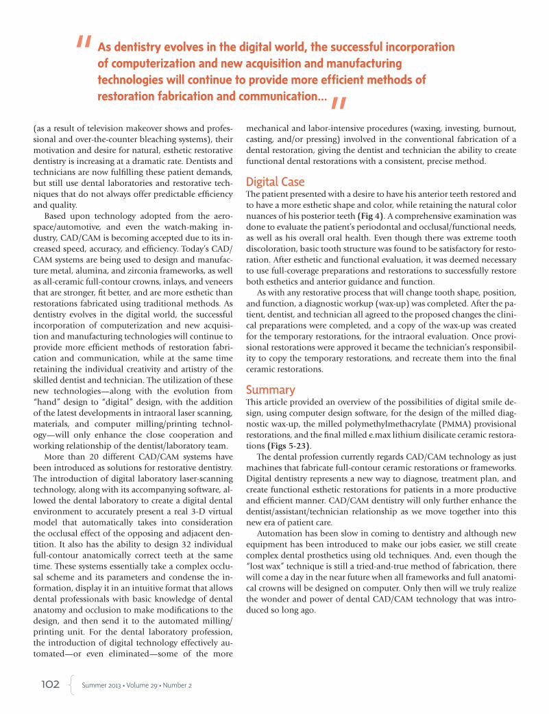
Digital CaseThe patient presented with a desire to have his anterior teeth restored and to have a more esthetic shape and color, while retaining the natural color nuances of his posterior teeth (Fig 4). A comprehensive examination was done to evaluate the patient’s periodontal and occlusal/functional needs, as well as his overall oral health. Even though there was extreme tooth discoloration, basic tooth structure was found to be satisfactory for resto-ration. After esthetic and functional evaluation, it was deemed necessary to use full-coverage preparations and restorations to successfully restore both esthetics and anterior guidance and function.
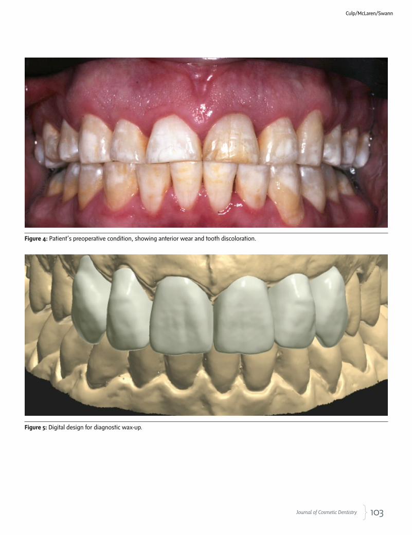
As with any restorative process that will change tooth shape, position, and function, a diagnostic workup (wax-up) was completed. After the pa-tient, dentist, and technician all agreed to the proposed changes the clini-cal preparations were completed, and a copy of the wax-up was created for the temporary restorations, for the intraoral evaluation. Once provi-sional restorations were approved it became the technician’s responsibil-ity to copy the temporary restorations, and recreate them into the final ceramic restorations.
SummaryThis article provided an overview of the possibilities of digital smile de-sign, using computer design software, for the design of the milled diag-nostic wax-up, the milled polymethylmethacrylate (PMMA) provisional restorations, and the final milled e.max lithium disilicate ceramic restora-tions (Figs 5-23).
The dental profession currently regards CAD/CAM technology as just machines that fabricate full-contour ceramic restorations or frameworks. Digital dentistry represents a new way to diagnose, treatment plan, and create functional esthetic restorations for patients in a more productive and efficient manner. CAD/CAM dentistry will only further enhance the dentist/assistant/technician relationship as we move together into this new era of patient care.
Automation has been slow in coming to dentistry and although new equipment has been introduced to make our jobs easier, we still create complex dental prosthetics using old techniques. And, even though the “lost wax” technique is still a tried-and-true method of fabrication, there will come a day in the near future when all frameworks and full anatomi-cal crowns will be designed on computer. Only then will we truly realize the wonder and power of dental CAD/CAM technology that was intro-duced so long ago.
As dentistry evolves in the digital world, the successful incorporation of computerization and new acquisition and manufacturing technologies will continue to provide more efficient methods of restoration fabrication and communication…
103 Journal of Cosmetic Dentistry
Culp/McLaren/Swann
Figure 4: Patient’s preoperative condition, showing anterior wear and tooth discoloration.
Figure 5: Digital design for diagnostic wax-up.
104 Summer 2013 • Volume 29 • Number 2
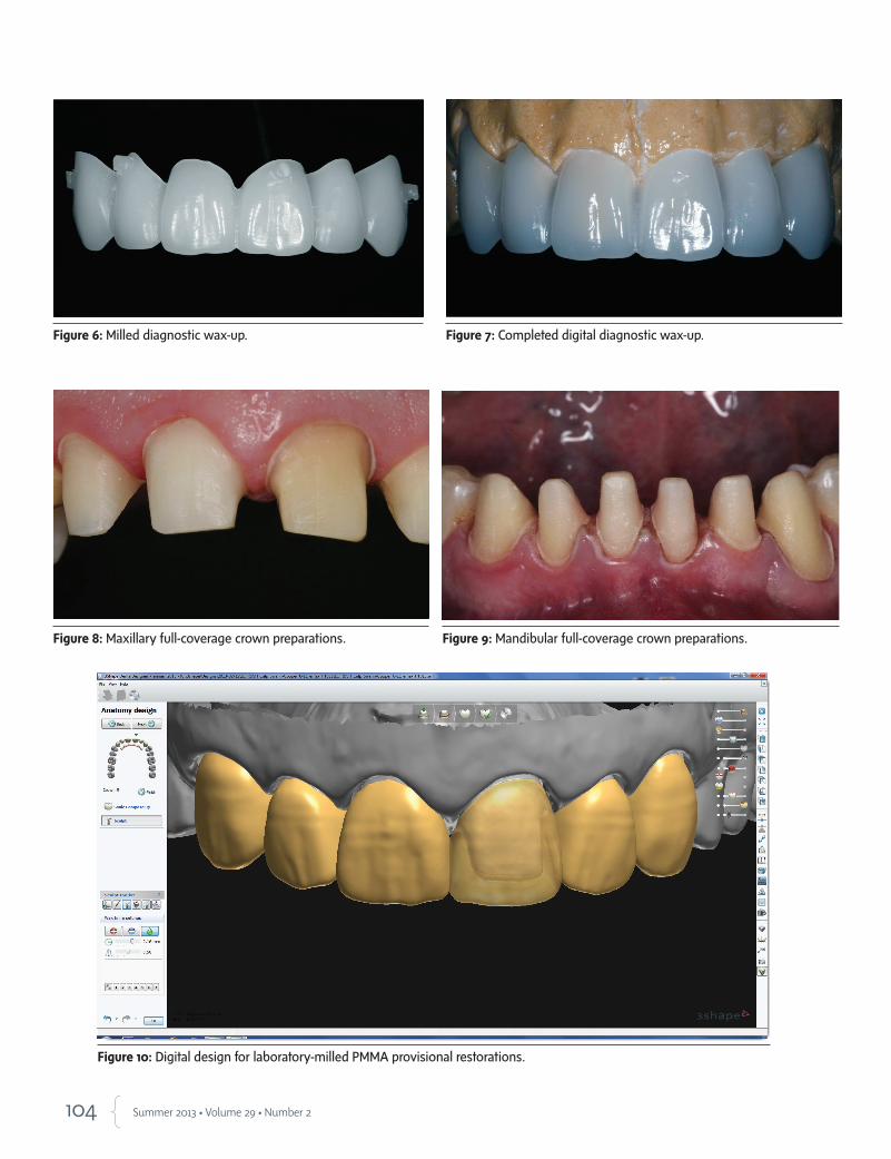
Figure 6: Milled diagnostic wax-up. Figure 7: Completed digital diagnostic wax-up.
Figure 8: Maxillary full-coverage crown preparations. Figure 9: Mandibular full-coverage crown preparations.
Figure 10: Digital design for laboratory-milled PMMA provisional restorations.
105 Journal of Cosmetic Dentistry
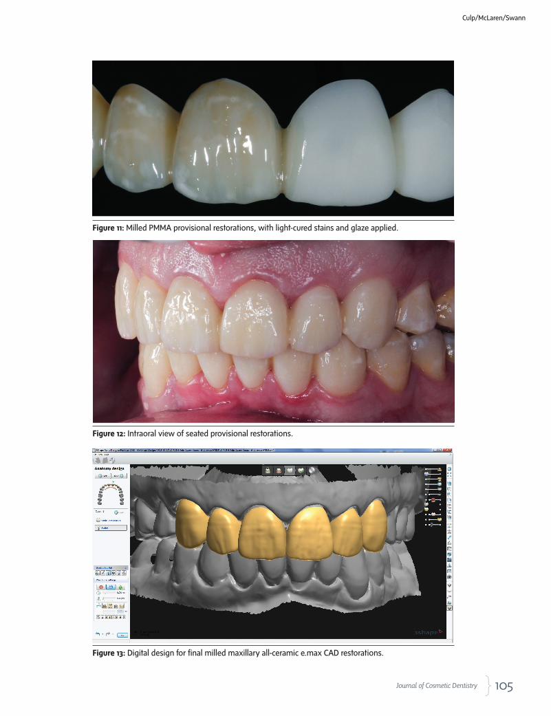
Figure 11: Milled PMMA provisional restorations, with light-cured stains and glaze applied.
Figure 12: Intraoral view of seated provisional restorations.
Culp/McLaren/Swann
Figure 13: Digital design for final milled maxillary all-ceramic e.max CAD restorations.
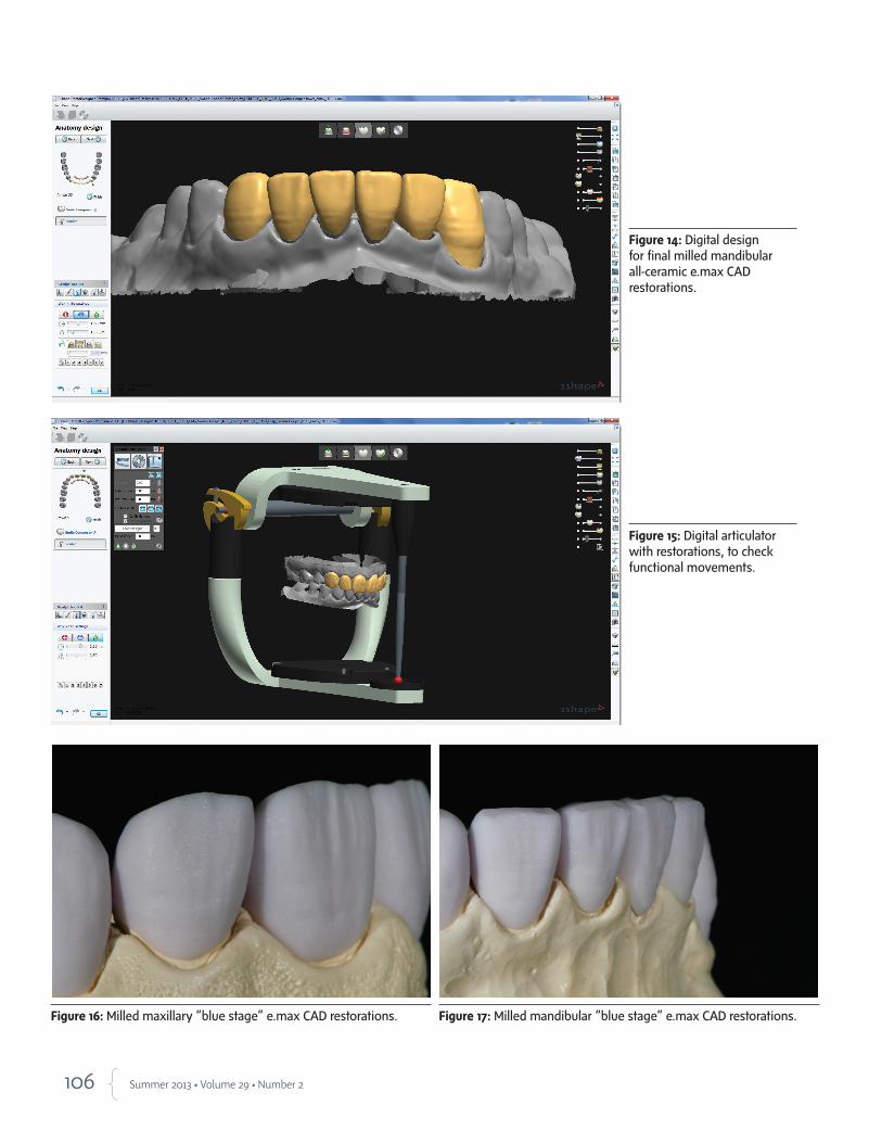
106 Summer 2013 • Volume 29 • Number 2
Figure 16: Milled maxillary “blue stage” e.max CAD restorations. Figure 17: Milled mandibular “blue stage” e.max CAD restorations.
Figure 14: Digital design for final milled mandibular all-ceramic e.max CAD restorations.
Figure 15: Digital articulator with restorations, to check functional movements.
107 Journal of Cosmetic Dentistry
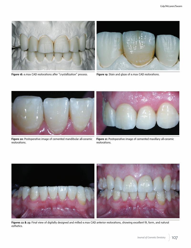
Figure 18: e.max CAD restorations after “crystallization” process. Figure 19: Stain and glaze of e.max CAD restorations.
Figure 20: Postoperative image of cemented mandibular all-ceramic restorations.
Figure 21: Postoperative image of cemented maxillary all-ceramic restorations.
Figures 22 & 23: Final view of digitally designed and milled e.max CAD anterior restorations, showing excellent fit, form, and natural esthetics.
Culp/McLaren/Swann
108 Summer 2013 • Volume 29 • Number 2
Acknowledgments
The clinical dentistry shown in this article was performed by Dr. Swann. The digital and technical dentistry was done by Mr. Culp.
References
1. Touchstone A, Nieting T, Ulmer N. Digital transition: the col-
laboration between dentists and laboratory technicians on CAD/
CAM restorations. J Am Dent Assoc. 2010 Jun;141 Suppl 2:15S-
9S.
2. McLaren EA, Rifkin R. Macroesthetics: facial and dentofacial
analysis. J Calif Dent Assoc. 2002 Nov;30(11):839-46.
3. American Academy of Cosmetic Dentistry (AACD). Diagnosis
and treatment evaluation in cosmetic dentistry: a guide to Ac-
creditation criteria. Madison (WI): AACD; 2001.
4. Kattadiyil MT, Goodacre CJ, Naylor WP, Maveli TC. Esthetic smile
preferences and the orientation of the maxillary occlusal plane.
J Prosthet Dent. 2012 Dec;108(6):354-61. doi: 10.1016/S0022-
3913(12)60192-9.
5. Greenberg JR, Bogert MC. A dental esthetic checklist for treat-
ment planning in esthetic dentistry. Compend Contin Educ
Dent. 2010 Oct;31(8):630-4, 636, 638.
6. McLaren EA, Tran Cao P. Smile analysis and esthetic design: “in
the zone.” Inside Dent. 2009;5(7):46-8.
7. Vig RG, Brundo GC. The kinetics of anterior tooth display. J Pros-
thet Dent. 1978;39:502-4.
8. Fradeani M. Esthetic analysis: a systematic approach to prosthetic
treatment. Hanover Park (IL): Quintessence Pub.; 2004.
9. Magne P, Gallucci GO, Belser UC. Anatomic crown width/length ratios of unworn
and worn maxillary teeth in with subjects. J Prosth Dent. 2003;89(5):453-61.
10. Preston JD. The Golden Proportion revisited. J Esthet Dent. 1993;5:247-51.
11. Ringer J. Digital smile enhancement: an essential modality for any successful cos-
metic practice. Dent Today. 2007 May;26(5):84, 86, 88-9.
12. Beuer F, Schweiger J, Edelhoff D, Sorensen JA. Reconstruction of esthetics with a
digital approach. Int J Periodontics Restorative Dent. 2011 Apr;31(2):185-93. jCD
Mr. Culp is an adjunct faculty member at the University of North Carolina (UNC) School of Dentistry. He is an instruc-tor at UNC, Chapel Hill, North Carolina, and practices in Dublin, California, and Raleigh, North Carolina.
Disclosure: Mr. Culp receives an honorarium from Ivoclar Vivadent.
Dr. McLaren is the director of the UCLA Center for Esthetic Dentistry. He maintains a private practice in Los Angeles, California.
Disclosure: Dr. McLaren did not report any disclosures.
Once provisional restorations were approved it became the technician’s responsibility to copy the temporary restorations, and recreate them into the final ceramic restorations.
Dr. Swann is a resident in the Department of Prosthodontics, UNC at Chapel Hill. Disclosure: Dr. Swann did not report any disclosures.


![Mandala Milis Culp[1] Copy](https://img.pdfslide.us/doc/110x75/540309238d7f72f24a8b47f3/mandala-milis-culp1-copy.jpg)