Embed Size (px)
Citation preview
ARTICLE OPEN
Small heat shock protein CRYAB inhibits intestinal mucosalinflammatory responses and protects barrier integrity throughsuppressing IKKβ activityWeimin Xu1, Yuegui Guo1, Zhenyu Huang1, Haoxin Zhao2, Mingxia Zhou3, Yuji Huang1, Dongpeng Wen1, Jinglue Song1, Zhehui Zhu1,Mingming Sun4, Chen-Ying Liu1, Yingwei Chen3, Long Cui1, Xiaolei Wang4, Zhanju Liu4, Yili Yang2 and Peng Du 1
Alpha B-crystallin (CRYAB) is an important member of the small heat shock protein family, and plays a protective and therapeuticrole in neurological inflammation. CRYAB expression was assessed in cultured HT29 and Caco-2 cells and inflamed mucosa ofpatients with inflammatory bowel disease (IBD) and colitis models in mice. Lentivirus-overexpressing and CRSIPR/Cas9 systemswere used in different cells to upregulate and silence CRYAB expression, respectively. Cell permeable recombined fusion proteinTAT-CRYAB was injected intraperitoneally into dextran sulfate sodium (DSS)- or 2,4,6-trinitrobenzene sulfonic acid (TNBS)-inducedcolitis in mice to assess its anti-inflammatory effects. CRYAB was found to be significantly decreased in the inflamed mucosa fromIBD patients and DSS-induced colitis in mice, and negatively correlated with the levels of TNF-α and IL-6, respectively. Enforcedexpression of CRYAB suppressed expression of proinflammatory cytokines (e.g., TNF-α, IL-6, IL-1β, and IL-8) via inhibiting the IKKcomplex formation, whereas lack of CRYAB expression markedly enhanced proinflammatory responses. Consistently, administrationof TAT-CRYAB fusion protein significantly alleviated DSS- or TNBS-induced colitis in mice and protected intestinal barrier integrity.CRYAB regulates inflammatory response in intestinal mucosa by inhibiting IKKβ-mediated signaling and may serve as a noveltherapeutic approach in the treatment of IBD.
Mucosal Immunology (2019) 12:1291–1303; https://doi.org/10.1038/s41385-019-0198-5
INTRODUCTIONInflammatory bowel disease (IBD) refers to the chronic inflamma-tory disease that affects the gastrointestinal tract, the mostcommon forms were the ulcerative colitis (UC) and Crohn’sdisease (CD).1 Noteworthily, the incidence rate of IBD has beenincreasing, in particular in the developing countries,2 and thedisease usually lasts for decades, characterized by alternatingperiods of the disease activity and remission,3,4 thus emphasizingthe urgent need for novel and more effective therapeutics.Extensive studies have indicated that the disease arises as a resultof the complicated interactions between the genetic andenvironmental factors, leading to dysregulated immunologicalresponses and tissue damages in the intestine.5–7 As such, theetiology and pathology of IBD depend upon the sensitivity of theintestine to inflammatory and immunological stimuli, particularly,the diets and food supplements ingested, and the intestinalmicroflora and their products.8
The rising incidence rate and long-lasting disease course havemade IBD a significant challenge for patients and physicians.Present therapeutics, including 5-aminosalicylic acids, glucocor-ticoids, and immunosuppressive and biological agents, areunsatisfactory or intolerable in a significant percentage ofpatients. It has been estimated that up to 25–30% of IBD
patients eventually need to undergo surgery due to theineffectiveness of drugs and disease complications.9 Therefore,it is warranted to search for novel candidate target genes foreffective clinical treatment.Alpha B-crystallin, also known as CRYAB or HSPB5, is initially
identified as a structural protein of the vertebrate eye lens, and isexpressed in a variety of tissues, including the heart, skeletalmuscles, brain, and fat.10–12 It is a member of small heat shockprotein family and functions as a molecular chaperon in order toprevent the aggregation of proteins denatured by stresses, suchas radiation and heat shock.10 Recent studies have reported thatincreased expression of CRYAB renders several highly invasiveand metastatic solid tumor cells,13–16 whereas degradation andphosphorylation of CRYAB during enterovirus infection facilitatesviral replication and induces viral pathogenesis.17 Interestingly,several studies have reported that CRAYB plays protective andtherapeutic roles in the development of neuroinflammationinduced by injury, infection, neurodegeneration, and autoimmu-nity.18–22
In this study, we determined the role of CRYAB in thepathogenesis and treatment of IBD. Taking advantage of intestinaltissues from IBD patients and dextran sulfate sodium (DSS)- and2,4,6-trinitrobenzene sulfonic acid (TNBS)-induced murine colitis
Received: 2 January 2019 Revised: 3 August 2019 Accepted: 12 August 2019Published online: 3 September 2019
1Department of Colorectal Surgery, Xinhua Hospital, Shanghai Jiaotong University School of Medicine, Shanghai 200092, China; 2Suzhou Institute of Systems Medicine, Center forSystems Medicine, Chinese Academy of Medical Sciences, Suzhou 215123, China; 3Department of Gastroenterology, Xinhua Hospital, School of Medicine, Shanghai Jiao TongUniversity, Shanghai 200092, China and 4Department of Gastroenterology, The Shanghai Tenth People’s Hospital, Tongji University, Shanghai 200072, ChinaCorrespondence: Zhanju Liu ([email protected]) or Yili Yang ([email protected]) or Peng Du ([email protected])These authors contributed equally: Weimin Xu, Yuegui Guo, Zhenyu Huang
www.nature.com/mi
© The Author(s) 2019

models, we demonstrated that expression of CRYAB wasdecreased in the inflamed mucosa of IBD patients and negativelycorrelated with the inflammatory reaction. We also found thatCRAYB inhibited inflammation by hampering IKKβ-mediatedsignaling, and that administration of TAT-CRYAB recombinedprotein alleviated DSS-induced intestinal mucosal inflammation inmice. These results indicate that CRYAB protects intestinal mucosafrom inflammation and provides the first evidence that exogenousCRYAB may act as a potential therapeutic approach for thetreatment of IBD.
MATERIALS AND METHODSPatients and specimensNon-inflamed and inflamed colon mucosa were collected from30 patients with active UC (A-UC) and 33 patients with activeCD (A-CD) who underwent surgery or colonic biopsy at theDepartment of Colorectal Surgery, Xinhua Hospital, ShanghaiJiaotong University School of Medicine (Shanghai, China) fromFebruary 2008 to March 2017. Moreover, normal colon tissueswere obtained from 45 patients who underwent colonic biopsyfor colon polyps as controls (HC). In addition, paired inflamedand distant non-inflamed colon tissues were collected from sixpatients with active UC, and six patients with active CD. Thediagnosis of CD and UC was based on clinical, endoscopic,radiological examination, and histological changes. The studywas approved by the Ethics Committee of Xinhua Hospital (No.XHEC-D-2018-054). All patients signed an informed consent priorto the study.
Cell culture and treatmentThe human monocyte cell line THP-1, colonic adenoma cell linesHT29 and Caco-2, colorectal adenocarcinoma cell lines HCT116and SW480, and HEK293 cell line were obtained from theAmerican Type Culture Collection (Manassas, VA, USA). These celllines were cultured with the Dulbecco’s modified Eagle’s medium(DMEM) supplemented with 10% fetal bovine serum (Gibco,Shanghai, China), 100 µg/ml of penicillin, and 100 U/ml ofstreptomycin at 37 °C with 5% CO2.
Enforced expression of CRYAB in intestinal epithelial cellsThe cDNA of CRYAB was cloned into puromycin-resistantlentiviral vector (pLVX-Puro). The lentivirus particles wereprepared using HEK293T cells according to the manufacturer’sprotocol (Takara, Beijing, China). THP-1, HT29, and Caco-2 cellswere then seeded into six-well culture plates and incubatedovernight, infected with CRYAB-expressing lentivirus, and werethen treated with puromycin to select the cells overexpressingCRYAB in a stable manner.
Silencing CRYAB expression using CRISPR/Cas9 systemThe guide RNA targeting CRYAB and Cas9 was expressed in HT29cells using the Lenti-CRISPR V2 vector. CRYAB-deficient cells werethen selected with puromycin, examined using qRT-PCR andimmunoblotting, and verified by genomic sequencing.
Immunohistochemistry and immunofluorescenceParaffin-embedded tissues were deparaffinized in xylene, rehy-drated with descending concentrations of ethanol, and theantigen was retrieved by heating in a citrate buffer (pH 6.0). Aftertreating with 3% hydrogen peroxide and 5% goat serum, theslides were incubated with specific antibodies at 4 °C overnight.After thorough washing with PBS, the slides were incubatedwith HRP-labeled secondary antibody for 30 min at roomtemperature and visualized with diaminobenzidine chromogen.For immunofluorescence staining, AlexaFluor 594-conjugatedand SABC-cy3-conjugated anti-rabbit IgGs were used as thesecondary antibodies.
Immunofluorescence staining of cultured cellsCells grown on the cover slides were fixed with cold 4%paraformaldehyde for 30 min, and were permeabilized by expos-ing to 0.3% Triton X-100 in cold PBS for 30 min. After blocking in5% bovine serum albumin (BSA) for 30 min at room temperature,the cells were incubated with anti-NF-κB p65 mAb (1:100, dilutedin 3% BSA) for 2 h at room temperature. After thorough washingwith PBS containing 0.1% Triton X-100, these cells were incubatedwith AlexaFluor 594-conjugated donkey anti-rabbit IgG (LifeTechnologies, Grand Island, NY, USA) in the dark for 1 h at roomtemperature, then counter-stained with 5 μg/ml DAPI, and wereeventually analyzed using a fluorescence microscopy.
Quantitative real-time PCR (qRT-PCR)The total RNA was extracted from the cultured cells and primarytissues using TRIzol reagent (Invitrogen, Carlsbad, CA, USA)according to the manufacturer’s instructions. Reverse transcriptionwas performed from 1 μg of the total RNA using PrimeScriptTM RTMaster Mix (Takara). qRT-PCR was carried out using the SYBRPremix ExTaqTM (Takara) and an Applied Biosystems 7500 Fastreal-time PCR system (公司). Relative expression of mRNA wasevaluated by 2−ΔΔCt method, and was normalized to theexpression of GAPDH. All experiments were repeated in triplicates.Sequences of PCR primers in this study are listed in Supplemen-tary Table 1.
ImmunoblottingCells were lysed with 1% NP40 lysis buffer supplemented withNaF, Na3VO4, and protease inhibitor cocktail. Lysates comprisingequal amounts of protein (20 μg/well) were loaded and separatedby SDS-PAGE, and were then transferred onto the nitrocellulosemembrane. The membrane was blocked with 5% BSA for at least1 h, and was then incubated with a specific antibody at 4 °Covernight. After washing the membranes three times with tris-borate saline containing 0.1% tween-20 for 5 min each, thespecific protein was visualized with HRP-conjugated secondaryantibody and enhanced chemiluminescence.
Co-immunoprecipitation assay (Co-IP)HEK293T cells were transfected with the plasmids expressing IκBα-Flag, Flag-IKKβ, and CRYAB-HA or the control plasmid pCDNA3.1as indicated. After 24 h of culture, the cells were lysed with 0.3%NP40 lysis buffer supplemented with NaF, Na3VO4, PMSF, andprotease inhibitor cocktail at 4 °C for 30 min. The lysates wereprepared by centrifugation at 12,000 rpm for 15 min at 4 °C, andwere then subjected to immunoprecipitation by incubating withanti-Flag or anti-HA antibodies conjugated with agarose for 3 h.Following extensive washes with TBST, the agarose beads werethen boiled with 2 × loading buffer for 5 min. The supernatantswere separated on SDS-PAGE, and were analyzed via immuno-blotting by using anti-HA and anti-FLAG antibodies.
ELISAHuman TNF-α, IL-6, IL-1β, and IL-8 in the culture supernatants weremeasured using enzyme-linked immunosorbent assay (ELISA) kitspurchased from Invitrogen with the Quick EIATM ELISA protocol,according to the manufacturer’s protocol.
Determination of IKKβ activityIKKβ activity was determined via the phosphorylation of specificpeptide (RRREEEEESAAA) in the presence and absence of a specificsmall molecular inhibitor (5-phenyl-3-ureido)-thiophene-2-carbox-amide, according to the manufacturer’s instructions from Shang-hai Haling Biotechnology (Shanghai, China).
Dual-luciferase reporter assaysThe plasmids pNFκB-Luc and (1 μg/3 well) and TK-Renilla (20 ng/well) were co-transfected into different cells cultured in a 24-well
Small heat shock protein CRYAB inhibits intestinal mucosal inflammatory. . .W Xu et al.
1292
Mucosal Immunology (2019) 12:1291 – 1303
1234567890();,:
plate with lipofectamine 2000 reagent (Invitrogen). After theindicated treatment, the cells were lysed with the passivelysis buffer, and the luciferases were evaluated using the dual-luciferase reporter assay kit (Promega, Madison, WI, USA)according to the manufacturer’s instruction.
Mice and DSS-induced murine colitis modelThe 6-week-old C57BL/6 male mice (20 g) were purchased fromthe Experimental Animal Center of the Chinese Academy ofSciences (Shanghai, China) and were maintained in the animalfacility of the Xinhua Hospital, Shanghai Jiaotong UniversitySchool of Medicine. To induce colitis, 8-week-old male mice werefed with 3.5% of DSS in drinking water and were monitored fortheir body weight, activity, and conditions, such as diarrhea andbloody stools. All mice were killed after 7-day induction. All animalexperimental procedures were approved by the LaboratoryAnimal Care and Welfare Committee of Xinhua Hospital (No.XHEC-F-2018-017).
2,4,6-trinitrobenzene sulfonic acid (TNBS)-induced colitisThe 8-week-old C57BL/6 male mice (25 g) were used for theinduction of TNBS-induced colitis. They were first sensitized with1% TNBS solution (presensitization solution) diluted from 5% TNBSsolution with acetone and olive oil mixture (4:1 volume ratio). Thepresensitization solution (100 μl) was applied to a 1 × 1 cm2
shaved area on the back of each mice. After 7 days, each mousewas anesthetized with 2.5% chloral hydrate solution and given150 μl 2.5% TNBS solution intrarectally by using a catheterthrough the anus. The activities and defecation of mice wereassessed every day for consecutive 3 days, then killed for furtherexamination.
Intestinal integrity examined by fluorescein isothiocyanate (FITC)-dextranFITC-dextran was utilized to examine the intestinal integrity inDSS and TNBS-induced mice colitis models. The mice were given150 μL (80 mg/mL) of FITC-dextran in sterile water at the last dayof induction (the seventh day of DSS induction or the third day ofTNBS induction). Blood samples (100 μL) were then collected fromthe tail vein to measure the amounts of FITC fluorescence(480 excitation/520 emission), which reflected the relativeepithelial permeability.
RNA and protein extraction from mouse colonMice were euthanized to dissect the colon and rectum.After extensive washing with pre-cooled PBS, the intestinaltissues were cut and homogenized to extract RNA with TRIzolreagent and proteins with RIPA lysis buffer supplemented withprotease inhibitors cocktail, respectively, using the methods asdescribed above.
Disease activity index (DAI) and histological pathological scoringTo assess DAI score of each mouse, body weight, stool character,and occult or rectal bleeding were monitored daily after DSS orTNBS administration, which were scored on a scale of 0–4. DAI foreach mouse was calculated by dividing the total scores by 3 aslisted in Supplementary Table 2.23 The histopathological scores ofmice with induced inflammations were assessed based onneutrophils infiltration, crypts, cross-section involvement, anderosion or ulceration formation by two independent pathologistson the scale of 0–3. The detailed scores for mice are listed inSupplementary Table 3.24
Production of TAT-CRYAB fusion proteinSynthetic DNA coding the TAT-PTD was inserted into the Nhe I siteof a pET28a vector. The CRYAB cDNA was then subcloned into theBamH I and EcoR I sites of the pET28a-PTD plasmid. Sequence ofthe resulted TAT-CRYAB-expressing construct was verified via DNA
sequencing. The production and purity of the recombinantprotein was examined by SDS-PAGE and Coomassie blue staining.
Statistical analysisThe data were presented as mean ± standard deviation (SD).Graph Pad Prism 5 software (San Diego, CA, USA) and SPSSStatistics version 19.0 software (IBM 2010, Chicago, IL, USA) wereused for statistical analyses. Unpaired Student’s t test was used tocompare the difference between two single groups. Pearson’scorrelation test was performed to analyze the relationshipbetween the levels of TNF-α and IL-6 and CRYAB expression inthe inflamed mucosa of IBD patients. All statistical tests were two-sided and considered as significant with a p-value of <0.05.
RESULTSCRYAB expression is significantly decreased in inflamed colonfrom IBD patientsTo study the molecular mechanisms of CRYAB involved in thedevelopment and pathogenesis of IBD, we initially examinedCRYAB expression in the inflamed colon from patients with A-UC(n= 30) and A-CD (n= 33) by qRT-PCR. A significant decrease inthe CRYAB mRNA was observed in the inflamed colon frompatients with A-UC and A-CD compared with that from the normalcolons (n= 45; Fig. 1a). Moreover, we also observed thatthe expression of IL-6 and TNF-α was notably increased in theinflamed tissues of UC and CD (Fig. 1a), and that their levels werenegatively correlated with the CRYAB expression in the inflamedcolon from patients with A-CD (for TNF-α, Spearman’s rankcorrelation coefficient, R=−0.4744, p= 0.0061; for IL-6, R=−0.4631, p= 0.0067; Fig. 1b). Nevertheless, no significant correla-tion was observed between TNF-α or IL-6 and CRYAB in theinflamed mucosa from A-UC patients, presumably due to the widedispersion in the levels of TNF-α and IL-6. We further examined theCRYAB expression by immunoblotting in paired, inflamed, andunaffected tissues from six UC and six CD patients. Whencompared with the paired normal mucosa, CRYAB was markedlydecreased in the inflamed mucosa of UC and CD patients (Fig. 1c).Furthermore, a significant increase in phosphorylated p65 andIκBα, indicative of activated proinflammatory NF-κB signaling, wasalso observed in these tissues (Fig. 1c). This was further confirmedby immunohistochemical analysis of the CD tissues. The levels ofCRYAB were found to be notably lower in the inflamed tissues ofCD patients compared with controls, which was also accompaniedby an increased infiltration of the macrophages and T lympho-cytes in inflamed mucosa (Fig. 1d). In addition, immunofluores-cence staining for intestinal epithelial marker CK18, macrophagemarker CD68, and CRYAB was performed and identified thatmacrophages but not intestinal epithelial cells mainly expressedCRYAB in inflamed tissues from CD patients (SupplementaryFig. 1). Detailed clinical characteristics of IBD patients are listed inSupplementary Table 4.
CRYAB inhibits production of proinflammatory cytokines by LPS-induced macrophagesTo examine whether CRYAB was able to affect the production ofproinflammatory cytokines, we cultured THP-1 cells in vitro andtransfected with CRYAB-expressing lentivirus (Fig. 2a). These cellswere then stimulated with LPS under different conditions in orderto assess the production of proinflammatory cytokines (e.g., TNF-α,IL-6, IL-1β, and IL-8) by qRT-PCR and ELISA. As shown in Fig. 2b, themRNA levels of these cytokines were rapidly induced in THP-1cells transfected with control lentiviruses, while they weresignificantly reduced in CRYAB-expressing lentivirus-transfectedcells. The same pattern of cytokine expression was found in thesecells when analyzed by ELISA (Fig. 2c).Moreover, we examined the effects of CRYAB on LPS-
stimulated phosphorylation of IκBα and p65 in THP-1 cells using
Small heat shock protein CRYAB inhibits intestinal mucosal inflammatory. . .W Xu et al.
1293
Mucosal Immunology (2019) 12:1291 – 1303

phosphorylated protein-specific immunoblotting. As presentedin Fig. 2d, CRYAB significantly reduced LPS-stimulated phosphor-ylation of IκBα and p65, in particular, during 1–6 h after thestimulation. Thus, CRYAB appears to block the activation of NF-κBand suppresses the production of proinflammatory cytokines inmacrophages at transcriptional levels.
CRYAB regulates immune response of HT29 and Caco-2 cells inresponse to TNF-αTo explore whether CRYAB is essential in regulating intestinal cellsin response to inflammatory stimulation, we examined CRYABexpression in HT29 and Caco-2 cells at the mRNA and proteinlevels. Both epithelial cell lines expressed moderate levels of
CRYAB at the baseline, which significantly decreased understimulation with TNF-α in a time-dependent manner (Fig. 3a),presumably recapitulating reduced CRYAB in intestinal tissuesfrom patients with UC and CD. Furthermore, we assessed the anti-inflammatory effects of enforced expression of CRYAB on HT29and Caco-2 cells in response to TNF-α (Fig. 3b). As presented inFig. 3c–f, TNF-α was found to promote the expression of TNF-α,IL-6, IL-1β, and IL-8 mRNAs in HT29 and Caco-2 cells. Whencompared with the cells infected with control lentivirus, CRYAB-expressing lentivirus-transfected cells expressed a markeddecrease of mRNAs of these cytokines after exposure to TNF-α,notably at 12–24 h. The inhibition of inflammatory cytokines inHT29 and Caco-2 cells by CRYAB overexpression was also
Fig. 1 CRYAB is decreased in the inflamed colon tissues from IBD patients. a qRT-PCR analysis of CRYAB, TNF-α, and IL-6 expression. RNA wasisolated from healthy controls (HC, n= 45), active UC patients (A-UC, n= 30), and active CD patients (A-CD, n= 33). CRYAB was significantlydecreased, whereas TNF-α and IL-6 were markedly increased in A-UC and A-CD than those in HC, (*p < 0.05, ***p < 0.001). b Relationshipsbetween TNF-α, IL-6, and CRYAB mRNA in inflamed mucosa from 33 A-CD. Significant correlations existed between the levels of TNF-α mRNAand CRYAB (Spearman’s rank correlation coefficient, R=−0.4744, p= 0.0061), and IL-6 and CRYAB expression (Spearman’s rank correlationcoefficient, R=−0.4631, p= 0.0067). c The expression of CRYAB, p65, p-p65, IκBα, and p-IκBα in the inflamed and unaffected tissues from sixpaired A-UC and six paired A-CD patients was examined by immunoblotting with specific antibodies. d Representative immunohistochemicalimages of CRYAB, CD68, CD3, and CD4 in the paired, inflamed, and unaffected tissues of the same CD patients (magnification: ×200, upperpanels; ×400, lower panels). All data are shown from three independent experiments
Small heat shock protein CRYAB inhibits intestinal mucosal inflammatory. . .W Xu et al.
1294
Mucosal Immunology (2019) 12:1291 – 1303

confirmed by ELISA (Fig. 3g). Moreover, we utilized the CRSPR/Cas9 system to downregulate CRYAB expression in HT29 cells(Supplementary Fig. 2A, B). Compared with controls, TNF-α-induced expression of TNF-α, IL-6, and IL-8 mRNAs wassignificantly increased in CRYAB-deficient cells (SupplementaryFig. 2C). Collectively, these data indicate that CRYAB is anendogenous anti-inflammatory molecule in regulating immuneresponses of intestinal epithelial cells.
CRYAB interacts with IKKβ and IKKα and inhibits IKKβ activityAs shown in Fig. 2d, ectopic expression of CRYAB in THP-1 cellsinhibited LPS-induced activation of the NF-κB pathway. We alsofound that TNF-α-induced phosphorylation and subsequentdownregulation of IκBα were markedly attenuated by enforced
expression of CRYAB in HT29 and Caco-2 cells (Fig. 4a). Consistentwith these changes, CRYAB expression decreased TNF-α-inducedluciferase expression driven by the NF-κB response element(Fig. 4b), resulting in blocking of translocation of p65 from thecytoplasm to the nucleus (Fig. 4c). Furthermore, the knockout ofCRYAB in HT29 and Caco-2 cells significantly increased the levelsof p-p65 and p-IκBα following TNF-α stimulation (SupplementaryFig. 2D). These results indicate that CRYAB inhibits intestinalinflammation by blocking the activation of the NF-κB pathway.As IκB kinase (IKK) β is crucial in phosphorylating IκB to activate
NFκB, we asked whether CRYAB could affect its action. As shownin Fig. 5a, transfection of IKKβ-expression plasmid into HCT116and SW480 cells led to p65 and IκB phosphorylation and IκBdegradation, whereas co-expression of CRYAB markedly reduced
Fig. 2 Enforced expression of CRYAB inhibits LPS-induced production of inflammatory cytokines by THP-1 cells. a Expression of CRYAB in THP-1 cells transfected with CRYAB-expressing lentivirus was detected by qRT-PCR and immunoblotting. b, c Cytokine production in THP-1-pLVXand THP-1-CRYAB cells after LPS (100 ng/ml) stimulation for indicated time. RNA was isolated to assess the mRNA levels of proinflammatorycytokines (e.g., TNF-α, IL-6, IL-1β, and IL-8) by qRT-PCR. b Cytokines in the supernatants were measured by EILSA. *p < 0.05, **p < 0.01, ***p <0.001. d Immunoblotting was performed to examine the levels of p-p65, p65, p-IκBα, and IκBα in THP-1-pLVX and THP-1-CRYAB cells after LPSstimulation for the indicated times. All data are shown from three independent experiments
Small heat shock protein CRYAB inhibits intestinal mucosal inflammatory. . .W Xu et al.
1295
Mucosal Immunology (2019) 12:1291 – 1303

Fig. 3 Enforced expression of CRYAB inhibits TNF-α-induced inflammatory response in HT29 and Caco-2 cells. a TNF-α decreased CRYAB inHT29 and Caco-2 cells. The cells were treated with TNF-α (10 ng/ml) for indicated times. CRYAB expression was assessed by qRT-PCR andimmunoblotting; p < 0.05, **p < 0.01, ***p < 0.001. b HT29 and Caco-2 cells were transfected by CRYAB-expressing lentivirus. CRYAB level wasassessed by qRT-PCR and immunoblotting; ***p < 0.001. c–f Expression of TNF-α, IL-6, IL-1β, and IL-8 was examined in CRYAB-overexpressingand control cells after exposed to TNF-α (10 ng/ml) for indicated times; *p < 0.05, **p < 0.01, ***p < 0.001. g Cytokines in the supernatants weremeasured by EILSA. **p < 0.01, ***p < 0.001. All data are shown from three independent experiments
Small heat shock protein CRYAB inhibits intestinal mucosal inflammatory. . .W Xu et al.
1296
Mucosal Immunology (2019) 12:1291 – 1303

these effects. Noteworthily, CRYAB expression did not affect TNF-α-induced IKKβ activation (phosphorylation) in colon cancer cells,indicating that CRYAB inhibits NF-κB activation by blocking actionof IKKβ.We used an in vitro assay system to measure the activity of IKKβ
from HT29 cells. Compared with unstimulated cells, TNF-α-stimulated HT29 cells had markedly increased IKKβ-specificactivity, and co-treatment of these cells with TAT-CRYABsignificantly decreased the activity (Fig. 5b). When HCT116 cellswere transfected with plasmids expressing HA-CRYAB and Flag-IKKβ, we found the co-immunoprecipitation of exogenouslyexpressed HA-CRYAB with Flag-IKKβ and vice versa (Fig. 5c, d).Similarly, CRYAB also bound with IKKα when overexpressed inHCT116 cells (Fig. 5e, f). We further assessed the interactions ofthese molecules by co-transfecting plasmids expressing Flag-IKKβ,HA-IKKα, and HA-CRYAB into HCT116 cells. As shown in Fig. 5g, h,the co-immunoprecipitation of Flag-IKKβ and HA-IKKα wasmarkedly reduced in the presence of HA-CRYAB and vice versa.These results indicated that CRYAB likely inhibits IKKβ activity viabinding to IKKβ and IKKα and interfering IKK complex formation.
Recombinant TAT-CRYAB protein has anti-inflammatory actionand alleviates DSS-induced colitis in miceTo explore whether CRYAB could be used as an anti-inflammatoryagent, the transduction domain of TAT, which is able to facilitatecell penetration of the recombinant heterogeneous proteins,25–27
was fused with CRYAB to generate a recombinant cell permeableCRYAB (Supplementary Fig. 3A) of near 29 KD (SupplementaryFig. 3B–C). HT29 and Caco-2 cells treated with recombined proteinshowed high levels of intracellular CRYAB (Fig. 6a). As presented inFig. 6b, TNF-α induced significantly less amounts of proinflamma-tory cytokines (e.g., TNF-α, IL-6, IL-1β, and IL-8) in TAT-CRYAB-treated HT29 or Caco-2 cells. Notably, recombined TAT-CRYABprotein also inhibited the phosphorylation of IκBα and p65, andblocked IκBα degradation in these cells (Fig. 6c).The ability of TAT-CRYAB to inhibit proinflammatory signal and
cytokine production encouraged us to determine whether therecombinant protein has an anti-inflammatory effect in an animalmodel of colitis. Mice fed with drinking water comprising of 3.5%DSS developed intestinal inflammation, manifested by a shor-tened colon, more severe colonic tissue damage and inflammatory
cells infiltration, when compared with controls (there were fivemice in the Water group and five mice in the DSS group,respectively; Supplementary Fig. 4A, B). Similar to the findings inUC and CD patients, CRYAB expression was significantly decreasedin inflamed intestines from DSS-fed mice, as evidenced byimmunoblotting (Supplementary Fig. 4C) and immunohistochem-ical analysis (Supplementary Fig. 4D).As presented in Fig. 7a, a time-dependent increase of CRYAB in
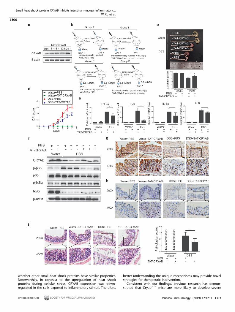
colon tissue from mice receiving intraperitoneal injection of TAT-CRYAB recombined protein was observed, indicating that therecombined protein might penetrate into the colon cells (onemouse per group). Detailed injection process of the recombinedprotein and induction by DSS are presented in Fig. 7b (therewere three mice in water+ PBS group, three mice in water+TAT-CRYAB group, four mice in DSS+ PBS group, and four micein DSS+ TAT-CRYAB group, respectively). In DSS-fed mice, thecolitis mice treated with TAT-CRYAB recombined protein for 7consecutive days appeared to be less severe compared withcontrols, as characterized by the shortened length of colons, adecrease of DAI score, and low levels of production ofinflammatory cytokines (TNF-α, IL-6, IL-1β, and IL-8) (Fig. 7c–e).Compared with untreated mice, the colon tissues from micereceiving TAT-CRYAB recombined protein showed prominentlyintensified levels of CRYAB, reduced p65 and IκBα phosphoryla-tion, and moderately decreased IκBα degradation (Fig. 7f). Similarresults were also obtained from the immunohistochemicalstaining for CRYAB (Fig. 7g). Immunohistochemical staining forTAT revealed that intestinal tissues from mice treated with TAT-CRYAB were TAT positive, but negative in PBS-treated mice,suggesting that the administrated TAT-CRYAB indeed penetratedinto the colon tissues (Fig. 7h). Moreover, hematoxylin–eosinstaining revealed that colon tissues from TAT-CRYAB-treatedmice had a decreased pathological score compared with controls(Fig. 7i). Collectively, these data demonstrate that recombinantTAT-CRYAB plays an anti-inflammatory role in protecting theintestines from inflammation.
Recombined TAT-CRYAB protein protects intestinal barrierintegrity from DSS-induced damageIt has been shown that disruption of the intestinal barrier plays acritical role in the development of intestinal inflammation, which
Fig. 4 CRYAB inhibits the activation of the NF-κB pathway in HT29 and Caco-2 cells. a TNF-α-induced alterations in p-p65, p65, p-IκBα, andIκBα were examined by immunoblotting in the CRYAB-overexpressing and control cells. b NF-κB-driven luciferase was inhibited by CRYAB.CRYAB-overexpressing and control cells were transfected with a NF-κB-driven luciferase reporter. Dual-luciferase assays were performed afterTNF-α stimulation (10 ng/ml) for indicated times. The firefly luciferase activity was measured with relative normalization for Renilla; *p < 0.05,**p < 0.01, ***p < 0.001. c Immunofluorescence staining of p65 in CRYAB-overexpressing and control cells after TNF-α treatment (10 ng/ml) forindicated times. Expression of CRYAB inhibited p65 nuclear localization. All data are shown from three independent experiments
Small heat shock protein CRYAB inhibits intestinal mucosal inflammatory. . .W Xu et al.
1297
Mucosal Immunology (2019) 12:1291 – 1303

is assessed by the amount of FITC-dextran in the blood after themouse was fed the compound in drink water. As shown in Fig. 8a,administration of TAT-CRYAB significantly reduced the intestinalepithelial permeability, and ameliorated the goblet cell destruc-tion in DSS-induced inflammatory intestines as evaluated byAlcian blue/periodic acid-Schiff staining (Fig. 8b). In the DSS-fedmice, there was also decreased ZO-1 expression, indicative of thedestruction of epithelial tight junction, which was alleviated byadministration of TAT-CRYAB (Fig. 8c). Colons from TAT-CRYAB-treated mice also contained higher proteins levels of E-cadherin(Fig. 8d). Immunofluorescence staining of E-cadherin, CK20, andMUC2 further revealed that mice treated with TAT-CRYAB had lessintestinal damage compared with those treated with PBS alone(Fig. 8e).Noteworthily, administration of TAT-CRYAB also reduced TNBS-
induced colitis in mice (there were three mice in absolute ethanol+ PBS group, three mice in absolute ethanol+ TAT-CRYAB group,
four mice in TNBS+ PBS group, and four mice in TNBS+ TAT-CRYAB group, respectively; Supplementary Fig. 5) and protectedthe intestinal epithelial cells and integrity (Supplementary Fig. 6).Therefore, these data indicate that CRYAB functions as an effectivetherapeutic agent for intestinal inflammation.
DISCUSSIONIn this study, we discovered that the small heat shock proteinCRYAB modulated the sensitivity of intestinal cells to inflammatorystimuli. Increased CRYAB could inhibit the inflammatoryresponses, while lack of CRYAB markedly amplified the response.We also demonstrated that CRYAB itself was downregulated by aproinflammatory cytokine TNF-α (Fig. 3a), presumably due toinitiating inflammatory response. Furthermore, CRYAB expressionwas significantly decreased in inflamed colon tissues of IBDpatients, which correlated with increased inflammatory cytokines.
Fig. 5 CRYAB interacts with IKKβ and IKKα and inhibits IKKβ activity. a CRYAB inhibited IKKβ-driven p65 phosphorylation, IκBαphosphorylation, and degradation. HCT116 and SW480 cells were transfected with control vector PRK7, Flag-IKKβ, or Flag-CRYAB plasmids,respectively. After 24 h of culture, the cells were lysed and immunoblotted with anti-FLAG, p65, p-p65, IκBα, p-IκBα, and p-IKKβ, respectively.b CRYAB inhibits the IKKβ activity. HT29 cell was treated with TNF-α (10 μg/ml) or control for 6 h in the absence or presence of pretreatment ofTAT-CRYAB recombined protein (5 μg/ml) for 12 h. Specific IKKβ activity was then measured using a peptide substrate and a small molecularIKKβ inhibitor; *p < 0.05, **p < 0.01. c, d Co-immunoprecipitation of IKKβ and CRYAB. HCT116 cells were transfected to express HA-CRYABand Flag-IKKβ. Cell lysates were immunoprecipitated with anti-HA beads and blotted with anti-FLAG antibody or immunoprecipitatedwith anti-FLAG beads and blotted with anti-HA antibody. e, f Co-immunoprecipitation of IKKα and CRYAB. Cell lysates wereimmunoprecipitated with anti-HA beads and blotted with anti-FLAG antibody or immunoprecipitated with anti-FLAG beads and blottedwith anti-HA antibody. g, h CRYAB inhibits the interaction between IKKα and IKKβ. HCT116 cells were co-transfected with plasmids expressingFlag-IKKβ, HA-IKKα, and HA-CRYAB. Co-immunoprecipitation of expressed Flag-IKKβ and HA-IKKα was significantly inhibited compared withthe cells without HA-CRYAB and vice versa. All data are shown from three independent experiments
Small heat shock protein CRYAB inhibits intestinal mucosal inflammatory. . .W Xu et al.
1298
Mucosal Immunology (2019) 12:1291 – 1303

Thus, our data indicate that CRYAB is crucial in the pathogenesis ofIBD and may serve as a precision medicine biomarker to assess thedisease and its response to anti-inflammatory effects.CRYAB is a component of the small heat shock protein family,
whose constituents are usually upregulated when cells areexposed to heat shock and other stresses, including accumulationof misfolded proteins in the cells. Noteworthily, heat shockproteins are often expressed at low-to-moderate levels in severalcells, and function as chaperons to prevent unwanted proteinaggregation. In this study, we observed that CRYAB inhibited thephosphorylation and degradation of IκBα, which is required for
the activation of NF-κB, a critical transcription factor for theproduction of multiple proinflammatory cytokines. We furtherdemonstrated that CRYAB did not affect the activation of IKK byinflammatory stimuli; however, it appeared to bind to IKKβ andinhibit its activity toward its substrate. We further found thatCRYAB also interacts with IKKα and apparently blocks theformation of IKK complex to reduce the IKKβ activity. As thesmall heat shock proteins are characterized by a conservedα-crystallin-domain that participates in their interaction withpartners, it is interesting to know in the future whether theα-crystallin-domain is involved in binding with IKKβ and IKKα and
Fig. 6 Recombinant TAT-CRYAB protein inhibits inflammatory response of intestinal epithelial cells. a HT29 and Caco-2 cells were incubatedwith TAT-CRYAB (500 ng/ml) for indicated times. CRYAB expression was assessed by immunoblotting. b TNF-α, IL-6, IL-1β, and IL-8 wereinhibited by TAT-CRYAB treatment. HT29 and Caco-2 cells were treated with TNF-α (10 μg/ml) for 6 h in the absence or presence ofpretreatment of recombinant TAT-CRYAB (5 μg/ml) for 12 h. The cytokine mRNAs were measured by qRT-PCR; *p < 0.05, **p < 0.01, ***p < 0.001.c The phosphorylation of p65 and IκBα was inhibited by TAT-CRYAB. The levels of p-p65, p65, p-IκBα, and IκBα in different treated cells wereanalyzed by immunoblotting with specific antibodies. All data are shown from three three independent experiments
Small heat shock protein CRYAB inhibits intestinal mucosal inflammatory. . .W Xu et al.
1299
Mucosal Immunology (2019) 12:1291 – 1303
whether other small heat shock proteins have similar properties.Noteworthily, in contrast to the upregulation of heat shockproteins during cellular stress, CRYAB expression was down-regulated in the cells exposed to inflammatory stimuli. Therefore,
better understanding the unique mechanisms may provide novelstrategies for therapeutic intervention.Consistent with our findings, previous research has demon-
strated that Cryab−/− mice are more likely to develop severe
Small heat shock protein CRYAB inhibits intestinal mucosal inflammatory. . .W Xu et al.
1300
Mucosal Immunology (2019) 12:1291 – 1303

Fig. 8 TAT-CRYAB administration ameliorates DSS-induced intestinal damages. Water+ PBS group: three mice; water+ TAT-CRYAB group:three mice; DSS+ PBS group: four mice; DSS+ TAT-CRYAB group: four mice. a Epithelial permeability was measured on day 7 post-DSStreatment by oral administration of FITC-dextran. ***p < 0.001. b Representative images of Alcian blue/periodic acid-Schiff staining of colontissues from mice of different groups (magnification: ×200, upper panels; ×400, lower panels). c Representative immunofluorescence imagesfor ZO-1 in colonic tissues from different groups of mice (magnification: ×400). d E-cadherin expression in colonic tissues from differentgroups was examined by immunoblotting; *p < 0.05. e Representative immunofluorescence images for E-cadherin, CK20, and MUC2 in colonictissues from different groups of mice (magnification: ×400). All data are shown from three independent experiments
Fig. 7 Administration of TAT-CRYAB alleviates DSS-induced intestinal mucosal inflammation in mice. a Administration of TAT-CRYABintraperitoneally increased CRYAB expression in the colon tissues. Each mouse was injected with 25 μg of TAT-CRYAB intraperitoneally, andkilled at indicated times to detect the levels of CRYAB in the colon (one mouse per group). b Schematic diagram of the TAT-CRYAB treatmentin DSS-induced colitis (water+ PBS group: three mice; water+ TAT-CRYAB group: three mice; DSS+ PBS group: four mice; DSS+ TAT-CRYABgroup: four mice). c TAT-CRYAB reduced colitis-induced shortening of colon length, and (d) DAI score; *p < 0.05, **p < 0.01. e TNF-α, IL-6, IL-1β,and IL-8 in DSS-induced colitis were reduced by TAT-CRYAB administration. The cytokines in the colon were assessed by qRT-PCR; *p < 0.05,**p < 0.01, ***p < 0.001. f CRYAB, p-p65, p65, p-IκBα, and IκBα in the colon were analyzed by immunoblotting with specific antibodies.g Representative immunohistochemical images of CRYAB expression in colons from different groups of mice (magnification: ×200, upperpanels; ×400, lower panels). h Representative immunohistochemical images of TAT-CRYAB expression examined by the anti-TAT antibody incolons from different groups of mice (magnification: ×200, upper panels; ×400, lower panels). i Representative images of hematoxylin–eosinstaining and pathological scores of colon tissues from DSS-exposed mice with or without TAT-CRYAB administration (magnification: ×200,upper panels; ×400, lower panels); *p < 0.05. All data are shown from three independent experiments
Small heat shock protein CRYAB inhibits intestinal mucosal inflammatory. . .W Xu et al.
1301
Mucosal Immunology (2019) 12:1291 – 1303

experimental autoimmune encephalomyelitis and have increasedimmune activation and intense central nervous system inflamma-tion.18 However, it was not determined whether there were anychanges in spontaneous or induced intestinal inflammation inCryab−/− mice. Therefore, it is interesting to further investigate therole of CRYAB in regulating mucosal inflammatory and immuneresponses using DSS- and TNBS-induced colitis model in Cryab−/−
mice, especially conditional knockout models to assess the roles ofCRYAB in myeloid cells and intestinal epithelial cells.TNF-α is crucial in the initiation and development of IBD.28–31
The successful therapy using various anti-TNF-α antibodiesrepresents a major breakthrough in the treatment of IBD, and italso propelled the development of novel biologicals targetingadditional cytokines and inflammation-related molecules.32 Theinfliximab ACCENT I clinical study, the CHARM study ofadalimumab and the vedolizumab GEMINI II study have shown acomplete 54-week remission rate of 16.7%, 25.2%, and 17.7%,respectively.33–35 It is conceivable that involvement of multipleproinflammatory cytokines in IBD patients at different stages ofdisease is responsible for the lack of effects. Thus, the ability ofCRYAB to inhibit IKKβ and the IKK complex formation and thenblock the production of multiple cytokines makes it a desirabletherapeutic approach for anti-inflammatory treatment in IBD(Fig. 9).It has been reported that the TAT-PDB domain is capable of
mediating fused protein into cells and affects the cellularfunctions, including reprogramming and ROS generation.36–39 Inthis study, we generated a novel TAT-CRYAB fusion protein, anddemonstrated for the first time that it not only had anti-inflammatory effect in the cultured cells but also markedly
alleviated DSS- and TNBS-induced colitis in mice, accompaniedby improving the colon length, and downregulating DAI scores,histopathological scores, and inflammatory cytokine levels. Inaddition, the administration of TAT-CRYAB protected the integrityof the intestinal barrier and prevented ulcerative formation inmurine colitis models. Thus, the recombinant protein is apromising novel therapeutic and needs to be further explored.
CONCLUSIONSCRYAB is significantly decreased in inflamed mucosa of IBDpatients and experimental colitis in mice. It appears to set thecellular sensitivity to inflammation. Enforced expression of CRYABinhibits IKKβ through hindering the formation of active IKKcomplex and exerts anti-inflammatory effect, whereas CRYABdeficiency enhances inflammatory response. Thus, decreasedCRYAB presumably contributes to the development of IBD. Inaddition, recombinant protein TAT-CRYAB possesses anti-inflammatory property and alleviates mucosal inflammation,indicating that the fusion protein could be a novel therapeuticagent for the treatment of IBD.
ACKNOWLEDGEMENTSThis work was supported by the grant from the National Natural Science Foundationof China (81873547, 81570474, 81630017, and 91740117) and CAMS Initiative forInnovative Medicine (CAMS-I2M, 2016-I2M-1-005).
AUTHOR CONTRIBUTIONSPeng Du is a guarantor of the article. Peng Du, Yili Yang, and Zhanju Liu conceivedand designed the study. Weimin Xu, Yuegui Guo, and Zhenyu Huang performed allexperiments. Weimin Xu wrote the final paper with input from Zhanju Liu, Yili Yang,and Peng Du. Haoxin Zhao, Mingxia Zhou, Yuji Huang, and Dongpeng Wen assistedwith qRT-PCR experiments about inflammatory cytokines. Jinglue Song, Zhehui Zhu,and Mingming Sun collaborated to collect intestinal biopsies of IBD patients. Chen-Ying Liu, Yingwei Chen, Long Cui, and Xiaolei Wang analyzed the data. All authorsreviewed the paper and approved the final version.
ADDITIONAL INFORMATIONThe online version of this article (https://doi.org/10.1038/s41385-019-0198-5)contains supplementary material, which is available to authorized users.
Competing interests: All authors have read and approved the paper, and none ofthem have any conflict of interests to declare.
Ethical approval: All procedures performed in studies involving human participantswere in accordance with the ethical standards of the institutional and/or nationalresearch committee and with the 1964 Helsinki declaration and its later amendmentsor comparable ethical standards. All animal experiments were in accordance with theU.K. Animals (Scientific Procedures) Act and its later amendments or comparableethical standards.
Publisher’s note: Springer Nature remains neutral with regard to jurisdictional claimsin published maps and institutional affiliations.
REFERENCES1. Kaplan, G. G. & Ng S. C. Understanding and preventing the global increase of
inflammatory bowel disease. Gastroenterology 152, 313–321.e312 (2017).2. Molodecky, N. A. et al. Increasing incidence and prevalence of the inflammatory
bowel diseases with time, based on systematic review. Gastroenterology 142,46–54 e42 (2012). quiz e30.
3. Podolsky, D. Inflammatory bowel disease. N. Engl. J. Med. 347, 417–429 (2002).4. Kaser, A., Zeissig, S. & Blumberg, R. S. Inflammatory bowel disease. Annu. Rev.
Immunol. 28, 573–621 (2010).5. Ng, S. C. et al. Geographical variability and environmental risk factors in inflam-
matory bowel disease. Gut 62, 630–649 (2013).6. Bernstein, C. & Shanahan, F. Disorders of a modern lifestyle: reconciling the
epidemiology of inflammatory bowel diseases. Gut 57, 1185–1191 (2008).
Fig. 9 Schematic diagram of CRYAB on the activation of the NF-κBpathway by TNF-α. On TNF-α stimulation, the TNF-α receptor isactivated, followed by the activation of TAK1, which is the crucialpivot for the important intersection of the NF-κB pathway activation.Thereafter, the activation of the IKK complex and subsequentdegradation of IκBα via the phosphorylation catalytic subunit IKKβare activated. According to the aforementioned results, we foundthat CRYAB does not affect the degradation of IKK complexes, butinhibits IKKβ activity via binding to IKKβ and IKKα and interfering IKKcomplex formation
Small heat shock protein CRYAB inhibits intestinal mucosal inflammatory. . .W Xu et al.
1302
Mucosal Immunology (2019) 12:1291 – 1303

7. Thia, K., Loftus, E., Sandborn, W. & Yang, S. An update on the epidemiology ofinflammatory bowel disease in Asia. Am. J. Gastroenterol. 103, 3167–3182 (2008).
8. Maloy, K. J. & Powrie, F. Intestinal homeostasis and its breakdown in inflammatorybowel disease. Nature 474, 298–306 (2011).
9. Marchioni Beery, R. & Kane, S. Current approaches to the management of new-onset ulcerative colitis. Clin. Exp. Gastroenterol. 7, 111–132 (2014).
10. Gruvberger-Saal, S. K. & Parsons, R. Is the small heat shock protein alphaB-crystallin an oncogene? J. Clin. Invest. 116, 30–32 (2006).
11. Lutsch, G. et al. Abundance and location of the small heat shock proteins HSP25and alphaB-crystallin in rat and human heart. Circulation 96, 3466–3476 (1997).
12. Iwaki, T., Kume-Iwaki, A. & Goldman, J. E. Cellular distribution of alpha B-crystallinin non-lenticular tissues. J. Histochem Cytochem. 38, 31–39 (1990).
13. Li, Q., Wang, Y., Lai, Y., Xu, P. & Yang, Z. HspB5 correlates with poor prognosis incolorectal cancer and prompts epithelial-mesenchymal transition through ERKsignaling. PLoS ONE 12, e0182588 (2017).
14. Shi, C., Yang, X., Bu, X., Hou, N. & Chen, P. Alpha B-crystallin promotes the invasionand metastasis of colorectal cancer via epithelial-mesenchymal transition. Bio-chem Biophys. Res. Commun. 489, 369–374 (2017).
15. Chen, D. et al. Alpha B-crystallin promotes the invasion and metastasis of gastriccancer via NF-kappaB-induced epithelial-mesenchymal transition. J. Cell Mol. Med.22, 3215–3222 (2018).
16. Huang, X. Y. et al. alphaB-crystallin complexes with 14-3-3zeta to induceepithelial-mesenchymal transition and resistance to sorafenib in hepatocellularcarcinoma. Hepatology 57, 2235–2247 (2013).
17. Fung, G. et al. Phosphorylation and degradation of alphaB-crystallin duringenterovirus infection facilitates viral replication and induces viral pathogenesis.Oncotarget 8, 74767–74780 (2017).
18. Ousman, S. S. et al. Protective and therapeutic role for alphaB-crystallin inautoimmune demyelination. Nature 448, 474–479 (2007).
19. Brownell, S., Becker, R. & Steinman, L. The protective and therapeutic function ofsmall heat shock proteins in neurological diseases. Front Immunol. 3, 74 (2012).
20. Whiston, E. A. et al. alphaB-crystallin protects retinal tissue during Staphylococcusaureus-induced endophthalmitis. Infect. Immun. 76, 1781–1790 (2008).
21. Arac, A. et al. Systemic augmentation of alphaB-crystallin provides therapeuticbenefit twelve hours post-stroke onset via immune modulation. Proc. Natl Acad.Sci. USA 108, 13287–13292 (2011).
22. Zhang, Y. et al. Activation of dopamine D2 receptor suppresses neuroin-flammation through alphaB-crystalline by inhibition of NF-kappaB nucleartranslocation in experimental ICH mice model. Stroke 46, 2637–2646 (2015).
23. Ito, R. et al. Interferon-gamma is causatively involved in experimental inflam-matory bowel disease in mice. Clin. Exp. Immunol. 146, 330–338 (2006).
24. Gupta, R. B. et al. Histologic inflammation is a risk factor for progression tocolorectal neoplasia in ulcerative colitis: a cohort study. Gastroenterology 133,1099–1105 (2007). quiz1340-1091.
25. Ziegler, A. & Seelig, J. High affinity of the cell-penetrating peptide HIV-1 Tat-PTDfor DNA. Biochemistry 46, 8138–8145 (2007).
26. Schwarze, S., Ho, A., Vocero-Akbani, A. & Dowdy, S. In vivo protein transduction:delivery of a biologically active protein into the mouse. Science 285, 1569–1572(1999).
27. Nagahara, H. et al. Transduction of full-length TAT fusion proteins into mam-malian cells: TAT-p27Kip1 induces cell migration. Nat. Med. 4, 1449–1452 (1998).
28. Matsuda, R. et al. Quantitive cytokine mRNA expression profiles in the colonicmucosa of patients with steroid naïve ulcerative colitis during active andquiescent disease. Inflamm. Bowel Dis. 15, 328–334 (2009).
29. Komatsu, M. et al. Tumor necrosis factor-alpha in serum of patients withinflammatory bowel disease as measured by a highly sensitive immuno-PCR. Clin.Chem. 47, 1297–1301 (2001).
30. Rioux, J. et al. Genomewide search in Canadian families with inflammatory boweldisease reveals two novel susceptibility loci. Am. J. Hum. Genet. 66, 1863–1870 (2000).
31. Hampe, J. et al. Linkage of inflammatory bowel disease to human chromosome6p. Am. J. Hum. Genet. 65, 1647–1655 (1999).
32. Melmed, G. & Targan, S. Future biologic targets for IBD: potentials and pitfalls.Nat. Rev. Gastroenterol. Hepatol. 7, 110–117 (2010).
33. Feagan, B. et al. Efficacy of vedolizumab in fistulising crohn’s disease: exploratoryanalyses of data from GEMINI 2. J. Crohns Colitis 12, 621–626 (2018).
34. Panaccione, R. et al. Adalimumab maintains remission of Crohn’s disease after upto 4 years of treatment: data from CHARM and ADHERE. Aliment Pharm. Ther. 38,1236–1247 (2013).
35. Cornillie, F. et al. Postinduction serum infliximab trough level and decrease of C-reactive protein level are associated with durable sustained response to inflix-imab: a retrospective analysis of the ACCENT I trial. Gut 63, 1721–1727 (2014).
36. Zhang, H. et al. Reprogramming of somatic cells via TAT-mediated proteintransduction of recombinant factors. Biomaterials 33, 5047–5055 (2012).
37. Pan, C., Jia, W., Lu, B. & Bishop, C. E. Expression of TAT recombinant Oct4, Sox2,Lin28, and Nanog proteins from baculovirus-infected Sf9 insect cells. Gene 556,245–248 (2015).
38. Lan, M. S., Chen, C., Saunee, N. A., Zhang, T. & Breslin, M. B. Expression of bio-logically active TAT-fused recombinant islet transcription factors. Life Sci. 114,45–50 (2014).
39. Park, H. et al. Alpha B-crystallin prevents the arrhythmogenic effects of particulatematter isolated from ambient air by attenuating oxidative stress. Toxicol. Appl.Pharm. 266, 267–275 (2013).
Open Access This article is licensed under a Creative CommonsAttribution 4.0 International License, which permits use, sharing,
adaptation, distribution and reproduction in anymedium or format, as long as you giveappropriate credit to the original author(s) and the source, provide a link to the CreativeCommons license, and indicate if changes were made. The images or other third partymaterial in this article are included in the article’s Creative Commons license, unlessindicated otherwise in a credit line to the material. If material is not included in thearticle’s Creative Commons license and your intended use is not permitted by statutoryregulation or exceeds the permitted use, you will need to obtain permission directlyfrom the copyright holder. To view a copy of this license, visit http://creativecommons.org/licenses/by/4.0/.
© The Author(s) 2019
Small heat shock protein CRYAB inhibits intestinal mucosal inflammatory. . .W Xu et al.
1303
Mucosal Immunology (2019) 12:1291 – 1303