Embed Size (px)
Citation preview

page 1
University Zurich Small Animal Surgery Clinic
Small animal surgery special course: Feline surgery, cruciate ligament rupture, problem solving
4th Continuing Education Course for Japan Small Animal Surgeons at the Small Animal Surgery Clinic, University of Zurich, Switzerland August 25 – 29, 2003

page 2 Contents Contents 2 Impressum 2 Program 3 AO course (feline) – Introduction to the exercises 4 The UniLock: A Locking Plate/Screw System 6 Intertarsal and tarsometatarsal luxation 8 Mandibular fractures 11 Maxillar fractures 13 Temporomandibular joint excision 14 Collateral ligament prosthesis in the carpus 16 Elbow luxations 19 Fractures of the humerus 22 Complications: orthopedics 24 Complications: soft tissues 26 Amputation of the tail and amputation of toes, Skin course III 29 Peripheral Nerve Injury – Focus Hindlimb 34 Skin tumors 37 Cranial cruciate ligament (CrCL) rupture: Anatomy, diagnosis, imaging, surgical approach, debridement, meniscectomy 41 Posterolateral capsuloraphy 45 Biomechanics of the stifle joint 46 Advancement of the tibial tuberosity for treatment of cranial cruciate deficient canine stifle 49
Impressum This handout was written by the assistants of the small animal surgery clinic. Editing work was done by Dr. D. Koch, corrections by Prof. P.M. Montavon. First edition, 2003. copyright University Zurich, 2003.

page 3 Program Mon Feline orthopedic AO course (procedures on the hindlimb) 0900
0915 1000 1100 1130 1300 1330 1700
- Introduction - Sacroiliac luxation - Acetabular fracture - Malleolar fractures - Introduction to locking systems - Introduction to the exercises - Rotation on 5 tables (4 rotations)
- Proximal femur fracture - femur fracture - transarticular tarsal ext fixator - intertarsal luxation - skull reconstruction
Montavon, Koch, Voss Voss Koch Montavon, Koch, Voss
Tue Feline orthopedics (procedures on the frontlimb and the axial skeleton) 0800 - 0900 - Rotation on 5 tables, ctd (1 rotation) Montavon, Koch, Voss 0900
0945 1030
- Maxilla fracture - Mandibula fracture - Temporomandibular joint excision
Keller Keller Haas
1115 1330 1430
- Carpal instability - Elbow luxation - humerus fracture, competition
Voss Voss Bass
1530 - Complications: orthopedics Dennler. Koch Wed Skin and radiology 0800 - Skin course 3 Schwandt 1030 - Radiology: interpretation of thorax and
abdomen radiographs (2 groups) Philipp
1400 – 2200 Excursion to Hergiswil De Robillard
Thu Problem solving, 3 groups, rotations: 0800 Introduction to peripheral nerve injury Steffen - Peripheral nerve injury; interpretation
of videos Steffen
- Degenerative problem: Elbow dysplasia, approach, surgery / arthroscopy
Damur
0900 – 1045 1100 – 1245 1400 – 1545 - Oncology: skin tumor, staging, CT,
lasersurgery, skin flaps Schwandt Melzer
1600 – 1700 - Complications : soft tissue Dennler, Koch
Fri Stifle day 0800
0900 1000 1100 1300 - 1630
- anatomy - approach, closure, meniscectomy - posterolateral capsulorrhaphy - Biomechanics - Tibia tuberosity advancement
Montavon Montavon Koch Tepic Guerrero, Montavon

page 4 AO course (feline) – Introduction to the exercises Daniel Koch, Dr. med. vet. ECVS, Pierre Montavon, Prof. Dr. med. vet. Consultants: D. Koch, A. Johnson, R. Vannini, J. Houlton, E. Asimus Sacroiliac luxation (feline) with a 2.0mm lag screw This is the luxation to stabilize in this exercise using a 2.0mm lag screw. Note the displacement of the iliac wing. The landmark for the incision is the tuber sacrale. The foam is incised on the dorsal aspect of the iliac crest. The self-retaining retractors are used to help maintain the exposure. A screw-hole is drilled in the sacral body taking care to avoid the sacral canal. The oscillating drill attachment may be used to avoid iatrogenic neural damage. After having drilled, measured and tapped the screw hole, drill an appropriate gliding hole in the ilium. The gliding hole is measured. The appropriate length 2.0mm screw is inserted through the gliding hole and its tip is directed into the tapped screw hole in the sacral body. The screw is tightened. Comminuted acetabular fracture (feline) with a 2.0 mm adaption plate This is the fracture to repair in this exercise. The landmarks for the incision are the wing of the ilium, the tuber ischium and the greater trochanter. The foam is incised vertically on the lateral aspect of the pelvis to expose the greater trochanter. Additional dissection to expose the acetabulum can include a trochanteric osteotomy. Further caudal exposure of the acetabulum can be obtained by tenotomy of the external rotator muscles of the hip. The intra articular fractures are reduced anatomically and temporarily fixed with two pairs of pointed reduction forceps. A 6 hole 2.0mm adaption plate is contoured to an intact acetabulum. The plate is applied to the fracture and acts in a tension band fashion. While alternatively tightening the screws, the fracture reduction the articular surface and the fixation are evaluated. Malleolar fractures (feline) with tension band wiring (1.0 K-wires, 0.6mm cerclage wire) These are the malleolar fractures to repair in this exercise. The landmarks for the repair of the lateral malleolar fracture are the distal fibula and the lateral metatarsal bones. The foam is incised on the lateral aspect of the talocrural joint. The fracture is reduced. Two 1.0mm K-wires are inserted from the lateral malleolus into the tibia. The tension band is completed with 0.6mm cerclage wire in a figure of 8 fashion. The excess K-wires are cut and bent. The landmarks for the repair of the medial malleolar fracture are the distal tibia and the central tarsal bone. The foam is incised on the medial aspect of the talocrural joint. The fracture is reduced. Two 1.0mm K-wires are inserted from the medial malleolus into the tibia. Aternatively, the K-wire may be introduced in a retrograde fashion. The tension band is completed with 0.6mm cerclage wire in a figure of 8 fashion. The excess K-wires are cut and bent.

page 5 Transarticular tarsal external fixator The tarsal joint may be temporarily stabilized with a type II external fixator. Fulls-pins are driven trough the distal tibia and half-pins are driven into the metatarsal bones from the medial and lateral side. The external fixator is completed with the according clamps and connecting bars. Femoral head fracture (feline) with 1.0mm K-Wires with double tip This is the fracture to repair in this exercise. The landmarks for the incision are the wing of the ilium, the tuber ischium and the greater trochanter. The foam is incised on the lateral aspect of the pelvis to expose the greater trochanter. Two or three 1.0 mm K-wires with double tip are inserted from the fracture surface into the femoral neck in a retrograde manner. The air drive is then connected to the opposite side of the K-wire. The K-wires are withdrawn until their tips are flush with the fracture surface. The fracture is reduced. The K-wires are driven gently into the femoral head. Penetration into the hip joint is avoided by comparing the length of the K-wire with an additional K-wire of similar length or a visual check. The K-wires are bent and cut at their exit on the lateral aspect of the femur. Alternatively, the K-wires can be driven in an antegrade manner starting from the level of the third trochanter directly across the fracture line. Another alternative is to use a ventral approach. The pins are driven from the articular surface into the femoral head and across the fracture into the femoral neck. The K-wires are cut short and countersunk beneath the articular surface. Femur fracture with UniLock The femur midshaft fracture is fixed with a 2.4 UniLock plate and locking screws according to the principles of locking systems (see introduction to UniLock by M. Keller and K. Voss). Skull reconstruction A defect in the maxillar, frontal or parietal bone is fixed with maxillofacial implants. According to the defect or the size of the skull, the 1.0 or the 1.3 system is chosen.

page 6 The UniLock: A Locking Plate/Screw System Marcel Keller, Dr. med. vet., Katja Voss, Dr. med. vet. Introduction The introduction of locking bone plate/screw systems has brought certain advantages in fracture fixation over other plating methods. Locking plate/screw systems act as internal fixators whose stability is given by the locking mechanism between the screw and the plate. The plate does not need to have intimate contact with the underlying bone, making exact plate contouring less crucial. A diminished contact between the plate and bone may also preserve the periosteal blood supply, thereby reducing the extent of bone resorption under the plate. Experimental studies have shown that locking plate/screw systems offer greater stability than standard reconstruction plates without locking screws, especially when only two screws are placed in each bone fragment. Materials/Properties The UniLock system is available as a 2.0-mm and a 2.4-mm bone plate/screw system with a locking mechanism between the plate and the screw (Fig. 1). For the 2.0-mm system, three plates of varying thickness are available, which accept the same 2.0-mm screws. For the 2.4-mm system, one plate with four different screws (a 2.4-mm and 3.0-mm locking screw, a 2.4-mm non-locking cortical screw and a 2.7-mm emergency screw) is available. All screws are self- tapping. The locking screws are applied using standard screw insertion techniques. An important attribute is the threaded drill guide, which centres the drill precisely over the plate hole and facilitates locking the screw to the plate. Indications In human medicine, the UniLock system is almost exclusively used for mandibular fracture fixation. In veterinary medicine, the possible applications of the system are much wider, given the small bones and joints in cats and dogs. The authors have
The threads in the screw hole of the plate and in the screw head comprise the locking mechanism in the UniLock system.
Preoperative stress radiographs reveal a dorsal proximal intertarsal instability. Postoperative view: The proximal screws are anchored in the talus. One distal screw is placed in the central tarsal bone and one in the fourth tarsal bone.

page 7 used the system in a variety of cases, including joint-near fractures, intertarsal instabilities, pelvic fractures and spinal subluxation. In the following, the application of the UniLock in three clinical cases is presented. Examples A cat was presented with a proximal intertarsal instability, due to rupture of the short ligaments (Fig. 2). Two parallel, two-holed, 2.0-mm UniLock plates were applied dorsally in an adaptive manner (Fig. 3). This permitted an immediate return to full function without the use of external splints as is generally necessary with other fixation methods and partial arthrodesis. A long, oblique fracture of the ilium, close to the acetabulum, in a mixed-breed dog was repaired with two 2.4-mm UniLock plates at the cranial and the caudal fracture ends (Fig. 4). The use of short plates facilitates maintaining the reduction of fractures while tightening the screws. Compared to traditional plating methods, shorter plates can be used with the locking system to attain adequate stability. A Dachshund was affected with cervical spinal instability following a C5-C6 ventral slot surgery for removal of a protruded intervertebral disc. Spinal stabilisation was performed using ventral plating with two 2.0-mm UniLock plates (Fig. 5). The small vertebral bodies only allow mini implants to be inserted. With the locking system, the use of short plates with monocortical screws is possible, which averts causing damage to the spinal cord. Conclusion The UniLock System has proved useful in a variety of applications in small animal surgery. Further experience with the system is necessary to delineate its field of indications.
Postoperative views of an oblique ilial fracture: The two short plates
bridge the cranial and caudal fracture ends. The borders of the ilium offer good cortical bone quality for screw purchase

page 8 Intertarsal and tarsometatarsal luxation Katja Voss, Dr. med. vet. Introduction The intertarsal and tarsometatarsal bones are connected by a large number of short ligaments. Hyperextension (plantar instability), hyperflexion (dorsal instability) and medial or lateral instabilities might occur at different joint levels after rupture of these ligaments. The lameness grade depends on the site and the grade of the instability. Dorsal ligament injuries usually do not cause a severe lameness, unless concurrent medial or lateral instability is present. In plantar instabilities the lameness is more severe and hyperextension is present when the cat is weight bearing. Abnormal mobility in the intertarsal or tarsometatarsal joints can usually be palpated. Radiographs are used to rule out concomitant fractures. The affected joint level is evaluated by stress radiographs in hyperextension, hyperflexion and varus and valgus position. Rupture of the plantar ligaments and fibrocartilage Rupture of the plantar ligaments and fibrocartilage results in hyperextension injury. These injuries are rare in cats. As the tension side is located on the plantar aspect of the tarsus, these injuries are treated with partial arthrodesis (Fig. 1). The plate can be placed medially or laterally, depending on concurrent medial or lateral instability. Alternatively, tarsometatarsal arthrodesis can be performed using crosspinning with two figure-8 wires applied laterally and medially in order to achieve interfragmentary compression. Rupture of the dorsal ligaments Rupture of the dorsal ligaments causes dorsal intertarsal or tarsometatarsal instability, which can be accompanied by medial or lateral instabilities. This type of injury is the most common cause for intertarsal or tarsometatarsal instability in our caseload of cats. Generally either the proximal
Figure 1. Partial tarsal arthrodesis with a lateral plate. In order to achieve a tension band effect, the plate can be twisted onto the plantar surface of the metatarsal bone. A figure-8 wire placed on the plantar side enhances stability.

page 9 intertarsal or the tarsometatarsal joint row is affected. Depending on the degree of instability conservative treatment with a splint or fixation with an adapation plate or a screw and wire technique is performed. Proximal intertarsal joint Two different types of dorsal injury occur at the proximal intertarsal joint. Rupture of the dorsal ligaments leads to a dorsal (hyperflexion) instability. Additional disruption of the talocalcaneal ligament leads to a luxation of the base of the talus. These injuries are treated by application of one or two miniplates in adaptation function. Locking screw and plate systems, such as Uni-Lock, are ideal for this purpose. The surgical approach is performed just above the instable joint row. For luxations of the base of the talus and for dorsomedial instabilities one 2,0 mm plate is placed dorsomedially (Fig 2). For stabilization of dorsolateral instabilities an additional plate between talus or calcaneus and fourth tarsal bone is used. The skin is closed routinely. No additional external stabilization is needed in the postoperative period. As the plates serve as a temporary fixation until fibrous healing has occurred, they can be removed after 3 to 4 months. Tarsometatarsal joint Dorsal tarsometatarsal instabilities are often accompanied by lateral or medial instabilities. They are also treated with the application of adapation plates as described above. Alternatively, the joints can be stabilized using a figure-8 cerclage wire or suture material, anchored around two screws (Fig. 3).
Figure 2. A luxation of the base of the talus has been stabilized with a 2,0 mm Uni-Lock plate. One screw is anchored in the talus and one in the central tarsal bone
Figure 3. A dorsomedial tarsometatarsal instability was stabilized using two miniscrews and a figure-8 wire. The proximal screw is placed in the central tarsal bone, the distal screw in the base of the second metatarsal bone. (Thanks to D. Damur, Chur)

page 10 Literature - Piermattei D.L. and Flo G.L. Handbook of Small Animal Orthopedics and Fracture Repair,
3rd ed., 1997, W.B. Saunders Company - Montavon et al., 1988: The Mini Instrument and Implant Set and its Clinical Application,
V.C.O.T.,1:44-51 - Keller M. and Voss K.: Uni-Lock – Applications in Small Animals, submitted for publication
in Dialogue, AO-publishing

page 11 Mandibular fractures Marcel Keller, Dr. med. vet. Fractures of the mandibula are usually caused by automobile or other forms of trauma and are characterized by swelling, deviation of the segments, malocclusion of the teeth, and blood stained saliva. With few exceptions, all jaw fractures are open and contaminated or infected. Systemic antibiotics are therefore recommended. Mandibular fractures accounted for 3 percent of all canine and 15 percent of all feline fractures in two studies. Symphyseal fractures were the most common injury in cats (73 percent), and fracture in the premolar region of the mandibular body the most common site in the dog. In general healing is rapid (3 to 5 weeks) in the rostral mandible, but more delayed (4 to 17 weeks) in the caudal region. The objective of treatment is restoration of functional occlusion by fixation that allows the animal to have sufficient use of the mouth to eat and drink following reduction and fixation. The tension band side of both jaws is the alveolar border, and fixation should be applied as close to this side as possible. Fractures of the mandibular symphysis are usually treated using an encircling wire. Fractures of the mandibular body can be treated using a variety of techniques. The simplest method is application of a tape muzzle in stable body fractures in the mid- and caudal regions. Unstable fractures are treated with interfragmentary wires, interdental wires, intramedullary pinning, intra-oral splints, external fixators, bone plates or a combination of the above. Most fractures of the vertical ramus can be treated conservatively by muzzling or interarcade wiring because of the extensive musculature covering the region. Various methods of internal fixation may be used when conservative treatment is not practical, including Kirschner wires, interfragmentary wire, and mini bone plates. Fractures of the condyle are usually not amenable to fixation due to the small size of the bone fragments. Initial conservative treatment is indicated; if good function does not return after removal of the muzzle/interarcade wires, then excision arthroplasty will permit good function. Fixation of a mandibular body fracture with an interfragmentary wire 1. Ventral approach to the body of the
mandibula. 2. Reduction of the fracture. 3. Two 0.8 mm drill holes are placed in the
mandibula:

page 12
- as close to the alveolar border as possible avoiding tooth roots. - perpendicular and about 5 mm from the fracture line
4. A raspatorium beneath the cerclage wire facilitates tightening of the cerclage wire. Fixation of a mandibular symphyseal fracture 1. Midline skin incision caudal to the symphysis. 2. Placement of two hypodermic needles through the skin
incision, lateral of the mandibular bone and caudal of the canine teeth.
3. Closing the mouth. 4. Tightening of the cerclage wire. 5. Do not suture the skin.
interarcade wiring tape muzzle
intraoral splinting external skeletal fixator

page 13 Maxillar fractures Marcel Keller, Dr.med.vet. Fractures of the maxilla are relatively rare compared to mandibular fractures. Vehicular trauma is the most common cause; therefore, associated life-threatening head trauma or trauma of other regions is common and must be addressed first. Maxillar fractures are usually readily diagnosed by observation and palpation. They are accompanied by bleeding from the nose and the mouth, swelling and varying degrees of malocclusion. The primary objective is reestablishment of dental occlusion and closing any communication between nasal passages and the mouth. In cases of severely comminuted fractures, dental occlusion must be used to assess accuracy of surgical reduction; therefore a tracheotomy or a pharyngotomy is useful for endotracheal intubation. Undisplaced maxillar fractures of the facial region often require no fixation at all. Closed reduction and taping or wiring the jaws together represents the next level of stabilization. When fragments are depressed into the nasal cavity, open approach and reduction is done by incision directly over the affected area. Most of these fractures are adequately stable once they are reduced and do not need fixation. Unstable simple fractures can be treated by interfragmentary wire or Kirschner wire stabilization. In the case of multiple fractures with marked displacement fixation with miniplates or maxillofacial plates is indicated. External skeletal fixators can also be used. Intraoral maxillary fractures usually need some type of stabilization. Different methods are described in the literature (intraoral splints, external skeletal fixators, plates, interfragmentary wire, Kirschner wire or a combination of the above). Midsagittal fracture of the hard palate is the most common injury of this area. When anatomical reduction can be accomplished tension band wiring is the procedure of choice. Biomechanically, the tension side of the maxilla is located orally, whereas the compression side is aborally. Therefore any fixation should be placed as near as possible to the oral side. In cases of multiple intraoral maxillary fractures miniplate fixation is the method of choice. Technique for midsagittal fracture of the hard palate 1. Place the cat in dorsal recumbency 2. One or two 0.8 mm K-wire will be driven transversely
through the nasal cavity, avoiding dental roots. Some bleeding of the nostrils may occur. The final configuration may be a cross or two parallel K-wires.
3. A 0.6 mm cerclage wire is placed around the K-wire to obtain interfragmentary compression.
4. The end of the K-wire is bent dorsally and cut. 5. The torn gingiva of palate can be sutured, although this is not necessary if the
fracture is well reduced.

page 14 Temporomandibular joint excision Barbara Haas, Dr. med.vet, Dipl. ECVS Temporomandibular joint ankylosis can occur as a posttraumatic complication, usually after a fracture involving the condylar process. Two types have been recognized: a. true or intracapsular ankylosis and b. false or extracapsular ankylosis. In the latter case, the temporomandibular joint may be unaffected, for example, if the ancylosis occurred between the zygomatic arch and the coronoid process. Both types are seen occasionally and are characterized by progressive inability to open the mouth. Treatment of a false ankylosis depends on the nature and location of the tissue interfering with the movement of the mandible and may include resection of the coronoid process, zygomatic arch or osteophytes. Treatment of a true ankylosis consists of a condylectomy and excision of all osteophytes. The prognosis is guarded because the cut bony surface may reankylose. In a review report of seven cats five were treated surgically and had a satisfactory outcome. Two cats that were treated conservatively by stretching of the jaw under anesthesia showed recurrence od the ankylosis. Make a skin incision along the ventral border of the caudal zygomatic arch, centered over the temporomandibular joint. Be sure to avoid the parotid duct and gland and facial nerve. Elevate the caudal periosteal insertion of the masseter muscle from the zygomatic arch to expose the joint capsule. Identify the joint by palpating it while an assistant moves the mandible. Incise the joint capsule between the meniscus and condyle and elevate the capsule. Identify the condylectomy site at the base of the condylar neck. First, resect the lateral portion of the condyle with a rongeur, then make a cut along the osteotomy line with high speed burr. Literature: L. Meomartino, G. Fatone, A. Brunetti, F. Lamagna, A. Potena; Temporomandibular ankylosis in the cat: a review of seven cases; JSAP 1999; vol 40, 7-10; M. Okumura, T. Kadosawa, T. Fujinaga; Surgical correction of temporomandibular joint ankylosis in two cats; Aust. Vet. J. 1999; Vol 77; 24-27; A.L.Johnson, D. Hulse; Temporomandibular joint; in Small Animal Surgery, T.W.Fossum, second edition 1043-1048; F.J.M. Verstraete, Maxillofacial fractures; in Textbook of small animal surgery, D. Slatter, third edition, 2204-2206

page 15

page 16 Collateral ligament prosthesis in the carpus Katja Voss, Dr. med. vet. Introduction Carpal ligament injuries are a rare condition in cats. Most of the literature on carpal anatomy and carpal ligamentous injuries has been focused on the dog. Carpal hyperextension injuries, radiocarpal luxations and luxation of the radial carpal bone have been described in the cat. Hyperextension injury due to disruption of the palmar fibrocartilage and ligaments may occur at the radiocarpal joint, although they are more common at the level of the carpometacarpal joint. Clinical results after attempts to repair the palmar ligaments and fibrocartilage are discouraging. Panarthrodesis has to be performed for hyperextension at the radiocarpal joint. Partial arthrodesis is the treatment of choice for hyperextension at the intercarpal and carpometacarpal levels. Rupture of the medial collateral ligament is generally involved in radiocarpal subluxation or luxation. Causes are falls from height or road traffic accidents. Abrasion injuries can accompany the latter. In cats anatomy of the medial collateral ligament is different to dogs, which has an influence on clinical symptoms and placement of implants. Anatomy In dogs the medial collateral ligament consists of a straight superficial and an oblique deep portion. On dissection of both carpi in 5 cats we found only one broad ligament, whose course corresponds to the deep ligament in dogs. It runs obliquely from dorsomedial on the distal radius to insert on the palmaromedial side of the radial carpal bone. The angle of the medial collateral ligament to the longitudinal axis of the radius is about 80 degrees. The tendon of the abductor pollicis longus muscle covers part of the radial collateral ligament near its proximal origin, before inserting at the proximal end of metacarpus I. Diagnosis Diagnosis of carpal injuries is made by palpation and by standard and stress radiographs. Clinical and stress radiographic findings with medial collateral ligament ruptures in cats seem different to those in dogs.
Figure 1. Cat with a medial collateral ligament rupture. Medial opening of the radiocarpal joint in valgus stress radiographs is not very pronounced.
Figure 2. The same cat as above. When applying varus stress, the radial carpal bone dislocates towards medial and palmar, showing radiocarpal subluxation

page 17 Due to the oblique course of the medial collateral ligament medial instability is often not the only clinical finding. Palmar displacement of the radial carpal bone occurs additionally. This dorsopalmar instability can be diagnosed in some clinical cases by eliciting a radiocarpal drawer sign. The test was confirmed in cat cadavers after severing the ligament. Some degree of hyperextension might be present as well during clinical examination. Stress radiograph findings do not seem to be consistent, probably depending on additional capsular or short ligament tears. Medial opening of the radiocarpal joint in valgus stress radiographs is usually less pronounced than in the dog (Fig. 1). In some of the cases medial and/or palmar displacement of the radial carpal bone or even complete radiocarpal luxation can be observed, when applying varus stress (Fig. 2). Medial collateral ligament prosthesis Collateral ligament prosthesis is best performed using two screws to anchor a figure-8 suture sling (Fig. 3). A medial approach to the radiocarpal joint is performed, leaving the tendon of the abductor pollicis muscle intact. Localization of screw placement should be aimed to correspond to the origin and insertion site of the medial collateral ligament. A 1,5 mm cortical screw is placed in the radial epiphysis in a dorsomedial to palmarolateral direction. The screw should not enter the ulna, as this restricts pronation and supination movements. Another 1,5 mm screw is placed from palmaromedial into the radial carpal bone. The radiocarpal joint is digitally reduced if luxated. Then the joint capsule is closed with polydioxanone 4-0 in an interrupted cruciate suture pattern. If possible a locking-loop suture with polydiaxanone 2-0 is used to repair remnants of the radial collateral ligament. For the ligament prosthesis a polypropylene 0 suture is placed under the tendon of the abductor pollicis longus muscle around the two screws in a figure-8 pattern and is then tightened. If flexion and extension is not restricted and stability is found to be satisfying, the knot is secured. Routine skin closue is performed.
Figure 3. The ligament prosthesis mimics the oblique course of the medial collateral ligament.

page 18 Postoperative treatment and complications Postoperatively the leg is immobilized in a splinted bandage for 4 weeks. Follow-up radiographs should be performed at 4 to 6 weeks to ensure radiocarpal joint integrity and to rule out screw migration. Screw migration is more likely to occur in the screw placed in the radial carpal bone. Interference of the screw head with the radial styloid or os carpale II might occur due to the limited space between those bones. If available a 1,0 mm Micro-Maxillofacial screw could be used instead a standard 1,5 mm screw. Prognosis for clinical outcome after radiocarpal subluxation or luxation seems to be good even in severe injuries, although some osteoarthritic changes are evident on radiographs. Therefore primary repair and prosthesis of the medial collateral ligament is preferred to pancarpal arthrodesis in cases where the palmar ligamentous support is intact. Literature - Pitcher G.D.C., 1996: Luxation of the radial carpal bone in a cat, JSAP, 37, 296-
295 - Piermattei D.L. and Flo G.L.: Handbook of Small Animal Orthopedics and Fracture
Repair, 3rd ed., 1997, W.B. Saunders Company - Montavon et al., 1988: The Mini Instrument and Implant Set and its Clinical
Application, V.C.O.T.,1:44-51

page 19 Elbow luxations Katja Voss, Dr. med. Vet. Introduction Trauma to the elbow, resulting in joint capsule, collateral ligament and anular ligament disruption might lead to traumatic elbow luxation. Generally, radius and ulna luxate towards laterally, although caudal or medial luxations can occur. In cats the radioulnar joint is often disrupted additionally, sometimes in combination with a proximal ulna fracture. This type of luxation is similar to Monteggia fractures, although in Monteggia fractures the humeroulnar joint is intact. Congenital elbow luxation or subluxation is rare in the cat. Anatomy In cats the medial collateral ligament consists of two branches. The caudal branch is broad and inserts on the proximal ulna, the cranial branch is connected to the interosseus ligament. The lateral collateral ligament inserts exclusively on the proximal radius. The anular ligament is closely associated with the cranial branch of the medial collateral ligament. Elbow luxations with intact radioulnar joint The elbow usually luxates towards lateral, because the shape of the medial epicondyle prevents medial luxation. Lateral elbow luxations are often accompanied by medial collateral ligament ruptures. Caudal and medial elbow luxations are rare. Significant soft tissue damage is necessary to allow the elbow to luxate caudally or medially. Usually, both the medial and lateral collateral ligaments are ruptured. In most of the acute luxations the elbow can be reduced in a closed manner (Fig. 1). After reduction medial and lateral stability is tested (Fig. 2). Conservative treatment with a spica splint for 10 days can be used in elbows that are stable after reduction. Longterm results are better with surgical treatment if the elbow is unstable.
Figure 1. First the anconeal process is placed back into position with the help of bone holding forceps pulling caudally. Then digital pressure is applied on the radial head and the leg is pronated while the elbow is slightly extended.

page 20 A medial approach is used when closed reduction is possible and the medial collateral ligament is ruptured. A lateral approach is used for open reduction and lateral ligament replacement. Ligament remnants are apposed if possible, using a locking loop suture pattern. The primary repair is augmented by a ligament prosthesis, consisting in a figure-8 suture sling around two 1,5 mm miniscrews. Polypropylene 0 is an adequate suture material and size. For a medial ligament prosthesis the proximal screw is anchored in the humeral condyle and the distal screw in the proximal ulna. For a lateral ligament prosthesis the proximal screw is placed in the humeral condyle and the distal screw in the radial head. Postoperatively, the elbow is immobilized in an extended position with a spica splint for 10 days. Alternatively, a transarticular external fixator can be temporarely applied in elbows with severe instability after reduction. Elbow luxations with disruption of the radioulnar joint Lateral elbow luxations might be accompanied by a separation of the radioulnar joint. This indicates rupture of the anular ligament and the proximal interosseus ligament. Proximal ulna fractures can occur simultaneously. In contrast to Monteggia fractures the humeroulnar joint is luxated as well. These luxations have to be treated surgically. The ulna can be repaired either with a miniplate or a caudally placed figure-8 hemicerclage wire, acting as a tension band (Fig. 3). After reduction of the humeroulnar joint and reduction of the radial head, the anular ligament is sutured if possible and the radial head is fixed to the
Figure 3
Figure 2. In cats normal range of motion is around 110° in supination and 70° in pronation. An increase in supination occurs with lateral collateral ligament rupture and an increase in pronation with medial collateral ligament rupture.

page 21 ulna with a screw (Fig. 3). The screw restricts the pronation and supination movements, which are even more important for cats than for dogs. The screw is therefore removed after 4 weeks, when fibrous healing has occured. Medial, and if necessary lateral ligament prosthesis, are performed as described above. Congenital elbow subluxation/luxation Congenital elbow deformation is very rarely diagnosed in cats. We are aware of only one case with bilateral caudolateral subluxation of the humeroradial joint. This condition is probably due to agenesia of the medial collateral ligament. The cat had walked normally until it sustained a proximal ulna fracture. As cats usually cope well with articular changes, the incidence of congenital deformities might be higher than it is actually diagnosed. Literature - Piermattei D.L. and Flo G.L.: Handbook of Small Animal Orthopedics and Fracture
Repair, 3rd ed., 1997, W.B. Saunders Company - Savoldelli D., Montavon P.M., Suter P.F., 1996: Die traumatische
Ellbogengelenkluxation bei Hund und Katze: perioperative Befunde, Schweiz. Arch. Tierheilk., 138, 387-391
- Schaeffer I.G.F. et al., 1999: Traumatic Luxation of the Elbow in 31 Dogs, V.C.O.T.,12:33-39

page 22 Fractures of the humerus Martin Bass, Dr. med. vet. Proximal humerus fractures Fractures of the proximal humerus are uncommon, but occasionally occur in immature animals through the proximal growth plate. Fractures through the growth plate may result from minimal external force and exhibit only slight displacement. Careful evaluation of the lateral radiograph and comparison to a radiograph of the contralateral limb may be needed to correctly diagnose these fractures. The fracture management and bone healing is influenced by the fast growth of the bone in the young animal. Non-union fractures are seldom, excessive callus possible. If the implants restrain the growth plate, the proliferation of the bone is stopped and progressive deformation is possible. These fractures of the proximal humerus in immature animals are fixed either by cross pins or parallel pins. In adult animals they are stabilized either with lag screws or by tension band wiring and parallel pinning. Shaft fractures The majority of the fractures involving the humerus are in the middle and distal third. Clinically, the ellbow is usually dropped and the paw is resting on its dorsal surface. Nerve injury, which occasionaly accompany humeral fractures, may occur at the fracture site or in the brachial plexus. Avulsion of the spinal nerves are also possible. Therefore a careful assessment of the animal’s status is essential. Fractures of the shaft are the indication for treatment with intramedullary pinning combined with a type I external fixator. This technique provides adequate rotational (external fixator) and axial (intramedullary pin) stability for the fracture healing. Compaired to the application of bone plates, this technique is less invasive as it respects

page 23 tissues and blood supply. Furthermore, external fixators are cheaper and can be applied with a minimal surgical instrumentation. Their removal is not invasive and can be accomplished either under deep sedation or short general anaesthesia. In cats and small dogs, transversal or oblique shaft fractures can be palpated. This makes close reduction occasionally possible. In open reduction, a craniolateral approach is usually performed. A medial approach is preferred in case of fractures occuring in the distal half of the shaft. Most supracondylar fractures pass through the supratrochlear foramen. In young animals, an epiphyseal separation may occur in association with a supracondylar fracture. Metaphyseal fractures with no involvement of supratrochlear foramen can also be seen. Open reduction with internal fixation provides early joint motion and weightbearing and produces the best results. Surgical exposure is through a medial or lateral approach. Condylus fractures Fractures of the lateral condyle of the humerus occur more frequently than medial condyle fractures. Forces transmitted along the radius largely affect the lateral condyle, creating shear forces and predisposing it to fracture. Radiographs of lateral condyle fractures usually reveal a subluxated elbow joint with cranial and lateral rotation of the fragment secondary to contaction of the extensor muscles. Fracture of the medial condyl causes caudal and medial displacement of the fragment. Lateral and medial condyle fractures are best managed by open reduction. Accurate anatomic reduction is paramount to successful repair of the articular surface. The fragment are stabilized temporarily with a bone clamp. The clamp is positioned to allow access to an area slightly distal and cranial to the epicondyles for placement of a transcondylar lag screw. An alternative technique predrilles the lateral or medial condyl fragment from the fracture site and uses the hole as a guide to drill the opposite condyle. With both techniques, a Kirschner wire is placed caudal to the screw head and is driven up the lateral or medial epicondyle to the opposite cortex to prevent rotation.

Page 24
Complications: orthopedics Daniel Koch, Dr. med. vet. ECVS Time of error Category Possible complication Possible prevention Possible salvage procedure
Before surgery Decision making With more than one limb affected:
Implants too weak for weight bearing Increase implant size Increase implant size
Implant breakage too to improper side of application
Respect tension side principle Apply plate on correct side
Stress protection due to too large implant
Use smaller implants Use flexible systems
Remove some screws, or: apply devices for indirect bone healing (external fixator, locking systems)
Concurrent disease Patient gets in trouble due to bad health after trauma
Delay orthopedic surgery Sustain patient with enteral feeding, infusion therapy, antibiotics and others
Cat has distended abdomen 3 weeks after surgery
Check for FIP Check for FIP
Diagnostic imaging Fractures only detected after surgery Always do a complete orthopedic examination Always take 2 planes
Go to the OR again !
During surgery Approach Delayed union, non union due to partial destruction of blood supply
take care of blood supply Leave implants in place Induce bone healing by bone morphogenic proteins
Nerve damage Learn more about anatomy Nerve suture Radial nerve damage: carpal arthodesis Give time for recovery
Use of hardware Wrong sized screws inserted Study your hardware store Leave as it is, some screws also fit with one size larger or smaller plates (DCP) Cancellous screws should be replaced, as there is danger of screw breakage.

page 25
Plate breakage due to improper bending
bend plate always between the holes and with proper instrumentation
Use new plate
Bone handling Screw loosening because of bone necrosis
Cool while drilling If construction is in danger: replace implant
Delayed or non-union, not enough callus formation
Handle bone with care, do not strip bone, do not use clamps
Revision surgery, use spongiosa graft or bone morphogenic proteins, or omentum to enhance bone healing.
Signs of osteomyelitis, sequestrum may be visible, or pus draining tracts
Handle bone with care Revision surgery, removal of all debris, stable fixation with bridging plate
Technical skill Screw loosening Cut thread carefully, always cool properly while drilling, use correct force to apply the screw
Replace screws, if construction is in danger
Screw in joint Learn more about anatomy, use a C-arm,
Replace screw immediately
Screws too long Measure length twice Leave the screws in place. Plate too long, empty screw holes at
the end Compare to x-ray Leave it in place, as long as it does
not disturb locomotion Pin tract infection Cooling while drilling
Predrilling of pins larger than 2.0mm
Replace pins
Closure Wound dehiscence over plate Advancement flap Secondary wound healing After surgery Home care Premature loosening of bandages,
implant breakage, wound infection Inform the owner about restriction, leash walk
Renewing of bandages
Achilles tendon shortening in young animals, while using a cast
90 degree angle of the tarsus, physiotherapy
Swelling, paleness and loss of sensation in the toes when a bandage is applied
Use proper bandage material, do not perform too much pressure
Change of bandage
Implant removal led to refracture Bridge plate over fracture Apply new plate

page 26
Complications: soft tissues Renate Dennler. Dr. med. vet. FVH

page 27
Complications in Skin Surgery
Wound healing
Perfusion Hemostasis
- Mechanical - Cautery
- Chemical
- Arterial supply - Venous return - Dead space
Discharge
Phases of woundhealing
- Coagulation - Inflammation - Proliferation - Maturation
Drainage Delayed closure Open treatment
Dehiscence Flaps Grafts
Underlying tissue
Decubital ulcer
Ischemic necrosis
Patient factors
Nutrition Immune system Polytrauma
Antibiotics
Supportive therapy

page 28
Complications in Gastrointestinal Surgery
Intestinal lesion
Enterotomy Enterectomy Suture technique
Vitality
Blood supply
Dehiscence
Stricture
Perfusion Perforation
Peritonitis
DIC Shock
Foreign body Intussuception Tumor Ulcer
Supportive therapy
Suture material
Omental patching Serosal patching

Page 29 Amputation of the tail and amputation of toes, Skin course III Christian Schwandt, Dr. med. vet. Introduction Due to various reasons an amputation of some smaller body parts may be necessary. Most often an accident is the cause of a lesion on the tail or at the toes. Tumors or severe infection may demand for surgery. In case of the tail, a lesion may be a large skin wound with loss of skin or a degloving injury; also luxation or fracture of a tail vertebra and total avulsion are commonly seen when the animal is hit by a vehicle or the tail is jammed into a door. Tail chasing, automutilation and recurrent severe bleeding of long tailed dogs caused by simple tail wagging are reasons to amputate the tail or at least a part of it. Cosmetic aspects are never an indication for docking the tail. A toe may be fractured and non-healing or simply luxated causing continuous pain during normal ambulation. Tumors, severe osteomyelitis and infection with clostridium tetany claim for amputation of the affected toe. Amputation of the tail: The surgical anatomy of the tail is best seen on a diagonal section over one of the vertebral bodies.
Knowing the location of the arteries that supply the tail is helpful before transection of the chosen vertebral body. The largest and most superficial arteries are the paired lateral-caudal-arteries and the unpaired median-caudal-artery. Smaller paired branches of the median-caudal-artery in the tail musculature are the dorsal-lateral-caudal-, the ventral-lateral-caudal- and the ventral-caudal-arteries. Blind ligation of these vessels with transfixation ligatures to occlude these vessels is helpful.

page 30 Before incising the skin the intervertebral spaces in front and after the chosen tail vertebra for transection are identified. Two v-shaped skin incisions are made at each side of the tail over the middle region of the vertebra with the tip of the v to the direction of the head of the animal and both dividing branches caudally one to the dorsum of the tail and the other to the ventral part. This pattern of incision creates two skin flaps, one dorsal and the other ventral. The dorsal one should be slightly bigger than the ventral one. In this case the suture line runs more ventrally and firstly is better protected and second the vertebra is covered by one single flap and no suture line is directly over the cut surface. The mayor vessels are ligated and the skin of both flaps is slightly reflected cranially by gentle blunt preparation with scissors. The musculature of the tail is sharply incised with the scalpel blade by a circular incision all around the vertebral body at the chosen place for transection. After bleeding vessels are occluded by cautery or ligation, the coccigeal vertebra is transected with bone scissors. The skin flaps are then apposed over the exposed bone by single subcutis stitches with small (~0.4) absorbable suture material and at last the skin is sutured with fine non-absorbable sutures. Postoperatively a wound dressing for two to three days should be applied to repel dirt from the environment and an Elizabethan collar is most often necessary to hinder the animal from licking the wound.

page 31 Amputation of a toe: Amputation of a toe is best performed at the height of the proximal phalanges. In this case the weight bearing digital pad can be preserved. The specific toe and the number of toes removed affect the ability of the animal to use the leg after surgery. Amputation where the third and the fourth toe can be preserved usually result in good function without resulting lameness. For surgical intervention the leg and the paw are routinely prepared. Because of a good blood supply to the toes a pressure tourniquet applied to the forearm may be helpful. It should not be longer in place than 20 minutes to avoid anemic tissue destruction distal to the tourniquet.
The blood supply to the paw is assured dorsally by the dorsal common digital arteries, which are located dorsally between each metacarpal/tarsal bone and branch at the height of the metarcarpo/metatarso-phalangeal joints to run along dorso-medial and dorso-lateral to each digit forming the axial and abaxial dorsal proper arteries of each digit extending to the claws. The ventral blood supply is slightly bigger and arises from the palmar/plantar common digital artery. These arteries run like the dorsal arteries between each metacarpal bone at the palmar/plantar side of the paw and also branch but slightly distal to the metarcarpo/metatarso-phalangeal joints forming the axial and abaxial palmar/plantar proper arteries of each digit, following the digital bones medially and laterally to the claw on the palmar/plantar side.
The interdigital skin fold of the toe to resect is incised up to middle of the proximal phalanx and around the toe, leaving the palmar/plantar digital pad untouched. Blood vessels are identified and ligated or cautered. Sharp dissection of tendons and tissues

page 32 around the proximal phalanx with a scalpel blade is performed and the bone is cut with bone scissors. The skin is then apposed over the exposed bone by single subcutis stitches with small (~0.4) absorbable suture material and at last the skin is sutured with fine non-absorbable sutures. After surgery a well-padded bandage is applied to the entire foot and changed as needed for several days. EXERCISE Instruments:
Scalpel Adson tissue forceps Metzenbaum scissors Majo scissors Bone scissors Needle holder Suture material
Technique: 1) Tail amputation:
• The animal is placed in ventral recumbence with the tail directed to the surgeon.
• The intervertebral spaces before and after the vertebra to be resected are identified by digital palpation.
• A v-shaped skin incision is performed on each side of the tail to create a dorsal and a ventral skin flap. Care is taken to create rounded flaps without a sharp beak dorsally and ventrally. The dorsal flap should be slightly bigger than the ventral one.
• Transfixation ligatures are placed around the lateral caudal and the median caudal artery with 3.0 silk.
• The flaps are bluntly elevated with Metzenbaum scissors over a short distance to facilitate closure afterwards.
• Circular sharp dissection of muscles and tendons around the vertebral body with the scalpel.
• Transection of the vertebral body with the bone scissors.
• Adaptation of both flaps with simple interrupted buried sutures in the subcuticular layer. At last simple single skin sutures.

page 33 2) Toe amputation:
• The animal is placed in ventral recumbence with the leg to be amputated on facing the surgeon.
• With digital palpation the proximal digit of the wanted toe is identified.
• The interdigital skin fold(s) of the toe is (are) incised with the scalpel blade and the identified arteries are ligated with 3.0 silk.
• The skin is further circular incised around the toe at the level of the diaphysis of the proximal digit while further arteries are identified and ligated. The ventral footpad is left intact.
• Tendons, ligaments and connective tissues are sharply dissected in a circular cut with the scalpel blade around the diaphysis of the proximal phalanx.
• The proximal phalanx is cut with bone scissors in the diaphysis.
• The skin is apposed with simple interrupted buried sutures in the subcuticular layer. At last simple single skin sutures are placed around the wound edges.
• A well-padded bandage is applied to the distal leg.

page 34 Peripheral Nerve Injury – Focus Hindlimb Frank Steffen, Dr.med.vet. Diplomate ECVN, Departement of Small Animals, University of Zurich Anatomy The lumbosacral plexus is derived from spinal segments L4-S2. The 4 main peripheral nerves to the pelvic limb and pelvic region include: Nerve roots of origin Femoral nerve L4,5 Obturator nerve L5,6 Sciatic nerve L6,7,S1,(S2) - peroneal L6,7 feline L6,7,S1 canine - tibial L6,7,S1,(S2) feline L6,7,S1, (S2) Pudendal nerve S1,2,3 Sciatic nerve: the intra-pelvic portion of the sciatic nerve is termed “lumbosacral trunk”. The cranial and caudal gluteal nerves arise from this trunk before it exits to the pelvis at the the greater sciatic notch to become the sciatic nerve (consisting of the tibial and peroneal nerve; macroscopically one nerve, but clearly two separate nerves at the microscopic level). The sciatic nerve divides in the peroneal and tibial nerve at the level of the stifle. A majority of peripheral nerve injuries are associated with pelvic fractures. Nerve structures lying close to the pelvis are the most important sites for serious nerve injury. These commonly affected nerves include:
- ventral branches of nerve roots L6, L7 and S1 - lumbosacral trunk
Mechanical injury can induce temporary or permanent motor and sensory deficits, depending on the site of the lesion and the likelihood of functional repair. Three types of nerve injuries can be indentified. In most spontaneous injuries a mixture of these types would be expected.
- Neuropraxia (localized conduction block, without structural nerve damage) - Axonotmesis (axonal disruption, sparing of neural connective tissue) - Neuronotmesis (partial or complete disruption with resulting Wallerian
degeneration) Many animals suffer severe pelvic fractures but never exhibit neurological signs. It is obviously useless to predict that a neurological complication will occur in any given case of a pelvic fracture. However, the fracture types (comminuted and unstable fx) should give the clinician high index of suspicion and should prompt him to a thorough neurolocial examination. If a sciatic deficit is detected, it is very likely that it is related to the fracture or luxation. A severe neurological deficit considerably complicates the

page 35 management of a dog/cat with pelvic fractures. Early recognition of neurological complications allow a proper client orientation (increased recovery period, additional nursing and physiotherapy care). Of 474 fractures or luxations occuring in dogs/cats, 26% involved the pelvis (Kolata and Johnson 1975). Another study recorded associated neurological deficits in 11% of animals who suffered pelvic fractures (Jacobson and Schrader 1987). Unstable and comminuted fracture types have the highest incidence of associated neural and soft tissue injury. One study revealed 40% neurological deficits in Type II pelvic fractures. Half were open book fractures involving the sacroiliac joint or sacral wing and half were segmented ilial shaft fractures (Sharp 1982). In general, the following three specific types of pelvic fractures have the highest incidence of sciatic type neurological deficits:
- sacroiliac luxations - longitudinal sacral fractures - ilial shaft fractures
Neural damage occurs usually either to the lumbosacral trunk or to the ventral branch of the S1-nerve, both of which lie in close proximity to these fracture sites and are poorly protected by surrounding soft tissue. The resultant injury causes a neurological deficit that is clinically indistinguishsable from an injury to the sciatic nerve itself. Some patients may show also mild sacral signs such as anal hyporeflexia. It the animal has urinary retention, it is likely that the S2-nerve root has been damaged (i.e. longitudinal fracture through sacral foramen). Other sacral signs such as perineal hypesthesia, decreased anal tone and sciatic type deficits may be present. Confirmation of a nerve injury at this location of the lumbosacral plexus is best obtained by EMG-mapping of muscle denervation. If the lesion involves the L6 spinal nerve and the lumbosacral trunk (gluteal and obturator nerves) denervation will occur in muscles supplied by both nerves (obturator nerve: adductor, pectineus and gracilis muscles; gluteal nerve: gluteal muscle group). These features serve to distinguish a lesion at this level from a lesion affecting the extrapelvic portion of the sciatic nerve. Therapy/prognosis General orthopedic recommendations as to wether perform internal fixation of the pelvic fx should be followed. Surgery will allow visualisation of the injured intra-pelvic nerve, but simple anastomosis is not feasible in crush or traction injuries. Recognition of a severed nerve trunk can justify limb ampution (always correlate the surgical finding with the clinical status of the nerve before amputation is performed!) Stabilisation of pelvic fx might be expected to reduce repeated trauma to the already damaged nerve trunks. This is not necessarily supported by the results of one survey:

page 36 The results were good to excellent in 11 of 13 dogs that underwent surgery, recovery occuring after 2-16 weeks of rehabilitation. However, 10 of 12 dogs that did not have surgery, had similar good outcomes over a 2-12 week period. This suggests, that the principal damage occurs at the time of the initial injury and that it does not seem to be exacerbated by unstable fractures. Overall, 81% of 34 dogs/cats had excellent or good recovery of neurological deficts following pelvic fractures (Jacobson and Schrader 1987). It is possible to cause iatrogenic damage to this area of the plexus during repair of pelvic or sacral fractures. In addition, the callus associated with a healing ilial shaft fracture can compress the nerves at this site. Therefore, it is necessary to identify neurological deficits perioperatively and early at presentation.

page 37 Skin tumors Katja Melzer, Dr. med. vet. Hemangioperycytoma
• Boxer, German SHepherd, Cocker SPaniel Irish Setter, Huskies, Fox Terrier, Collie, Beagle.
• Solitary, firm, invasive, often ulcerated, slow growing. Usually on extremities.
• Local control very difficult. • Mitotic index (< 9) best indicator for metastasis. Metastasis very rare,
usually to regional lymph node, lung and liver. • Tumor present for <2months have 41 months disease free interval after
surgery and radiation therapy, compared to 21 if >2 months.
Mammary tumors: • 50%malignant, Spaniel, Poodle, Dachshund • Strong reduction by early ovariohysterectomy, before 1st estrus: 0.05%,
after 1st: 8%, and after 2nd: 26%, compared to intact dogs. Later OHE reduces risk for development of benig tumors
• Progesterone increases mitogenic activity of mammary cells • Estrogens and progesterone receptors only in half of the malignant
tumors. • Lumpectomy in small (<0.5cm) tumors
Mammectomy in centrally located, fixed tumors Regional Mastectomy: lymphatic drainage between all glands possible (1, 2 and 3 en bloc or 3, 4 and 5 en bloc) Uni-or bilateral mastectomy as prevention for occurrence of new tumors or recurrence Removal of axillary lymph node only if enlarged, inguinal if mammary complex 5 involved
• Chemotherapy: doxorubicin, cisplatin. Only minimal antitumor activity • Hormonal therapy (Tamoxifen): controversial. In receptor positive tumors
of possible benefit • Important prognostic factors: Tumor size (<3cm), invasion, lymph node
involvement, lymphoid reactivity, inflammatory carcinoma (poor), ulceration (poor), sarcoma (poor)
• Feline mammary tumors: in 85% malignant.
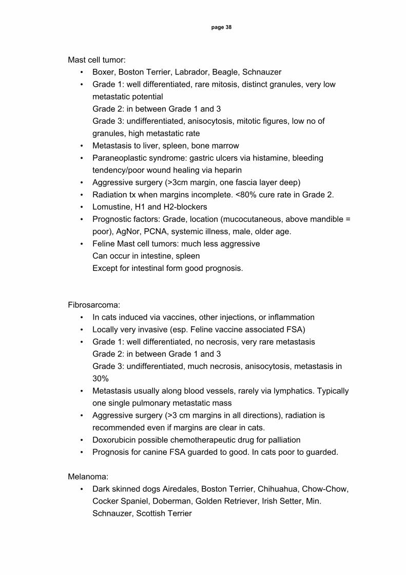
page 38 Mast cell tumor:
• Boxer, Boston Terrier, Labrador, Beagle, Schnauzer • Grade 1: well differentiated, rare mitosis, distinct granules, very low
metastatic potential Grade 2: in between Grade 1 and 3 Grade 3: undifferentiated, anisocytosis, mitotic figures, low no of granules, high metastatic rate
• Metastasis to liver, spleen, bone marrow • Paraneoplastic syndrome: gastric ulcers via histamine, bleeding
tendency/poor wound healing via heparin • Aggressive surgery (>3cm margin, one fascia layer deep) • Radiation tx when margins incomplete. <80% cure rate in Grade 2. • Lomustine, H1 and H2-blockers • Prognostic factors: Grade, location (mucocutaneous, above mandible =
poor), AgNor, PCNA, systemic illness, male, older age. • Feline Mast cell tumors: much less aggressive
Can occur in intestine, spleen Except for intestinal form good prognosis.
Fibrosarcoma:
• In cats induced via vaccines, other injections, or inflammation • Locally very invasive (esp. Feline vaccine associated FSA) • Grade 1: well differentiated, no necrosis, very rare metastasis
Grade 2: in between Grade 1 and 3 Grade 3: undifferentiated, much necrosis, anisocytosis, metastasis in 30%
• Metastasis usually along blood vessels, rarely via lymphatics. Typically one single pulmonary metastatic mass
• Aggressive surgery (>3 cm margins in all directions), radiation is recommended even if margins are clear in cats.
• Doxorubicin possible chemotherapeutic drug for palliation • Prognosis for canine FSA guarded to good. In cats poor to guarded.
Melanoma: • Dark skinned dogs Airedales, Boston Terrier, Chihuahua, Chow-Chow,
Cocker Spaniel, Doberman, Golden Retriever, Irish Setter, Min. Schnauzer, Scottish Terrier

page 39
• Fairly benign in skin, very malign subungual or in oral cavity. Mitotic index best prognostic indicator.
• Wide surgical excision • Chemotherapy not evaluated. • Prognosis: good (benign mass) to poor (30 weeks survival)
Squamous cell carcinoma: • Scottish terrier, Pekinese, Boxer, Poodle, Dachshund, white haired cats.
Exposure to sun light. • Erosive or proliferative. • Rare metastasis, usually to lymph nodes, rarely to lungs. • Hypercalcemia described in cats. • Early and complete excision with 1-3 cm margins • Radiation of nasal planum SSC is successful in 83% of cats, but dogs
have only 3 months disease fee interval. Subungual SSC has good prognosis (75% 1-year survival, 60%2-year survival) with wide surgical excision.
• Photodynamic therapy for early non-invasive lesions. • Chemotherapy systemically gives only short-term control. Intralesional is
more successful. Piroxicam results in partial response.

page 40 Fibrosarcoma Melanoma Adenocarcinoma Squamous Cell Carcinoma Hemangiopericytoma Mast Cell Tumor Staging MCT FSA SCC HPC Melanoma AdenoCA Blood analysis ! ! ! ! ! !
Buffy coat analysis " # # # # #
Bone marrow aspirate ! # # # # #
Bone marrow biopsy " # # # # #
Fine needle aspirate reg. lymph node ! ! ! ! ! !
Biopsy punch # ! # ! # !
Biopsy excisional # # # # # #
Thoracic radiographs ! ! ! ! ! !
Abdominal radiographs # # # # # #
Abdominal ultrasound ! # # # # !
Skin scraping # # " # # #
Regional CT scan # ! # # # #
" Possible, but not optimal ! Should be performed

page 41 Cranial cruciate ligament (CrCL) rupture: Anatomy, diagnosis, imaging, surgical approach, debridement, meniscectomy Pierre M. Montavon, Prof. Dr. med. vet., Daniel A. Koch. Dr. med. vet. ECVS Anatomy The CrCL runs from the caudomedial part of the lateral condyle of the femur diagonally across the intercondylar fossa to the cranial intercondyloid area of the tibia. The caudal cruciate ligament runs from the lateral surface of the medial femoral condyle caudodistally to the lateral edge of the popliteal notch of the tibia. The cruciate ligaments consist of two anatomically and functionally different parts. The CrCL has a stronger caudolateral and somewhat smaller craniomedial part. In extension of the stifle joint, both parts are taut. In flexion, the caudolateral band is loose whereas the craniomedial band is taut. The cruciate ligaments receive their blood supply from vessels of the synovial tissue ensheating them. As the stifle flexes, the lateral collateral ligament relaxes as a result of its attachments. This allows caudal displacement of the lateral femoral condyle and internal rotation of the tibia. The reverse occurs in extension. The lateral and medial menisci are semilunar, fibrocartilaginous discs. The lateral meniscus is slightly greater than the medial one. The transverse intermeniscal ligament is a band between their cranial horns. The lateral meniscus has a cranial and a caudal tibial ligament and a meniscofemoral ligament. This makes it more mobile with repect to the femur. The medial mensicus has a cranial und caudal tibial ligament and an attachment to the medial collateral ligament. This causes less mobility with respect to the femur and full load transmission may be placed on the caudal rim of the medial meniscus. It is the craniomedial part of the CrCL which provides the primary check against hyperextension and cranial displacement of the tibia. Secondary constraints againts cranial movement are provided by the joint capsule, the menisci, the collateral ligaments, the muscle forces and the shape of the tibial plateau. Diagnosis Risk patients for CrCL ruptures are dogs between 7 and 10 years of age, female gender, obesity, high body weight or preexisting medial patellar luxation. Typical anamnesis includes initial non-weight bearing lameness, clinical improvement after some weeks and signs of degenerative joint disease.

page 42 Stifle laxity is palpated in upright position and lateral recumbency. A positive cranial drawer sign or tibial compression test is diagnostic for CrCL rupture. Increased internal rotation and crepitus are the most common associated findings. Incomplete CrCL ruptures do not always lead to a positive drawer or tibial compression test. Eventually, pain in hypereytension or a slight drawer sign in flexion my be elicted. False negative joint laxity results are obtained in dogs with heavy muscle tone or severe degenerative joint disease with capsule fibrosis. Young dog normally have in contrast some degree of stifle laxity. Imaging Radiographs of the stifle are taken to rule other abnormalities than a CrCL rupture. Signs associated with CrCL may be joint effusion, cranial displacement of the tibia, bulging of the joint capsule, calcification after meniscal or ligament injuries, and signs of degenerative joint disease. Radiographs of the entire tibia are taken to plan the surgery and to give a prognosis concerning long term outcome. Synovial fluid assessment, scintigraphy and positive contrast arthrography add little information to the diagnosis, whereas MRI is the method of choice in human knee disorders. Partial tears of the CrCL are best diagnosed with arthroscopy or explorative arthrotomy. Surgical approach: lateral arthrotomy The approach can be performed from the medial or the lateral side. Tension on the skin in the popliteal area allows retractionof the wound edges, which fascilitates the subcutaneous incision. Depending on the intervention, the further preparation and incision can be centred on the height of the joint (from patella to tibial tuberosity in order to inspect meniscus and CrCL). If a larger approach is desired, the incision is prolonged proximally to the supratrochanteric region. The lateral fascia is incised first at a minimal distance of 5mm from the patella in order to avoid the patellar wings. Further distally, ths incision follows the patellar ligament. The joint capsule is opened with a No. 15 blade. Care must be taken to not sever the intraarticular tendon of the long digital extensor muscle. The extruding synovialis must be sucked up. Complete evacuation of the joint capsule is

page 43 achieved by gentle pressure on the caudal parts of it. A piece of the infrapatellar fat pad can be removed while coagulating the vessels. By means of this, the overview can be ameliorated. Debridement, cleaning up The remnants of the ruptured CrCL must be removed in order to avoid impingement between the bony joint surfaces. A No. 11 blade is taken. Proximally, the blade is guided along the intercondylar surface of the lateral condyle. So, the caudal cruciate ligament is spared. The distal part of the CrCL is severed from the tibia under cranial retraction of the medial meniscus. Osteophytes in the supratrochlear region can be removed with a scalpel blade or a synovectomy rongeur. Meniscectomy The joint is extended with the help of a stifle distractor, a Hohmann retractor or a curved tissue forceps. Most meniscal injuries can be found in caudal horn of the medial meniscus. Damaged parts of the meniscus are removed, which is called partial meniscectomy. It is performed by releasing the meniscus, first medial and caudal from the medial collateral ligament, and second severing it at the medial caudal meniscotibial ligament. Subtotal synovectomy The subtotal synovectomy is performed with a synovectomy rongeur. Inflamed synovium, especially in the retropatellar region (plica synovialis) is removed. A similar effect on the medial side can be achieved by a medial arthrotomy. Closure of the capsule The joint is closed with absorbable, monofilamentous suture material, size 2-0 and half-curved, cutting needle. The fibrous joint capsule is closed with a mattress pattern. Contact of the suture material with the trochlear cartilage must be avoided to rule out erosion damages.

page 44 Instrumentation for stifle surgery - Bard-Parker scalpel holder with blades no. 10, 11, 15 - Mayo scissors, straight - Adson Brown or Adson forceps - Senn-Miller retractor - Finger-Meyerding (blades 13 and 25m) - Finger-Hohmann-lever - Stifle distractor - synovectomy rongeur - Russian tissue forceps - oscillating saw - oscillating drill device - electrocautery - Ringer lactate solution - material for bandaging

page 45 Posterolateral capsuloraphy Daniel Koch, Dr. med. vet. ECVS, Pierre M. Montavon, Prof. Dr. med. vet. Introduction: With the posterolateral capsulraphy (Hohn 1974), the drawer sign following a cranial cruciate ligament rupture and the internal rotation of the tibia while extending the stifle (screw home mechanism) are reduced. The raphy can only be applied on the lateral side. The tension of the joint capsule stabilizes the joint, since the caudally bulging femoral condyle pushes against the capsule. The technique is indicated with toy breeds, which suffer from patellar luxation and cranial cruciate ligament rupture. Technique: The fascia of the biceps muscle is incised and detached from the tuber Gerdy. After retraction, the fibular nerve and the lateral fabella are identified. The joint capsule is liberated from the lateral femoral condyle. The fibular nerve, the lateral collateral ligament and the popliteal artery are avoided. The condylus and the distally running tendon of the popliteal muscle are palpated. A curved hemostat is placed in the intercondylr region, in order to retract and avoid damage to the popliteal artery. The joint capsule is incised horizontally on the height of the most prominent bulging of the femoral condyle. The collateral ligament and the popliteal tendon are not severed. Two to three mattress sutures are preplaced. The needle is best directed through each layer separately, in order to achieve a maximal imbrication. The sutures are tied and secured with the tibia in exoratation and the stifle in flexion.
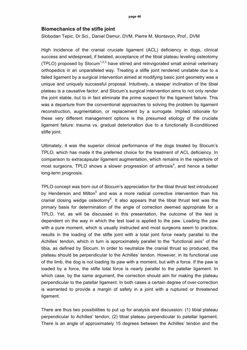
page 46 Biomechanics of the stifle joint Slobodan Tepic, Dr.Sci., Daniel Damur, DVM, Pierre M. Montavon, Prof., DVM High incidence of the cranial cruciate ligament (ACL) deficiency in dogs, clinical success and widespread, if belated, acceptance of the tibial plateau leveling osteotomy (TPLO) proposed by Slocum1,2,3 have stirred and reinvigorated small animal veterinary orthopedics in an unparalleled way. Treating a stifle joint rendered unstable due to a failed ligament by a surgical intervention aimed at modifying basic joint geometry was a unique and uniquely successful proposal. Intuitively, a steeper inclination of the tibial plateau is a causative factor, and Slocum’s surgical intervention aims to not only render the joint stable, but to in fact eliminate the prime suspect for the ligament failure. This was a departure from the conventional approaches to solving the problem by ligament reconstruction, augmentation, or replacement by a surrogate. Implied rationale for these very different management options is the presumed etiology of the cruciate ligament failure: trauma vs. gradual deterioration due to a functionally ill-conditioned stifle joint. Ultimately, it was the superior clinical performance of the dogs treated by Slocum’s TPLO, which has made it the preferred choice for the treatment of ACL deficiency. In comparison to extracapsular ligament augmentation, which remains in the repertoire of most surgeons, TPLO shows a slower progression of arthrosis4, and hence a better long-term prognosis. TPLO concept was born out of Slocum’s appreciation for the tibial thrust test introduced by Henderson and Milton5 and was a more radical corrective intervention than his cranial closing wedge osteotomy6. It also appears that the tibial thrust test was the primary basis for determination of the angle of correction deemed appropriate for a TPLO. Yet, as will be discussed in this presentation, the outcome of the test is dependent on the way in which the test load is applied to the paw. Loading the paw with a pure moment, which is usually instructed and most surgeons seem to practice, results in the loading of the stifle joint with a total joint force nearly parallel to the Achilles’ tendon, which in turn is approximately parallel to the “functional axis” of the tibia, as defined by Slocum. In order to neutralize the cranial thrust so produced, the plateau should be perpendicular to the Achilles’ tendon. However, in its functional use of the limb, the dog is not loading its paw with a moment, but with a force. If the paw is loaded by a force, the stifle total force is nearly parallel to the patellar ligament. In which case, by the same argument, the correction should aim for making the plateau perpendicular to the patellar ligament. In both cases a certain degree of over-correction is warranted to provide a margin of safety in a joint with a ruptured or threatened ligament. There are thus two possibilities to put up for analysis and discussion: (1) tibial plateau perpendicular to Achilles’ tendon; (2) tibial plateau perpendicular to patellar ligament. There is an angle of approximately 15 degrees between the Achilles’ tendon and the

page 47 patellar ligament (with the stifle in extension). In both cases, one can attempt to influence either the plateau or the tendon/ligament. Slocum’s TPLO turns the plateau, orienting it with respect to Achilles’ tendon. A cranial closing wedge osteotomy, abandoned by Slocum, has been reinvented and is also practiced, and advocated as an alternative way to tilt the plateau, wherein in fact it mostly turns the Achilles’ tendon. TPLO can of course be performed with a smaller angle of correction aiming at perpendicularity to the patellar ligament. The fourth possibility, that of correcting the angle of the patellar ligament, is well known in orthopedics, but to our knowledge has not been used for ACL deficiency. And yet, it may be the simplest procedure to perform, with major benefits expected in terms of reduced morbidity and improved short and long-term performance. Slocum’s publications have in fact laid out basic mechanics of the stifle, but rather arbitrarily, he decided that the tibia is a simple axially loaded truss. Determination of true joint forces, in spite of many decades of research in biomechanics, remains an illusive task, mostly for the problems of muscle redundancy and co-contraction. The later has subtle and difficult to objectively assess functional / physiological causes. The arguments presented here are based on greatly simplified mechanics of the stifle, and should be used only for sorting out different possibilities when contemplating surgical interventions. Yet the need to perform them is such that we cannot wait to figure it all out. And for the benefit of many dogs and his fellow surgeons treating them, it is good that Slocum did not wait either. His insistence on the strictest adherence to the rules for execution of TPLO has perhaps prevented variations which otherwise might have suggested that the corrective angle is excessive. For that we have only anecdotal evidence. Yet one of the main drawbacks of TPLO may be caused by the excessive angle of correction: the most obvious, and generally overlooked fact is that the joint congruity is drastically changed by placing it on an average into 25 degrees of extra flexion. It may well be that the medial meniscal release is mandated by its impingement caused not by the tibial thrust (which should be well compensated), but by artificially increased flexion of the joint itself. If the total stifle joint force is in fact oriented by about 15 degrees more cranial than presumed by Slocum, the evidence for the causative role of the increased tibial plateau angle becomes even more convincing. Morris and Lipowitz7 have measured this angle to be about 18 degrees in controls and about 24 degrees in dogs with failed ACL. If the reference is shifted by some 15 degrees, this would suggest 3 degrees in controls and 9 degrees in failed ACL cases. Instead of a 30% increased load responsible for the failure, we would be looking at a 300% percent increased load. And if the functional reference of true relevance is the angle of the patellar ligament, instead of a tibial axis, we may find a further cause for ACL failure in the formation of the tibial tuberosity. A steeper slope of the plateau and/or a less cranially prominent tibial tuberosity will increase tension in the cranial cruciate ligament, and may consequently predispose for its injury. Surgical management of the cranial cruciate deficiency could then rationally involve a reduction of the slope of the plateau and/or a cranial advancement of the tibial tuberosity.

page 48 If only a crude approximation to the true biomechanical status of an active musculato-skeletal linkage, simple static analysis of the stifle joint may prove its utility in at least three ways: (i) by guiding the search for underlying causes of the unusually high incidence of the cranial cruciate ligament deficiency; (ii) by providing more precise, and rationally based, means of determining the parameters for the currently practiced surgical interventions; (iii) by inspiring new management procedures. References: 1. Slocum B, Devine T, JAVMA, 1983;183:456-459 2. Slocum B, Devine Slocum T, Vet Clin of North Am, Small Anim Pract, 1993;23:777-
795 3. Slocum B, Devine Slocum T, 10th ESVOT Congress, Munich, March 2000: 60-62 4. Lazar TP, Berry CR, deHaan JJ, Peck JN, 11th ACVS Symp, Chicago, Oct 2001,
Abstr 48 5. Henderson R, Milton J, J Am Anim Hosp Assoc, 1978;14: 474-479 6. Slocum B, Devine T, JAVMA, 1984;184:564-569 7. Morris E, Lipowitz AJ, JAVMA, 2001; 218, 363-366

page 49 Advancement of the tibial tuberosity for treatment of cranial cruciate deficient canine stifle Pierre M. Montavon, Prof. Daniel M. Damur, Dr med. vet. FVH, Slobodan Tepic Dr. Sc., Tomas Guerrero, Dr. med. vet. Introduction The introduction of biomechanical considerations has influenced the treatment of cranial cruciate deficient stifles in dogs. Recent techniques modify the basic joint geometry in an attempt to neutralize the tibiofemoral shear force, which appears to be responsible for the overload of the cranial cruciate ligament. The better long-term prognosis offered by the slower postoperative progression of arthrosis in comparison to previous techniques is considered as a clinical success by a majority of surgeons. Conformational malalignment entities have also been evidenced and surgical limb realignment is indicated in these clinical cases. The procedure presented here consists in advancing the tibial tuberosity, in order to position the patellar ligament perpendicularly to the tibial plateau, thereby reducing to zero the tibiofemoral shear force and easing the function of the deficient cranial cruciate. The lesser invasive technique reduces operative time and perioperative morbidity. Respecting the normal range of flexion of the stifle should make a meniscal release, hence a loss of intraarticular caudal support, not necessary. Decreased retropatellar pressure could alleviate the sulcus chondromalacia present in about 30% of the cases. These advantages should improve the short and long-time results of the surgical treatment of cranial cruciate deficient stifle. Preoperative planning Mediolateral radiographs of the stifle in extension, avoiding the cranial drawer phenomenon in the presence of total rupture of the cranial cruciate ligament are necessary to figure out the angle necessary to bring the patellar ligament perpendicularly to the tibial plateau. The patellar ligament is represented by its cranial border, the orientation of the tibial plateau by a line passing through both tibial origins of the cranial and caudal cruciate ligaments.

page 50 Surgical technique Arthroscopy or medial arthrotomy are
performed in case of total cranial cruciate
ligament rupture to explore the stifle joint
and treat eventual meniscal lesions.
Transverse osteotomy of the tibial
tuberosity is carried through its distal
extremity to the cranial borders of the
menisci. A bone spacing structure of
desired size is inserted into the distracted
osteotomy in order to advance the tibial
tuberosity, giving its new position to the
patellar ligament. The tibial tuberosity is
fixed to the tibia with a tension band. The
wound is closed in appositional manner
after mobilizing the edges in order to cover
the implants.
Results Failure to maintain the advancement of the patellar ligament has been encountered, resulting from excessive tension and compression forces for the internal fixation repair. In all other cases, clinical results are satisfactory as documented by force plate gait analysis. No additional postoperative meniscal damages have been observed at this date. The results are encouraging and the technique will be further developed.