Embed Size (px)
Citation preview

BONE MATRIX
Tim Arnett
Department of Anatomy andDevelopmental Biology
University College London

Treated with bleach (hypochlorite) to digest collagen
leaves mineral component intact
Treated with hydrochloric acidto dissolve mineral
leaves collagen component intact
Collagen shrinkage on drying
Bone is made of mineral+ organic (mainly collagen)
components

Bone matrix
· bone is a composite material
· organic component is ~90% type I collagen- structural protein providing strength, flexibility
· remaining 10% of organic component is complex mixture of proteins, including growth factors, osteocalcin, osteonectin, osteopontin, glycoproteins; all produced by osteoblasts, along with type I collagen

Bone Matrix - 2
· organic matrix is mineralised after secretion
– unmineralised matrix = “osteoid”
· mineral component is hydroxyapatite – Ca10 (PO4)6 (OH)2
- tiny crystals impregnate & surround collagen fibres- provides rigidity, resistance to compression
· mineralisation of osteoid is dependent on the hormonally active form of vitamin D (1,25-dihydroxyvitamin D)
· vitamin D deficiency results in failure to mineralise– leads to rickets in children, osteomalacia in adults

Two types of bone
Woven (immature, fracture)
large, rounded osteocytes
osteocytes irregularly spaced
randomly oriented collagen fibres
variable collagen fibre diameter
rapid matrix mineralisation
forms rapidly
rapid turnover
Lamellar (mature, adult)
smaller, flattened osteocytes
osteocytes regularly spaced
collagen fibres show regular, “plywood” orientation – confers strength
regular collagen fibre diameter
delayed matrix mineralisation (few days)
forms slowly
slow turnover

Collagen
• stable, structural ECM protein
• collagen molecule = triple-stranded helix (3 �-chains)
• 25% of total body protein (mainly type I)
MAIN COLLAGEN TYPES…..
TYPE I: (90% of total): fibrils � fibres bone, skin, tendon, ligaments, cornea, internal organs
TYPE II: fibrils (form meshwork) not visible with light microscopecartilage, intervertebral disc, vitreous humour
TYPE III: fibrils → fibres (‘reticular’ - coarse mesh)skin, blood vessels, internal organs
TYPE IV: sheet-like networkbasal laminae

Collagen biosynthesis
Alberts et al – Molecular Biology of the Cell, Ed. 2

Amino acid composition of collagen chains
Alb
erts
et a
l–M
olec
ular
Bio
logy
of t
he C
ell,
Ed.
3
…Gly-X-Y…
X or Y frequently proline

Hydroxylated amino acids in collagen
Alberts et al – Molecular Biology of the Cell, Ed. 3

Pro-collagen
Alberts et al – Molecular Biology of the Cell, Ed. 3

“¼ stagger” model of collagen molecule alignment in fibrils
Alberts et al – Molecular Biology of the Cell, Ed. 3

Extracellular assembly of individual collagen molecules into fibrils- ‘quarter stagger’ model
Alberts et al – Molecular Biology of the Cell, Ed. 3

Covalent links between and within collagen molecules formed by action of lysyl oxidase on lysine and OH-lysine residues
Alberts et al – Molecular Biology of the Cell, Ed. 3
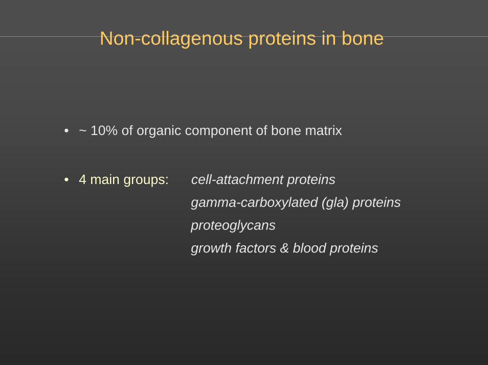
Non-collagenous proteins in bone
• ~ 10% of organic component of bone matrix
• 4 main groups: cell-attachment proteins
gamma-carboxylated (gla) proteins
proteoglycans
growth factors & blood proteins

Non-collagenous proteins in bone
• Thrombospondin (450 kD) - influences cell attachment - strong Ca2+ bindingFibronectin (440 kD) - influences cell attachment & spreading; contains RGD sequence; binds collagen
• Osteopontin (50 kD) - phosphorylated; contains RGD sequence; influences cell attachment & spreading; strong Ca2+ binding; not bone-specificBone sialoprotein (50 kD) - also affects cell attachment; strong Ca2+ binding; bone-specific
• Osteonectin (SPARC) (35 kD) - most abundant NCP: ~2% total protein in developing bone; binds collagen & hydroxyapatite; promotes calcium phosphate deposition in vitro
• Osteocalcin (BGP) (6kD) - contains γ-carboxy glutamyl residues (Ca2+ binding); bone-specific protein; low affinity Ca2+ binding; synthesis stimulated by 1,25(OH)2 vitamin D. Matrix gla-protein (9kD) - not bone-specific
• Proteoglycan I (biglycan) (170 kD) - 2 chondroitin sulphate chains; mainly in developing bone, function unknownProteoglycan II (decorin) (120 kD) - 1 chondroitin sulphate chain; present in all bone; binds collagen, may modulate fibrillogenesis
• Blood proteins (~25% of NCP) - including α2HS glycoprotein… and growth factors, including IGF-I, IGF-II, BMPs, PDGF, FGFs, TGFβ….

γ-carboxy-glutamic acid
H O
N C C
H CH2
CO O -
CO O -
Ca ++
γ-carboxylation is vit Kdependent process →

Alberts et al – Molecular Biology of the Cell, Ed. 3

Proteoglycans
Cartilage: aggrecan
Bone: decorin
Alberts et al – Molecular Biology of the Cell, Ed. 3

Targeted disruption of non-collagenous protein genesie, “knockouts”
Osteocalcin - increased osteoblast activity (bone formation) in knockout mice
Biglycan - (binds collagen + TGFβ) knockout mice show reduced growth rate + develop osteoporotic phenotype; double KO with decorin → more osteoporosis, premature osteoarthritis, abnormal collagen fibril formation
MGP - matrix γ-carboxyglutamic acid (gla) protein knockout mouse exhibits extensive, lethal calcification + cartilaginous metaplasia of media of all elastic arteries
Osteopontin - knockout mice show altered wound healing; resistance to osteoporotic effects of ovariectomy; reduced tumour metastasis; reduced inflammatory arthritis…

Naturally occurring ‘knockouts’
Osteogenesis imperfecta
• diverse family of genetic disorders
• mutations of α1 and α2 chains of type I collagen
Severity varies greatly, depending on the type of mutation, from prenatal lethality through impaired stature / tooth development and deformity to minimal deformity, sometimes with hearing loss or blue sclerae