Embed Size (px)
Citation preview
Slide 1 of 23
Copyright Pearson Prentice Hall
Biology

Slide 2 of 23
Copyright Pearson Prentice Hall
36–3 The Integumentary System
36–3 The Integumentary System
Slide 3 of 23
Copyright Pearson Prentice Hall
The skin, hair, nails, and a variety of glands make up the integumentary system.
The skin is the largest organ in the body.

36–3 The Integumentary System
Slide 4 of 23
Copyright Pearson Prentice Hall
What are the functions of the integumentary system?
36–3 The Integumentary System
Slide 5 of 23
Copyright Pearson Prentice Hall
The integumentary system: • serves as a barrier against infection
and injury. • helps to regulate body temperature. • removes waste products from the body. • provides protection against ultraviolet
radiation from the sun.
36–3 The Integumentary System
Slide 6 of 23
Copyright Pearson Prentice Hall
The Skin
The Skin
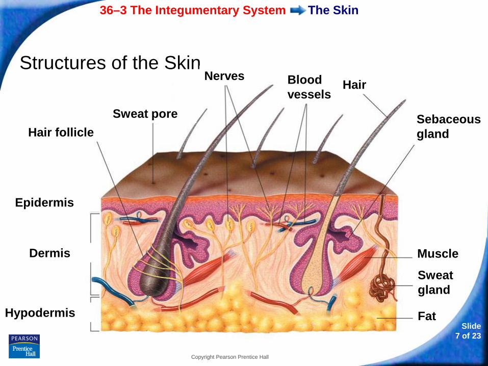
The skin is made up of two main layers—the epidermis and the dermis.
Beneath the dermis is a layer of fat (hypodermis) and loose connective tissue that insulates the body.
36–3 The Integumentary System
Slide 7 of 23
Copyright Pearson Prentice Hall
The Skin
Structures of the Skin
Epidermis
Dermis
Hypodermis
Hair follicle Sweat pore
Nerves
Muscle
Sweat gland
Fat
Sebaceous gland
Hair Blood vessels

36–3 The Integumentary System
Slide 8 of 23
Copyright Pearson Prentice Hall
The Skin
Epidermis
The outer layer of the skin is the epidermis.
The epidermis has two layers.
• The outer layer is made up of dead cells.
• The inner layer is made up of living cells.

36–3 The Integumentary System
Slide 9 of 23
Copyright Pearson Prentice Hall
The Skin
Cells in the inner layer undergo rapid cell division, producing new cells that push older cells to the surface of the skin.
Older cells flatten and their organelles disintegrate.
Older cells also begin making keratin, a tough, fibrous protein.
When these cells die, they form a waterproof covering on the skin’s surface.

36–3 The Integumentary System
Slide 10 of 23
Copyright Pearson Prentice Hall
The Skin
The epidermis also contains melanocytes, which are cells that produce melanin, a dark brown pigment.
Melanin protects the skin from sun damage.
Differences in skin color result from different amounts of melanin and where melanocytes are distributed.
36–3 The Integumentary System
Slide 11 of 23
Copyright Pearson Prentice Hall
The Skin
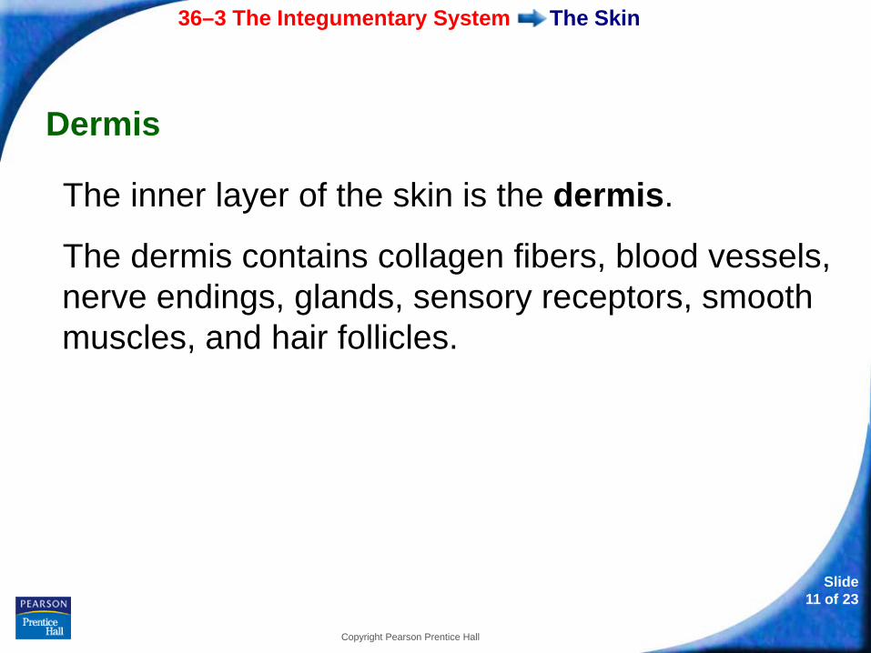
Dermis
The inner layer of the skin is the dermis.
The dermis contains collagen fibers, blood vessels, nerve endings, glands, sensory receptors, smooth muscles, and hair follicles.

36–3 The Integumentary System
Slide 12 of 23
Copyright Pearson Prentice Hall
The Skin
The dermis contains two major types of glands:
• sweat glands
• sebaceous, or oil, glands
36–3 The Integumentary System
Slide 13 of 23
Copyright Pearson Prentice Hall
The Skin
If your body gets too hot, sweat glands produce sweat.
When sweat evaporates, it cools the body.
Sweat also gets rid of wastes from the blood, along with water.
36–3 The Integumentary System
Slide 14 of 23
Copyright Pearson Prentice Hall
The Skin
Sebaceous glands produce an oily secretion called sebum.
Sebum spreads out along the surface of the skin and helps to keep the skin flexible and waterproof.
36–3 The Integumentary System
Slide 15 of 23
Copyright Pearson Prentice Hall
Hair and Nails
Hair
Hair covers most body surfaces. Hair:
• protects the scalp from ultraviolet light from the sun.
• provides insulation from the cold.
• prevents dirt and other particles from entering the body.
36–3 The Integumentary System
Slide 16 of 23
Copyright Pearson Prentice Hall
Hair and Nails
Hair is produced by hair follicles, which are tubelike pockets of epidermal cells that extend into the dermis.
An individual hair is a column of cells that have filled with keratin and died.
The oily secretions of sebaceous glands help maintain the condition of each individual hair.
36–3 The Integumentary System
Slide 17 of 23
Copyright Pearson Prentice Hall
Hair and Nails
Nails
Nails grow from rapidly dividing cells in the nail root.
The nail root is located near the tips of the fingers and toes.
During cell division, cells fill with keratin and produce a platelike nail that covers and protects the fingertips and toes.

- or - Continue to: Click to Launch:
Slide 18 of 23
Copyright Pearson Prentice Hall
36–3
Slide 19 of 23
Copyright Pearson Prentice Hall
36–3
Keratin provides
a. insulation.
b. a waterproof covering.
c. pigmentation.
d. protection from UV radiation.
Slide 20 of 23
Copyright Pearson Prentice Hall
36–3
The dermis contains two types of glands: sweat glands and
a. sebaceous glands.
b. pigment glands.
c. hair follicles.
d. dermal glands.
Slide 21 of 23
Copyright Pearson Prentice Hall
36–3
All of the following are found in the dermis EXCEPT
a. nerve endings.
b. blood vessels.
c. sebaceous glands.
d. melanocytes.
Slide 22 of 23
Copyright Pearson Prentice Hall
36–3
The function of melanin is to
a. help control the rate of heat loss by the skin.
b. produce sweat.
c. produce a waterproof covering on the surface of the skin.
d. absorb harmful UV radiation.
Slide 23 of 23
Copyright Pearson Prentice Hall
36–3
The basic structure of human hair and nails is
a. melanin.
b. sebum.
c. keratin.
d. dermal cells.

END OF SECTION