Embed Size (px)
Citation preview
lable at ScienceDirect
Sleep Medicine Reviews 50 (2020) 101249
Contents lists avai
Sleep Medicine Reviews
journal homepage: www.elsevier .com/locate /smrv
CLINICAL REVIEW
Polysomnographic features of pregnancy: A systematic review
Corrado Garbazza a, b, c, *, Sandra Hackethal a, Silvia Riccardi a, Christian Cajochen b, c,Alessandro Cicolin d, Armando D'Agostino e, Fabio Cirignotta f, Mauro Manconi a
a Sleep and Epilepsy Center, Neurocenter of Southern Switzerland, Civic Hospital of Lugano (EOC), Via Tesserete 46, Lugano, CH-6903, Switzerlandb Centre for Chronobiology, University of Basel, Basel, Switzerlandc Transfaculty Research Platform Molecular and Cognitive Neurosciences, University of Basel, Basel, Switzerlandd Sleep Medicine Center, Neuroscience Department, AOU Citt�a della Salute e della Scienza - Molinette, Universit�a di Torino, Torino, Italye Department of Health Sciences, Universit�a degli Studi di Milano, Milan, Italyf DIBINEM, University of Bologna, Bologna, Italy
a r t i c l e i n f o
Article history:Received 29 July 2019Received in revised form23 October 2019Accepted 27 November 2019Available online 5 December 2019
Keywords:PregnancyPolysomnographySleep stagesOSASleep-disordered breathingAdverse fetal outcomesHypertensive disease of pregnancyGestational diabetes mellitus
* Corresponding author. Sleep and Epilepsy CentSwitzerland, Civic Hospital of Lugano (EOC), Via TesSwitzerland.
E-mail address: [email protected] (C. Garb
https://doi.org/10.1016/j.smrv.2019.1012491087-0792/© 2019 Elsevier Ltd. All rights reserved.
s u m m a r y
Symptoms of sleep disturbances are common among pregnant women and generally worsen acrossgestation. Pregnancy-related sleep disorders are not only associated with a poor quality of life of theaffected mothers, but also with adverse perinatal outcomes, including perinatal depression, gestationaldiabetes, preeclampsia, and preterm birth. The current knowledge about the impact of sleep disordersduring pregnancy largely derives from the results of sleep surveys conducted in various populations.However, the number of studies examining changes in objective sleep variables during pregnancy viapolysomnography has progressively increased in recent years.
Here we systematically reviewed the polysomnographic studies available in the literature with the aimto describe the sleep pattern and to identify possible markers of sleep disruption in pregnant women.
Based on our analysis, subjective worsening of sleep quality across gestation is related to objectivechanges in sleep macrostructure, which become particularly evident in the third trimester. Pregnancy perse does not represent an independent risk factor for developing major polysomnography-assessed sleepdisorders in otherwise healthy women. However, in women presenting predisposing factors, such asobesity or hypertension, physiological changes occurring during pregnancy may contribute to the onsetof pathological conditions, especially sleep-disordered breathing, which must be carefully considered.
© 2019 Elsevier Ltd. All rights reserved.
Introduction
Pregnancy is a physiological condition of relatively short dura-tion in a woman's life, but characterized by profound biologicalchanges, which have a significant influence on sleep [1].
The typically increased secretion of several hormones acrosspregnancy considerably impacts on both the circadian and ho-meostatic components of sleep regulation, leading to modificationsof sleep architecture [2]. In human studies, non-rapid eye move-ment sleep (NREM) has been shown to be enhanced by proges-terone and prolactin [3,4], while rapid eye movement sleep (REM)is decreased by progesterone and increased by estrogens [5,6].Oxytocin peaks during the night, promoting uterine contractionsleading to sleep fragmentation [2]. Cortisol and growth hormone
er, Neurocenter of Southernserete 46, Lugano, CH-6903,
azza).
levels are also elevated, affecting sleep quality and inducing day-time sleepiness [2].
Besides hormones, other factors contribute to sleep disruptionduring pregnancy: gastroesophageal reflux, affecting up to 75% ofpregnant women [7]; nocturnal micturition, due to an increase inovernight sodium excretion [8]; anatomical changes related to thegrowing uterus and increased body weight [9]. Moreover, iron andfolate deficiency may play a role in the occurrence of sleep-relatedmovement disorders in pregnant women [10,11].
Subjectively reported sleep disturbances are very commonduring pregnancy, with increasing rates from the first (13%), to thesecond (19%), and third (66%) trimester of gestation [12,13]. Arecent meta-analysis showed that 46% of women experience poorsleep quality during pregnancy, with an average score of thePittsburgh Sleep Quality Index (PSQI) of 6.4 (95% CI, 5.3e6.85) andwith a worsening trend from the 2nd to the 3rd trimester by anaverage of 1.68 points (95% CI, 0.42e2.94) [14]. While at earlygestational age women mainly attribute sleep problems to nausea/
Abbreviations
AHI Apnea-hypopnea indexBMI Body mass indexCHTN Chronic hypertensionCPAP Continuous positive airway pressureEEG ElectroencephalographyEMG ElectromyogramEOG ElectrooculogramESS Epworth sleepiness scaleGA Gestational ageGCT Glucose challenge testGDM Gestational diabetes mellitusGHTN Gestational hypertensionHDP Hypertensive disease of pregnancyNREM Non-rapid eye movement sleepODI Oxygen desaturation indexOGTT Oral glucose tolerance testOSA Obstructive sleep apnea
PaO2 Partial pressure of oxygenPE PreeclampsiaPLMS Periodic leg movements during sleepPLMSI Periodic leg movements during sleep indexPSG PolysomnographyPSQI Pittsburgh sleep quality indexRDI Respiratory disturbance indexREM Rapid eye movement sleepRLS Restless legs syndromeSaO2 Oxygen saturationSDB Sleep-disordered breathingSE Sleep efficiencySOL Sleep onset latencySWS Slow wave sleepTCO2 Transcutaneous CO2
TIB Time spent in bedTST Total sleep timeWASO Wake after sleep onset
C. Garbazza et al. / Sleep Medicine Reviews 50 (2020) 1012492
vomiting, urinary frequency, and backpain [15], in late gestation upto 69.9% of women report difficulty in maintaining sleep, 34.8%early morning awakenings, and 23.7% difficulty falling asleep [16],mainly due to fetal movements, heartburn, cramps or tingling inthe legs, and shortness of breath [13,17e19]. By the end of preg-nancy almost all women suffer from recurrent and long wake ep-isodes during the night [17,20].
Self-reported sleep duration also declines across pregnancy [21].Moreover, objectively assessed sleep duration andquality are relatedto age and ethnicity, with non-Hispanic black and Asian womenhaving the shortest sleep duration, and younger pregnant womenhaving the highest amount of wake after sleep onset (WASO), thelowest sleep efficiency (SE), and the latest sleep midpoint [22].
To date, the available literature on sleep during pregnancy ismostly based on subjective information from screening question-naires or interviews [14,19]. However, in recent years, an increasingnumber of studies investigated sleep in pregnant women objectively,by using polysomnography (PSG) or actigraphy. Sleep parametersderived from actigraphy may significantly differ from those obtainedby PSG recordings and should therefore be interpreted with caution[23]. Thus, PSG remains the gold standard for sleep depiction, beingthe only reliable tool to precisely describe sleep macro- and micro-structure, correctly estimate respiratoryandmotor events, and permitan accurate identification of pregnancy-related sleep disorders.
We here present the first systematic review of polysomno-graphic studies conducted in pregnant women, with the aim toprovide a detailed overview about the intrinsic, objective featuresof sleep in normal, healthy pregnancy, as well as in some typicalpregnancy-related complications.
Methods
We performed a systematic review of the literature by searchingfor studies reporting objective sleep parameters obtained by PSG inpregnant women until February 1, 2019. The review process fol-lowed the PRISMA statement guidelines [24]. The completedPRISMA checklist can be found in the Supplementary materialsection (Table S1).
Search strategy
The terms ‘pregnancy’ OR ‘gestation’ AND ‘polysomnography’OR ‘PSG’ were searched in the databases Medline, Scopus and
Embase. The search terms had to be included in the Title, Abstractor Keyword section of the articles. The first author reviewed theautomatically generated list of items and classified every manu-script, based on its abstract, as “eligible”, “not eligible” and “maybeeligible”, according to the selection criteria described below. Arti-cles considered “not eligible”were excluded from a further analysis.Afterward, the first and second authors independently examinedthe “eligible” and “maybe eligible” full-text articles in a blindedfashion, to determine whether they met the criteria to be includedin the review. The inter-rater agreement calculated as Cohen'skappa coefficient (k) was 0.92. In case of disagreement, they con-sulted the senior author (MM) for a final decision.
Selection criteria
The following criteria were applied:
1) Sleep assessment: only studies reporting PSG data recordedduring pregnancy and using a minimal montage of at least oneEEG channel either in mono- or bipolar, electrooculogram(EOG), chin electromyogram (EMG) were included. Studiesbased on other objective sleep assessment methods than PSG(e.g., actigraphy or polygraphy) or using subjective tools (e.g.,questionnaires) were excluded;
2) Number of nights recorded: at least one full night PSG recording3) Sample size: only studies with a sample size of �10 women4) Language: English;5) Type of study: original studies on human subjects; no single
case reports, reviews, commentaries/letters, editorial, confer-ence abstracts;
6) Control group: studies including either a control group (healthypregnant or non-pregnant women) or without a control groupwere included.
Additionally, the authors went through the reference lists of theselected articles to identify further studies. Unpublished manu-scripts were not included.
Quality assessment
The quality assessment of the studies included in the systematicreview was performed using the Newcastle-Ottawa scale (NOS)adapted for cross-sectional studies (according to Herzog et al. [25]),
C. Garbazza et al. / Sleep Medicine Reviews 50 (2020) 101249 3
cohort studies, and caseecontrol studies (available at http://www.ohri.ca/programs/clinical_epidemiology/oxford.asp). The NOS con-sists of several items included in three domains (selection of thestudy groups, comparability of the groups, and outcome/exposureassessment). Each item is evaluated based on a ‘star system’. Trialsincluded in our review were evaluated using the Cochrane Col-laboration's tool for assessing risk of bias in randomized trials [26]and the Cochrane Collaboration's risk of bias in non-randomizedstudies (ROBINS-I) [27]. The results of the quality assessment ofall included studies are summarized in Table S2 (supplementarymaterial).
Statistical analysis
Mean and standard deviation of longitudinal studies reportingtotal sleep time (TST) and SE were pooled in order to evaluatechanges in these sleep variables from the first to the third trimesterof gestation. Weighted mean difference (WMD) with 95% confi-dence interval (95% CI) was used to estimate absolute differences ofcontinuous outcomes. I2-statistics was adopted to measure thepercentage of variance attributable to study heterogeneity
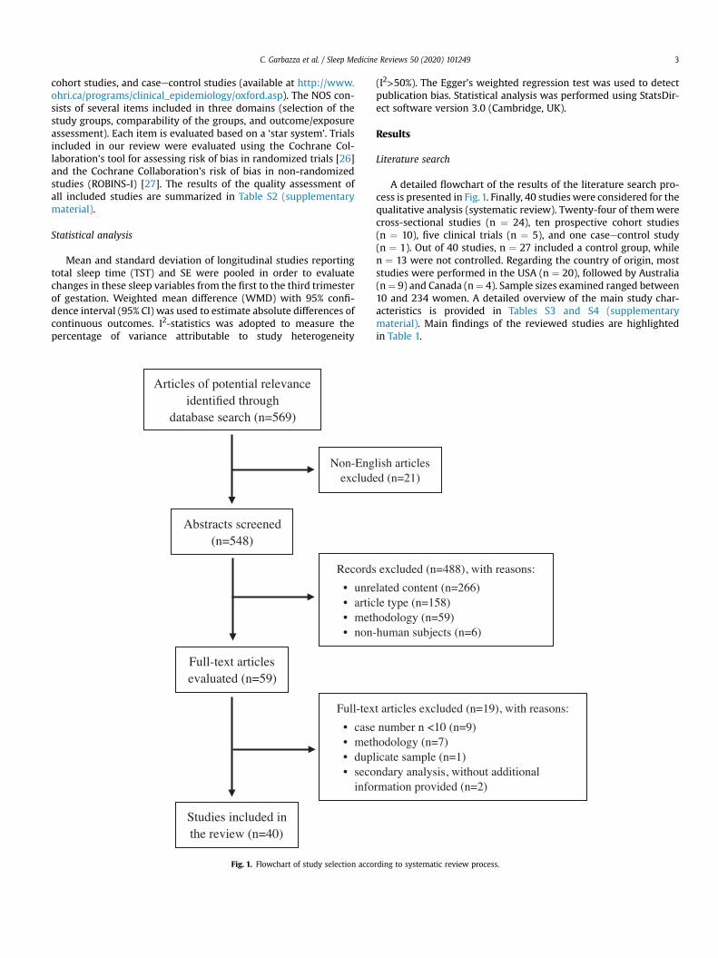
Fig. 1. Flowchart of study selection acco
(I2>50%). The Egger's weighted regression test was used to detectpublication bias. Statistical analysis was performed using StatsDir-ect software version 3.0 (Cambridge, UK).
Results
Literature search
A detailed flowchart of the results of the literature search pro-cess is presented in Fig.1. Finally, 40 studies were considered for thequalitative analysis (systematic review). Twenty-four of themwerecross-sectional studies (n ¼ 24), ten prospective cohort studies(n ¼ 10), five clinical trials (n ¼ 5), and one caseecontrol study(n ¼ 1). Out of 40 studies, n ¼ 27 included a control group, whilen ¼ 13 were not controlled. Regarding the country of origin, moststudies were performed in the USA (n ¼ 20), followed by Australia(n¼ 9) and Canada (n¼ 4). Sample sizes examined ranged between10 and 234 women. A detailed overview of the main study char-acteristics is provided in Tables S3 and S4 (supplementarymaterial). Main findings of the reviewed studies are highlightedin Table 1.
rding to systematic review process.
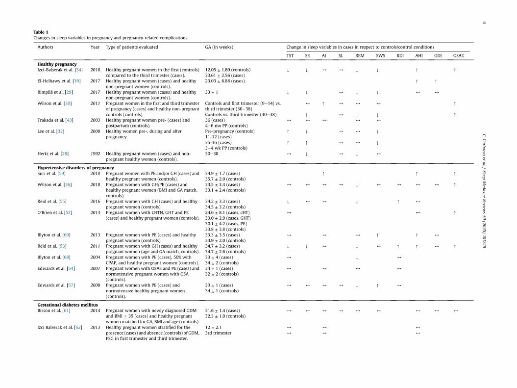
Table 1Changes in sleep variables in pregnancy and pregnancy-related complications.
Authors Year Type of patients evaluated GA (in weeks) Change in sleep variables in cases in respect to controls/control conditions
TST SE AI SL REM SWS RDI AHI ODI OSA%
Healthy pregnancyIzci-Balserak et al. [34] 2018 Healthy pregnant women in the first (controls)
compared to the third trimester (cases).12.05 ± 1.80 (controls)33.61 ± 2.56 (cases)
Y Y 4 4 Y Y [ [
El-Helbawy et al. [39] 2017 Healthy pregnant women (cases) and healthynon-pregnant women (controls).
23.03 ± 8.88 (cases) [ [
Rimpil€a et al. [29] 2017 Healthy pregnant women (cases) and healthynon-pregnant women (controls).
33 ± 1 Y Y 4 Y Y 4 4
Wilson et al. [30] 2011 Pregnant women in the first and third trimesterof pregnancy (cases) and healthy non-pregnantcontrols (controls).
Controls and first trimester (9e14) vs.third trimester (30e38)
4 [ 4 4 4 [
Controls vs. third trimester (30e38) Y 4 Y Y [
Trakada et al. [43] 2003 Healthy pregnant women pre- (cases) andpostpartum (controls).
36 (cases)4e6 mo PP (controls)
4 4 4 4 4
Lee et al. [32] 2000 Healthy women pre-, during and afterpregnancy.
Pre-pregnancy (controls)11-12 (cases)
[ Y 4 4 Y
35-36 (cases)3e4 wk PP (controls)
[ [ 4 4 Y
Hertz et al. [28] 1992 Healthy pregnant women (cases) and non-pregnant healthy women (controls).
30e38 4 Y 4 Y 4
Hypertensive disorders of pregnancySuri et al. [59] 2018 Pregnant womenwith PE and/or GH (cases) and
healthy pregnant women (controls).34.9 ± 1.7 (cases)35.7 ± 2.0 (controls)
[ [ [
Wilson et al. [56] 2018 Pregnant women with GH/PE (cases) andhealthy pregnant women (BMI and GA match,controls).
33.5 ± 3.4 (cases)33.1 ± 2.4 (controls)
4 4 4 4 Y 4 4 4 4 [
Reid et al. [55] 2016 Pregnant women with GH (cases) and healthypregnant women (controls).
34.2 ± 3.3 (cases)34.5 ± 3.2 (controls)
Y 4 4 Y [ 4
O'Brien et al. [52] 2014 Pregnant women with CHTN, GHT and PE(cases) and healthy pregnant women (controls).
24.6 ± 8.1 (cases, cHT)33.0 ± 2.9 (cases, GHT)30.1 ± 4.2 (cases, PE)33.8 ± 3.8 (controls)
4 4 [
Blyton et al. [69] 2013 Pregnant women with PE (cases) and healthypregnant women (controls).
33.3 ± 3.5 (cases)33.9 ± 2.0 (controls)
4 4 4 [ [ 4
Reid et al. [53] 2011 Pregnant women with GH (cases) and healthypregnant women (age and GA match, controls).
34.7 ± 3.2 (cases)34.7 ± 2.6 (controls)
Y Y 4 Y 4 [ [ 4 [
Blyton et al. [68] 2004 Pregnant women with PE (cases), 50% withCPAP, and healthy pregnant women (controls).
33 ± 4 (cases)34 ± 2 (controls)
4 Y 4
Edwards et al. [54] 2001 Pregnant women with OSAS and PE (cases) andnormotensive pregnant women with OSA(controls).
34 ± 1 (cases)32 ± 2 (controls)
4 4 4 4
Edwards et al. [57] 2000 Pregnant women with PE (cases) andnormotensive healthy pregnant women(controls).
33 ± 1 (cases)34 ± 1 (controls)
4 4 4 4 Y [ 4
Gestational diabetes mellitusBisson et al. [61] 2014 Pregnant women with newly diagnosed GDM
and BMI � 35 (cases) and healthy pregnantwomenmatched for GA, BMI and age (controls).
31.6 ± 1.4 (cases)32.3 ± 1.0 (controls)
4 4 4 4 4 4 4 4 4
Izci Balserak et al. [62] 2013 Healthy pregnant women stratified for thepresence (cases) and absence (controls) of GDM,PSG in first trimester and third trimester.
12 ± 2.1 4 4 4
3rd trimester 4 4 4
C.Garbazza
etal./
SleepMedicine
Reviews50
(2020)101249
4
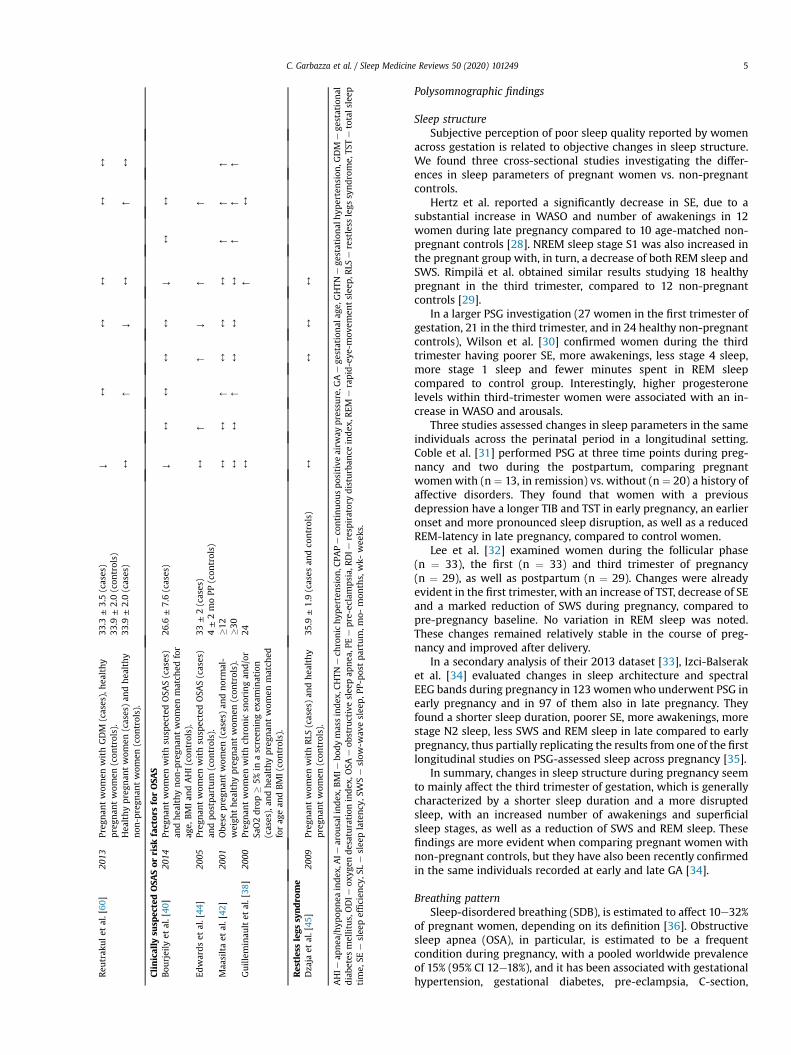
Reu
trak
ulet
al.[60
]20
13Preg
nan
twom
enwithGDM
(cases),hea
lthy
pregn
antwom
en(con
trols).
33.3
±3.5(cases)
33.9
±2.0(con
trols)
Y4
44
44
Hea
lthypregn
antwom
en(cases)an
dhea
lthy
non
-pregn
antwom
en(con
trols).
33.9
±2.0(cases)
4[
Y4
[4
Clinically
susp
ectedOSA
Sorrisk
factors
forOSA
SBou
rjeily
etal.[40
]20
14Preg
nan
twom
enwithsu
spectedOSA
S(cases)
andhea
lthynon
-pregn
antwom
enmatch
edfor
age,
BMIan
dAHI(con
trols).
26.6
±7.6(cases)
Y4
44
4Y
44
Edwardset
al.[44
]20
05Preg
nan
twom
enwithsu
spectedOSA
S(cases)
andpostpartum
(con
trols).
33±2(cases)
4±2moPP
(con
trols)
4[
[Y
[[
Maa
siltaet
al.[42
]20
01Obe
sepregn
antwom
en(cases)an
dnormal-
weigh
thea
lthypregn
antwom
en(con
trols).
�12
44
[4
44
[[
[
�30
44
[4
44
[[
[
Guillem
inau
ltet
al.[38
]20
00Preg
nan
twom
enwithch
ronic
snoringan
d/or
SaO2drop�
5%in
ascreen
ingex
amination
(cases),an
dhea
lthypregn
antwom
enmatch
edforag
ean
dBMI(con
trols).
244
[4
Restlesslegs
syndro
me
Dzaja
etal.[45
]20
09Preg
nan
twom
enwithRLS
(cases)an
dhea
lthy
pregn
antwom
en(con
trols).
35.9
±1.9(cases
andco
ntrols)
44
44
AHIe
apnea
/hyp
opnea
index
,AIe
arou
salindex
,BMIe
bodymassindex
,CHTN
ech
ronichyp
ertension
,CPA
Peco
ntinuou
spositiveairw
aypressure,G
Aege
stational
age,GHTN
ege
stational
hyp
ertension
,GDM
ege
stational
diabe
tesmellitus,ODIe
oxyg
endesaturation
index
,OSA
eob
structiveslee
pap
nea
,PEe
pre-eclam
psia,RDI e
resp
iratorydisturban
ceindex
,REM
erapid-eye
-mov
emen
tslee
p,R
LSe
restless
legs
syndrome,
TSTe
totalsleep
time,
SEe
slee
pefficien
cy,S
Le
slee
platency
,SW
Se
slow
-wav
eslee
p,P
P-postpartum,m
o-mon
ths,wk-
wee
ks.
C. Garbazza et al. / Sleep Medicine Reviews 50 (2020) 101249 5
Polysomnographic findings
Sleep structureSubjective perception of poor sleep quality reported by women
across gestation is related to objective changes in sleep structure.We found three cross-sectional studies investigating the differ-ences in sleep parameters of pregnant women vs. non-pregnantcontrols.
Hertz et al. reported a significantly decrease in SE, due to asubstantial increase in WASO and number of awakenings in 12women during late pregnancy compared to 10 age-matched non-pregnant controls [28]. NREM sleep stage S1 was also increased inthe pregnant group with, in turn, a decrease of both REM sleep andSWS. Rimpil€a et al. obtained similar results studying 18 healthypregnant in the third trimester, compared to 12 non-pregnantcontrols [29].
In a larger PSG investigation (27 women in the first trimester ofgestation, 21 in the third trimester, and in 24 healthy non-pregnantcontrols), Wilson et al. [30] confirmed women during the thirdtrimester having poorer SE, more awakenings, less stage 4 sleep,more stage 1 sleep and fewer minutes spent in REM sleepcompared to control group. Interestingly, higher progesteronelevels within third-trimester women were associated with an in-crease in WASO and arousals.
Three studies assessed changes in sleep parameters in the sameindividuals across the perinatal period in a longitudinal setting.Coble et al. [31] performed PSG at three time points during preg-nancy and two during the postpartum, comparing pregnantwomenwith (n ¼ 13, in remission) vs. without (n ¼ 20) a history ofaffective disorders. They found that women with a previousdepression have a longer TIB and TST in early pregnancy, an earlieronset and more pronounced sleep disruption, as well as a reducedREM-latency in late pregnancy, compared to control women.
Lee et al. [32] examined women during the follicular phase(n ¼ 33), the first (n ¼ 33) and third trimester of pregnancy(n ¼ 29), as well as postpartum (n ¼ 29). Changes were alreadyevident in the first trimester, with an increase of TST, decrease of SEand a marked reduction of SWS during pregnancy, compared topre-pregnancy baseline. No variation in REM sleep was noted.These changes remained relatively stable in the course of preg-nancy and improved after delivery.
In a secondary analysis of their 2013 dataset [33], Izci-Balseraket al. [34] evaluated changes in sleep architecture and spectralEEG bands during pregnancy in 123 womenwho underwent PSG inearly pregnancy and in 97 of them also in late pregnancy. Theyfound a shorter sleep duration, poorer SE, more awakenings, morestage N2 sleep, less SWS and REM sleep in late compared to earlypregnancy, thus partially replicating the results from one of the firstlongitudinal studies on PSG-assessed sleep across pregnancy [35].
In summary, changes in sleep structure during pregnancy seemto mainly affect the third trimester of gestation, which is generallycharacterized by a shorter sleep duration and a more disruptedsleep, with an increased number of awakenings and superficialsleep stages, as well as a reduction of SWS and REM sleep. Thesefindings are more evident when comparing pregnant women withnon-pregnant controls, but they have also been recently confirmedin the same individuals recorded at early and late GA [34].
Breathing patternSleep-disordered breathing (SDB), is estimated to affect 10e32%
of pregnant women, depending on its definition [36]. Obstructivesleep apnea (OSA), in particular, is estimated to be a frequentcondition during pregnancy, with a pooled worldwide prevalenceof 15% (95% CI 12e18%), and it has been associated with gestationalhypertension, gestational diabetes, pre-eclampsia, C-section,
C. Garbazza et al. / Sleep Medicine Reviews 50 (2020) 1012496
postoperative wound complication, and pulmonary edema [37].Moreover, OSA is related to an increased risk for preterm birth(aOR ¼ 1.62) and neonatal intensive care unit admission(aOR ¼ 1.28) [37]. Based on these findings, the analysis of respira-tory parameters in pregnant women has become the main target ofsleep research studies.
Guilleminaut et al. [38] screened 267 healthy pregnant womenwith a normal BMI (23.7 ± 0.8 kg/m2 at study entry) regarding thepresence of daytime sleepiness and snoring. A selected subgroupbased on stratified questionnaire results (n ¼ 26) underwentovernight PSG. None of the subjects showed an apnea-hypopneaindex (AHI) > 5/h but chronic snorers presented breathing abnor-malities such as esophageal pressure crescendos in S1 and S2 andabnormal sustained effort during SWS, which were associated withhigher systolic and diastolic blood pressure increases, as well as anon-dipper profile in the 24-hour blood pressure (24 h-BP) re-cordings (six out of 13 snorers).
Small cross-sectional studies in pregnant women compared tonon-pregnant controls also reported slightly decreased mean andminimumoxygen saturation (SaO2) values but no differences in AHIand/or oxigen desaturation index (ODI) or transcutaneous carbondioxide (TcCO2) levels [28,29].
However, El-Helbawy et al. [39], examining 30 primiparouspregnant women vs. 30 age-matched non-pregnant controlsfound a higher mean AHI (4.38/h ± 4.45 vs. 1.77/h ± 1.2), ODI(3.72/h ± 4.03 vs. 2.27/h ± 1.11), and snoring index (8.19/h ± 6.87vs. 1.08/h ± 1.75) in the pregnant group. Among pregnantwomen, 36.7% had a mild OSA and 53.3% were snorers. Patientswith OSA had a significantly higher GA, BMI, a larger neckcircumference, a higher ODI, flow limitation index, snoring index,and Epworth sleepiness scale (ESS) score compared to healthysubjects. GA and BMI, in particular, emerged as independent riskfactors for OSA during pregnancy, with odds ratio of 2.23 and4.99 respectively.
Izci-Balserak et al. [34], by applying a longitudinal design,found a statistically significant increase in AHI (2.09/h ± 3.17 vs.3.41/h ± 4.60, p < 0.002) and OSA cases [AHI>5 events/h; n ¼ 14(11.38%) vs. n ¼ 26 (26.80%), p < 0.004] during late compared toearly pregnancy.
An elevated BMI has been often associated with a higher risk fordeveloping SDB during gestation. To assess pregnancy as an inde-pendent risk factor for SDB, Bourjeily et al. performed a thirdtrimester PSG in obese pregnant women (BMI 44.1 ± 6.9) comparedto BMI- and age-matched non-pregnant controls [40,41]. AHI andoxygen desaturation showed no differences between groups, with8/25 within the case group qualifying as OSA (AHI�5/h). However,pregnant women had significantly more flow limitations duringTST and in each sleep stage compared to controls.
Maasilta et al. [42], compared obese with normal weightwomen, during early and late pregnancy. They found no differencein sleep structure, but an increase in AHI (1.7/h vs. 0.2/h; p < 0.05),RDI (7.4/h vs. 0.8/h; p < 0.001), ODI (5.3/h vs. 0.3/h p < 0.005) andsnoring time (32% vs.1%, p < 0.001), as well as aworsening in sleep-related breathing parameters in the obese group.
Pien et al. [33] studied 105women (mean BMI 33.4 ± 6.4) duringthe first and third trimester of pregnancy. The mean AHI increasedacross gestation from 2.07 events/h to 3.74 events/h. BMI andmaternal age at the beginning of pregnancy positively correlatedwith the occurrence of OSA in late pregnancy. Moreover, in a sec-ondary analysis of the same cohort [34], including 123 womenrecorded in early and 97 also in late pregnancy, the authors re-ported a higher AHI (3.41/h ± 4.6 vs. 2.09/h ± 3.17) and a higherperiodic leg movements during sleep index (PLMSI) index (5.62/h ± 12.65 vs. 2.47/h ± 6.23) in late compared to early pregnancy.Also in this cohort, the increase in AHI was conceivably related to
the increase in BMI, with values of 30.56 ± 7.22 kg/m2 in early vs.33.3 ± 6.25 kg/m2 in late pregnancy (p < 0.001).
Trakada et al. [43] studied 11 healthy pregnant women at 36 wkof gestation and again at 4e6mo postpartumwith measurement ofPartial pressure of oxygen (PaO2) every two hours. The AHI wassignificantly lower during late pregnancy compared to postpartum(5.81/h ± 2.1 vs. 12.1/h ± 2.7, p < 0.05) with a longer mean durationof apneas/hypopneas in the postpartum period. The overall meanSaO2 (%) did not differ between the two time-points, but the meanPaO2 (%) in supine position was significantly lower in the antenatalperiod compared to the one in postnatal period (90.1 ± 0.6 vs.99.2 ± 0.4, p < 0.001). Edwards et al. [43] investigated 10 pregnantwomen (BMI 30 ± 3 kg/m2) with OSA diagnosed in the thirdtrimester with a second PSG 4mo after delivery (BMI 27 ± 3 kg/m2).Women were treated with CPAP until delivery. The postnatal re-cordings showed consistent improvement of both AHI (63/h ± 15vs. 18/h ± 4, p ¼ 0.03) and minimum SaO2 (86% ± 2% vs. 91% ± 1%,p ¼ 0.01) with reduced severity of blood pressure responses toapneas (170e180 mmHg vs. 130e140 mmHg), contrasting results ofprevious studies [43]. No significant relationship between changesin either weight or BMI from the antenatal to the postnatal sleepstudies and changes in AHI were found.
Periodic limb movements during sleep (PLMS)Few studies reported data on PLMS [28e30,34]. Most of them
found no [28e30] or only a clinically non-significant increase [34]of the PLMS-Index measured across normal gestation or comparedto non-pregnant controls.
Dzaja et al. [45] studied 10 pregnant women with RLS and 9without RLS around the 36th wk of gestation and 12 wk post-partum. Women with RLS showed more PLMS before (F1,13 ¼ 6.11,p < 0.05) and after delivery (F1,13 ¼ 3.21, p < 0.1) than controls. Inparticular, during pregnancy, PLMS were significantly morefrequent in the RLS group in wake (F1,13 ¼ 7.19, p < 0.05), S1(F1,13 ¼ 11.72, p ¼ 0.005), and S2 (F1,13 ¼ 4.87, p < 0.05) sleep stage.Interestingly, subjects affected by RLS also had higher bloodestradiol levels during pregnancy compared to controls. However,there was no correlation between PLMS index and RLS severitywithin the RLS group. Overall, PLM activity showed a negativecorrelation with estradiol levels in RLS patients (r ¼ �0.66,p < 0.05), but not in the control group (r ¼ 0.03, ns).
Subjective and other objective sleep assessment vs.polysomnography
Zhu et al. [23] analyzed the agreement between actigraphy andPSG in estimating basic key sleep parameters (TST, SE, WASO andsleep onset latency (SOL)) of 38 healthy pregnant women duringthe third trimester. The best correspondence to PSG-derived pa-rameters was obtained by using the 10 immobile/mobile minutesfor sleep onset/end with an activity threshold of 10 (10-by-10),while the default scoring setting (10-by-40) provided significantlydifferent results from the PSG (p < 0.01).
By examining possible discrepancies between subjectively re-ported and objective PSG parameters in 33 women in the thirdtrimester of gestation,16 in the first trimester, and 15 non-pregnantwomen, Wilson et al. [46] found that the first group slightly over-estimated TST, whereas the second and third groups tended tounderestimate TST. Sleep latency was overestimated by all groupsand corresponded closest to the first epoch of 10min uninterruptedsleep or first epoch of SWS.
The same group later screened 380 pregnant women during thesecond trimester bymeans of the Berlin Questionnaire (BQ) and theMultivariable Apnea Risk Index (MAP-Index) [47]. Forty-threeparticipants repeated the questionnaires and additionally under-went PSG at 37 wk of gestation, which in 15 cases (35%) showed an
C. Garbazza et al. / Sleep Medicine Reviews 50 (2020) 101249 7
RDI>5/h. Overall, both the BQ and the MAP-index had low tomoderate predictive value and were judged inadequate asscreening instruments for SDB in pregnancy.
In a recent secondary analysis of their previous longitudinalstudy [33], Balserak et al. [48] tested the predictive value for OSA ofthe Sleep Apnea Symptom Score (SASS) vs. a combined modelincorporating questionnaire data with clinical measures, in 94women not meeting the diagnostic criteria for OSA according toPSG in the first trimester of gestation. In the third trimester, 17women (15.98%) had incident OSA (AHI�5 events/h). The meanSASS administered in the first trimester showed acceptable validityand reliability to predict OSA. However, when adding maternal age,BMI, and bedpartner-reported information, the combined modelperformed better than the SASS alone in predicting OSA.
Finally, Sharkey et al. [49] and O'Brien et al. [50] tested thevalidity of two portable devices, compared to PSG, for the assess-ment of SDB during pregnancy. Both the Apnea Risk EvaluationSystem (ARES) [49] and the Watch-PAT-200 wrist-worn screeningdevice [50] showed good sensitivity and specificity in the identifi-cation of SDB among pregnant women.
Polysomnographic findings in pregnancy-related complications
It is estimated that about 5% of all pregnancies are affected bythe occurrence of gestational complications, ranging from minordiseases to potentially life-threatening conditions for both motherand fetus [51].
Sleep disorders during pregnancy may play a role in inducing orexacerbating gestational complications, but these, in turn, may alsodeteriorate sleep. Few PSG studies addressed the topic of sleep inwomen affected by typical pregnancy-related complications, suchas gestational hypertension (GHTN), preeclampsia (PE), andgestational diabetes (GDM), with the aim to shed more light on thebidirectional relationship between sleep and health problemsoccurring during pregnancy.
Hypertensive disease of pregnancy (HDP)Three studies examined sleep in HDP. O'Brien et al. [52] studied
51 pregnant hypertensive women, of which 59% with chronic hy-pertension (CHTN), 23% GHTN and 18% PE, compared to 16pregnant healthy women. Subjects belonging to the hypertensivegroup had a mean BMI>30 kg/m2 vs. 28.1 ± 4.7 of the controlsubjects. Snoring was significantly more reported by hypertensivewomen (n ¼ 31; 61%) compared to controls (n ¼ 3; 19%). Snoringhypertensive women had a significantly higher AHI (19.9/h ± 34.1vs. 3.4/h ± 3.1, p ¼ 0.013), a significantly lower SpO2% nadir(86.4% ± 6.6 vs. 90.2% ± 3.5, p ¼ 0.021) and were significantlymore likely to have undiagnosed OSA (AHI�5; 53% vs. 24%,p ¼ 0.03), than non-snoring hypertensive women. Thus, the au-thors pointed out that pregnant women presenting with a combi-nation of hypertension and snoring are at risk of developing OSAwith clinically significant oxyhemoglobin desaturation.
Reid et al. [53] investigated 34 obese pregnant women withGHTN, compared to 26 healthy pregnant women. Sleep-disorderedbreathing (SDB) was significantly more frequent in the casescompared to the controls (53% and 12% respectively, p < 0.001).Nocturnal blood-pressure monitoring showed no group specificdifferences in hemodynamic response to respiratory eventsincluding flow limitations, contrasting the results from anotherrecent study by Edwards et al. [54]. However, in both groups, upperairway obstructive events of any severity were associated with asubstantial transient blood pressure response, as shown by a latersecondary analysis of the dataset [55].
Wilson et al. [56] compared obese women with HDP withnormotensive pregnant controls matched for BMI, age and
gestational age. SDB was found to be more common (52.5% vs.37.5%) and more severe (35% vs. 15% of subjects with RDI>10/h,p ¼ 0.039) in the HDP vs. control group, although RDI did not differ(p ¼ 0.20) between groups.
Pre-eclampsia (PE)Four studies examined the sleep pattern of women with PE.
Edwards et al. [57] performed third trimester PSG in 25 womensuffering from PE and 17 healthy pregnant control subjects. Pre-eclamptic patients showed an increase in SWS (43 ± 3% vs.21 ± 2%, p < 0.001), a longer REM sleep latency (205 ± 23 vs.92 ± 11 min, p < 0.001) and a reduced REM-sleep percentage(10 ± 2% vs. 18 ± 1%, p < 0.001). REM-related sleep changes werepossibly due to clonidine medication in the patient group.
In a later study, the same authors performed PSG in pregnantwomen in the third trimester suffering from OSA (n ¼ 10) or bothOSA and PE (n ¼ 10) [54]. Blood-pressure responses to obstructiverespiratory events during sleep were significantly increased in pa-tients affected by both conditions, compared to normotensive OSApatients. No significant difference between groups in heart rateresponse was found, but, as compared to control OSA patients,heart rate did not show any modification during sleep and wake-fulness in PE patients, suggesting a possible alteration in the normalpattern of reduced sympathetic tone during NREM sleep in thisgroup.
Guilleminaut et al. [58] performed PSG in 12 women with riskfactors for PE (hypertension, obesity and prior PE). None of themhad oxygen desaturations >3% but all participants showed signifi-cant SDB (mean RDI 8.5/h ± 2.6). All women received nasal CPAPtreatment for the remainder of pregnancy, which was effective inalleviating SDB symptoms and ameliorating blood pressure controlin patients with pre-existing hypertension, but did not preventnegative pregnancy outcomes associated with obesity and PE.
Suri et al. [59] conducted a prospective PSG study in 40 patientswith PE or GHTN aged 25.3 ± 3.9 y (mean GA 34.9 ± 1.7 wk) and 60healthy pregnant controls aged 25 ± 3.5 y (mean GA 35.7 ± 2.0 wk).Pre-pregnancy and present BMI, as well as AHI, snoring, systolic(SBP), diastolic (DBP), and mean (MBP) blood pressures weresignificantly higher in cases than in controls. SDB was morefrequent (p¼ 0.018; OR 13.1) andmore severe (p¼ 0.001; OR 1.8) inhypertensive pregnant women vs. healthy pregnant women, evenafter controlling for pre-pregnancy BMI. AHI was significantlyassociated with blood pressure, even after adjustment for BMI.Therefore, the authors concluded that not only obesity may play arole in the causation of hypertension and SDB, but also SDB may beimplicated in the development of hypertension (r ¼ 0.612;p ¼ 0.01).
Hyperglycemia and gestational diabetes mellitus (GDM)Three studies examined sleep in women with GDM. Reutrakul
et al. [60] analyzed PSG features of healthy pregnant women,pregnant women with GDM and non-pregnant healthy controls(n ¼ 15 individuals for each group). When comparing pregnantwomenwith and without GDM, the first group showed a lower TST(median 397 vs. 464min, p < 0.02), a higher AHI (median 8.2 vs. 2.0,p < 0.05) and a higher prevalence of OSA (73% vs. 27%, p < 0.01). Inmultivariate analysis, after adjusting for pre-pregnancy BMI, thediagnosis of OSA was associated with GDM (OR 6.60). In pregnantwomen, a higher AI was significantly associated with higher HbA1cand fasting glucose levels, which, in turn, were positively associatedwith ODI.
By contrast, Bisson et al. [61], evaluating sleep characteristics ofpregnant women with and without GDM, found no statisticallysignificant differences between groups regarding sleep structure,breathing variables, and movement parameters.
C. Garbazza et al. / Sleep Medicine Reviews 50 (2020) 1012498
In their large prospective PSG study, Izci Balserak et al. [62]examined the correlation between SDB and glucose tolerance(measured with Oral glucose tolerance test (OGTT) at enrollment)in a cohort of 104 pregnant women, recorded in the first and thirdtrimester (83 women). No differences in sleep structure andbreathing parameters were found between the hyperglycemia(Glucose challenge test (GCT)�135, n ¼ 11) and normoglycemia(GCT<135, n ¼ 93) groups. Although RDI and flow-limitations werenot reported, symptoms of SDB (snoring 9.3% vs. 45.5%, p < 0.01;daytime nap duration 1.49 ± 1.3 h vs. 2.27 ± 1.4, p ¼ 0.07; MAP-index 0.52 ± 0.8 vs. 1.53 ± 1.1, p < 0.01) rather than objectivebreathing parameters were associated with maternal impairedglucose tolerance.
Adverse fetal outcomesSahin et al. [63] performed third trimester PSG in 35 healthy
pregnant women who reported frequent snoring. Among the fourwomen, who were found to suffer from OSA (mean BMI 37.5 ± 8.4,mean AHI 13.5 ± 5.5), two also had GDM and one cardiovasculardisease. Three of them showed fetal heart deceleration accompa-nying maternal desaturation and their neonates had lower APGARscores, as well as birth weights compared to those from womenwithout OSA.
More recently, Pamidi et al. [64] explored the relationship be-tween PSG-diagnosed SDB in the third trimester of pregnancy anddelivery of small for GA infants (SGA, defined as growth <10thpercentile for the corresponding GA) in a prospective cohort study of234 women. Twenty-seven (12%) women delivered a SGA infant.SDB symptoms in the third trimester were found to be not predictiveof delivering an SGA infant and their overall sensitivity and speci-ficity for predicting a PSG-based diagnosis of SDB was also poor. Bycontrast, a PSG-based diagnosis of SDB in the third gestationaltrimester was associated with a significantly increased odds ofdelivering an SGA baby (using a AHI cut-off of 10 events/h, OR 2.65).
In their prospective study, Fung et al. [65] investigated the ef-fects of maternal OSA on fetal growth. Of 371 screened women, 41patients (n ¼ 26 high-risk and n ¼ 15 low-risk) underwent PSGduring the second trimester and subsequent fetal growth assess-ment in the third trimester. Fourteen women received a PSG-confirmed diagnosis of OSA (RDI>5/h). The remaining 27 subjectsrepresented the control group. Impaired fetal growth was observedin 43% of cases, vs. 11% of non-OSA controls (RR 2.67; 1.25e5.7;p ¼ 0.04). Logistic regression analysis identified OSA (OR 6;1.2e29.7, p ¼ 0.03) and BMI (OR 2.52; 1.09e5.80, p ¼ 0.03) as sig-nificant predictor of fetal growth restriction. However, whenadjusting for BMI in multivariate analysis, the association did notreach statistical significance (OR 5.3; 0.93e30.34, p ¼ 0.06).
Finally, Kneitel et al. [66] compared women without OSA ortreated with PAP-therapy in a retrospective caseecontrol setting.There was no difference between the percentage of infants withgrowth restriction (SGA, <10th percentile) from women with orwithout OSA, although in logistic models the presence of OSA waspredictive of slowing fetal growth in the third trimester.
Interventional approaches in pregnancy
CPAP during pregnancyAs for non-pregnant women, CPAP is generally considered the
first-line therapy for pregnant women affected by OSA. Threestudies objectively assessed the effects of CPAP on sleep duringpregnancy.
Guilleminaut et al. [67] treated 12 women with OSA with nasalCPAP (mean AHI 21 events/h, mean RDI 33 events/h). Full PSG wasperformed at study entry, during CPAP titration, and repeated at 6mo of GA. An additional home monitoring of cardio-respiratory
variables was conducted at 8 mo GA. From the first to the secondPSG recording, a moderateworsening of PLM score and snoring wasnoted in three women, and CPAP pressure had to be increased in sixcases. Subjective measures of sleepiness, fatigue and snoringimproved significantly compared to study entry and CPAP showedoverall a good compliance and safety.
Blyton et al. [68] studied the effect of CPAP treatment on bloodpressure, heart rate and cardiac stroke volume in 24 women withsevere PE, who were randomly assigned to either receive nasalCPAP (n ¼ 12) or no treatment (n ¼ 12). PSG was performed on twoconsecutive nights (baseline and treatment). Objective sleep fea-tures were compared between groups and to a healthy pregnantcontrol group (n¼ 15). The amount of REM sleep (%) was reduced inPE women regardless of treatment status compared to controlsubjects (23 ± 3, 12 ± 6, and 12 ± 7% of TST in control, no-CPAP, andCPAP subjects, respectively, p < 0.001). The RDI was slightlyincreased in both PE groups compared to control subjects (9 ± 4events/h, 19 ± 10 events/h, and 22 ± 23 events/h in control, non-CPAP, and CPAP subjects, respectively, p ¼ 0.10) and all casesshowed upper-airway flow limitations. Pre-eclamptic women hadincreased daytime BP, a reversed nocturnal BP decrement and asignificantly lower heart rate in NREM, as well as a significantdecrease of cardiac stroke volume during sleep compared to controlcases. Furthermore, total peripheral vascular resistance washeightened, and cardiac output reduced. All the above-mentionedvariables improved or normalized with CPAP treatment.
The same authors [69] also performed third trimester PSG in 20womenwith PE and 20 healthy pregnant women (BMI 31.9 ± 3.2 vs.30.6 ± 2.5 kg/m2, p ¼ 0.15). Preeclamptic patients showed signifi-cantly more flow limitations, higher AHI and increased number ofoxygen desaturation especially during REM sleep, compared to con-trols. Fetal wellbeing, measured by movement pattern and hiccups,was also significantly reduced in PE patients and responded to CPAP.
Positional therapyIn the third trimester, themajority of pregnant women spend up
to 25% of TST in supine position [70e72], which is considered to bea risk factor for still births (SB), with an attributable risk between3.7% and 37% [73,74]. Avoiding supine position during sleep inpregnant women could therefore significantly reduce the occur-rence of late SB.
Kember et al. [75] performed a two-night, in-lab, PSG studywitha cross-over design in 20 pregnant women in the third trimester, inorder to evaluate the effect of a positional therapy device (Pre-naBelt), compared to a sham device, in discouraging healthypregnant women to sleep in supine position. Considering allavailable recordings (n¼ 40 nights), themedian percentage of sleeptime spent in supine position was reduced from comparable lowbaseline values of 16.4% on the sham night to 3.5% on the PrenaBeltnight (p ¼ 0.03), with overall good compliance and tolerability.Sleep macrostructure and sleep-related breathing and movementparameters did not significantly differ between groups andremained within the normal range, as expected in this low-riskpopulation.
Quantitative analysis (meta-analysis)
Five studies reporting TST and six reporting SE were selected forinclusion in the meta-analysis. Results are presented as forest plotsin Figs. 2 and 3. TST was overall significantly reduced from the firstto the third trimester of pregnancy by 26.8 min (pooled WMD, 95%CI¼ 12.14e41.56). Similarly, SE was reduced between first and thirdtrimester by 4% (pooled WMD, 95% CI ¼ 1.50e6.65). A significantstatistical heterogeneity between studies was found for both sleepparameters evaluated (I2>50%). Egger's test detected no significant
Fig. 2. Forest plot of studies reporting TST included in meta-analysis.
Fig. 3. Forest plot of studies reporting SE included in meta-analysis.
C. Garbazza et al. / Sleep Medicine Reviews 50 (2020) 101249 9
publication bias for studies reporting TST (p > 0.1), but a possiblepublication bias for studies reporting SE (p ¼ 0.072).
Discussion
Changes in sleep structure during pregnancy, as objectivelymeasured by PSG, mainly consist in a reduction of sleep duration(TST), due to an increase of WASO, and in a transition from N3 andREM sleep to more superficial NREM sleep stages (N1, N2) [28,29].As a result, mean SE is diminished and sleep is perceived as non-restorative across gestation [30].
These findings become particularly evident in the thirdtrimester and are confirmed both by studies comparing pregnantwith age-matched non-pregnant women, and by a recent largeanalysis of PSG data collected among the same mothers duringearly and late pregnancy [34].
Hormonal variations can only partially explain these alterations,which should instead be ascribed to a series of concurrent factors,including anatomical/mechanical changes and psychological vari-ables [31].
Objective sleep alterations occurring during pregnancy areprecisely detected by PSG and might be relevant in guidingappropriate therapeutic strategies for women reporting poorsleep quality and insomnia symptoms across gestation. Previousresearch has shown that, in untreated women, actigraphy-assessed sleep variables tend to worsen from pregnancy topostpartum [76] and that cognitive behavioral therapy forinsomnia (CBT-I) can significantly reduce insomnia symptoms andimprove objectively measured sleep variables, such as SE, SOL,and TIB in pregnant women [77]. However, since objective TSTseems to be not significantly modified by CBT-I [77], the benefitsof using this treatment during pregnancy need to be furtherstudied and evaluated.
According to the PSG studies published so far, there is poorevidence of an increased occurrence of SDB among healthy,normal-weight pregnant women without risk factors, suggestingthat pregnancy per se does not necessarily predispose to majorchanges in sleep-related respiratory parameters. However, a dete-rioration of the respiratory pattern during pregnancy, with a higherAHI, ODI, snoring time and incidence of OSA, particularly in the
Practice points
Sleep disturbances during pregnancy are common andmay
sometimes require a full polysomnographic assessment in
order to:
1. correctly identify pregnancy-related sleep disorders ac-
cording to current diagnostic criteria
2. avoid under- or overestimation of self-reported sleep
problems
3. establish an early treatment of pathological conditions,
which represent a risk factor for the health of the fetus
and the mother
C. Garbazza et al. / Sleep Medicine Reviews 50 (2020) 10124910
third trimester, is generally a common finding in studies analyzingSDB in at-risk pregnant women. Besides gestational age, some pre-existing conditions, such as a BMI in the range of obesity, a largerneck circumference, as well as higher maternal age at pregnancyonset, should be carefully considered as possible risk factors for thedevelopment of OSA during pregnancy [33,34,75].
Also, in most studies, the classical PSG parameters consideredfor diagnosing OSA in the non-pregnant population, such as AHIand ODI, show no significant differences or are only slightlyincreased, without reaching a pathological threshold (i.e., AHI�5)in pregnant women [38]. This raises the question whether thecurrent diagnostic criteria for OSA also apply to pregnancy, andwhether other respiratory markers, such as airflow limitations andsnoring, may be more reliable in identifying possible borderlinepathological conditions, which may predispose to pregnancy-related adverse cardiovascular or other health outcomes [40].Future research should be therefore focused on better definingnormative PSG values for SDB during pregnancy based on largedatasets.
The frequency and characteristics of PLMS during pregnancy areclearly underinvestigated, but the available studies did not show arelevant increase of PLMS-Index in women across gestation orcompared to non-pregnant controls. This is a surprising finding,considering the high prevalence of RLS during pregnancy [78] andthat up to 80% of patients with RLS also have increased PLMS [79].In fact, the only small PSG study examining pregnant women withRLS confirmed these to have more PLMS before and after deliverythan healthy pregnant controls [45].
Subjective tools to evaluate sleep characteristics or to screen forSDB in pregnant women must be carefully interpreted, due to theirgenerally lower accuracy compared to PSG. Some of these in-struments may be implemented by adding information provided bythe women themselves or their bed partners, which may criticallyimprove the sensibility of the tools [47,48]. This suggests that thecreation and validation of new questionnaires specifically targetingthe pregnant population are recommended for future researchstudies.
Actigraphy represents a valid objective alternative to PSG forassessing some fundamental sleep parameters, such as TST, SE orWASO. However, its accuracy in comparison to PSG seems to beclearly influenced by the basic settings of the devices used,which would require to be validated during pregnancy [23],paying particular attention to the late GA, when mobility isreduced.
Single validation studies of a few portable devices for detectingSDB in pregnant women have shown a good diagnostic sensibilityof these instruments with respect to PSG [49,50].
Hypertensive disorder of pregnancy (HDP), independently fromits nature, is associated with snoring, and women affected by bothconditions are more likely to have a higher AHI and to suffer fromOSA [52]. Moreover, obesity may play a significant role in predis-posing pregnant women with HDP to develop SDB and BMI shouldbe therefore carefully accounted for when evaluating pregnanthypertensive individuals.
Pregnant women affected by PE not only show alterations ofsleep structure, with increased SWS and REM sleep latency, aswell as reduced REM sleep percentage, but are also more likelyto suffer from snoring and SDB. In particular, PE patients have ahigher AHI, AI, and a lower minimum oxygen desaturation,which all positively correlate with blood pressure parameters,even after adjustment for BMI, and predispose to poor maternaland fetal outcomes [58].
Data regarding the association between gestational diabetes andaltered sleep pattern or SDB are scarce and not consistent. However,the largest study on maternal hyperglycemia conducted so far
showed that, also in this case, the traditional parameters used todiagnose SDB may not differ between patients and healthy controlsubjects, even if symptoms of respiratory disturbances during sleepmay be significantly more frequent in patients with impairedglucose tolerance [62].
A few studies examined the association between OSA andsevere perinatal outcomes, such as impaired fetal growth or thedelivery of SGA newborns. In particular, a PSG-based diagnosis ofSDB in the third gestational trimester was associated with asignificantly increased odds of delivering an SGA baby [64].However, BMI seems, once again, to critically influence the valueof OSA as predictor of fetal growth restriction [80]. In general,further evidence especially regarding the role of mild OSA as riskfactor for pregnancy-related complication, as well as the efficacyof CPAP therapy in preventing such complications is warranted.
To date, interventional studies evaluating the effects of CPAP onpregnancy-related OSA and PE by using PSG are lacking, but overallsupporting the use of this type of non-invasive ventilation, which isgenerally well tolerated and remarkably contributes to improvesubjective sleep quality and daytime symptoms, as well as objectivesleep and hemodynamic parameters [67,68].
Some limitations of the present analysis should be considered.First, some studies only report parts of their polysomnographicresults, generally those addressing the specific research question,or possibly only the positive ones. Also, main characteristicsconsiderably differ between studies, e.g., regarding design, popu-lation, sample size, time of pregnancy and criteria used for PSGscoring. This large heterogeneity makes it difficult to draw defini-tive conclusions about most outcome parameters and to pool thedata in order to perform a meta-analysis.
Finally, articles reporting PSG results from a sample <10women,as well as reducedmontage polygraphic studies (without EEG, EMGand EOC derivations) were excluded from the present review.Although PSG is the most accurate method for sleep depiction, itsuse in the clinical setting may be complicated by the limitedavailability, the necessary technical equipment, and the elevatedcosts. Also, it must be considered that PSG data obtained from asingle recording might be biased by an habituation effect (alsocalled “first night effect”), especially if the sleep study was notperformed in the home environment [81].
In conclusion, a growing number of PSG studies are providingfurther knowledge on the intrinsic features of sleep during preg-nancy, thus contributing to better describe changes in objectivesleep variables occurring in pregnant women beyond subjectivereports. Portable devices will help to collect large-scale data infuture research, but efforts are needed in designing more homog-enous and comparable studies, in order to maximize the informa-tion gained from the results and to better understand the clinicalvalue of using PSG during pregnancy.
Research agenda
Future research studies using polysomnography in preg-
nant women should be preferably aimed at:
1. evaluating changes in sleep variables within the same
women at different time points before, during, and after
pregnancy by adopting longitudinal study designs
2. establishing an expert consensus on the minimal poly-
somnographic parameters to be reported in studies on
pregnancy and on the sleep scoring criteria to be
adopted
3. creating large datasets in order to define normative poly-
somnographic values per each trimester of pregnancy
4. developing algorithms based on combined information
from PSG and questionnaires, in order to better predict
pregnancy-related clinical outcomes
5. further validating the accuracy of portable polygraphy
devices vs standard polysomnography
6. evaluating the efficacy of CBT-I and CPAP during preg-
nancy for the treatment of insomnia and sleep-disor-
dered breathing, respectively
C. Garbazza et al. / Sleep Medicine Reviews 50 (2020) 101249 11
Conflicts of interest
The authors have no conflicts of interest to disclose.
Acknowledgements
This work is supported by a grant of the Swiss National ScienceFoundation (SNSF 320030_160250/1) and a grant of the Ministerodella Salute, Italy (“Perinatal depression: chronobiology, sleep-related risk factor and light therapy”, project code: PE-2011-02348727). The authors would like to thank Dr. Giorgio Treglia, MD,for his expert advice regarding the methodology of systematic re-views and meta-analyses.
Appendix A. Supplementary data
Supplementary data to this article can be found online athttps://doi.org/10.1016/j.smrv.2019.101249.
References
[1] Pengo MF, Won CH, Bourjeily G. Sleep in women across the life span. Chest2018;154(1):196e206 [Internet].
*[2] Bourjeily G. Sleep disorders in pregnancy. Obstet Med 2009;2(3):100e6.[3] Driver HS, Dijk DJ, Werth E, Biedermann K, Borb�ely AA. Sleep and the sleep
electroencephalogram across the menstrual cycle in young healthy women.J Clin Endocrinol Metab 1996;81(2):728e35.
[4] Frieboes RM, Murck H, Stalla GK, Antonijevic IA, Steiger A. Enhanced slowwave sleep in patients with prolactinoma. J Clin Endocrinol Metab1998;83(8):2706e10.
[5] Thomson J, Oswald I. Effect of oestrogen on the sleep, mood, and anxiety ofmenopausal women. BMJ 1977;2(6098):1317e9.
[6] Schiff I, Regestein Q, Tulchinsky D, Ryan KJ. Effects of estrogens on sleep andpsychological state of hypogonadal women. JAMA 1979;242(22):2405e14.
[7] Habr F, Raker C, Lin CL, Zouein E, Bourjeily G. Predictors of gastroesophagealreflux symptoms in pregnant women screened for sleep disordered breath-ing: a secondary analysis. Clin Res Hepatol Gastroenterol 2013 Feb;37(1):93e9.
* The most important references are denoted by an asterisk.
[8] Parboosingh J, Doig A. Studies of nocturia in normal pregnancy. J ObstetGynaecol Br Commonw 1973;80(10):888e95.
[9] Goldsmith LT, Weiss G, Steinetz BG. Relaxin and its role in pregnancy.Endocrinol Metab Clin N Am 1995;24(1):171e86.
[10] Lee KA, Zaffke ME, Baratte-Beebe K. Restless legs syndrome and sleepdisturbance during pregnancy: the role of folate and iron. J Women's HealthGend Based Med 2001;10(4):335e41.
[11] Manconi M, Govoni V, De Vito A, Economou NT, Cesnik E, Casetta I, et al.Restless legs syndrome and pregnancy. Neurology 2004;63(6):1065e9.
[12] Reichner CA. Insomnia and sleep deficiency in pregnancy. Obstet Med2015;8(4):168e71.
[13] Schweiger MS. Sleep disturbance in pregnancy. A subjective survey. Am JObstet Gynecol 1972;114(7):879e82.
*[14] Sedov ID, Cameron EE, Madigan S, Tomfohr-Madsen LM. Sleep quality duringpregnancy: a meta-analysis. Sleep Med Rev 2018;38:168e76.
[15] Okun ML, Buysse DJ, Hall MH. Identifying insomnia in early pregnancy:validation of the insomnia symptoms questionnaire (ISQ) in pregnantwomen. J Clin Sleep Med 2015;11(6):645e54.
[16] Marques M, Bos S, Soares MJ, Maia B, Pereira AT, Valente J, et al. Is insomnia inlate pregnancy a risk factor for postpartum depression/depressive symp-tomatology? Psychiatry Res 2011;186(2e3):272e80.
[17] Mindell JA, Jacobson BJ. Sleep disturbances during pregnancy. J ObstetGynecol Neonatal Nurs 2000;29(6):590e7.
[18] Hutchison BL, Stone PR, McCowan LME, Stewart AW, Thompson JMD,Mitchell EA. A postal survey of maternal sleep in late pregnancy. BMCPregnancy Childbirth 2012;12(1):144.
[19] Mindell JA, Cook RA, Nikolovski J. Sleep patterns and sleep disturbancesacross pregnancy. Sleep Med 2015;16(4):483e8.
[20] Hedman C, Pohjasvaara T, Tolonen U, Suhonen-Malm AS, Myllyl€a VV. Effectsof pregnancy on mothers' sleep. Sleep Med 2002;3(1):37e42.
[21] Rawal S, Hinkle SN, Zhu Y, Albert PS, Zhang C. A longitudinal study of sleepduration in pregnancy and subsequent risk of gestational diabetes: findingsfrom a prospective, multiracial cohort. Am J Obstet Gynecol 2017;216(4).399.e1-e8.
[22] Reid KJ, Facco FL, Grobman WA, Parker CB, Herbas M, Hunter S, et al. Sleepduring pregnancy: the nuMoM2b pregnancy and sleep duration and conti-nuity study. Sleep 2017;40(5).
[23] Zhu B, Calvo RS, Wu L, Simon L, Shah K, Piano M, et al. Objective sleep inpregnant women: a comparison of actigraphy and polysomnography. SleepHeal 2018;4(5):390e6.
[24] Moher D, Shamseer L, Clarke M, Ghersi D, Liberati A, Petticrew M, et al.Preferred reporting items for systematic review and meta-analysis protocols(PRISMA-P) 2015 statement. Syst Rev 2015;4(1):1.
[25] Herzog R, �Alvarez-Pasquin MJ, Díaz C, Del Barrio JL, Estrada JM, Gil �A. Arehealthcare workers' intentions to vaccinate related to their knowledge, beliefsand attitudes? A systematic review. BMC Public Health 2013 Feb 19;13:154.
[26] Higgins JP, Altman DG, Gøtzsche PC, Jüni P, Moher D, Oxman AD, et al. TheCochrane Collaboration's tool for assessing risk of bias in randomised trials.BMJ 2011 Oct 18;343:d5928.
[27] Sterne JA, Hern�an MA, Reeves BC, Savovi�c J, Berkman ND, Viswanathan M,et al. ROBINS-I: a tool for assessing risk of bias in non-randomised studies ofinterventions. BMJ 2016 Oct 12;355:i4919.
[28] Hertz G, Fast A, Feinsilver SH, Albertario CL, Schulman H, Fein AM. Sleep innormal late pregnancy. Sleep 1992;15(3):246e51.
[29] Rimpil€a V, Jernman R, Lassila K, Uotila J, Huhtala H, M€aenp€a€a J, et al. Upper-airway flow limitation and transcutaneous carbon dioxide during sleep innormal pregnancy. Sleep Med 2017;36:67e74.
[30] Wilson DL, Barnes M, Ellett L, Permezel M, Jackson M, Crowe SF. Decreasedsleep efficiency, increased wake after sleep onset and increased corticalarousals in late pregnancy. Aust New Zeal J Obstet Gynaecol 2011;51(1):38e46.
[31] Coble PA, Reynolds III CF, Kupfer DJ, Houck PR, Day NL, Giles DE. Childbearingin women with and without a history of affective disorder. II. Electroen-cephalographic sleep. Compr Psychiatr 1994;35(3):215e24.
[32] Lee KA, Zaffke ME, McEnany G. Parity and sleep patterns during and afterpregnancy. Obstet Gynecol 2000;95(1):14e8.
[33] Pien GW, Pack AI, Jackson N, Maislin G, Macones GA, Schwab RJ. Risk factorsfor sleep-disordered breathing in pregnancy. Thorax 2014;69(4):371e7.
*[34] Izci-Balserak B, Keenan BT, Corbitt C, Staley B, Perlis M, Pien GW. Changes insleep characteristics and breathing parameters during sleep in early and latepregnancy. J Clin Sleep Med 2018;14(7):1161e8.
[35] Brunner DP, Münch M, Biedermann K, Huch R, Huch A, Borb�ely AA. Changesin sleep and sleep electroencephalogram during pregnancy. Sleep1994;17(7):576e82.
[36] Louis JM, Koch MA, Reddy UM, Silver RM, Parker CB, Facco FL, et al. Predictorsof sleep-disordered breathing in pregnancy. Am J Obstet Gynecol2018;218(5). 521.e1-e12.
[37] Liu L, Su G, Wang S, Zhu B. The prevalence of obstructive sleep apnea and itsassociation with pregnancy-related health outcomes: a systematic reviewand meta-analysis. Sleep Breath 2019;23(2):399e412.
*[38] Guilleminault C, Querra-Salva M-A, Chowdhuri S, Poyares D. Normal preg-nancy, daytime sleeping, snoring and blood pressure. Sleep Med 2000;1(4):289e97.
[39] El-Helbawy R, Elmahalawy I, Shaheen A, Ibrahim RA. Obstructive sleep apneain pregnancy. Is it a new syndrome in obstetrics? Egypt. J Chest Dis Tuberc2017;66(4):651e6.
C. Garbazza et al. / Sleep Medicine Reviews 50 (2020) 10124912
*[40] Bourjeily G, Fung JY, Sharkey KM, Walia P, Kao M, Moore R, et al. Airflowlimitations in pregnant women suspected of sleep-disordered breathing.Sleep Med 2014;15(5):550e5.
[41] Bourjeily G, Sharkey KM, Mazer J, Moore R, Martin S, Millman R. Central sleepapnea in pregnant women with sleep disordered breathing. Sleep Breath2015;19(3):835e40.
[42] Maasilta P, Bachour A, Teramo K, Polo O, Laitinen LA. Sleep-related disor-dered breathing during pregnancy in obese women. Chest 2001;120(5):1448e54.
[43] Trakada G, Tsapanos V, Spiropoulos K. Normal pregnancy and oxygenationduring sleep. Eur J Obstet Gynecol Reprod Biol 2003;109(2):128e32.
[44] Edwards N, Blyton DM, Hennessy A, Sullivan CE. Severity of sleep-disorderedbreathing improves following parturition. Sleep 2005;28(6):737e41.
[45] Dzaja A, Wehrle R, Lancel M, Pollm€acher T. Elevated estradiol plasma levels inwomen with restless legs during pregnancy. Sleep 2009;32(2):169e74.
[46] Wilson DL, Fung A, Walker SP, Barnes M. Subjective reports versus objectivemeasurement of sleep latency and sleep duration in pregnancy. Behav SleepMed 2013;11(3):207e21.
[47] Wilson DL, Walker SP, Fung AM, O'Donoghue F, Barnes M, Howard M. Can wepredict sleep-disordered breathing in pregnancy? The clinical utility ofsymptoms. J Sleep Res 2013;22(6):670e8.
[48] Balserak BI, Zhu B, Grandner MA, Jackson N, Pien GW. Obstructive sleepapnea in pregnancy: performance of a rapid screening tool. Sleep Breath2019;23(2):425e32.
[49] Sharkey KM, Waters K, Millman RP, Moore R, Martin SM, Bourjeily G. Vali-dation of the apnea risk evaluation System (ARES) device against laboratorypolysomnography in pregnant women at risk for obstructive sleep apneasyndrome. J Clin Sleep Med 2014 May 15;10(5):497e502.
[50] O'Brien LM, Bullough AS, Shelgikar AV, Chames MC, Armitage R, Chervin RD.Validation of watch-PAT-200 against polysomnography during pregnancy.J Clin Sleep Med 2012;8(3):287e94.
[51] Savitz DA, Hertz-Picciotto I, Poole C, Olshan AF. Epidemiologic measures ofthe course and outcome of pregnancy. Epidemiol Rev 2002;24(2):91e101.
*[52] O'Brien L, Bullough A, Chames M, Shelgikar A, Armitage R, Guilleminualt C,et al. Hypertension, snoring, and obstructive sleep apnoea during pregnancy:a cohort study. BJOG 2014;121(13):1685e93.
[53] Reid J, Skomro R, Cotton D, Ward H, Olatunbosun F, Gjevre J, et al. Pregnantwomen with gestational hypertension may have a high frequency of sleepdisordered breathing. Sleep 2011;34(8):1033e8.
[54] Edwards N, Blyton DM, Kirjavainen TT, Sullivan CE. Hemodynamic responsesto obstructive respiratory events during sleep are augmented in womenwithpreeclampsia. Am J Hypertens 2001;14(11 I):1090e5.
[55] Reid J, Glew RA, Mink J, Gjevre J, Fenton M, Skomro R, et al. Hemodynamicresponse to upper airway obstruction in hypertensive and normotensivepregnant women. Can Respir J 2016;2016:9816494.
[56] Wilson DL, Walker SP, Fung AM, Pell G, O'Donoghue FJ, Barnes M, et al. Sleep-disordered breathing in hypertensive disorders of pregnancy: a BMI-matched study. J Sleep Res 2018;27(5):e12656.
[57] EdwardsN,BlytonDM,KesbyGJ,Wilcox I, SullivanCE.Pre-eclampsia isassociatedwith marked alterations in sleep architecture. Sleep 2000;23(5):619e25.
*[58] Guilleminault C, Palombini L, Poyares D, Takaoka S, Huynh NT-L, El-Sayed Y.Pre-eclampsia and nasal CPAP: Part 1. Early intervention with nasal CPAP inpregnant women with risk-factors for pre-eclampsia: preliminary findings.Sleep Med 2007;9(1):9e14.
[59] Suri J, Suri JC, Arora R, Gupta M, Adhikari T. The impact of sleep-disorderedbreathing on severity of pregnancy-induced hypertension and feto-maternal outcomes. J Obstet Gynaecol India 2018:1e11.
[60] Reutrakul S, Zaidi N, Wroblewski K, Kay HH, Ismail M, Ehrmann DA, et al.Interactions between pregnancy, obstructive sleep apnea, and gestationaldiabetes mellitus. J Clin Endocrinol Metab 2013;98(10):4195e202.
[61] Bisson M, S�eri�es F, Gigu�ere Y, Pamidi S, Kimoff J, Weisnagel SJ, et al. Gesta-tional diabetes mellitus and sleep-disordered breathing. Obstet Gynecol2014;123(3):634e41.
*[62] Izci Balserak B, Jackson N, Ratcliffe SA, Pack AI, Pien GW. Sleep-disorderedbreathing and daytime napping are associated with maternal hyperglycemia.Sleep Breath 2013;17(3):1093e102.
[63] Sahin FK, Koken G, Cosar E, Saylan F, Fidan F, Yilmazer M, et al. Obstructivesleep apnea in pregnancy and fetal outcome. Int J Gynecol Obstet2008;100(2):141e6.
[64] Pamidi S, Marc I, Simoneau G, Lavigne L, Olha A, Benedetti A, et al. Maternalsleep-disordered breathing and the risk of delivering small for gestationalage infants: a prospective cohort study. Thorax 2016;71(8):719e25.
[65] Fung AM, Wilson DL, Lappas M, Howard M, Barnes M, O'Donoghue F, et al.Effects of maternal obstructive sleep apnoea on fetal growth: a prospectivecohort study. PLoS One 2013;8(7):e68057.
[66] Kneitel AW, Treadwell MC, O'Brien LM. Effects of maternal obstructive sleepapnea on fetal growth: a case-control study. J Perinatol 2018;38(8):982e8.
*[67] Guilleminault C, Kreutzer M, Chang JL. Pregnancy, sleep disordered breathingand treatment with nasal continuous positive airway pressure. Sleep Med2004;5(1):43e51.
[68] Blyton DM, Sullivan CE, Edwards N. Reduced nocturnal cardiac outputassociated with preeclampsia is minimized with the use of nocturnal nasalCPAP. Sleep 2004;27(1):79e84.
[69] Blyton DM, Skilton MR, Edwards N, Hennessy A, Celermajer DS, Sullivan CE.Treatment of sleep disordered breathing reverses low fetal activity levels inpreeclampsia. Sleep 2013;36(1):15e21.
[70] O'Brien LM, Warland J. Typical sleep positions in pregnant women. EarlyHum Dev 2014;90(6):315e7.
[71] McIntyre JPR, Ingham CM, Hutchinson BL, Thompson JMD, McCowan LM,Stone PR, et al. A description of sleep behaviour in healthy late pregnancy,and the accuracy of self-reports. BMC Pregnancy Childbirth 2016;16(1):115.
[72] Warland J, Dorrian J. Accuracy of self-reported sleep position in late preg-nancy. In: Gamble KL, editor. PLoS One, vol. 9; 2014, e115760.
[73] Stacey T, Thompson JMD, Mitchell EA, Ekeroma AJ, Zuccollo JM,McCowan LME. Association between maternal sleep practices and risk of latestillbirth: a case-control study. BMJ 2011;342:d3403.
[74] Heazell A, Li M, Budd J, Thompson J, Stacey T, Cronin RS, et al. Associationbetween maternal sleep practices and late stillbirth - findings from a still-birth case-control study. BJOG 2018;125(2):254e62.
[75] Kember AJ, Scott HM, O'Brien LM, Borazjani A, Butler MB, Wells JH, et al.Modifying maternal sleep position in the third trimester of pregnancy withpositional therapy: a randomised pilot trial. BMJ Open 2018;8(8):e020256.
[76] Signal TL, Gander PH, Sangalli MR, Travier N, Firestone RT, Tuohy JF. Sleepduration and quality in healthy nulliparous and multiparous women acrosspregnancy and post-partum. Aust N Z J Obstet Gynaecol 2007 Feb;47(1):16e22.
[77] Tomfohr-Madsen LM, Clayborne ZM, Rouleau CR, Campbell TS. Sleeping fortwo: an open-pilot study of cognitive behavioral therapy for insomnia inpregnancy. Behav Sleep Med 2017 Sep-Oct;15(5):377e93.
[78] Chen SJ, Shi L, Bao YP, Sun YK, Lin X, Que JY, et al. Prevalence of restless legssyndrome during pregnancy: a systematic review and meta-analysis. SleepMed Rev 2018 Aug;40:43e54.
[79] Montplaisir J, Boucher S, Poirier G, Lavigne G, Lapierre O, Lesp�erance P.Clinical, polysomnographic, and genetic characteristics of restless legs syn-drome: a study of 133 patients diagnosed with new standard criteria. MovDisord 1997 Jan;12(1):61e5.
[80] Fung AM, Wilson DL, Barnes M, Walker SP. Obstructive sleep apnea andpregnancy: the effect on perinatal outcomes. J Perinatol 2012;32(6):399e406.
[81] Newell J, Mairesse O, Verbanck P, Neu D. Is a one-night stay in the lab reallyenough to conclude? First-night effect and night-to-night variability in pol-ysomnographic recordings among different clinical population samples.Psychiatry Res 2012 Dec 30;200(2e3):795e801.