Embed Size (px)
Citation preview
10/31/2014
1
Cindy A. Ryan The Procter & Gamble Company
2014 PCPC Science Symposium
Skin Sensitization: Development of Alternative Methods
The “3 Rs” of Alternatives
Refinement – alleviate or minimize pain and distress and enhance well-being
Reduction – comparable information with fewer or more information with same
Replacement – achieve information without the use of animals
Strategy for Development of Test Methods for Skin Sensitization
NH2
NH2
T
DC
NH2
NH2
GP Tests, HRIPT
LLNA
Skin penetration
Protein binding
DC activation
1° T reaction

10/31/2014
2
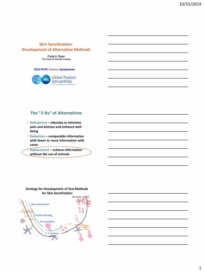
Adverse Outcome Pathway and Predictive Testing
Chemical Structure & Properties
Organism Response
Organ Response
Cellular Response
Molecular Initiating
Event
1. Skin Penetration
2. Electrophilic substance:
directly or via auto-oxidation or metabolism
3-4. Haptenation: covalent
modification of epidermal proteins
5-6. Activation of epidermal
keratinocytes & Dendritic cells
7-8. Presentation of haptenated protein by Dendritic cell resulting
in activation & proliferation of specific
T cells
9-11. Allergic Contact Dermatitis: Epidermal
inflammation following re-exposure to
substance due to T cell-mediated cell death
Key Event 1 Key Events 2 + 3 Key Event 4 Adverse Outcome
In chemico models
In silico models SAR/ QSAR
In vitro cell-based models
Reactivity Assays Modified version of flow diagram from ‘The Adverse
Outcome Pathway for Skin Sensitisation initiated by
Covalent Binding to Proteins’, OECD report
In Silico Methods: Structure Activity Relationships (SAR/QSAR)
SAR are useful for estimating the toxicity of a chemical when actual data are lacking
Find structurally similar chemicals (structural analogs) for which data exist, and then to relate those data to the chemical of interest
Examples
DEREK
ToxTree
TIMES-SS
OECD Toolbox
In Silico Methods: Skin Penetration
Epidermal bioavailability is a pre-requisite for skin sensitization
Amount in epidermis rather than total systemic uptake
Depending on phys/chem properties of the chemical
Lipophilicity/ hydrophilicity, MW, etc.
Example
Kasting Toxicokinetic model (Adv. Drug Deliv. Rev.2013, 65: 221–236.)
10/31/2014
3
Adverse Outcome Pathway and Predictive Testing
Chemical Structure & Properties
Organism Response
Organ Response
Cellular Response
Molecular Initiating
Event
1. Skin Penetration
2. Electrophilic substance:
directly or via auto-oxidation or metabolism
3-4. Haptenation: covalent
modification of epidermal proteins
5-6. Activation of epidermal
keratinocytes & Dendritic cells
7-8. Presentation of haptenated protein by Dendritic cell resulting
in activation & proliferation of specific
T cells
9-11. Allergic Contact Dermatitis: Epidermal
inflammation following re-exposure to
substance due to T cell-mediated cell death
Key Event 1 Key Events 2 + 3 Key Event 4 Adverse Outcome
In chemico models
In silico models SAR/ QSAR
In vitro cell-based models
Modified version of flow diagram from ‘The Adverse
Outcome Pathway for Skin Sensitisation initiated by
Covalent Binding to Proteins’, OECD report
AREc32 [CXR Bio.]
DPRA [P&G]
PPRA [P&G]
Chemical reactivity
Chemical-Protein Reactivity: Skin Sensitization
Nucleophilic-electrophilic interaction:
Hapten
E
:Nu
The correlation of skin protein reactivity and skin sensitization is well established and has been known for many years. (Landsteiner and Jacobs, 1936; Dupuis and Benezra, 1982; Lepoittevin et al, 1998)
Hapten Pro/Pre-Hapten
Protein
Protein
O
O
F
F
F
O
O
F
F
F
HAPTENIZATION
Covalent Protein Modification: Key step in the induction of skin sensitization
Mechanism-based classification for skin sensitizers (modified by Aynur et al. 2007)
Sensitizing chemical applied to the skin
Electrophiles (hapten)
Michael acceptor domain
SNAr domain
SN2 domain
Schiff base
domain
Acylating agent
domain
Proelectrophile (non-electrophile)
Via abiotic transformation (pre-haptens)
Via metabolic transformation (pro-haptens)
Chemical is not reactive Chemical is reactive
Mechanism of reaction Allergen
10/31/2014
4
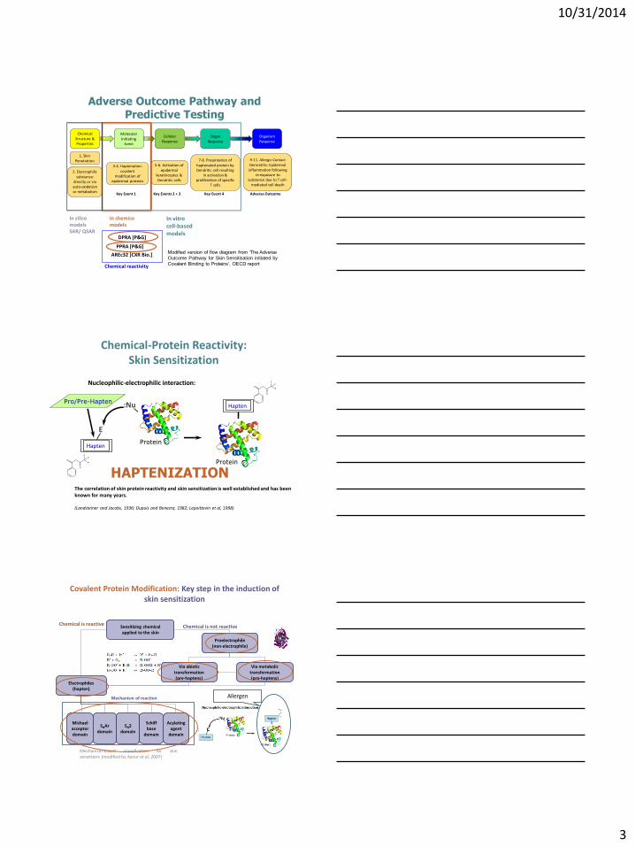
Screening method for evaluation skin sensitization potential (haptens, prehaptens)
Direct Peptide Reactivity Assay (DPRA)
• The reactivity is quantified based on the percentage of peptide depletion (HPLC/PDA)
Incubation for 24 h, 25°C (dark)
Cysteine (Ac-RFAACAA-COOH)
Lysine (Ac-RFAAKAA-COOH)
1:10 at pH 7.4
1:50 at pH 10.2
Synthetic model peptides in buffer
Test chemical in solvent
O
N
N+
O
O-
O
N
N+ O
O-
O
O
N
N+
O
-O
O
In Chemico: Direct Peptide Reactivity Assay (DPRA) Method
HPLC/PDA
Calculation of peptide depletion
In Chemico: Direct Peptide Reactivity Assay (DPRA) Method
Prediction Model based on Cys 1:10 and Lys 1:50 (n=81)
NS/W/M/S
Test (29 / 11 / 3 / 0)
Total Sample (29 / 15 / 20 / 17)
Minimal Reactivity (26 / 5 / 1 / 0)
Low Reactivity (3 / 6 / 2 / 0)
Avg Score < 6.376%
Avg Score < 22.62% Avg Score > 22.62%
Test (0 / 4 / 17 / 17)
Moderate Reactivity (0 / 1 / 6 / 3)
High Reactivity (0 / 3 / 11 / 14)
Avg Score < 42.47%
Avg Score > 6.376% Avg Score > 42.47%
Non-sensitizing Sensitizing
10/31/2014
5
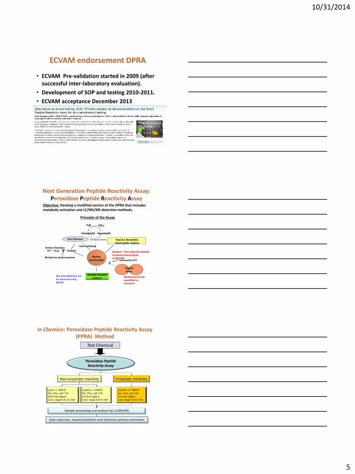
ECVAM endorsement DPRA
• ECVAM Pre-validation started in 2009 (after successful inter-laboratory evaluation).
• Development of SOP and testing 2010-2011.
• ECVAM acceptance December 2013
Principle of the Assay
Objective: Develop a modified version of the DPRA that includes metabolic activation and LC/MS/MS detection methods.
Next Generation Peptide Reactivity Assay: Peroxidase Peptide Reactivity Assay
Test Chemical
Peptide-chemical ADDUCT
Readout = Non-adducted peptide monomer measured by LC/MS/MS
Monitored but not quantified vs monomer
X reversed by DTT
Auto-oxidation
Not quantified but can be observed using MALDI
Reactive Metabolite (electrophilic hapten)
P e r o x i d a s e ( O ) P e r o x i d a s e ( R )
H 2 O 2 H 2 O
Fe2+ + H2O2 Oxidant
Blocked by desferroxamine
X Fenton Chemistry
Peptide (nucleophile)
Peptide Dimer
Test Chemical
Peroxidase Peptide Reactivity Assay
Lysine (- HRP/P) Pot. Phos. (pH 7.4) 25% final organic Conc. range 0.01-25 mM
Enzymatic reactivity Non-enzymatic reactivity
Cysteine (- HRP/P) Pot. Phos. (pH 7.4) 1% final organic Conc. range 0.04-5 mM
Cysteine (+ HRP/P) Pot. Phos. (pH 7.4) 1% final organic Conc range 0.04-5 mM
Sample processing and analysis by LC/MS/MS
Data reduction, hazard prediction and reactivity potency estimation
In Chemico: Peroxidase Peptide Reactivity Assay (PPRA) Method
10/31/2014
6
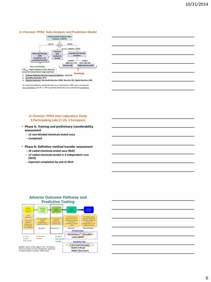
Quantify and Classify ‘positives’
Minimally Reactive (MR)
(reactivity is not quantified by EC25)
Analyze peptide depletion data (Cysteine + HRP/P)
DPMAX*
>24.9% <15.1%
1. Analyze depletion data for hazard prediction: (yes/no) 2. Quantify reactivity: EC25 3. Classify chemicals: Minimally Reactive (MR), Reactive (R), Highly Reactive (HR)
For hazard prediction, chemicals that were classified as ‘MR’ were considered non-sensitizers and ‘R’ or ‘HR’ classified chemicals were considered sensitizers.
Highly Reactive (HR) Reactive (R)
Lowest EC25 <0.1 mM
Lowest EC25 >0.1 mM
*DPMAX - highest depletion value observed across the concentration range examined.
Interpret data
(analyze & classify)
15.1% < DPMAX < 24.9%
In Chemico: PPRA Data Analysis and Prediction Model
Non-sensitizing
Sensitizing
• Phase A: Training and preliminary transferability assessment – 12 non-blinded chemicals tested once
– Completed
• Phase B: Definitive method transfer assessment – 24 coded chemicals tested once (BLR)
– 12 coded chemicals tested in 3 independent runs (WLR)
– Expected completion by end of 2014
In Chemico: PPRA Inter-Laboratory Study 3 Participating Labs (1 US, 2 European)
Adverse Outcome Pathway and Predictive Testing
Chemical Structure & Properties
Organism Response
Organ Response
Cellular Response
Molecular Initiating
Event
1. Skin Penetration
2. Electrophilic substance:
directly or via auto-oxidation or metabolism
3-4. Haptenation: covalent
modification of epidermal proteins
5-6. Activation of epidermal
keratinocytes & Dendritic cells
7-8. Presentation of haptenated protein by Dendritic cell resulting
in activation & proliferation of specific
T cells
9-11. Allergic Contact Dermatitis: Epidermal
inflammation following re-exposure to
substance due to T cell-mediated cell death
Key Event 1 Key Events 2 + 3 Key Event 4 Adverse Outcome
In chemico models
In silico models SAR/ QSAR
In vitro cell-based models
Modified version of flow diagram from ‘The Adverse
Outcome Pathway for Skin Sensitisation initiated by
Covalent Binding to Proteins’, OECD report
KeratinoSensTM [Givaudan]
LuSens [BASF]
Keratinocytes
PBMDC [Beiersdorf]
h-CLAT [KAO/Shiseido]
MUSST [L’Oreal]
Dendritic Cells
10/31/2014
7
Cell-Based In Vitro Test Methods
KeratinoSensTM Protocol developed by A. Natsch
(Givaudan)
The Keap1 – Nrf2 – ARE signaling pathway of cells specifically
responds to electrophiles
Nrf2
Keap1
SH SH SH
ARE-regulated gene ARE DNA Antioxidant response element
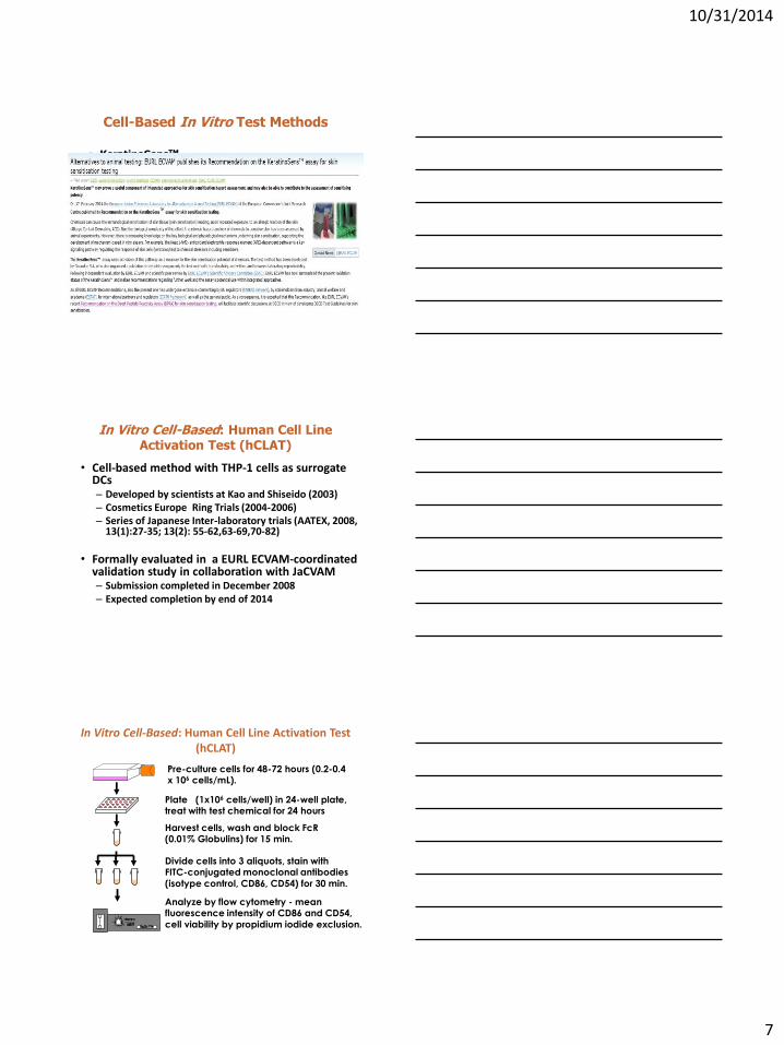
In Vitro Cell-Based: Human Cell Line Activation Test (hCLAT)
• Cell-based method with THP-1 cells as surrogate DCs – Developed by scientists at Kao and Shiseido (2003) – Cosmetics Europe Ring Trials (2004-2006) – Series of Japanese Inter-laboratory trials (AATEX, 2008,
13(1):27-35; 13(2): 55-62,63-69,70-82)
• Formally evaluated in a EURL ECVAM-coordinated validation study in collaboration with JaCVAM – Submission completed in December 2008 – Expected completion by end of 2014
In Vitro Cell-Based: Human Cell Line Activation Test (hCLAT)
Plate (1x10⁶ cells/well) in 24-well plate, treat with test chemical for 24 hours
Pre-culture cells for 48-72 hours (0.2-0.4 x 10⁶ cells/mL).
Harvest cells, wash and block FcR (0.01% Globulins) for 15 min.
Divide cells into 3 aliquots, stain with FITC-conjugated monoclonal antibodies
(isotype control, CD86, CD54) for 30 min.
Analyze by flow cytometry - mean fluorescence intensity of CD86 and CD54,
cell viability by propidium iodide exclusion.
10/31/2014
8
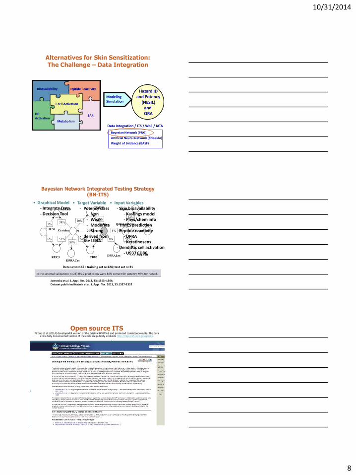
Alternatives for Skin Sensitization: The Challenge – Data Integration
Hazard ID and Potency
(NESIL) and QRA
Bioavailability
SAR
Peptide Reactivity
T cell Activation
Metabolism
DC Activation
Modeling Simulation
Data Integration / ITS / WoE / IATA
Bayesian Network (P&G)
Artificial Neural Network (Shiseido)
Weight of Evidence (BASF)
IC50
KEC1.5
7%
6%
KEC3
Cysteine
59%
55%
DPRACys
39%
CD86
LLNA
DPRALys
24% 20% 8%
16%
59%
Bioavailability
57% 5% 20%
20% 36%
TIMES logKow
Cfree
AUC120
Jaworska et al. J. Appl. Tox. 2013, 33: 1353–1364; Dataset published Natsch et al. J. Appl. Tox. 2013, 33:1337-1352
Data set n=145 : training set n=124; test set n=21
In the external validation ( n=21) ITS-2 predictions were 86% correct for potency, 95% for hazard.
• Graphical Model - Integrate Data - Decision Tool
• Target Variable - Potency class
- Non - Weak - Moderate - Strong
- derived from the LLNA
• Input Variables - Skin bioavailability
- Kastings model - Phys/chem info
- TIMES prediction - Peptide reactivity
- DPRA - Keratinosens
- Dendritic cell activation - U937 CD86
Bayesian Network Integrated Testing Strategy (BN-ITS)
Open source ITS Pirone et al. (2014) developed R version of the original BN ITS-2 and produced consistent results. The data
and a fully documented version of the code are publicly available http://ntp.niehs.nih.gov/go/its.
10/31/2014
9
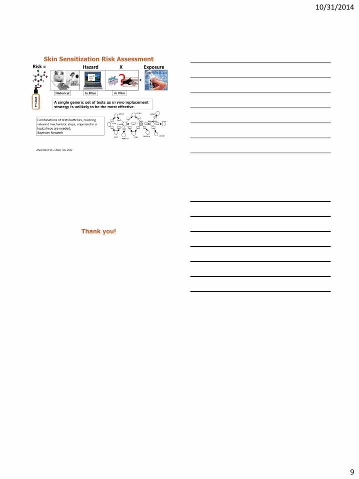
Risk =
Pro
du
ct
X
Hazard X Exposure
Historical In Silico In Vivo
Skin Sensitization Risk Assessment
In Vitro
? A single generic set of tests as in vivo replacement strategy is unlikely to be the most effective.
Combinations of tests batteries, covering relevant mechanistic steps, organized in a logical way are needed: Bayesian Network
IC50
KEC1.5
7%
6%
KEC3
Cysteine
59%
55%
DPRACys
39%
CD86
LLNA
DPRALys
24% 20% 8%
16%
59%
Bioavailability
57% 5% 20%
20% 36%
TIMES logKow
Cfree
AUC120
Jaworska et al. J. Appl. Tox. 2013
Thank you!

![Butylparaben [CAS No. 94-26-8] Review of Toxicological ... · volunteers, butylparaben (≤5%) produced no irritation or sensitization when applied to the skin. Photocontact sensitization](https://img.pdfslide.us/doc/110x75/5f023d627e708231d40345b0/butylparaben-cas-no-94-26-8-review-of-toxicological-volunteers-butylparaben.jpg)