Embed Size (px)
Citation preview
Skin and Soft-Tissue Infections
IMPETIGO, ABSCESSES, CELLULITIS, AND ERYSIPELA
1
2
Objectives1. Describe the anatomical structure of skin and soft tissues.
2. Differentiate the various types of skin and soft tissue infections and there
clinical presentation.
3. Name bacteria commonly involved in skin and soft tissue infections
4. Describe the pathogenesis of various types of skin and soft tissue infections
5. Recognize specimens that are acceptable and unacceptable for different types
of skin and soft tissue infections
6. Describe the microscopic and colony morphology and the results of
differentiating bacteria isolates in addition to other non-microbiological
investigation
7. Discuss antimicrobial susceptibility testing of anaerobes including methods and
antimicrobial agents to be tested.
8. Describe the major approaches to treat of skin and soft tissue infections
either medical or surgical.
Introduction Common Can be mild to moderate or sever
muscle or bone and lungs or heart valves infection .
Staphylococcus aureus and streptococcus are the most cause
Emerging antibiotic resistance among Staphylococcus aureus (methicillin
resistance) Streptococcus pyogenes
(erythromycin resistance) 4
Key to developing an adequate differential diagnosis requires
History patient’s immune status, the geographical locale,
travel history, recent trauma or surgery, previous antimicrobial therapy, lifestyle, and animal exposure or bites
Physical examination severity of infection
Investigation CBCs, Chemistry Swab, biopsy or aspiration Radiographic procedures
Level of infection and the presence of gas or abscess. Diagnostic and therapeutic
Surgical exploration or debridement Medical treatment5
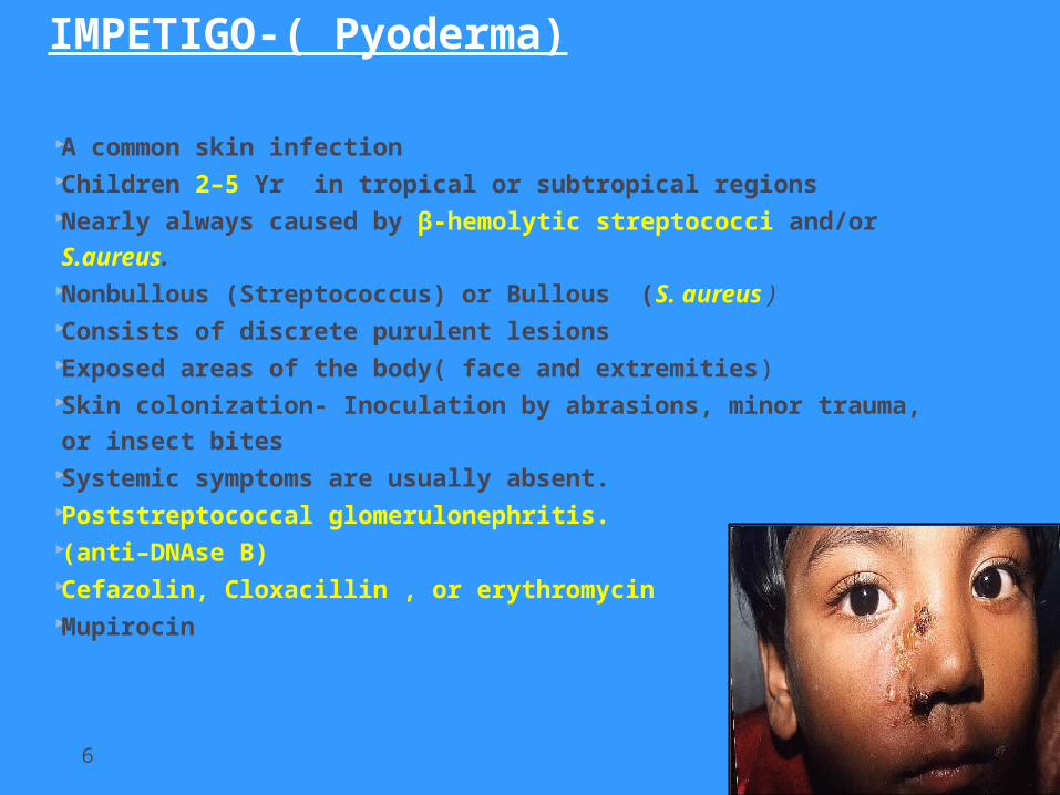
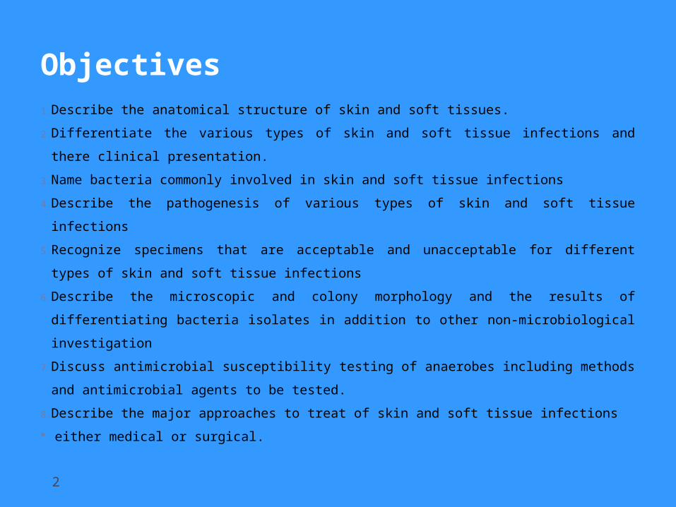
IMPETIGO-( Pyoderma)
A common skin infection Children 2–5 Yr in tropical or subtropical regions Nearly always caused by β-hemolytic streptococci and/or
S.aureus. Nonbullous (Streptococcus) or Bullous (S. aureus ) Consists of discrete purulent lesions Exposed areas of the body( face and extremities) Skin colonization- Inoculation by abrasions, minor
trauma, or insect bites Systemic symptoms are usually absent. Poststreptococcal glomerulonephritis. (anti–DNAse B) Cefazolin, Cloxacillin , or erythromycin Mupirocin
6
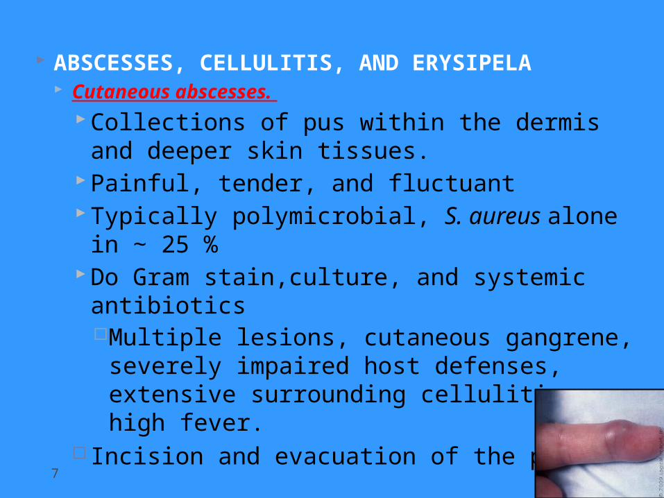
ABSCESSES, CELLULITIS, AND ERYSIPELA Cutaneous abscesses.
Collections of pus within the dermis and deeper skin tissues.
Painful, tender, and fluctuantTypically polymicrobial, S. aureus alone in
∼ 25 %Do Gram stain,culture, and systemic
antibioticsMultiple lesions, cutaneous gangrene,
severely impaired host defenses, extensive surrounding cellulitis or high fever.
Incision and evacuation of the pus7

Furuncles and carbuncles. Furuncles (or “boils”) are infections of the
hair follicle (folliculitis ), usually caused by S. aureus, in which suppuration extends through the dermis into the subcutaneous tissue
Carbuncle- extension to involve several adjacent follicles with coalescent inflammatory mass - back of the neck especially in diabetics
Larger furuncles and all carbuncles require incision and drainage.
Systemic antibiotics are usually unnecessary8
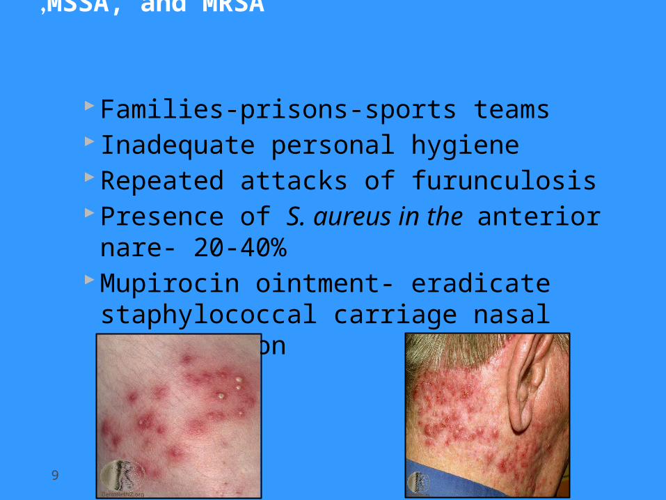
Outbreaks of furunculosis caused by MSSA, and MRSA,
9
Families-prisons-sports teams Inadequate personal hygieneRepeated attacks of furunculosisPresence of S. aureus in the anterior
nare- 20-40%Mupirocin ointment- eradicate
staphylococcal carriage nasal colonization
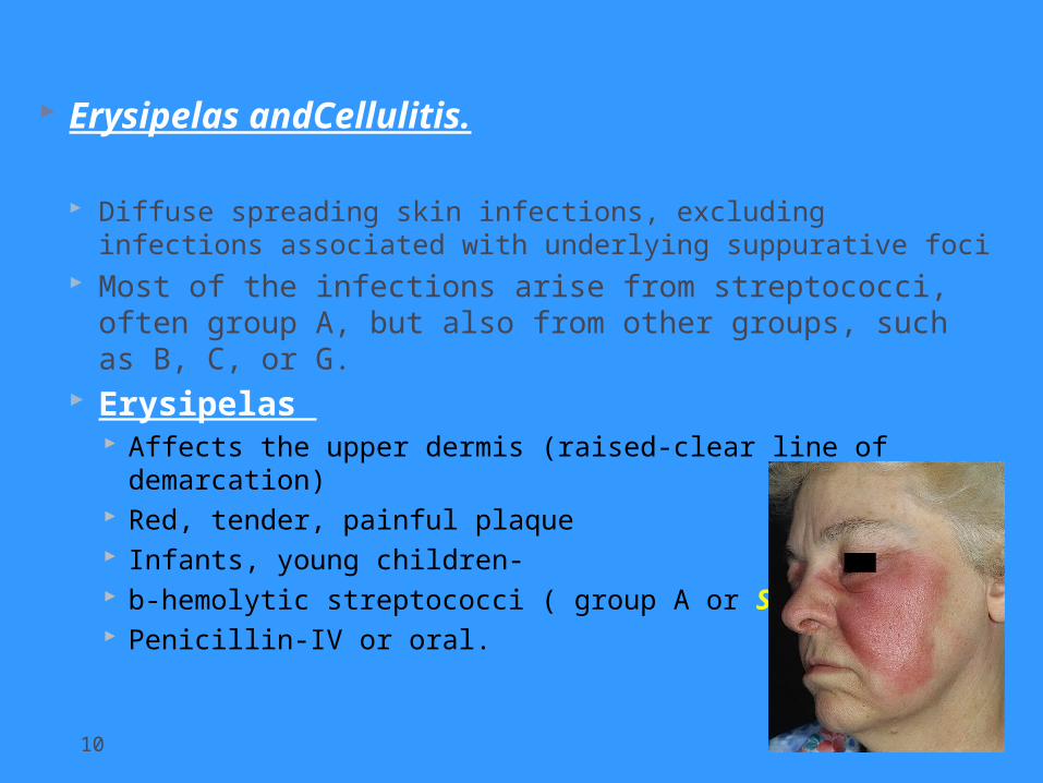
Erysipelas andCellulitis.
Diffuse spreading skin infections, excluding infections associated with underlying suppurative foci
Most of the infections arise from streptococci, often group A, but also from other groups, such as B, C, or G.
Erysipelas Affects the upper dermis (raised-clear line of demarcation) Red, tender, painful plaque Infants, young children- b-hemolytic streptococci ( group A or S. pyogenes.) Penicillin-IV or oral.
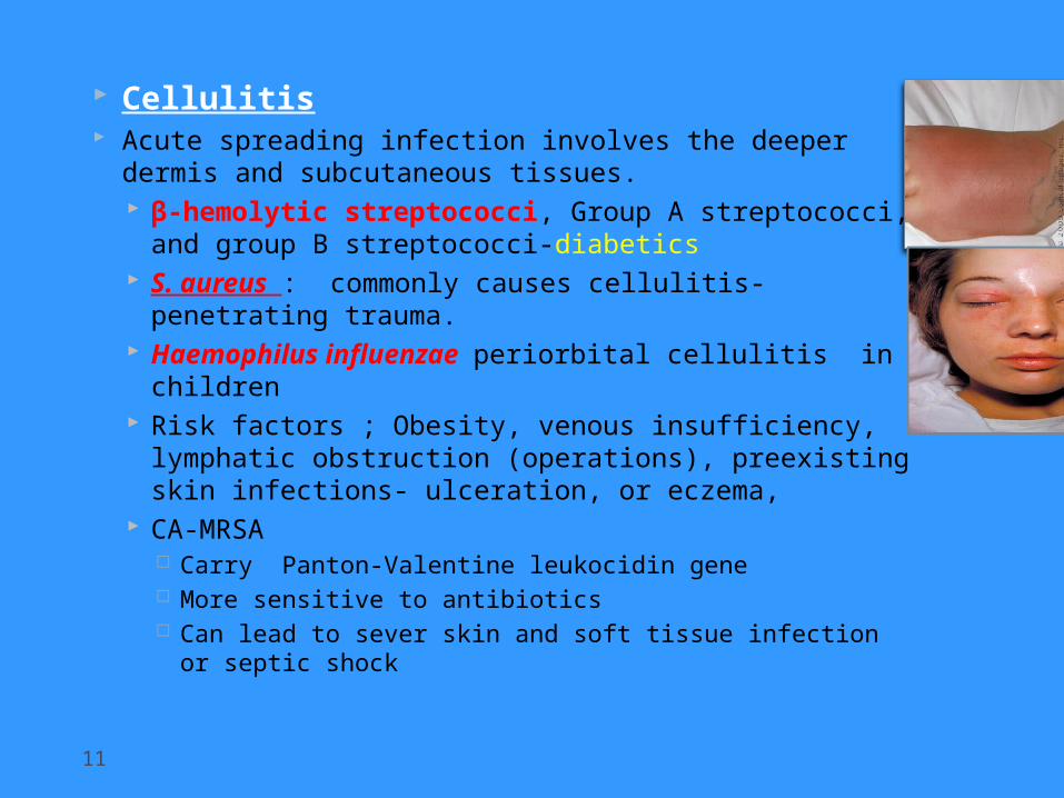
Cellulitis Acute spreading infection involves the deeper
dermis and subcutaneous tissues. β-hemolytic streptococci, Group A streptococci,
and group B streptococci-diabetics S. aureus : commonly causes cellulitis-
penetrating trauma. Haemophilus influenzae periorbital cellulitis in
children Risk factors ; Obesity, venous insufficiency,
lymphatic obstruction (operations), preexisting skin infections- ulceration, or eczema,
CA-MRSA Carry Panton-Valentine leukocidin gene More sensitive to antibiotics Can lead to sever skin and soft tissue infection or
septic shock
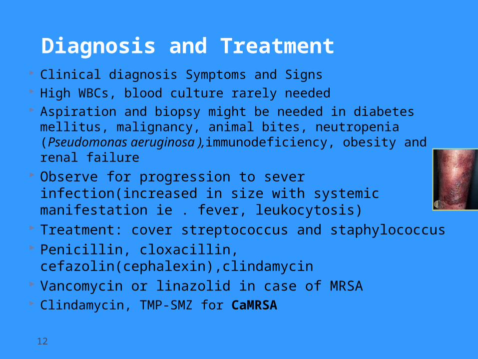
Diagnosis and Treatment Clinical diagnosis Symptoms and Signs High WBCs, blood culture rarely needed Aspiration and biopsy might be needed in diabetes
mellitus, malignancy, animal bites, neutropenia (Pseudomonas aeruginosa ),immunodeficiency, obesity and renal failure
Observe for progression to sever infection(increased in size with systemic manifestation ie . fever, leukocytosis)
Treatment: cover streptococcus and staphylococcus Penicillin, cloxacillin,
cefazolin(cephalexin),clindamycin Vancomycin or linazolid in case of MRSA Clindamycin, TMP-SMZ for CaMRSA
12
Necrotizing fasciitis
Flesh-eating disease
13
Introduction
14
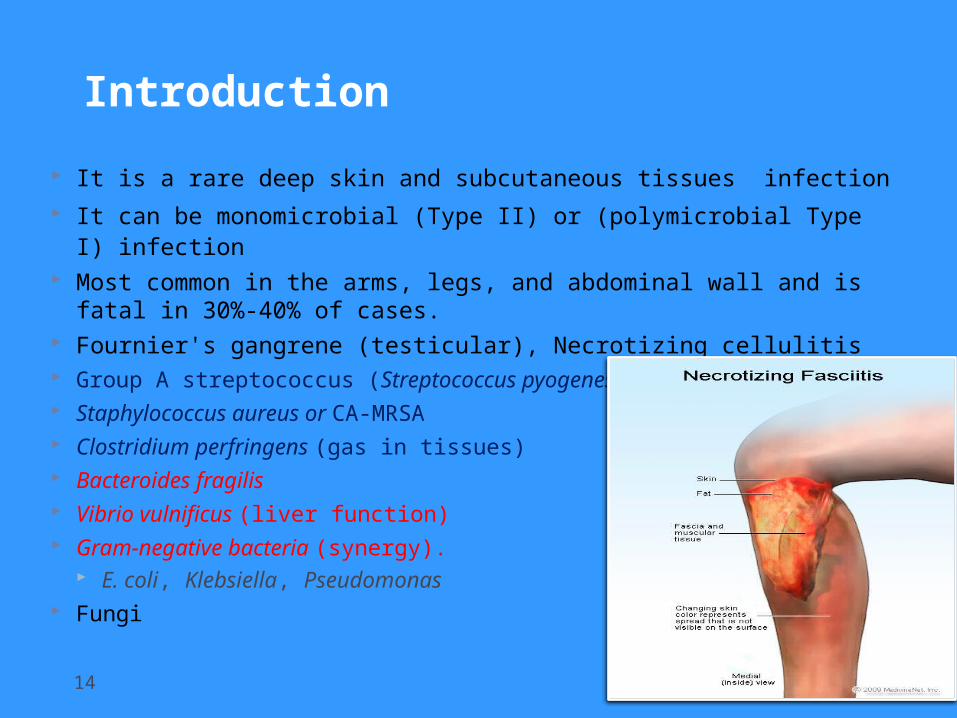
It is a rare deep skin and subcutaneous tissues infection It can be monomicrobial (Type II) or (polymicrobial Type I)
infection Most common in the arms, legs, and abdominal wall and is
fatal in 30%-40% of cases. Fournier's gangrene (testicular), Necrotizing cellulitis Group A streptococcus (Streptococcus pyogenes) Staphylococcus aureus or CA-MRSA Clostridium perfringens (gas in tissues) Bacteroides fragilis Vibrio vulnificus (liver function) Gram-negative bacteria (synergy).
E. coli, Klebsiella, Pseudomonas Fungi
Risk factors
15
Immune-suppression Chronic diseases: ( diabetes, liver and kidney diseases,
malignancy Trauma:(laceration, cut, abrasion, contusion, burn, bite,
subcutaneous injection, operative incision) Recent viral infection rash (chickenpox) Steroids Alcoholism Malnutrition Idiopathic
Pathophysiology
16
Destruction of skin and muscle by releasing toxins Streptococcal pyogenic exotoxins Superantigen
Non-specific activation of T-cells Overproduction of cytokines Severe systemic illness (Toxic shock syndrome)
Signs and symptoms
17
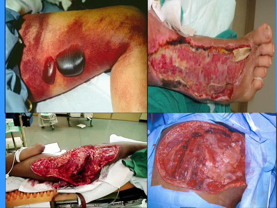
Rapid progression of sever pain with fever , chills (typical)
Swelling , redness, hotness, blister, gas formation, gangrene and necrosis
Blisters with subsequent necrosis , necrotic eschars Diarrhea and vomiting (very ill) Shock organ failure Mortality as high as 73 % if untreated
Diagnosis
19
A delay in diagnosis is associated with a grave prognosis and increased mortality
Clinical-high index of suspicion
Blood tests CBC-WBC , differential , ESR BUN (blood urea nitrogen)
Surgery debridement- amputation Radiographic studies
X-rays : subcutaneous gases Doppler CT or MRI
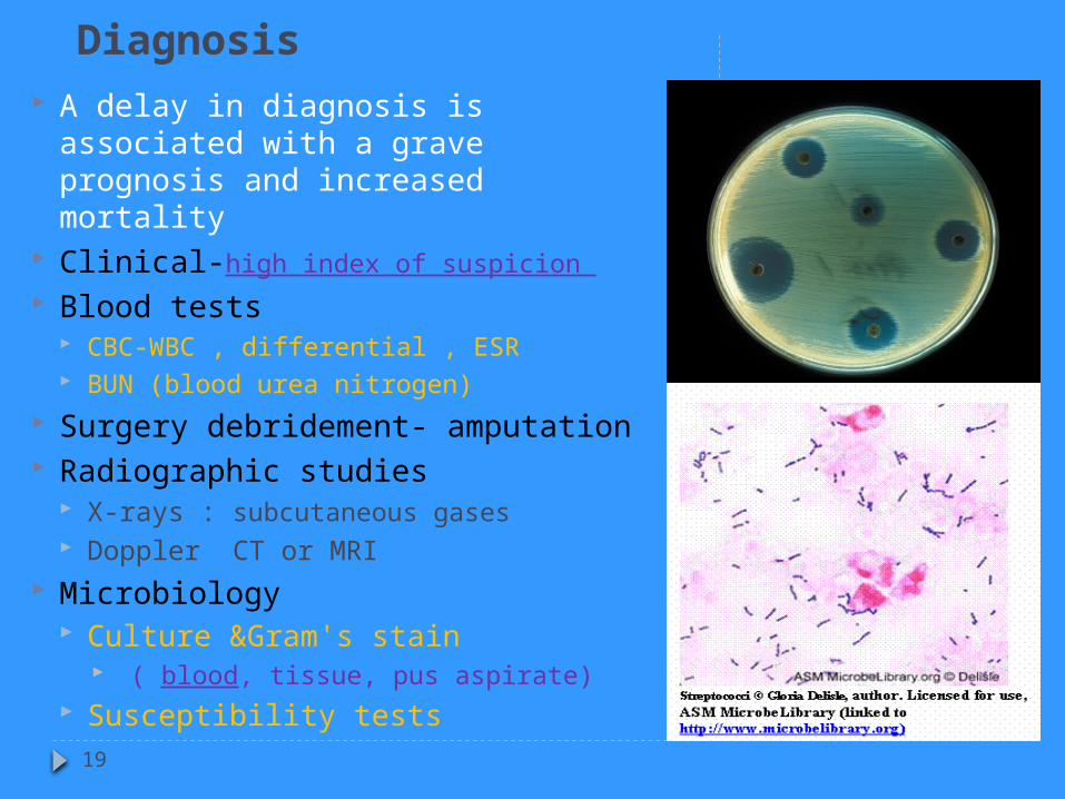
Microbiology Culture &Gram's stain
( blood, tissue, pus aspirate) Susceptibility tests
Treatment
20
If clinically suspected patient needs to be hospitalized OR require admission to ICU
Start intravenous antibiotics immediately Antibiotic selection based on bacteria suspected broad spectrum antibiotic combinations against
methicillin-resistant Staphylococcus aureus (MRSA) anaerobic bacteria Gram-negative and gram-positive bacilli
Surgeon consultation Extensive Debridement of necrotic tissue and collection of tissue
samples Can reduce morbidity and mortality

Treatment Antibiotics combinations
Penicillin-clindamycin-gentamicin Ampicillin/sulbactam Cefazolin plus metronidazol Piperacillin/tazobactam Clostridium perfringens - penicillin G
Hyperbaric oxygen therapy (HBO) treatment
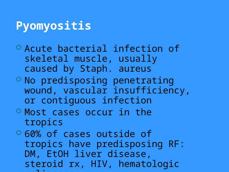
Pyomyositis
Acute bacterial infection of skeletal muscle, usually caused by Staph. aureus
No predisposing penetrating wound, vascular insufficiency, or contiguous infection
Most cases occur in the tropics 60% of cases outside of tropics have
predisposing RF: DM, EtOH liver disease, steroid rx, HIV, hematologic malignancy
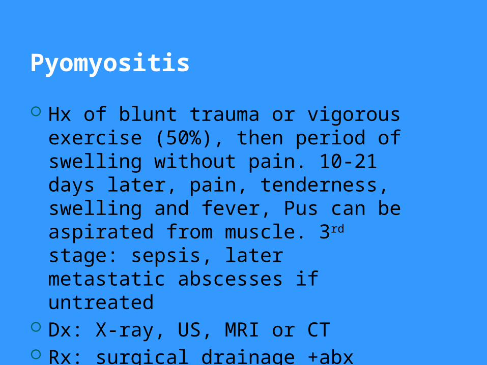
Pyomyositis
Hx of blunt trauma or vigorous exercise (50%), then period of swelling without pain. 10-21 days later, pain, tenderness, swelling and fever, Pus can be aspirated from muscle. 3rd stage: sepsis, later metastatic abscesses if untreated
Dx: X-ray, US, MRI or CT Rx: surgical drainage +abx
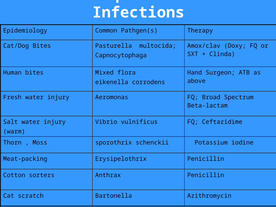
Other Specific Skin InfectionsEpidemiology Common Pathgen(s) Therapy
Cat/Dog Bites Pasturella multocida;Capnocytophaga
Amox/clav (Doxy; FQ or SXT + Clinda)
Human bites Mixed floraeikenella corrodens
Hand Surgeon; ATB as above
Fresh water injury Aeromonas FQ; Broad Spectrum Beta-lactam
Salt water injury(warm)
Vibrio vulnificus FQ; Ceftazidime
Thorn , Moss sporothrix schenckii Potassium iodine
Meat-packing Erysipelothrix Penicillin
Cotton sorters Anthrax Penicillin
Cat scratch Bartonella Azithromycin
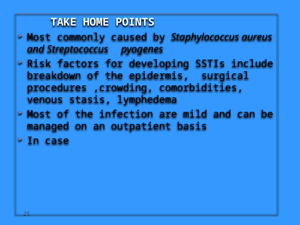
TAKE HOME POINTS Most commonly caused by Staphylococcus
aureus and Streptococcus pyogenes Risk factors for developing SSTIs include
breakdown of the epidermis, surgical procedures ,crowding, comorbidities, venous stasis, lymphedema
Most of the infection are mild and can be managed on an outpatient basis
In case
26
Most SSTIs can be managed on an outpatient basis, although patients with evidence of rapidly progressive infection, high fevers, or other signs of systemic inflammatory response should be monitored in the hospital setting.
Superficial SSTIs typically do not require systemic antibiotic treatment and can be managed with topical antibiotic agents, heat packs, or incision and drainage.
Systemic antibiotic agents that provide coverage for both Staphylococcus aureus and Streptococcus pyogenes are most commonly used as empiric therapy for both uncomplicated and complicated deeper infections.
TAKE HOME POINTS


![STREPTOCOCCUS - Omeo]Webomeoweb.com/documenti/biblioteca/streptococcus.pdf · •Streptococcus pneumoniae – ... •Impetigo (Streptococcal pyoderma) - purulent with crusting •Cellulitis](https://img.pdfslide.us/doc/110x75/5c89e10609d3f232478b7a2e/streptococcus-omeo-streptococcus-pneumoniae-impetigo-streptococcal.jpg)