Embed Size (px)
Citation preview
REVIEW
Skin and diabetes mellitus: what do we know?
Fabio Quondamatteo
Received: 22 June 2013 /Accepted: 29 October 2013 /Published online: 7 December 2013# Springer-Verlag Berlin Heidelberg 2013
Abstract Diabetes mellitus (DM) is becoming increasinglyprevalent worldwide. Although major complications of thiscondition involve kidney, retina and peripheral nerves, theskin of diabetic patients is also frequently injured. Hence,interest is mounting in the definition of the structural andmolecular profile of non-complicated diabetic skin, i.e., beforeinjuries occur. Most of the available knowledge in this areahas been obtained relatively recently and, in part, derives fromvarious diabetic animal models. These include both insulin-dependent and insulin-resistant models. Structural work inhuman diabetic skin has also been carried out by means oftissue samples or of non-invasive methods. Indications haveindeed been found for molecular/structural changes in diabeticskin. However, the overall picture that emerges is heteroge-neous, incomplete and often contradictory and many ques-tions remain unanswered. This review aims to detail, as muchas possible, the various pieces of current knowledge in asystematic and synoptic manner. This should aid the identifi-cation of areas in which key questions are still open and moreresearch is needed. A comprehensive understanding of thisfield could help in determining molecular targets for theprevention and treatment of skin injuries in DM and markersfor the monitoring of cutaneous and systemic aspects of thedisease. Additionally, with the increasing development ofnon-invasive optics-based deep-tissue-imaging diagnostictechnologies, precise knowledge of cutaneous texture andmolecular structure becomes an important pre-requisite forthe use of such methods in diabetic patients.
Keywords Non-complicated skin . Diabetes mellitus .
Cutaneous structure .Molecular composition . Diabetic skin
Abbreviations
AGEs Advanced glycation endproductsAX AlloxanBB rats Bio-breeding ratsBM Basement membranecm-OCT Correlation mapping optical coherence
tomographyCYP Cytochrome P450DM Diabetes mellitusDTA Diphteria toxin A-chainECM Extracellular matrixGFOGER Glycine—phenylanaline—hydrohyproline—
glycine—glutamic acid—arginineGLUT4 Glucose transporter 4HFD High-fat dietMMPs Matrix metalloproteinasesNOD mice Non-obese-diabetic miceOCT Optical coherence tomographyOLETF rats Otsuka Long Evans Tokushima fatty ratsRGD Arginine—glycine—aspartic acidSAF Skin autofluorescenceSTZ StreptozotocinTEWL Trans-epidermal water lossTIMP Tissue inhibitor of matrix metalloproteinasesTSNO mice Tsumura-Suzuki non-obesity miceTSOD mice Tsumura-Suzuki obese diabetic miceUCP1 Uncoupling protein 1
General aspects of diabetes mellitus
Diabetes mellitus (DM) is a chronic and systemic conditioncharacterised by hyperglycaemia and severe complicationssuch as retinopathy, nephropathy and neuropathy (American
F. Quondamatteo (*)Skin and ECM Research Group—Anatomy, NUI Galway, AnatomyBuilding, University Road, Galway, Irelande-mail: [email protected]
Cell Tissue Res (2014) 355:1–21DOI 10.1007/s00441-013-1751-2
Diabetes Association 2013). Clinically, the most commonforms are type 1 DM (insulin-dependent) and type 2 DM(non-insulin-dependent, ∼90 % DM), the latter of which isassociated with obesity and insulin resistance (Zimmet et al.2001; Lam and LeRoith 2012). Prevalence of DM is constant-ly increasing. In 2011, the total worldwide population affectedby the disease was reported to be in excess of 350,000,000 andthis figure will increase to around half a billion by 2030 (Lamand LeRoith 2012). Such an order of magnitude was predictedat the beginning of the 2000s, when DM was defined as “oneof the main threats to human health of the 21st century”(Zimmet et al. 2001). Accordingly, DM has a high socio-economic impact. For example, the financial burden of apatient over the age of 65 with type 2 DM has been estimatedto be nearly double that of patients in the same age groupwithout type 2 DM (O’Shea et al. 2013).
Skin involvement in DM
The skin (Fig. 1) is variably but often, affected during DM.Reported figures indicate that about 1/3 to nearly all diabeticpatients experience cutaneous complications (Aye andMasson 2002; Wohlrab et al. 2007; Bristow 2008; vanHattem et al. 2008). The most severe cutaneous lesions arechronic ulcers that form as a consequence of the poor healingpotential of diabetic skin (Falanga 2005). These can frequent-ly become infected, thus leading to amputation (Ngo et al.2005; Eaglstein and Callen 2009). In the Republic of Irelandin 2009, for example, the total number of lower limb amputa-tions specifically related to DM was 175.7 per 100,000 dia-betic patients, compared with lower limb amputations notrelated to DM, which had a frequency of 9.2 per 100,000non-diabetic patients (Buckley et al. 2012).
A large amount of work on skin in DM has primarily beenfocused on the management of the diabetic foot and on exper-imental wound healing (e.g., Greenhalgh et al. 1990; Aye andMasson 2002; Dinh and Veves 2005; Rodgers et al. 2006;Schramm et al. 2006; Berdal et al. 2011; Bermudez et al. 2011;Zhao et al. 2012). Conversely, non-injured skin in DM isrelatively understudied. As a consequence, knowledge of thein vivo structure and molecular composition of non-injureddiabetic skin is fragmentary, most being relatively recent andto a wide extent derived from various diabetic animal models.This body of work provides an indication for molecular/structural cutaneous changes in DM. However, actualmolecular/structural features of diabetic skin are currentlypoorly defined and a number of questions remain unanswered.
This review will focus on the structural and molecularknowledge of non-injured diabetic skin. It attempts to gathervarious pieces of current data and to collate them systemati-cally into a synoptic view (Tables 1, 2). This will help toidentify areas in which key questions are still open and more
research is needed. Other aspects, such as clinical condi-tions of dermatologic interest associated with DM anddiabetic wound healing will only be referred to in orderto place the actual content of the review into its appropri-ate context. For a more comprehensive overview of theseaspects, the reader can refer to a broad body of existingliterature (e.g., Greenhalgh et al. 1990; Lipsky et al. 2000;Aye and Masson 2002; Dinh and Veves 2005; Rodgerset al. 2006; Schramm et al. 2006; Wohlrab et al. 2007;Bristow 2008; Larsen et al. 2008; van Hattem et al. 2008;Berdal et al. 2011; Bermudez et al. 2011; Ragunatha et al.2011; Zhao et al. 2012; Behm et al. 2012). The reviewbegins with a brief introduction to the diabetic animalmodels used for skin analysis. The outcome of the indi-vidual skin studies will then be discussed in the subse-quent sections, in the context of the main topic, startingfrom the epidermal barrier and systematically moving(anatomically speaking) deeper in the direction of thedermis and dermal microvessels.
Animal models used in the study of diabetic skin
Since the end of the nineteenth century, when vonMering and Minkowski carried out the first radical pan-createctomy on dogs (von Mering and Minkowski 1890),a series of animal models for the study of DM have beendeveloped (Rees and Alcolado 2005; Chatzizgeorgiouet al. 2009; King 2012). In this section, models that have beenused in investigations into non-injured skin in DM will bebriefly introduced. For more details and for a wider view onthe use of animal models in DM, the reader can refer to severalexcellent review papers (e.g., Rees and Alcolado 2005;Chatzizgeorgiou et al. 2009; King 2012).
Type 1 diabetes models. The generation of insulin-dependent DM in experimental animals for skin studies hastypically been carried out by means of streptozotocin (STZ) oralloxan (AX) treatment.
STZ treatment This drug leads to β-cell destruction and, thus,the animals develop insulopaenia and hyperglycaemia(Chatzizgeorgiou et al. 2009; King 2012). However, STZtreatment can also be followed by islet regeneration, withobvious consequences on glycaemia and this must be takeninto account when using this model (King 2012). In studies ofnon-injured skin, STZ treatment is the most frequently usedamong the insulin-dependent models and has been employedin rats (Craig et al. 1998; Kiliçaslan and Özer 2009; Chen et al.2010; Takahashi and Takasu 2011; Tellechea et al. 2013),mice (Sakai et al. 2003; Tellechea et al. 2013) and baboons(Heffernan et al. 1996).
2 Cell Tissue Res (2014) 355:1–21
AX treatment This drug is also delivered to β-cells where ittriggers their destruction (Rees and Alcolado 2005;Chatzizgeorgiou et al. 2009; King 2012). In studies on non-injured skin, AX treatment has been used in rabbits (Tellecheaet al. 2013) and in mice (Sakai et al. 2003; Ye et al. 2013).However, in the paper of Sakai et al. (2003) in which both AXand STZ models have been investigated, the results on skin
structure do not consistently refer to the AX model, whereasthey are consistently described in relation to the STZ model.This is probably because DM induction is more complicatedfollowing AX treatment as a result of its higher toxicity (King2012; Sakai et al. 2003).
In general, despite being suitable for generating insulin-dependent diabetic animals, neither model has a strong
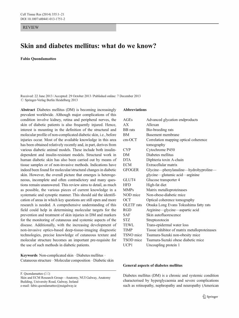
Fig. 1 Skin overview (a) showing a representation of general cutaneousorganisation according to Simpson et al. (2011). The skin is composed ofthe epidermis and dermis separated (and united) by the epidermal base-ment membrane (BM ). The epidermis is mainly composed ofkeratinocytes that organise themselves to form four layers, namely (fromthe BM to the skin surface), the basal layer or stratum basale (BL),spinous layer or stratum spinosum (SL ), granular layer or stratumgranulosum (GL) and cornified layer or stratum corneum (CL or SC).This representation gives a general orientation for Figs. 2, 3, 4 and 5 inwhich the skin structure is presented in more detail in the individualregions corresponding to those in the red frames . Intercalated withkeratinocytes, other cell types are present in the epidermal layer, e.g.,dendiritc cells (red cell). These cells extend their processes between thekeratinocytes and play an important role in host defence. For simplicity,other cell types usually found in the epidermis (e.g., melanocytes) areomitted; only dendritic cells are depicted here, as they will be discussedwith regard to the barrier function of skin. b–e Skin samples stained byhaematoxylin-eosin (HF hair follicles with associated sebaceous glands,E epidermis, D dermis, S subcutis, SC stratum corneum). b , c Murineback skin. d , e Human skin, mammary region. Note that, although thegeneral structure of the epidermis also applies to murine skin, the layersare much less distinct in the murine epidermis than in human skin at thelight microscope level (cf. c , e). In human skin at the light microscope
level, a basal layer can be recognised (e , white arrowhead) composed ofcuboidal cells. Towards the skin surface (just underneath the cornifiedlayer), a granular layer (e , black/white arrowheads) can be seen in whichthe cells are flat with more intense staining in their cytoplasm attributableto fine granula. Between the two layers, the spinous layer (intermediatelayer) has cells that are quadrangular/polygonal that tend to become flattowards the skin surface. In the dermis of human skin, two distinct layerscan be observed at the light microscope level: a more superficial layer,namely the papillary layer (P) and a deeper layer called the reticular layer(R). The papillary layer contains a much finer network of fibrillar extra-cellular matrix components, which become coarser and denser in thereticular layer. Although also in murine skin the fibres tend to becomelarger and more densely packed in the deep dermis (as seen ultrastructur-ally), such a distinction into two layers is not obvious at the lightmicroscope level (cf. b , d). f Two mast cells stained by alcian blue(arrows) between a number of other cells (mostly fibroblasts, see redcell nuclei) in murine dermis. g , h Higher power views of papillary andreticular dermis, respectively, of human skin (same sample as in d , e)stained by the trichromic method to highlight the fibrous components ofthe connective tissue, namely collagen (green) and elastic fibres (darkblue/grey, arrowheads). Note the difference in size of the fibrous com-ponents and the organisation of the overall network. Bars 30 μm
Cell Tissue Res (2014) 355:1–21 3
autoimmune component (Chatzizgeorgiou et al. 2009), whichotherwise characterises the vast majority of human type 1 DM(American Diabetes Association 2013). Nevertheless, in sev-eral settings that do not particularly focus on the immuneaspects of the disease, both STZ and AX models are widely
used as the model of choice. Models with spontaneous auto-immune destruction of β-cells exist, e.g., the non-obese-diabetic (NOD) mouse and the bio-breeding (BB) rat. How-ever, although these models bear the autoimmune componentthat is an important aspect of the human disease, they also
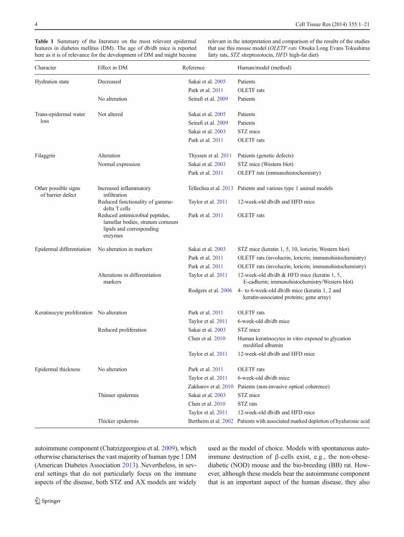
Table 1 Summary of the literature on the most relevant epidermalfeatures in diabetes mellitus (DM). The age of db/db mice is reportedhere as it is of relevance for the development of DM and might become
relevant in the interpretation and comparison of the results of the studiesthat use this mouse model (OLETF rats Otsuka Long Evans Tokushimafatty rats, STZ streptozotocin, HFD high-fat diet)
Character Effect in DM Reference Human/model (method)
Hydration state Decreased Sakai et al. 2005 Patients
Park et al. 2011 OLETF rats
No alteration Seirafi et al. 2009 Patients
Trans-epidermal waterloss
Not altered Sakai et al. 2005 Patients
Seirafi et al. 2009 Patients
Sakai et al. 2003 STZ mice
Park et al. 2011 OLETF rats
Filaggrin Alteration Thyssen et al. 2011 Patients (genetic defects)
Normal expression Sakai et al. 2003 STZ mice (Western blot)
Park et al. 2011 OLEFT rats (immunohistochemistry)
Other possible signsof barrier defect
Increased inflammatoryinfiltration
Tellechea et al. 2013 Patients and various type 1 animal models
Reduced functionality of gamma-delta T cells
Taylor et al. 2011 12-week-old db/db and HFD mice
Reduced antimicrobial peptides,lamellar bodies, stratum corneumlipids and correspondingenzymes
Park et al. 2011 OLETF rats
Epidermal differentiation No alteration in markers Sakai et al. 2003 STZ mice (keratin 1, 5, 10, loricrin; Western blot)
Park et al. 2011 OLETF rats (involucrin, loricrin; immunohistochemistry)
Park et al. 2011 OLETF rats (involucrin, loricrin; immunohistochemistry)
Alterations in differentiationmarkers
Taylor et al. 2011 12-week-old db/db & HFD mice (keratin 1, 5,E-cadherin; immunohistochemistry/Western blot)
Rodgers et al. 2006 4– to 6-week-old db/db mice (keratin 1, 2 andkeratin-associated proteins; gene array)
Keratinocyte proliferation No alteration Park et al. 2011 OLETF rats
Taylor et al. 2011 6-week-old db/db mice
Reduced proliferation Sakai et al. 2003 STZ mice
Chen et al. 2010 Human keratinocytes in vitro exposed to glycationmodified albumin
Taylor et al. 2011 12-week-old db/db and HFD mice
Epidermal thickness No alteration Park et al. 2011 OLETF rats
Taylor et al. 2011 6-week-old db/db mice
Zakharov et al. 2010 Patients (non-invasive optical coherence)
Thinner epidermis Sakai et al. 2003 STZ mice
Chen et al. 2010 STZ rats
Taylor et al. 2011 12-week-old db/db and HFD mice
Thicker epidermis Bertheim et al. 2002 Patients with associatedmarked depletion of hyaluronic acid
4 Cell Tissue Res (2014) 355:1–21
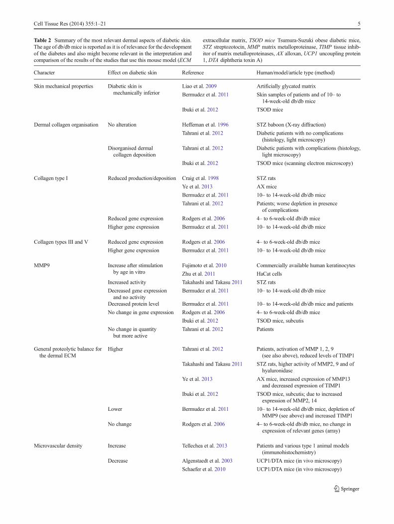
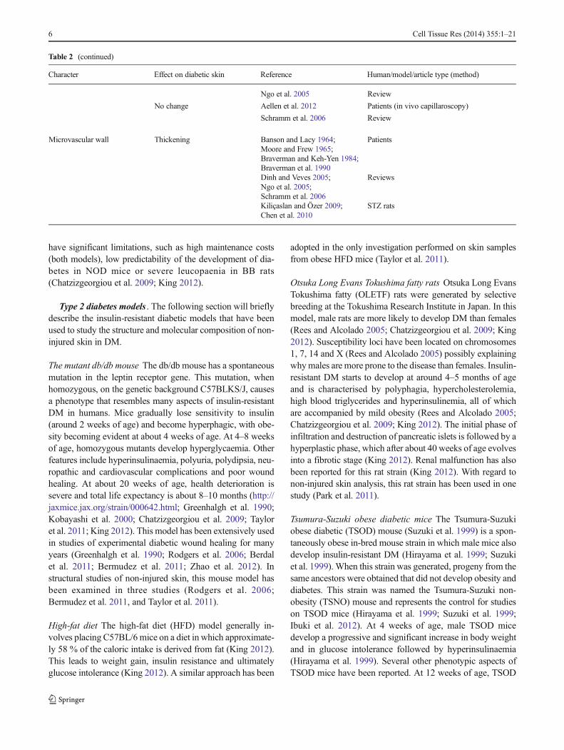
Table 2 Summary of the most relevant dermal aspects of diabetic skin.The age of db/dbmice is reported as it is of relevance for the developmentof the diabetes and also might become relevant in the interpretation andcomparison of the results of the studies that use this mouse model (ECM
extracellular matrix, TSOD mice Tsumura-Suzuki obese diabetic mice,STZ streptozotocin, MMP matrix metalloproteinase, TIMP tissue inhib-itor of matrix metalloproteinases, AX alloxan, UCP1 uncoupling protein1, DTA diphtheria toxin A)
Character Effect on diabetic skin Reference Human/model/article type (method)
Skin mechanical properties Diabetic skin ismechanically inferior
Liao et al. 2009 Artificially glycated matrix
Bermudez et al. 2011 Skin samples of patients and of 10– to14-week-old db/db mice
Ibuki et al. 2012 TSOD mice
Dermal collagen organisation No alteration Heffernan et al. 1996 STZ baboon (X-ray diffraction)
Tahrani et al. 2012 Diabetic patients with no complications(histology, light microscopy)
Disorganised dermalcollagen deposition
Tahrani et al. 2012 Diabetic patients with complications (histology,light microscopy)
Ibuki et al. 2012 TSOD mice (scanning electron microscopy)
Collagen type I Reduced production/deposition Craig et al. 1998 STZ rats
Ye et al. 2013 AX mice
Bermudez et al. 2011 10– to 14-week-old db/db mice
Tahrani et al. 2012 Patients; worse depletion in presenceof complications
Reduced gene expression Rodgers et al. 2006 4– to 6-week-old db/db mice
Higher gene expression Bermudez et al. 2011 10– to 14-week-old db/db mice
Collagen types III and V Reduced gene expression Rodgers et al. 2006 4– to 6-week-old db/db mice
Higher gene expression Bermudez et al. 2011 10– to 14-week-old db/db mice
MMP9 Increase after stimulationby age in vitro
Fujimoto et al. 2010 Commercially available human keratinocytes
Zhu et al. 2011 HaCat cells
Increased activity Takahashi and Takasu 2011 STZ rats
Decreased gene expressionand no activity
Bermudez et al. 2011 10– to 14-week-old db/db mice
Decreased protein level Bermudez et al. 2011 10– to 14-week-old db/db mice and patients
No change in gene expression Rodgers et al. 2006 4– to 6-week-old db/db mice
Ibuki et al. 2012 TSOD mice, subcutis
No change in quantitybut more active
Tahrani et al. 2012 Patients
General proteolytic balance forthe dermal ECM
Higher Tahrani et al. 2012 Patients, activation of MMP 1, 2, 9(see also above), reduced levels of TIMP1
Takahashi and Takasu 2011 STZ rats, higher activity of MMP2, 9 and ofhyaluronidase
Ye et al. 2013 AX mice, increased expression of MMP13and decreased expression of TIMP1
Ibuki et al. 2012 TSOD mice, subcutis; due to increasedexpression of MMP2, 14
Lower Bermudez et al. 2011 10– to 14-week-old db/db mice, depletion ofMMP9 (see above) and increased TIMP1
No change Rodgers et al. 2006 4– to 6-week-old db/db mice, no change inexpression of relevant genes (array)
Microvascular density Increase Tellechea et al. 2013 Patients and various type 1 animal models(immunohistochemistry)
Decrease Algenstaedt et al. 2003 UCP1/DTA mice (in vivo microscopy)
Schaefer et al. 2010 UCP1/DTA mice (in vivo microscopy)
Cell Tissue Res (2014) 355:1–21 5
have significant limitations, such as high maintenance costs(both models), low predictability of the development of dia-betes in NOD mice or severe leucopaenia in BB rats(Chatzizgeorgiou et al. 2009; King 2012).
Type 2 diabetes models . The following section will brieflydescribe the insulin-resistant diabetic models that have beenused to study the structure and molecular composition of non-injured skin in DM.
The mutant db/db mouse The db/db mouse has a spontaneousmutation in the leptin receptor gene. This mutation, whenhomozygous, on the genetic background C57BLKS/J, causesa phenotype that resembles many aspects of insulin-resistantDM in humans. Mice gradually lose sensitivity to insulin(around 2 weeks of age) and become hyperphagic, with obe-sity becoming evident at about 4 weeks of age. At 4–8 weeksof age, homozygous mutants develop hyperglycaemia. Otherfeatures include hyperinsulinaemia, polyuria, polydipsia, neu-ropathic and cardiovascular complications and poor woundhealing. At about 20 weeks of age, health deterioration issevere and total life expectancy is about 8–10 months (http://jaxmice.jax.org/strain/000642.html; Greenhalgh et al. 1990;Kobayashi et al. 2000; Chatzizgeorgiou et al. 2009; Tayloret al. 2011; King 2012). This model has been extensively usedin studies of experimental diabetic wound healing for manyyears (Greenhalgh et al. 1990; Rodgers et al. 2006; Berdalet al. 2011; Bermudez et al. 2011; Zhao et al. 2012). Instructural studies of non-injured skin, this mouse model hasbeen examined in three studies (Rodgers et al. 2006;Bermudez et al. 2011, and Taylor et al. 2011).
High-fat diet The high-fat diet (HFD) model generally in-volves placing C57BL/6 mice on a diet in which approximate-ly 58 % of the caloric intake is derived from fat (King 2012).This leads to weight gain, insulin resistance and ultimatelyglucose intolerance (King 2012). A similar approach has been
adopted in the only investigation performed on skin samplesfrom obese HFD mice (Taylor et al. 2011).
Otsuka Long Evans Tokushima fatty rats Otsuka Long EvansTokushima fatty (OLETF) rats were generated by selectivebreeding at the Tokushima Research Institute in Japan. In thismodel, male rats are more likely to develop DM than females(Rees and Alcolado 2005; Chatzizgeorgiou et al. 2009; King2012). Susceptibility loci have been located on chromosomes1, 7, 14 and X (Rees and Alcolado 2005) possibly explainingwhymales are more prone to the disease than females. Insulin-resistant DM starts to develop at around 4–5 months of ageand is characterised by polyphagia, hypercholesterolemia,high blood triglycerides and hyperinsulinemia, all of whichare accompanied by mild obesity (Rees and Alcolado 2005;Chatzizgeorgiou et al. 2009; King 2012). The initial phase ofinfiltration and destruction of pancreatic islets is followed by ahyperplastic phase, which after about 40 weeks of age evolvesinto a fibrotic stage (King 2012). Renal malfunction has alsobeen reported for this rat strain (King 2012). With regard tonon-injured skin analysis, this rat strain has been used in onestudy (Park et al. 2011).
Tsumura-Suzuki obese diabetic mice The Tsumura-Suzukiobese diabetic (TSOD) mouse (Suzuki et al. 1999) is a spon-taneously obese in-bred mouse strain in which male mice alsodevelop insulin-resistant DM (Hirayama et al. 1999; Suzukiet al. 1999). When this strain was generated, progeny from thesame ancestors were obtained that did not develop obesity anddiabetes. This strain was named the Tsumura-Suzuki non-obesity (TSNO) mouse and represents the control for studieson TSOD mice (Hirayama et al. 1999; Suzuki et al. 1999;Ibuki et al. 2012). At 4 weeks of age, male TSOD micedevelop a progressive and significant increase in body weightand in glucose intolerance followed by hyperinsulinaemia(Hirayama et al. 1999). Several other phenotypic aspects ofTSOD mice have been reported. At 12 weeks of age, TSOD
Table 2 (continued)
Character Effect on diabetic skin Reference Human/model/article type (method)
Ngo et al. 2005 Review
No change Aellen et al. 2012 Patients (in vivo capillaroscopy)
Schramm et al. 2006 Review
Microvascular wall Thickening Banson and Lacy 1964;Moore and Frew 1965;Braverman and Keh-Yen 1984;Braverman et al. 1990
Patients
Dinh and Veves 2005;Ngo et al. 2005;Schramm et al. 2006
Reviews
Kiliçaslan and Özer 2009;Chen et al. 2010
STZ rats
6 Cell Tissue Res (2014) 355:1–21
mice have a significantly higher body weight, subcutaneousfat level and fasting glucose than TSNO mice (Ibuki et al.2012). High non-fasting glucose levels have been reported at13 weeks of age and progressively increasing levels have beenmeasured at 16 and 24 weeks (Hirayama et al. 1999). Iizukaet al. (2005) performed extensive analysis at 6, 12 and18 months of age and reported several features of DM inTSOD compared with TSNO mice, including nephropaticand neuropathic complications (Iizuka et al. 2005). The reasonwhy TSOD become diabetic whereas the other in-bred strain(TSNO) does not is not fully understood. However, TSODmice have altered gene expression of hypothalamic neuropep-tides and a reduction in nucleobindin 2 protein levels com-pared with TSNO mice (Miyata et al. 2012) and alteredhepatic CYP expression (Kudo et al. 2009). Additionally, inresponse to insulin stimulation, TSOD mice have a reducedability to translocate GLUT4 to the membrane in skeletalmuscle and adipose cells (Miura et al. 2001). Finally, a geneticanalysis performed on TSOD mice has revealed the presenceof three major loci associated with DM on chromosomes 1, 2and 11 (Hirayama et al. 1999). DM in TSOD mice does notprogress to high levels of severity and these mice retainnormal life expectancy and fertility (Hirayama et al. 1999).One structural study on non-injured skin has examined thismodel (Ibuki et al. 2012).
UCP1/DTA mice These mice were generated by the selectiveexpression of the diphtheria toxin A (DTA) chain in brownadipose tissue under the control of the uncoupling protein 1(UCP1) promoter. This results in brown adipose tissue deple-tion and obesity (Lowell et al. 1993). During the second post-natal month, mice develop hyperphagia, followed by obesityand high levels of cholesterol, triglycerides, glucose and insulin,although they retain fertility (Lowell et al. 1993). UCP-DTAmice have been used in two studies related to skin vasculaturein DM (Algenstaedt et al. 2003; Schaefer et al. 2010).
The epidermal barrier in diabetic skin
An ongoing discussion relates to whether diabetic patients aremore susceptible to infections and/or to a worse outcome ofsuch diseases. Indeed, comorbidity might contribute to thehigher infection risk in DM, even though its impact is difficultto assess (Jackson 2005; Knapp 2012). However, a number oflarge studies conducted in the United States, Canada, Australiaand The Netherlands suggests that diabetic patients do indeedhave a higher risk of infectious diseases, higher mortality andhigher frequency of complications and hospitalisation attribut-able to infections (Geerlings et al. 2000; Shah and Hux 2003;Muller et al. 2005; Hamilton et al. 2013; Suaya et al. 2013).Moreover, success in treating Staphylococcus aureus infectionsby vancomicin and linezolid is less likely in diabetic patients
compared with non-diabetic subjects (Lipsky et al. 2011).Interestingly, the level of Staphylococcus aureus has beenreported to increase in non-lesional skin of the plantar surfaceof the feet of diabetic patients when compared with non-diabetic individuals (Redel et al. 2013) and that its cutaneoustropism is facilitated by an epidermal barrier defect (Wankeet al. 2013). Therefore, the question arises as to whether DM isassociated with damage to the epidermal barrier (Fig. 2).
The diabetic condition does not seem to increase trans-epidermal water loss (TEWL) in patients (Koivukangas et al.1999; Sakai et al. 2005; Seirafi et al. 2009). In particular, Sakaiet al. (2005) reported that a decreased state of skin hydration isassociated with high fasting plasma glucose but not with anincrease in TEWL; they concluded that dehydration is asso-ciated with the state of hyperglycaemia but not with a defect inthe epidermal barrier (Sakai et al. 2005). Interestingly, thesame authors also reported a decrease in TEWL in associationwith higher levels of glycated haemoglobin and concludedthat this is an effect of ageing unrelated to DM (Sakai et al.2005). The authors also correlated lipid alterations in thestratum corneum with ageing and suggested thathyperglycaemia accelerates dehydration, which would other-wise occur in ageing and that sebum alterations probablycontribute to this effect (Sakai et al. 2005). Interestingly, amore recent study reported that, whereas a decrease in sebumlevels occurs on the forehead of diabetic patients, no alter-ations occur in the hydration status of the skin of diabetics(Seirafi et al. 2009). Possibly, climatic circumstances and theethnic backgrounds of the patients examined in the two stud-ies account for these discrepancies. However, given the lack ofincrease in TEWL in non-injured skin of diabetic patients, theconclusion was drawn, in both studies, that no barrier defectoccurs in DM (Sakai et al. 2005; Seirafi et al. 2009).
Interestingly, in human skin, an association betweengenetic defects in filaggrin (a component of theepidermal barrier; McAleer and Irvine 2013) and diabeteshas been made among the Danish population (Thyssenet al. 2011). This suggests a possible barrier defect inhumans and contradicts the above studies. In support ofthis notion, a recent study that analysed, for the first time,the level of inflammation in diabetic skin, reported in-creased cellular infiltration in samples of human skin(both type 1 and 2 DM) and in samples of various animalmodels of insulin-dependent DM (Tellechea et al. 2013).This obviously suggests that a more permissive barrier inDM exists, rather than an unaltered one. However, nostructural studies have ever been performed in humandiabetic skin to address the issue of the barrier specifical-ly. In contrast, some structural work has been carried outin rodents (STZ mouse, Sakai et al. 2003; OLETF rat,Park et al. 2011). Both these studies reported normalfilaggrin expression and higher dehydration of the stratumcorneum of the diabetic animals without a significant
Cell Tissue Res (2014) 355:1–21 7
increase of basal TEWL (Sakai et al. 2003; Park et al.2011). Whereas Sakai et al. (2003) concluded that thediabetic condition does not determine epidermal barrierinjury, Park et al. (2011), in the type 2 rat model, reportedadditional structural alterations that might be indicative ofa defective epidermal barrier. Specifically, they describeda reduction in lamellar bodies and in lipids of the stratumcorneum, with marked decreases in the synthesis of cho-lesterol and fatty acids and a corresponding decrease inexpression for rate-limiting enzymes for epidermal lipidsynthesis (Park et al. 2011). These findings were
complemented by a decrease in antimicrobial peptidesand delayed barrier recovery after tape stripping of theepidermis in the diabetic rats; both effects are more pro-nounced in animals with higher glycated haemoglobin(Park et al. 2011). These data suggest impairment of theepidermal barrier, damage that can even be present in theabsence of increased basal TEWL.
Normal TEWL is not, indeed, synonymous with a healthyskin barrier. In contrast, TEWL is one of the parameters thatcan be measured to identify potential barrier disruptions(Proksch et al. 2008). Specifically, the superficial layers of
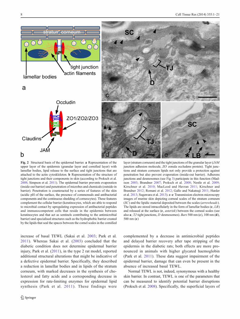
Fig. 2 Structural basis of the epidermal barrier. a Representation of theupper layer of the epidermis (granular layer and cornified layer) withlamellar bodies, lipid release to the surface and tight junctions that areattached to the actin cytoskleleton. b Representation of the structure oftight junctions and their components in skin (according to Proksch et al.2008; Simpson et al. 2011). The epidermal barrier prevents evaporation(inside-out barrier) and penetration of microbes and chemicals (outside-inbarrier). Penetration is counteracted by a series of features of the skin(acidic pH of the surface, the presence of commensals and antibacterialcomponents and the continuous shedding of corneocytes). These featurescomplement the cellular barrier (keratinocytes, which are able to respondto microbial contact by upregulating expression of antibacterial peptidesand immunocompetent cells that reside in the epidermis betweenkeratinocytes and that act as sentinels contributing to the antimicrobialbarrier) and specialised structures such as the hydrophobic barrier createdby the lipids that seal the spaces between the cornel scales in the cornified
layer (stratum corneum) and the tight junctions of the granular layer (JAMjunction adhesion molecule, ZO zonula occludens protein). Tight junc-tions and stratum corneum lipids not only provide a protection againstpenetration but also prevent evaporation (inside-out barrier). Adherensjunctions and desmosomes (see Fig 3) participate in this function (Mad-ison 2003; Brandner 2007; Proksch et al. 2008; Nestle et al. 2009;Kirschner et al. 2010; MacLeod and Havran 2011; Kirschner andBrandner 2012; Romani et al. 2012; Gallo and Nakatsuji 2011; Harderet al. 2013; Sugawara et al. 2013). c–e Transmission electron microscopyimages of murine skin depicting corneal scales of the stratum corneum(SC) and the lipidic material deposited between the scales (arrowheads).The lipids are stored intracellularly in the form of lamellar bodies (e , LB)and released at the surface (e , asterisk) between the corneal scales (seealso a ; TJ tight junctions,D desmosomes). Bars 500 nm (c), 100 nm (d),500 nm (e)
8 Cell Tissue Res (2014) 355:1–21
the epidermis not only prevent evaporation but also act as abarrier to penetration and, as such, perturbation of the lattermight not be reflected in TEWL measurements (Prokschet al. 2008). Moreover, TEWL is not only dependent onbarrier disruption but also on other factors such as gender,age, location, diurnal and seasonal variations (Black et al.2000; Chilcott and Farrar 2000) and, most importantly, bloodflow (Proksch et al. 2008), which is notoriously compromisedin diabetic skin (Rendell et al. 2003; Schramm et al. 2006; Ngoet al. 2005; Dinh and Veves 2005). In other words, a healthycutaneous blood flow is a necessary driving force to promoteevaporation, which in turn is prevented or limited by theepidermal barrier. Therefore, in DM, even in the presence ofa defective epidermal barrier, increased TEWL might not bemanifest because of defective cutaneous circulation. For thisreason, the lack of increased TEWL should not be taken as anabsolute indicator for barrier function in DM.
Unaltered TEWL accompanied by structural defects in thebarrier also occurs in senescent human skin. Here, combinedalterations in sweating, cutaneous blood flow and temperaturemight affect TEWL, thus masking barrier defects, which arevisible at a structural level (Ghadially et al. 1995). Likewise,in the Filaggrin null mouse, although TEWL is unaltered, amore permissive barrier to penetration is present (Kawasakiet al. 2012).
These considerations raise two fundamental questions: (1)whether, in human diabetic skin, other possible signs of a barrierdefect might indeed exist, as is the case in the rat and (2) whethersigns of barrier damage might be masked by the defectivecutaneous microcirculation, as is the case in senescent skin.
Other important signs of barrier defect in OLETF rats in-clude reduced levels of antimicrobial peptides (Park et al.2011). In other diabetic models (db/db mouse with overt DMand HFD mouse), Taylor et al. (2011) reported defects inepidermal gamma-delta Tcells. Given that gamma-delta Tcellscarry out their antimicrobial action at the front line of theepidermal barrier (Nestle et al. 2009; MacLeod and Havran2011), defects in these cells might provide a further sign ofbarrier deficit. Finally, another important structural element ofthe epidermal barrier is the presence of functional tight junc-tions in the epidermis (Brandner 2007; Kirschner et al. 2010;Kirschner and Brandner 2012). Nothing is known about cuta-neous tight junctions in DM. The fact that these structures aregenerally associated with the prevention of excessive evapora-tion (Brandner 2007), which has been reported not to occur inDM, might be a reason for the lack of interest shown in tightjunctions in diabetic skin. However, as discussed above, unal-tered TEWL inDMmight not exclude other barrier defects and,to further complicate the scenario, the direct correlation be-tween TEWL and epidermal barrier function has beenquestioned (Chilcott et al. 2002). Additionally, claudin-1 defi-ciency in mice is detrimental not only to epidermal tight junc-tions but also to the overall organisation of the upper epidermis
(Sugawara et al. 2013). All these considerations suggest that thesituation is intricate and that an assessment of cutaneous tightjunctions in DM might add valuable elements to the overalldiscussion of the integrity of the epidermal barrier.
In summary, the functional status of the epidermalbarrier in DM still needs to be fully clarified. Whereasa consensus seems to have been reached regarding theabsence of excessive TEWL in DM, signs of barrierdamage have been reported. Elucidation of the integrityof the epidermal barrier might improve our understanding ofthe risk of infection in DM.
Stability of junctional complexes and physical integrityof the epidermis in DM
The physical integrity of the epidermis is an important pre-requisite for all skin functions. Junctional structures (Figs. 3, 4)connect keratinocytes with each other and to the basementmembrane (BM), thus ensuring integrity and stability of theepidermal layer. Diabetic patients can experience a non-inflammatory blistering condition known as bullosisdiabeticorum or diabetic bullae. These blisters occur spontane-ously, i.e., without apparent trauma, mostly on the lower limbsand the separation can be intra- or subepidermal (Lipsky et al.2000; Aye and Masson 2002; Ngo et al. 2005; Wohlrab et al.2007; Bristow 2008; Larsen et al. 2008; VanHattem et al. 2008;Behm et al. 2012). This condition has also been described in apre-diabetic state (Lopez et al. 2009). Blistering in DM isgenerally considered to be rare, self-resolving and benign,affecting only about 0.5 % of diabetic patients (Aye andMasson 2002; Ngo et al. 2005; Wohlrab et al. 2007; Bristow2008; Van Hattem et al. 2008; Ragunatha et al. 2011; Behmet al. 2012). However, despite this relatively small percentage,given the worldwide dimension of DM, diabetic bullae mightnevertheless affect two and half million people by 2030. In anycase, their rarity and harmlessness have been questioned, assevere consequences such as chronic and necrotic ulcerationshave been reported (Lipsky et al. 2000; Larsen et al. 2008).Although the aetiology of this condition is unknown, amicrovascular cause is suspected (Ngo et al. 2005). Alterna-tively, as reported by Bristow (2008), diabetic patients mightintrinsically have lower epidermal stability. Cadherin disar-ray has been reported in diabetic mice (Taylor et al. 2011).Likewise, a reduction in the gene expression of type XVIIand type IV collagen has been reported in normal skin ofyoung db/db mice at a pre-diabetic or early diabetic stage(Rodgers et al. 2006). These two collagen types are typicalcomponents of hemidesmosomes and of the BM, respective-ly (Quondamatteo 2002; Gordon and Hahn 2010; vanAgtmael and Bruckner-Tuderman 2010; Behrens et al.2012). However, no studies have examined the fine structureof the cellular junctions and of the dermal epidermal junction
Cell Tissue Res (2014) 355:1–21 9
in diabetic skin as yet. Moreover, reduced deposition inheparan sulphate proteoglycan, i.e., another fundamentalBM component (Quondamatteo 2002; Behrens et al. 2012),has been described in the epidermal BM of diabetic patientswith nephropathy (van der Pijl et al. 1998). However, itsdepletion in the dermal-epidermal junction of diabeticpatients has been reported to be more a result of thenephropathy itself rather than of the diabetic condition(van der Pijl et al. 1998). Interestingly, subepidermalblisters in diabetic patients are often associated withthe occurrence of nephropathy (Ngo et al. 2005).
Indeed, some molecular signs related to structurescontributing to epidermal stability have been described.However, whether corresponding structural defects thatpre-dispose to a less stable epidermis also occur in DMis unknown.
Keratinocyte proliferation, epidermal thicknessand keratinocyte differentiation in DM
As mentioned previously, neither OLETF rats (Park et al.2011) nor STZ-treated mice (Sakai et al. 2003) showalterations in cutaneous filaggrin in their diabetic skin.The above studies have further examined additional dif-ferentiation markers such involucrin and loricrinimmunohistochemically (Park et al. 2011) or keratin-1,-5 and -10 and loricrin by Western blotting (Sakai et al.2003). Apart from some aberrant reactivity of the anti-loricrin antibodies with small peptides in the stratumcorneum extracts of STZ-treated mice at 10 and 80 weeks(Sakai et al. 2003), neither study has found substantialalterations in such differentiation parameters (Sakai et al.2003; Park et al. 2011).
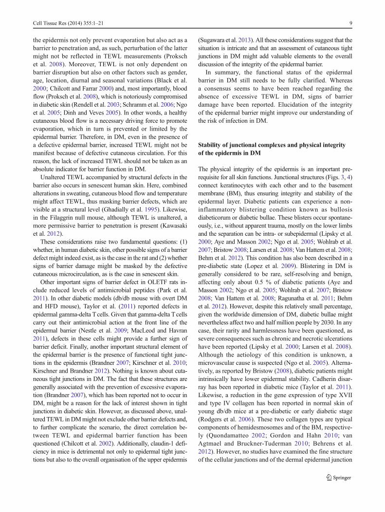
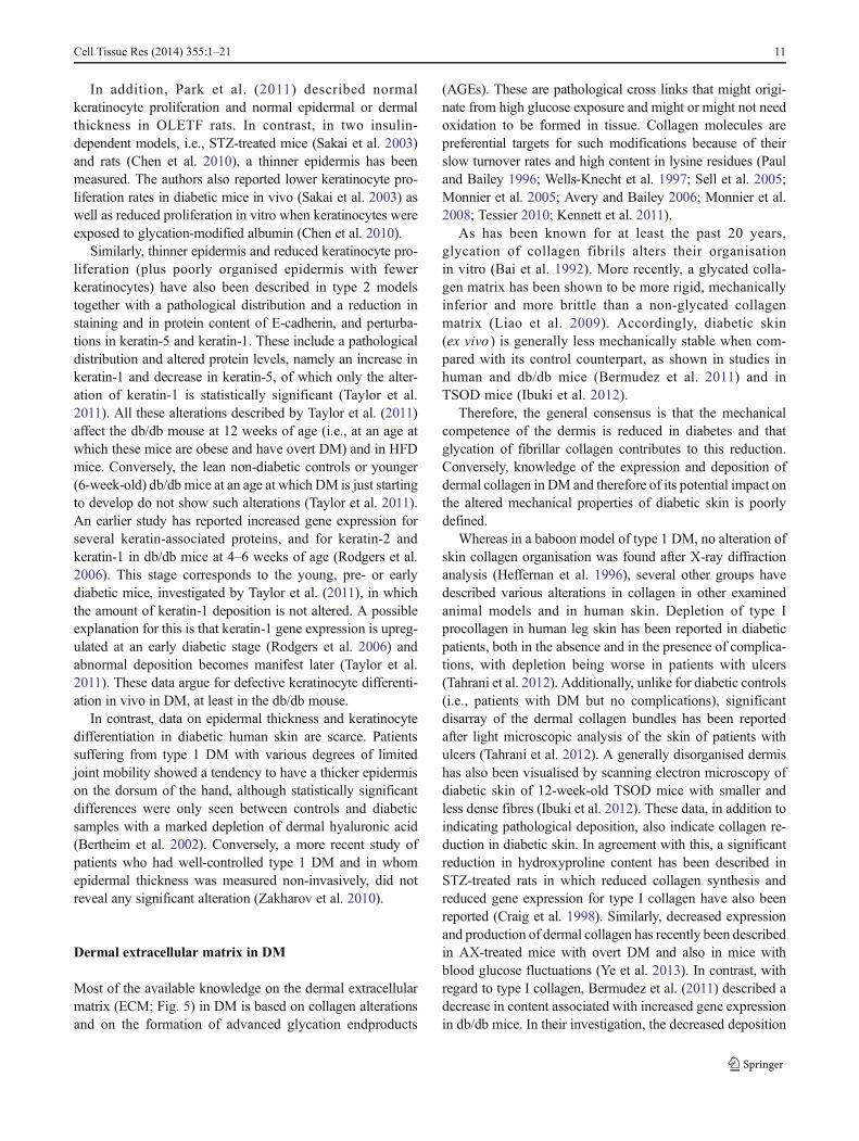
Fig. 3 Intercellular connections of the intermediate epidermal layers. aRepresentation of the intermediate epidermal layer (spinous layer). Me-chanically stable intercellular connections are primarily provided bydesmosomes linked intracellularly to the tonofilaments (grey). The cellmembrane located between desmosomes contains adherens junctions thatform a belt-like cell-cell junction connected intracellularly with the actincytoskeleton (orange dashed line) that contribute to the maintenance ofthe epidermal structure (Vasioukhin et al. 2001; Simpson et al. 2011). bRepresentation of adherent junctions (i) and desmosomes (ii) and of theirmolecular components (according to Simpson et al. 2011). c–e Electronmicrographs of murine skin showing cell-cell borders of keratinocytes (N
nuclei of keratinocytes) from the intermediate layers of murine epidermis(equivalent of the spinous layer). Desmosomes are evident (arrows) andtonofilaments (TF) converge onto them forming an inner dense plaque(d , asterisk). The darker line near the inner dense plaque is the outerdense plaque (open arrowheads). According to the current model, this isthe location at which the cytoplasmic domains of desmosomal cadherins(desmoglein and desmocollin) interact with desmoplakins via binding toplakophilin and plakoglobin (Desai et al. 2009). The portions of themembrane between two desmosomes are covered by adherent junctions(solid arrowheads). Bars 500 nm (c),100 nm (d , e)
10 Cell Tissue Res (2014) 355:1–21
In addition, Park et al. (2011) described normalkeratinocyte proliferation and normal epidermal or dermalthickness in OLETF rats. In contrast, in two insulin-dependent models, i.e., STZ-treated mice (Sakai et al. 2003)and rats (Chen et al. 2010), a thinner epidermis has beenmeasured. The authors also reported lower keratinocyte pro-liferation rates in diabetic mice in vivo (Sakai et al. 2003) aswell as reduced proliferation in vitro when keratinocytes wereexposed to glycation-modified albumin (Chen et al. 2010).
Similarly, thinner epidermis and reduced keratinocyte pro-liferation (plus poorly organised epidermis with fewerkeratinocytes) have also been described in type 2 modelstogether with a pathological distribution and a reduction instaining and in protein content of E-cadherin, and perturba-tions in keratin-5 and keratin-1. These include a pathologicaldistribution and altered protein levels, namely an increase inkeratin-1 and decrease in keratin-5, of which only the alter-ation of keratin-1 is statistically significant (Taylor et al.2011). All these alterations described by Taylor et al. (2011)affect the db/db mouse at 12 weeks of age (i.e., at an age atwhich these mice are obese and have overt DM) and in HFDmice. Conversely, the lean non-diabetic controls or younger(6-week-old) db/db mice at an age at which DM is just startingto develop do not show such alterations (Taylor et al. 2011).An earlier study has reported increased gene expression forseveral keratin-associated proteins, and for keratin-2 andkeratin-1 in db/db mice at 4–6 weeks of age (Rodgers et al.2006). This stage corresponds to the young, pre- or earlydiabetic mice, investigated by Taylor et al. (2011), in whichthe amount of keratin-1 deposition is not altered. A possibleexplanation for this is that keratin-1 gene expression is upreg-ulated at an early diabetic stage (Rodgers et al. 2006) andabnormal deposition becomes manifest later (Taylor et al.2011). These data argue for defective keratinocyte differenti-ation in vivo in DM, at least in the db/db mouse.
In contrast, data on epidermal thickness and keratinocytedifferentiation in diabetic human skin are scarce. Patientssuffering from type 1 DM with various degrees of limitedjoint mobility showed a tendency to have a thicker epidermison the dorsum of the hand, although statistically significantdifferences were only seen between controls and diabeticsamples with a marked depletion of dermal hyaluronic acid(Bertheim et al. 2002). Conversely, a more recent study ofpatients who had well-controlled type 1 DM and in whomepidermal thickness was measured non-invasively, did notreveal any significant alteration (Zakharov et al. 2010).
Dermal extracellular matrix in DM
Most of the available knowledge on the dermal extracellularmatrix (ECM; Fig. 5) in DM is based on collagen alterationsand on the formation of advanced glycation endproducts
(AGEs). These are pathological cross links that might origi-nate from high glucose exposure and might or might not needoxidation to be formed in tissue. Collagen molecules arepreferential targets for such modifications because of theirslow turnover rates and high content in lysine residues (Pauland Bailey 1996; Wells-Knecht et al. 1997; Sell et al. 2005;Monnier et al. 2005; Avery and Bailey 2006; Monnier et al.2008; Tessier 2010; Kennett et al. 2011).
As has been known for at least the past 20 years,glycation of collagen fibrils alters their organisationin vitro (Bai et al. 1992). More recently, a glycated colla-gen matrix has been shown to be more rigid, mechanicallyinferior and more brittle than a non-glycated collagenmatrix (Liao et al. 2009). Accordingly, diabetic skin(ex vivo ) is generally less mechanically stable when com-pared with its control counterpart, as shown in studies inhuman and db/db mice (Bermudez et al. 2011) and inTSOD mice (Ibuki et al. 2012).
Therefore, the general consensus is that the mechanicalcompetence of the dermis is reduced in diabetes and thatglycation of fibrillar collagen contributes to this reduction.Conversely, knowledge of the expression and deposition ofdermal collagen in DM and therefore of its potential impact onthe altered mechanical properties of diabetic skin is poorlydefined.
Whereas in a baboon model of type 1 DM, no alteration ofskin collagen organisation was found after X-ray diffractionanalysis (Heffernan et al. 1996), several other groups havedescribed various alterations in collagen in other examinedanimal models and in human skin. Depletion of type Iprocollagen in human leg skin has been reported in diabeticpatients, both in the absence and in the presence of complica-tions, with depletion being worse in patients with ulcers(Tahrani et al. 2012). Additionally, unlike for diabetic controls(i.e., patients with DM but no complications), significantdisarray of the dermal collagen bundles has been reportedafter light microscopic analysis of the skin of patients withulcers (Tahrani et al. 2012). A generally disorganised dermishas also been visualised by scanning electron microscopy ofdiabetic skin of 12-week-old TSOD mice with smaller andless dense fibres (Ibuki et al. 2012). These data, in addition toindicating pathological deposition, also indicate collagen re-duction in diabetic skin. In agreement with this, a significantreduction in hydroxyproline content has been described inSTZ-treated rats in which reduced collagen synthesis andreduced gene expression for type I collagen have also beenreported (Craig et al. 1998). Similarly, decreased expressionand production of dermal collagen has recently been describedin AX-treated mice with overt DM and also in mice withblood glucose fluctuations (Ye et al. 2013). In contrast, withregard to type I collagen, Bermudez et al. (2011) described adecrease in content associated with increased gene expressionin db/db mice. In their investigation, the decreased deposition
Cell Tissue Res (2014) 355:1–21 11
of type I collagen is also accompanied by an increased depo-sition of type III collagen in diabetic skin, resulting in areduced collagen I/III ratio (Bermudez et al. 2011). A lowerI/III collagen ratio has been associated with reduced connec-tive tissue stability, as indicated by studies on hernias (Klingeet al. 2006) and, in agreement with this, the presence of typeIII collagen has been associated with smaller collagen fibrilsin developing tendon (Birk and Mayne 1997). In this context,the finding of Bermudez et al. (2011) of the reduced collagenI/III ratio might also contribute to explaining the lower me-chanical stability of diabetic skin. This further suggests thatthe reduced mechanical competence of diabetic skin is notsolely caused by collagen glycation and AGE formation.Additionally, the finding that a decrease in type I collagencontent is associated with higher gene expression and lower
matrix proteolysis (Bermudez et al. 2011) indicates that aprimary defect in the deposition of collagen I occurs inDM. As a consequence of the reduced ECM deposition,proteolytic activity will be slowed or stopped and com-pensatory gene expression will be triggered. This sce-nario however will not apply to other studies that, inaddition to a reduction of dermal collagen, have alsodescribed lower collagen I expression (Craig et al.1998; Ye et al. 2013). Notably, the last-mentioned stud-ies have involved insulin-dependent models, whereasBermudez et al. (2011) investigated the db/db mouse,which is insulin-resistant. This raises the question as towhether differences in the homeostasis of collagensmight exist between insulin-dependent and insulin-resistant DM.
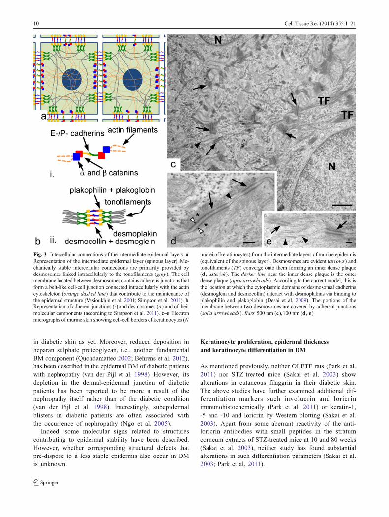
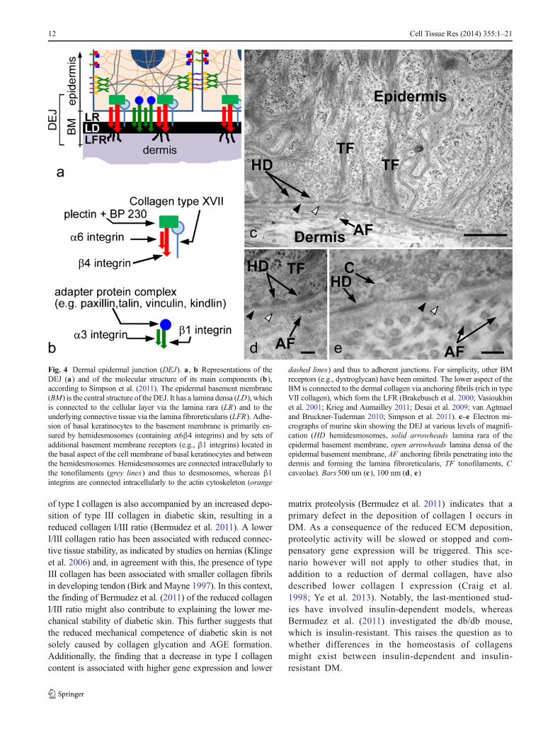
Fig. 4 Dermal epidermal junction (DEJ). a , b Representations of theDEJ (a) and of the molecular structure of its main components (b),according to Simpson et al. (2011). The epidermal basement membrane(BM) is the central structure of the DEJ. It has a lamina densa (LD), whichis connected to the cellular layer via the lamina rara (LR) and to theunderlying connective tissue via the lamina fibroreticularis (LFR). Adhe-sion of basal keratinocytes to the basement membrane is primarily en-sured by hemidesmosomes (containing α6β4 integrins) and by sets ofadditional basement membrane receptors (e.g., β1 integrins) located inthe basal aspect of the cell membrane of basal keratinocytes and betweenthe hemidesmosomes. Hemidesmosomes are connected intracellularly tothe tonofilaments (grey lines) and thus to desmosomes, whereas β1integrins are connected intracellularly to the actin cytoskeleton (orange
dashed lines) and thus to adherent junctions. For simplicity, other BMreceptors (e.g., dystroglycan) have been omitted. The lower aspect of theBM is connected to the dermal collagen via anchoring fibrils (rich in typeVII collagen), which form the LFR (Brakebusch et al. 2000; Vasioukhinet al. 2001; Krieg and Aumailley 2011; Desai et al. 2009; van Agtmaeland Bruckner-Tuderman 2010; Simpson et al. 2011). c–e Electron mi-crographs of murine skin showing the DEJ at various levels of magnifi-cation (HD hemidesmosomes, solid arrowheads lamina rara of theepidermal basement membrane, open arrowheads lamina densa of theepidermal basement membrane, AF anchoring fibrils penetrating into thedermis and forming the lamina fibroreticularis, TF tonofilaments, Ccaveolae). Bars 500 nm (c), 100 nm (d , e)
12 Cell Tissue Res (2014) 355:1–21
Bermudez et al. (2011) also described the increasedexpression of collagen III and V in the skin of the diabeticmouse. In another study on the same mouse model (db/db), Rodgers et al. (2006) noted reduced gene expressionfor various collagens, including I, III, V, VI and XIV(Rodgers et al. 2006). Notably, whereas the db/db micestudied by Rodgers et al. (2006) were aged 4–6 weeks,corresponding to an early or even pre-diabetic stage, theage group investigated by Bermudez et al. (2011), was10–14 weeks, i.e., an age when the diabetic condition isobvious. This could explain some of the differences ingene expression seen in the two laboratories.
As mentioned above, Rodgers et al. (2006) also foundreduced gene expression of type XIV collagen. This is afibril-associated collagen (Krieg and Aumailley 2011;Gordon and Hahn 2010) thought to participate in the con-trol of the size of collagen fibrils by preventing the fusionof fibrils (Gordon and Hahn 2010). Therefore, a drop in theexpression of a negative regulator of collagen fibre
thickness (such as type XIV collagen) would seem tocontradict the presence of thin fragile collagen fibres asdescribed in the diabetic TSOD mouse (Ibuki et al. 2012).This might further complicate the issue of the regulation ofcollagen deposition and expression in diabetic skin.
Taken together, although AGEs surely play a prominentrole in the collagen damage in the diabetic skin, this body ofliterature suggests that the deregulation of mechanisms oftranscription and the deposition of fibrillar collagens in thedermal matrix also contribute to the poor mechanical compe-tence of skin in DM.
The effects of AGE formation on the ECM however gobeyond alterations of a biomechanical nature. Cross links suchas glucosepane might also involve arginine residues and theselatter could be part of the RGD (arginine—glycine—asparticacid) or GFOGER (glycine—phenylanaline—hydrohyproline—glycine—glutamic acid—arginine) domains of ECM proteins(Monnier et al. 2005; Sell et al. 2005; Avery and Bailey 2006;Monnier et al. 2008). Given that such residues are known
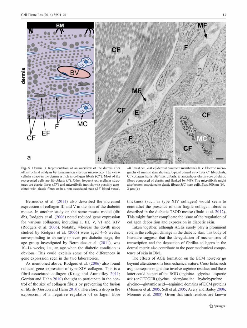
Fig. 5 Dermis. a Representation of an overview of the dermis afterultrastructural analysis by transmission electron microscopy. The extra-cellular space in the dermis is rich in collagen fibrils (CF). Most of therepresented cells are fibroblasts (F). Other frequent extracellular struc-tures are elastic fibres (EF) and microfibrils (not shown) possibly asso-ciated with elastic fibres or in a non-associated state (BV blood vessel,
MC mast cell, BM epidermal basement membrane). b , c Electron micro-graphs of murine skin showing typical dermal structures (F fibroblasts,CF collagen fibrils,MF microfibrils, E amorphous elastin core of elasticfibres composed of elastin and flanked by MF). The microfibrils mightalso be non-associated to elastic fibres (MC mast cell). Bars 500 nm (b),2 μm (c)
Cell Tissue Res (2014) 355:1–21 13
ECM protein sequences recognised by integrins (Leitingerand Hohenester 2007; Barczyk et al. 2010), their alterationmight interfere with ECM/integrin interactions and, ultimate-ly, with the biological effects of dermal ECM. This obviouslyopens a new avenue of interest in the study of the dermalmatrix in DM. A notion that has become increasingly obviousin the ECM field over the past 20 years is that the biologicalfunction of the ECM depends more on the overall mixture andinteractions of matrix components and thus on thesuprastructure of the matrix, rather than on the individualand possibly more abundant components (Bruckner 2010).Hence, molecules considered to be “minor” might be ofcrucial importance and might be determinants of differentialfunctions between two otherwise similar matrices (Bruckner2010). For a comprehensive understanding of the biologicalrole of the ECM in a given compartment (e.g., the dermis),investigations of its detailed composition are therefore funda-mental. This becomes even more relevant in the dermal ECMin diabetes, given that the diabetic condition is able to interferewith the ECM-integrins interactions and thus with the biolog-ical action of the dermal ECM. Therefore, a thorough under-standing of the dermal matrix in diabetes should deliveranswers regarding its possible pathogenetic role in skindamage in DM.
Additionally, AGEs, other than having the potential forinterfering with the ECM, possess intrinsic biological activityby acting on cellular receptors (Schmidt et al. 1992; Brett et al.1993; Fujimoto et al. 2010; Zhu et al. 2011). Thus, theglycated matrix can have an impact on dermal fibroblasts(Liao et al. 2009; Pageon 2010) and keratinocytes (Fujimotoet al. 2010; Zhu et al. 2011). Interestingly, both the studies ofFujimoto et al. (2010) and Zhu et al. (2011) refer to an AGE-dependent increase in matrix metalloproteinase-9 (MMP-9) inhuman keratinocytes, which in vivo is reported to be signifi-cantly decreased in quantity in human diabetic skin by someauthors (Bermudez et al. 2011) or unaltered in quantity butmore active by others (Tahrani et al. 2012).
In general, MMPs play a prominent role in controllingECM remodelling and, in this way, these enzymes also controlthe composition and therefore the function, of the ECM (Luet al. 2011). At least six studies have addressed MMPs indiabetic skin in vivo. In most of the cases, a higher proteolyticstatus has been described in human skin (Tahrani et al. 2012),in STZ-treated rats (Takahashi and Takasu 2011), in AX-treated mice (Ye et al. 2013) and in the subcutis of TSODmice (Ibuki et al. 2012). Ye et al. (2013) also showed that thehigher proteolytic status attributable to DM is exacerbated incases of blood glucose fluctuations (Ye et al. 2013). In con-trast, in db/db mice of a young age (early DM or pre-disease),no changes in the expression of genes relevant to ECMremodelling have been found (Rodgers et al. 2006), whereasin older db/db mice (notably diabetic), a net decrease inproteolytic activity has been reported (Bermudez et al. 2011).
Another important component of the human dermalmatrix, namely hyaluronic acid, has been studied in hu-man skin in patients affected by insulin-dependent DM. Aconsiderable reduction in hyaluronic acid, particularly inthe region of the dermal epidermal junction, has beenfound in the dermis of patients with low joint mobility,whereas in patients with little or no impairment of jointmobility, hyaluronic acid distribution predominantly re-sembles that of the normal condition (Bertheim et al.2002). Interestingly, an increase in hyaluronidase has beendescribed in the skin of STZ-treated rats (Takahashi andTakasu 2011).
Taken together, neither the regulation of collagen synthesisand production, nor matrix remodelling seems to be uniformin individual reports on diabetic skin in vivo. Additionalfactors that differentiate the DM types and the various modelsmight have an influence on collagen homeostasis.
Dermal microvessels and their BM in diabetic skin
Similar to the retina, glomeruli and endoneurium, the skin inDM is affected by microangiopathy with defective vasodila-tion which is exacerbated by neuropathy (Rendell et al. 2003;Schramm et al. 2006; Ngo et al. 2005; Dinh and Veves 2005).This can result in poor skin nutrition and is believed tocontribute to a number of cutaneous injuries, including thepoor regeneration potential of diabetic skin (Falanga 2005;Ngo et al. 2005). On a structural note, similarly to the otherdiabetic microangiopathy sites, thickened microvascular BMshave also been reported in skin (Banson and Lacy 1964;Moore and Frew 1965; Braverman and Keh-Yen 1984;Braverman et al. 1990; Dinh and Veves 2005; Ngo et al.2005; Schramm et al. 2006; Kiliçaslan and Özer 2009; Chenet al. 2010). Such microvascular alterations have been pro-posed to be the hallmark of accelerated aging of diabetic skin(Braverman and Keh-Yen 1984). At the same time, suchmicrovascular thickening is thought to be detrimental to va-sodilation, nutrient diffusion and leucocyte migration andmight account for impaired wound closure and increasedwound infection (Dinh and Veves 2005; Schramm et al.2006). Despite this concept, a closer association betweenBM thickening and microvascular dysfunction has yet tobe made. For example, nothing is known regarding pos-sible alterations in the composition of the BMs in cutane-ous microvessels, alterations that might, in turn, accountfor their dysfunction in DM. In this context, the db/dbmouse, which presents several characteristics of type 2DM, including delayed wound healing (Greenhalgh et al.1990; Kobayashi et al. 2000) and which is widely used indiabetic wound healing studies (Greenhalgh et al. 1990;Rodgers et al. 2006; Berdal et al. 2011; Bermudez et al.2011; Zhao et al. 2012), might represent a useful
14 Cell Tissue Res (2014) 355:1–21
comparison. However, the cutaneous microvascular struc-tural phenotype of this mouse model has never beenstudied. Indeed, addressing this aspect should add valu-able data in understanding the correlation between poten-tial structural and/or molecular aberrations in skinmicrovessels and poor wound healing in DM.
Another important component of the microvascularwall is represented by the pericytes. Upon injury, thesecells interact with endothelial cells, connective tissue andinfiltrating inflammatory cells and are, thus, a centralpoint of interchange of various signals during tissue repair(Dulmovits and Herman 2012). Therefore, perturbationsin the pericytes of skin microvessels might affect woundhealing potential. Pericyte loss in microvessels of diabeticskin has been reported in the literature (Dulmovits andHerman 2012) together with morphological alterationsrelated to the excessive deposition of BM material in themicrovascular wall (Braverman et al. 1990). The numberof contact points between endothelial cells and pericytesis markedly diminished in DM and this enhances thephysical separation between the two cell types. Hence,in diabetic pericytes, cell protrusions trying to make con-tact with endothelial cells are thinner and longer(Braverman et al. 1990). This renders the pericyte/endothelial interactions more difficult and might negative-ly impact on pericyte functionality, thus contributing tomicrovascular dysfunction in DM and especially inwound healing.
Reports differ regarding the density and size of cutaneousmicrovessels in DM (Algenstaedt et al. 2003; Ngo et al. 2005;Schramm et al. 2006; Aellen et al. 2012; Tellechea et al.2013). Moreover, the specific impact of hyperglycaemia onsuch parameters is not completely clear. Reduced capillarydensity has, indeed, been described both in correlation to highlevels of glycaemia and in pre-diabetes in the same mousemodel of type 2 DM (Algenstaedt et al. 2003; Schaefer et al.2010). The same studies describe a general increase in capil-lary size possibly attributable to a loss of smaller vessels(Algenstaedt et al. 2003; Schaefer et al. 2010). A recentinvestigation of skin samples of patients and of three differentanimal models of insulin-dependent DM found an increasedvessel density in all cases (Tellechea et al. 2013). Sangiorgiet al. (2010) showed vascular regression, disarray, disruptionand occlusion in microvessels of diabetic skin obtained froman amputation, i.e., most likely from poorly controlled DM. Incontrast, an absence of alterations in skin capillary densityin vivo has been reported in diabetic patients with well-controlled hypertension (Aellen et al. 2012). However, dia-betic patients with higher glycated haemoglobin have a lowermicrovascular density (Aellen et al. 2012). This supports theidea that higher glycaemia can indeed contribute to the wors-ening of the cutaneous microcirculation in DM. Interestingly,in young db/db mice (i.e., at a pre- or early diabetic stage),
gene expression of type XV collagen is increased (Rodgerset al. 2006). Collagen XV is one of the possible progenitors ofthe anti-angiogenic molecule endostatin (Sasaki et al. 2000;Gordon and Hahn 2010). Whether this plays a role in theoverall scenario of poor vascularisation is unknown. At thesame time, this and/or other similar, yet unidentified,features might contribute to the entire microvascular def-icit, and be further pathogenetic factors in addition to theaccumulation of AGE in the vascular wall. Moreover,knowledge of dermal microvascular BM in DM (beyondthickening) is scarce and the potential impact of BMalterations in compromising microvascular function re-mains theoretical. Deeper comprehension of the structureof the microvascular BM and of the composition of der-mal capillaries in DM will contribute to a better under-standing of cutaneous microangiopathy and will furtheradd to the clarification of the pathogenesis of poor woundhealing and other cutaneous manifestations caused bypoor skin nutrition.
Outlook
Given its relatively easy accessibility, both for taking smallbiopsies and for non-invasive sampling, the skin has an enor-mous diagnostic potential in providing biomarkers for DM.Skin biopsies have some limitations, because of patient recov-ery (Paliwal et al. 2013), which can be critical in DM. Despitethis, biopsy-based studies in diabetic patients can be success-fully carried out under well-controlled conditions (Tahraniet al. 2012; Tellechea et al. 2013) demonstrating the feasibilityof this approach.
Biomarkers in DM are scarce. One of the major candidatesis the non-fluorescent AGE glucosepane (Monnier et al. 2005;Sell et al. 2005). Although it has recently been associated withlong-term complications in type 1 DM (Monnier et al. 2013),an understanding of its full potential and its possible pathoge-netic role in skin damage in DM is still in its infancy. Anotherrecent study has related the depletion of cutaneousprocollagen I specifically to DM, with this beingmoremarkedin patients with foot ulcers than in non-complicated DM(Tahrani et al. 2012). As a further biopsy-based tool, the studyof skin fibroblasts is being evaluated for monitoring type 1DM (Millioni et al. 2012).
In order to lessen patient burden, especially in those pa-tients who need several daily checks, non-invasive approachesare presently under development (for details, see Kottmanet al. 2012; Sivanandam et al. 2012; Vashist 2012;Mayrovitz et al. 2013; Pleitez et al. 2013) or, as for skinautofluorescence (SAF), have been intensively evaluated(e.g., Lyons et al. 1991; Monnier et al. 1999; Maynard et al.2007; Gerrits et al. 2008a, 2008b; Stirban et al. 2008;Ghazaryan et al. 2012; Tanaka et al. 2012; Noordzij et al.
Cell Tissue Res (2014) 355:1–21 15

2012; Mascai et al. 2013). A fundamental limitation of thesemethods is the signal:noise ratio attributable to the biophysi-cal, molecular and structural characteristics of the skin(Mulder et al. 2006; Vashist 2012). SAF for example hasrecently been successfully used to mirror the diabetic state ina well-controlled patient cohort after exclusion of potentialinterfering factors, e.g., smoking or kidney failure (Mascaiet al. 2013). Otherwise, SAF appears to be an indicator forcumulative hyperglycaemic damage and a complication
predictor, rather than a specific index of the glycaemia(Lyons and Basu 2012).
Finally, the skin is notably the gateway to the body for anumber of non-invasive imaging technologies (for a review,see Daly and Leahy 2013). These depend on the passage ofsignals through the cutaneous layers and, therefore, thebiochemical/biophysical features of skin components mighttheoretically interfere with them and produce artifacts. Forexample, optical coherence tomography (OCT), which is
Fig. 6 OCT scan of a pin-prickwound of the forearm. a Pin-prickwound on the dorsal aspect of aforearm. b Structural OCT B-Scan of the non-wounded site. cStructural OCT B-Scan of thewound site. The images werekindly provided by Prof. MartinLeahy, Physics, NUI Galway
Fig. 7 Skin plexus and its visualisation by means of cm-OCT of skin atvarious levels. a–d Normal skin. e–h Pin-prick wound. This techniqueallows to image movement and to exclude the structure at the same time.Therefore, all the structural parts of the tissue, with the exception of theblood vessels (red to orange) that contain cells in movement, can becomputationally blanked out. Furthermore, it is possible to scan and slicethe tissue at various levels. a , e A three-dimensional view of the vascularplexus. The epidermis (still visible at the margin of the field) wasotherwise excluded, as was the case for the rest of the skin structures,with the exception of the vascular profile. b , f Relatively superficial slices
of the papillary region of the dermis in which the tips of theblood vessels are visible in the papillae (red /orange dots ). Theremaining images show deeper slices at the level of the superfi-cial plexus, which is located between the bottom region of thepapillary dermis and the top region of the reticular dermis. Notethe nearly horizontal course of the vessels. The slices in d , h areat approximately 200–300 μm from the level of those in b , f ,whereas those in c , d are at an intermediate level (green arrowprick wound plexus). The images were kindly provided by Prof.Martin Leahy, Physics, NUI Galway
16 Cell Tissue Res (2014) 355:1–21
based on skin penetration by optic signals and on the detectionof reflected and backscattered light (Marschall et al. 2011), hasshown that the presence of gel or glycerol, instead of air,between the light source and the skin surface improves thevisualisation of blood vessels (Liew et al. 2011). Similarresults have been obtained by using a skin phantom (Liewet al. 2011). This means that changes in the layers crossed bythe optic signals can affect the clarity of the recording from thedeeper regions (i.e., the vessels). OCT and its variants arestrongly emerging in dermatological research (Daly andLeahy 2013; Enfield et al. 2011; Zafar et al. 2013; Zam et al.2013), enabling visualisation of the epidermis with a distinctdermal epidermal junction (see Fig. 6). A variant of thisdeveloped by Martin Leahy and his group (correlation map-ping OCT, cm-OCT) enables, via a similar principle, themapping of blood vessels in the skin at various levels of tissuedepth (Enfield et al. 2011; Zafar et al. 2013; Zam et al. 2013,see also Fig. 7). The use of these non-invasive diagnostic toolswill presumably increase in the near future. Since structural/molecular changes in skin can interfere with optical signalsand pose a serious limitation to their use, this issue should beaddressed for the correct interpretation of the readings. In thecase of DM in which skin alterations are still poorly defined, aprecise understanding of the actual texture and composition ofdiabetic skin will contribute to overcome such limitations,thereby supporting and promoting the use of non-invasivediagnostics in diabetic patients.
Concluding remarks
Interest is increasing in the structure and molecular composi-tion of non-injured diabetic skin, as shown by the fact that alarge part of the available knowledge is relatively recent.Although skin alterations clearly occur in DM, the overallpicture is not uniform. A consensus seems to have beenattained regarding some features (e.g., diabetic skin is me-chanically less competent), whereas others (e.g., the epidermalbarrier) are still unclear. Underlying pathogenetic mechanismshave not been fully elucidated. Further open questions includethe impact of hyperglycaemia (severity or duration) or ofanimal models. Addressing the above issues should help toidentify potential markers or therapeutic targets for the pre-vention and monitoring of DM and of its complications.Finally, a full understanding of skin structure in DM is crucialfor the interpretation of non-invasive deep-tissue diagnosticsin which optic signals crossing skin layers might be distortedby altered tissue texture or composition.
Acknowledgments The author is grateful to Prof. Peter Dockery(Anatomy, NUI Galway, Ireland), Prof. Martin Leahy (Physics, NUIGalway, Ireland) and Prof. Cord Brakebusch (BRIC, University of Co-penhagen, Denmark) for encouragement, critical reading and helpful
discussions in the preparation of the manuscript. The author also thanksProf. Martin Leahy, in particular, for providing expertise and support innon-invasive tissue imaging and for the corresponding images shown inFigs. 6, 7. The author is grateful to Prof. Cord Brakebusch for providingthe murine tissue samples from which the micrographs shown in thispaper were taken. The human tissue was obtained from the SurgeryDepartment (University Hospital Galway), after previous ethical approvalto F.Q. and informed patient consent and with the ongoing support ofDr. Manvydas Varžgalis, Mr. Karl Sweeney and Prof. Micheal Kerinwhom the author wishes to thank. All the histological processing andimaging related to the samples shown in Figs. 1, 2, 3, 4, 5 were carried outat the Centre for Microscopy and Imaging of NUI Galway with the muchappreciated contribution of NUI Galway Anatomy staff members, post-graduates and undergraduate students to whom the author is particularlygrateful (Mr. Pierce Lalor,Mr.Mark Canney,Mr. Ian O’Brian,Ms. AlannaStanley, Ms. Kristin Kloke, Ms. Sinead Coen, Ms. Orla Hennessy, Ms.Orla Cullivan and Dr. Manvydas Varžgalis). Finally, the author wishesparticularly to thank Ms. Orla Cullivan and Ms. Orla Hennessy for theirvaluable help in proof reading and in improving the readability of themanuscript.
References
Aellen J, Dabiri A, Heim A, Liaudet L, Burnier M, Ruiz J, Feihl F,Waeber B (2012) Preserved capillary density of dorsal finger skinin treated hypertensive patients with or without type 2 diabetes.Microcirculation 19:554–562
Algenstaedt P, Schaefer C, Biermann T, Hamann A, Schwazloh B, GretenH, Ruther W, Hansen-Algenstaedt N (2003) Microvascular alter-ations in diabetic mice correlate with level of hyperglycemia.Diabetes 52:542–549
American Diabetes Association (2013) Diagnosis and classification ofdiabetes mellitus. Diabetes Care 36:S67–S74
Avery NC, Bailey AJ (2006) The effects of the Maillard reaction on thephysical properties and cell interactions of collagen. Pathol Biol 54:387–395
AyeM,Masson EA (2002) Dermatological care of the diabetic foot. Am JClin Dermatol 3:463–474
Bai P, Phua K, Hardt T, Cernadas M, Brodsky B (1992) Glycation alterscollagen fibril organisation. Connect Tissue Res 28:1–12
Banson BB, Lacy PE (1964) Diabetic microangiopathy in human toeswith emphasis on the ultrastructural change in dermal capillaries.Am J Pathol 45:41–58
Barczyk M, Carracedo S, Gullberg D (2010) Integrins. Cell Tissue Res339:269–280
Behm B, Schreml S, Landthaler M, Babilas P (2012) Skin signs indiabetes mellitus. J Eur Acad Dermatol Venereol 26:1203–1211
Behrens DT, Villone D, Koch M, Brunner G, Sorokin L, Robenek H,Bruckner-Tuderman L, Bruckner P, Hansen U (2012) The epidermalbasement membrane is a composite of separate laminin- or collagenIV-containing networks connected by aggregated perlecan, but notby nidogens. J Biol Chem 287:18700–18709
Berdal M, Appelbom HI, Eikrem JH, Lund A, Busund L, Hanes R,Seljelid R, Jenssen T (2011) Aminated β-1,3-D-glucan has a dose-dependent effect on wound healing in diabetic db/db mice. WoundRep Reg 19:579–587
Bermudez DM, Herdrich BJ, Xu J, Lind R, Beason DP, Mitchell ME,Soslowsky LJ, Liechty KW (2011) Impaired biomechanical proper-ties of diabetic skin. Am J Pathol 178:2215–2222
Bertheim U, Engstorm-Laurent A, Hofer P, Hallgren P, Asplund J,Hellstrom S (2002) Loss of hyaluronan in the basement membranezone of the skin correlates to the degree of stiff hands in diabeticpatients. Acta Derm Venereol 82:329–334
Cell Tissue Res (2014) 355:1–21 17
Birk D, Mayne R (1997) Localization of collagen types I, III andV during tendon development. Changes in collagen types Iand III are correlated with changes in fibril diameter. Eur JCell Biol 72:352–361
Black D, Del Pozo A, Lagarde JM, Gall Y (2000) Seasonal variability inthe biophysical properties of stratum corneum from different ana-tomical sites. Skin Res Technol 6:70–76
Brakebusch C, Grose R, Quondamatteo F, Ramirez A, Jorcano JJ, PirroA, Svensson M, Herken R, Sasaki T, Timpl R, Werner S, Fässler R(2000) Skin and hair follicle integrity is crucially dependent on β1integrin expression on keratinocytes. EMBO J 19:3990–4003
Brandner JM (2007) Pores in the epidermis: aquaporins and tight junc-tions. Int J Cosmet Sci 29:413–422
Braverman IM, Keh-Yen A (1984) Ultrastructural abnormalities of themicrovasculature and elastic fibres in the skin of juvenile diabetics. JInvest Dermatol 82:270–274
Braverman IM, Sibley J, Keh-YenA (1990)Ultrastructural analysis of theendothelial-pericyte relationships in diabetic cutaneous vessels. JInvest Dermatol 95:147–153
Brett J, Schmidt AM, Yan SD, Weidman E, Pinsky D, Nowygrod R,NeeperM, Przysiecki C, ShawA,Migheli A, Stern D (1993) Surveyof the distribution of a newly characterised receptor for advancedglycation end products in tissues. Am J Pathol 143:1699–1712
Bristow I (2008) Non-ulcerative skin pathologies of diabetic foot.Diabetes Metab Res Rev 24:S84–S89
Bruckner P (2010) Suprastructures of extracellular matrices: paradigms offunctions controlled by aggregates rather than molecules. CellTissue Res 339:7–18
Buckley CM, O’Farrell A, Canavan RJ, Lynch AD, De La Harpe DV,Bradley CP, Perry IJ (2012) Trends in the incidence of lowerextremity amputations in people with and without diabetes overfive-year period in the Republic of Ireland. PLOS ONE 7:e41492
Chatzizgeorgiou A, Halapas A, Kalafatakis K, Kamper E (2009) The useof animal models in the study of diabetes mellitus. In Vivo 23:245–258
Chen X, Lin W, Lu S, Xie T, Kui G, Shi Y, Zou J, Liu Z, Liao W (2010)Mechanisitic study of endogenous skin lesions in diabetic rats. ExpDermatol 19:1088–1095
Chilcott RP, Farrar R (2000) Biophysical measurements of human fore-arm skin in vivo: effects of site, gender, chirality, and time. Skin ResTechnol 6:64–69
Chilcott RP, Dalton CH, Emmanuel AJ, Allen CE, Bradley ST (2002)Transepidermal water loss does not correlate with skin barrier func-tion in vitro. J Invest Dermatol 118:871–875
Craig RG, Yu Z, Xu L, Barr R, Ramamurthy N, Boland J, Schneir M,Golub LM (1998) A chemically modified tetracycline inhibitsstreptozotocin-induced diabetic depression of collagen synthesisand steady-state type I procollagen mRNA. Biochim Biophys Acta1402:250–260
Daly SM, LeahyMJ (2013) “Go with the flow”: a review of methods andadvances in blood flow imaging. J Biophotonics 6:217–255
Desai BV, Harmon RM, Green KJ (2009) Desmosomes at a glance. J CellSci 122:4401–4407
Dinh TL, Veves A (2005) A review of the mechanisms implicated in thepathogenesis of the diabetic foot. Int J Low ExtremWounds 4:154–159
Dulmovits BM, Herman IM (2012) Microvascular remodelling andwound healing: a role for pericytes. Int J Biochem Cell Biol 44:1800–1812
Eaglstein WH, Callen JP (2009) Dermatologic comorbidities of diabetesmellitus and related issues. Arch Dermatol 145:467–469
Enfield J, Jonathan E, Leahy M (2011) In vivo imaging of the microcir-culation of the volar forearm using correlation mapping opticalcoherence tomography (cmOCT). Biomed Opt Exp 2:1184–1193
Falanga V (2005) Wound healing and its impairment in the diabetic foot.Lancet 366:1736–1743
Fujimoto E, Kobayashi T, Fujimoto N, Akiyama M, Tajima S, Nagai R(2010) AGE-modified collagens I and III induce keratinocyte ter-minal differentiation through AGE receptor CD36: epidermal-dermal interaction in acquired perforating dermatosis. J InvestDermatol 130:405–414
Gallo RL, Nakatsuji T (2011) Microbial symbiosis with the innate de-fense system of the skin. J Invest Dermatol 131:1974–1980
Geerlings SE, Stolk RP, Camps MJL, Netten PM, Hoekstra JBL, BouterKP, Bravenboer B, Collet JT, Jansz AR, Hoepelman ALM (2000)Asymptomatic bacteriuria may be considered a complication inwomen with diabetes. Diabetes Care 23:744–749
Gerrits EG, Lutgers HL, Kleefstra N, Graaff R, Groenier KH, Smit AJ,Gans RO, Bilo HJ (2008a) Skin autofluorescence: a tool to identifytype 2 diabetic patients at risk for developing microvascular com-plications. Diabetes Care 31:517–520
Gerrits EG, Lutgers HL, Kleefstra N, Groenier KH, Smit AJ, Gans RO,Bilo HJG (2008b) Skin advanced glycation end product ac-cumulation is poorly reflected by glycemic control in type 2diabetic patients (ZODIAC-9). J Diabetes Sci Technol 2:572–577
Ghadially R, Brown BE, Sequiera-Martin SM, Feingold K, Elias PM(1995) The aged epidermal permeability barrier: structural, func-tional, and lipid biochemical abnormalities in human and a senes-cent murine model. J Clin Invest 95:2281–2290
Ghazaryan AA, Hu P-S, Chen S-J, Tan H-Y, Dong C-Y (2012)Spatial and temporal analysis of skin glycation by the use ofmultiphoton microscopy and spectroscopy. J Dermatol Sci65:189–195
Gordon MK, Hahn RA (2010) Collagens. Cell Tissue Res 339:247–257Greenhalgh DG, Sprugel KH,MurrayMJ, Ross R (1990) PDGF and FGF
stimulate wound healing in the genetically diabetic mouse. Am JPathol 136:1235–1246
Hamilton E, Martin N, Makepeace A, Sillars BA, Davis WA, Davis TME(2013) Incidence and predictors of hospitalization for bacterialinfection in community-based patients with type 2 diabetes: theFreemantle diabetes study. PLOS ONE 8:e60502
Harder J, Schröder J-M, Gläser R (2013) The skin surface as antimicro-bial barrier: present concepts and future outlooks. Exp Dermatol 22:1–5
Heffernan S, James V, Zilkens R, Kirwan P, Birrell A, McLennan S,Hennessy A, Gillin A, Horvath J, Tiller D, Yue D, Turtle J(1996) Changes of extracellular matrix in a baboon (Papiohadryas ) model of insulin dependent diabetes: studies usingelectron microscopy and X-ray diffraction techniques.Diabetes Res Clin Pract 34:65–72
Hirayama I, Yi Z, Izumi S, Arai I, Suzuki W, Nagamachi Y, Kuwano H,Takeuchi, Izumi T (1999) Genetic analysis of obese diabetes in theTSOD mouse. Diabetes 48:1183–1191
Ibuki A, Akase T, Nagase T, Minematsu T, Nakagami G, Horii M, SagaraH, Komeda T, Kobayashi M, Shimada T, Aburada M, Yoshimura K,Sugama J, Sanada H (2012) Skin fragility on obese mice: possibleinvolvement of elevated oxidative stress upregulation of matrixmetalloproteinases. Exp Dermatol 21:178–183
Iizuka S, Suzuki W, Tabuchi M, Nagata M, Imamura S, Kobayashi Y,Kanitani M, Yanagisawa T, Kase Y, Takeda S, Aburada M,Takahashi KW (2005) Diabetic complications in a new animalmodel (TSOD mouse) of spontaneous NIDDM with obesity. ExpAnim 54:71–83
Jackson LA (2005) Evaluating diabetes mellitus as a risk factor forcommunity-acquired infections. Clin Infect Dis 41:289–290
Kawasaki H, Nagao K, Kubo A, Hata T, Shimizu A, Mizuno H, YamadaT, Amagai M (2012) Altered stratum corneum barrier and enhancedpercutaneous immune response in filaggrin-null mice. J Allergy ClinImmunol 129:1538–1546
Kennett EC, Chuang CY, Degendorfer G, Whitekock JM, DaviesMJ (2011) Mechanisms and consequences of oxidative
18 Cell Tissue Res (2014) 355:1–21
damage to extracellular matrix. Biochem Soc Trans 39:1279–1287
Kiliçaslan SMS, Özer C (2009) Effects of benfluorex—vitamin C sup-plementation on cutaneous capillaries of diabetic rats. Cell Biol Int33:442–446
King AJ (2012) The use of animal models in diabetes research. Br JPharm 166:877–894
Kirschner N, Brandner JM (2012) Barriers and more: functions of tightjunction proteins in the skin. Ann N YAcad Sci 1257:158–166
Kirschner N, Houdek P, Fromm M, Moll I, Brandner JM (2010) Tightjunctions form a barrier in human epidermis. Eur J Cell Biol 89:839–842
Klinge U, Binnebösel M, Mertens PR (2006) Are collagens the culprit inthe development of incisional and inguinal hernia diseases? Hernia10:472–477
Knapp S (2012) Diabetes and infection: is there a link? A mini review.Gerontology 26:1–6
Kobayashi K, Forte TM, Taniguchi S, Ishida B, Oka K, Chan L (2000) Thedb/db a model for diabetic dyslipidemia: molecular charectarisationand effects of western diet feeding. Metabolism 49:22–31
Koivukangas V, Annala AP, Salmela PI, Oikarinet A (1999) Delayedrestoration of epidermal barrier function after blister injury in pa-tients with diabetes mellitus. Diabet Med 16:563–567
Kottman J, Rey JM, Luginbuhl J, Reichmann E, Sigrist MW (2012)Glucose sensing in human epidermis using mid-infrared photo-acoustic detection. Biomed Opt Expr 3:667–680
Krieg T, Aumailley M (2011) The extracellular matrix of the dermis:flexible structures with dynamic functions. Exp Dermatol 20:689–695
KudoT, Shimada T, Toda T, Igeta S, SuzukiW, Ikarashi N, OchiaiW, ItoaK, Aburada M, Sugiyama K (2009) Altered expression of CYP inTSODmice: a model of type 2 diabetes and obesity. Xenobiotica 39:889–902
Lam DW, LeRoith D (2012) The worldwide diabetes epidemic. CurrOpin Endocrinol Diabetes Obes 19:93–96
Larsen K, Jensen T, Karlsmark T, Holstein PE (2008) Incidence ofbullosis diabeticorum—a controversial cause of chronic foot ulcer-ation. Int Wound J 5:591–596
Leitinger B, Hohenester E (2007) Mammalian collagen receptors. MatrixBiol 26:146–155
Liao H, Zakhaleva J, Chen W (2009) Cells and tissue interactions withglycated collagen and their relevance to delayed diabetic woundhealing. Biomaterials 30:1689–1696
Liew YM, McLaughlin RA, Wood FM, Sampson DD (2011) Reductionof image artifacts in three-dimensional optical coherence tomogra-phy of skin in vivo. J Biomed Opt 16:116018
Lipsky BA, Baker PD, Ahroni JH (2000) Diabetic bullae: 12 cases ofpurportedly rare cutaneous disorder. Int J Dermatol 39:196–200
Lipsky BA, Itani KMF, Weigelt JA, Joseph W, Paap CM, Reisman A,Myers DE, Huang DB (2011) The role of diabetes mellitus in thetreatment of skin and skin structure infections caused bymethicillin-resistant Saphylococcus aureus : results from three randomised con-trolled trials. Int J Infect Dis 15:e140–e146
Lopez PR, Leicht S, Sigmon JR, Stigall L (2009) Bullosis diabeticorumassociated with a prediabetic state. South Med J 102:643–644
Lowell BB, S-Susulic V, Hamann A, Lawitts JA, Himms-Hagen J, BoyerBB, Kozak LP, Flier JS (1993) Development of obesity in transgenicmice after genetic ablation of brown adipose tissue. Nature 366:740–742
Lu P, Takai K, Weaver VM, Werb Z (2011) Extracellular matrix degra-dation and remodeling in development and disease. Cold SpringHarb Perspect Biol 3:pii: a005058
Lyons TJ, Basu A (2012) Biomarkers in diabetes: hemoglobin A1c,vascular and tissue markers. Transl Res 159:303–312
Lyons TJ, Bailie KE, Dyer DG, Dunn JA, Baynes JW (1991) Decrease inskin collagen glycation with improved glycemic control in patients
with insulin-dependent diabetes mellitus. J Clin Invest 87:1910–1915
MacLeod AS, Havran W (2011) Functions of skin resident γδ T cells.Cell Mol Life Sci 68:2399–2408
Mascai E, Takats Z, Derzbach L, Korner A, Vasarhelyli (2013)Verification of skin autofluorescence values by mass spectrometryin adolescents with type 1 diabetes: brief report. Diabetes TechnolTher 15:1–4
Madison KC (2003) Barrier function of the skin: “La raison d’etre” of theepidermis. J Invest Dermatol 121:231–241
Marschall S, Sander B, Mogensen M, Jørgensen TM, Andersen PE(2011) Optical coherence tomography—current technology and ap-plications in clinical and biomedical research. Anal Bioanal Chem400:2699–2720
Maynard JD, Rohrscheib M, Way JF, Nguyen CM, Ediger MN (2007)Non-invasive type 2 diabetes screening. Superior sensitivity tofasting plasma glucose and A1C. Diabetes Care 30:1120–1124
Mayrovitz HN, McClymont A, Pandya N (2013) Skin tissue waterassessed via tissue dielectric constant measurements in personswith and without diabetes mellitus. Diabetes Technol Ther 15:1–6
McAleer MA, Irvine AD (2013) The multifunctional role of filaggrin inallergic skin disease. J Allergy Clin Immunol 131:280–291
Millioni R, Puricelli L, Iori E, Trevisan R, Tessari P (2012) Skin fibro-blasts as a tool for identifying the risk of nephropathy in the type 1diabetic population. Diabetes Metab Res Rev 28:62–70
Miura T, Suzuki W, Ishihara E, Arai I, Ishida H, Seino Y, Tanigawa K(2001) Impairment of insulin-stimulated GLUT4 translocation inskeletal muscle and adipose tissue in the Tsumura Suzuki obesediabeticmouse: a new genetic animalmodel of type 2 diabetes. Eur JEndocrinol 145:785–790
Miyata S, Yamada N, Kawada T (2012) Possible involvement of hypo-thalamic nucleobindin-2 in hyperphagic feeding in Tsumura-Suzukiobese diabetes mice. Biol Pharmacol Bull 35:1784–1793
Monnier VM, Bautista O, Kenny D, Sell DR, Fogarty J, Dahms W,Cleary PA, Lachin J, Genuth S (1999) Skin collagen glycation,glycoxidation, and crosslinking are lower in subjects with long-term intensive versus conventional therapy of type 1 diabetes.Diabetes 48:870–880
Monnier VM,Mustata GT, Biemel KL, Reihl O, Lederer MO, Zhenyu D,Sell DR (2005) Cross-linking of the extracellular matrix by theMaillard reaction in aging and diabetes. An update on “a puzzlenearing resolution”. Ann N YAcad Sci 1043:533–544
Monnier VM, Sell DR, Dai Z, Nemet I, Collard F, Zhang J (2008) Therole of the Amadori product in the complications of diabetes. Ann NYAcad Sci 1126:81–88
Monnier VM, Sell DR, Strauch C, SunW, Lachin JM, Cleary PA, GenuthS (2013) The association between skin collagen glucosepane andpast progression of microvascular and neuropathic complications intype 1 diabetes. J Diabetes Complications 27:141–149
Moore JM, Frew IDO (1965) Peripheral vascular lesion in diabetesmellitus. BMJ 2:19–23
Mulder DJ, Water T, Lutgers HL, Graaff R, Gans RO, Zijlistra F, Smit AJ(2006) Skin autofluorescence, a novel marker for glycemic andoxidative stress-derived advanced glycation endproducts: an over-view of current clinical studies, evidence, and limitations. DiabetesTechnol Ther 8:523–535
Muller LMAJ, Gorter KJ, Goudzwaard WL, Schellevis FG,Hoepelma IM (2005) Increased risk of common infectionsin patients with type1 and type2 diabetes mellitus. Clin InfectDis 41:281–288
Nestle FO, Di Meglio P, Qin J-Z, Nickoloff B (2009) Skin immunesentinels in health and disease. Nat Rev Immunol 9:679–691
Ngo BT, Hayes KD, DiMiao DJ, Srinivasan SK, Huerter CJ, Rendell MS(2005) Manifestations of cutaneous diabetic microangiopathy. Am JClin Dermatol 6:225–237
Cell Tissue Res (2014) 355:1–21 19
Noordzij MJ, Mulder DJ, Oomen PHN, Brouwer T, Jager J, CastroCabezas M, Lefrand JD, Smit AJ (2012) Skin autofluorescenceand risks of micro- and macrovascular complications in patientswith type 2 diabetes mellitus—a multicentre study. Diabet Med29:1556–1561
O’Shea M, Teeling M, Bennett K (2013) The prevalence and ingredientcost of chronic comorbidity in the Irish elderly population withmedication treated type 2 diabetes: a retrospective cross sectionalstudy using a national pharmacy claims database. BMC Health ServRes 13:23
PageonH (2010) Reaction of glycation and human skin: the effects on theskin and its components, reconstructed skin as model. Pathol Biol58:226–231
Paliwal S, Hwang BH, Tsai KY, Mitragotri S (2013) Diagnostic oppor-tunities based on skin biomarkers. Eur J Pharm Sci 50:546-556
Park HY, Kim HJ, Jung M, Chung CH, Hasham R, Park CS, Choi EH(2011) A long-standing hyperglycaemic condition impairs skinbarrier by accelerating skin ageing process. Exp Dermatol 20:969–974
Paul RG, Bailey AJ (1996) Glycation of collagen: the basis of its centralrole in the late complications of ageing and diabetes. Int J BiochemCell Biol 28:1297–1310
Pleitez MA, Lieblein T, Bauer A, Hertzberg O, von Lilienfeld-Toal H,Mäntele W (2013) In vivo noninvasive monitoring of glucoseconcentration in human epidermis by mid-infrared pulsedphotoaucstic spectroscopy. Anal Chem 15:1013–1020
Proksch E, Brandner JM, Jensen J-M (2008) The skin: an indispensablebarrier. Exp Dermatol 17:1063–1072
Quondamatteo F (2002) Assembly, stability and integrity of basementmembranes in vivo. Histochem J 34:369–381
Ragunatha S, Anitha B, Inamadar AC, Palit A, Devarmani SS (2011)Cutaneous disorders in 500 diabetic patients attending diabeticclinic. Indian J Dermatol 56:160–164
Redel H, Gao Z, Aleksyenko AV, Zhou Y, Perez-Perez GI, Weinstock G,Sodegren E, Blaser MJ (2013) Quantitation and composition ofcutaneous microbiota in diabetic and non-diabetic men. J InfectDis 207:1105–1114
ReesDA, Alcolado JC (2005)Animal models of diabetes mellitus. DiabetMed 22:359–370
Rendell M, Saxena S, Shah D (2003) Cutaneous blood flow and periph-eral resistance in type II diabetes compared to intermittent claudica-tion patients. Int J Angiol 12:166–171
Rodgers KE, Ellefson DD, Espinosa T, Hsu Y, Di Zerega G, Mehiran-Shai R (2006) Expression of intracellular filament, collagen, andcollagenase genes in diabetic and normal skin after injury. WoundRepair Regen 14:298–305
Romani N, Brunner PM, Stingl G (2012) Changing views of the role ofLangerhans cells. J Invest Dermatol 132:872–881
Sakai S, Endo Y, Ozawa N, Sugawara T, Kusaka A, Sayo T, Tagami H,Inoue S (2003) Characteristics of the epidermis and stratumcorneum of hairless mice with experimentally induced diabetesmellitus. J Invest Dermatol 120:79–85
Sakai S, Kikuchi K, Satoh J, Tagami H, Inoue S (2005) Functionalproperties of stratum corneum in patients with diabetes mellitus:similarities to senile xerosis. Br J Dermatol 153:319–323
Sangiorgi S,Manelli A, ReguzzoniM, RongaM, Protasoni M, Dell’OrboC (2010) The cutaneous microvascular architecture of human dia-betic toe studied by corrosion casting and scanning electron micros-copy analysis. Anat Rec 293:1639–1645
Sasaki T, Larsson H, Tisi D, Claesson-Welsh L, Hohenester E,Timpl R (2000) Endostatins derived from collagens XV andXVIII differ in structural and binding properties, tissue dis-tribution and anti-angionenic activity. J Mol Biol 01:1179–1190
Schaefer C, Biermann T, SchroederM, Fuhrhop I, Niemeier A, RutherW,Algenstaedt P, Hansen-Algenstaedt N (2010) Early microvascular
complications of prediabetes in mice with impaired glucose toler-ance in dyslipidemia. Acta Diabetol 47:S19–S27
Schmidt AM, Vianna M, Gerlach M, Brett J, Ryan J, Kao J, Esposito C,Hegarty H, Hurley W, Clauss M, Wang F, Pan Y-CE, Tsang TC,Stern D (1992) Isolation and characterization of two binding pro-teins for advanced glycosylation end products from bovine lungwhich are present on the endothelial cell surface. J Biol Chem267:14987–14997
Schramm JC, Dinh T, Veves A (2006) Microvascular changes in thediabetic foot. Int J Low Extrem Wounds 5:149–159
Seirafi H, Farsinejad K, Firooz A, Davoudi SM, Robati RM, Hoseini MS,Ehasani AH, Sadr B (2009) Biophysical characteristics of skin indiabetes: a controlled study. J Eur Acad Dermatol Venereol 23:146–149
Sell DR, Biemel KM, Reihl O, Lederer MO, Strauch CM, Monnier VM(2005) Glucosepane is a major protein cross-link of the senescenthuman extracellular matrix. J Biol Chem 280:12310–12315
Shah BR, Hux JE (2003) Quantifying the risk of infectious diseases forpeople with diabetes. Diabetes Care 26:510–513
Simpson CL, Patel DM, Green KJ (2011) Deconstructing the skin:cytoarchitectural determinants of epidermal morphogenesis. NatRev Mol Cell Biol 12:565–580
Sivanandam S, Anburajan M, Venkartraman B, Menaka M, Sharath D(2012) Medical thermography: a diagnostic approach for type dia-betes based on non-contact infrared thermal imaging. Endocrine 42:343–351
Stirban A, Nandrean S, Negrean M, Koschinsky T, Tschoepe D (2008)Skin autofluorescence increases postprandially in human subjects.Diabetes Technol Ther 10:200–205
Suaya JA, Eisenberg DF, Fang C, Miller LG (2013) Skin and soft tissueinfections and associated complications among commercially in-sured patients aged 0–64 years with and without diabetes in theUS. PLOS ONE 8:e60057
Sugawara T, Iwamoto N, Akashi M, Kojima T, Hisatsune J, Sugai M,Furuse M (2013) Tight junction dysfunction in the stratumgranulosum leads to aberrant stratum corneum barrier function inclaudin-1 deficient mice. J Dermatol Sci 70:12–18
Suzuki W, Iizuka S, Tabuchi M, Funo S, Yanagisava T, Kimura M, SatoT, Endo T, Kawamura H (1999) A new mouse of spontaneousdiabetes derived from ddY strain. Exp Anim 48:181–189
Tahrani AA, ZengW, Sakher J, PiyaMK, Hughes S, DubbK, StevensMJ(2012) Cutaneous structural and biochemical correlates of footcomplications in high-risk diabetes. Diabetes Care 35:1913–1918
Takahashi N, Takasu S (2011) A close relationship between type 1diabetes and vitamin A deficiency and matrix metalloproteinaseand hyaluronidase activities in skin tissues. Exp Dermatol 20:899–904
Tanaka K, Tani Y, Asai J, Nemoto F, Kusano Y, Suzuki H, Hayshi Y,Asahi K, Nakayama M, Miyata T, Watanabe T (2012) Skin autoflu-orescence is associated with severity of vascular complications inJapanese patients with type 2 diabetes. Diabet Med 29:492–500
Taylor KR, Costanzo AE, Jameson JM (2011) Dysfunctional γδ T cellscontribute to impaired keratinocyte homeostasis in mouse models ofobesity. J Invest Dermatol 131:2409–2418
Tellechea A, Kafanas A, Leal EC, Tecilazich F, Kuchibhotla S, AusterME, Kontoes I, Paolino J, Carvalho E, Nabzdyk LP, Veves A (2013)Increased skin inflammation and blood vessel density in human andexperimental diabetes. Int J Low Extrem Wounds 12:4–11
Tessier FJ (2010) The Maillard reaction in the human body. The maindiscoveries and factors that affect glycation. Pathol Biol 58:214–219
Thyssen JP, Linneberg A, Carlsen BC, Johansen JD, Engkilde K, HansenT, Pociot F, Pedersen O,MeldgaardM, Scecsi PB, Stender S,MenneT (2011) A possible association between a dysfunctional skin barrier(filaggrin null-mutation status) and diabetes: a cross-sectional study.BMJ Open 1:e000062
Van Agtmael T, Bruckner-Tuderman L (2010) Basement membranes andhuman disease. Cell Tissue Res 339:167–188
20 Cell Tissue Res (2014) 355:1–21
Van der Pijl JW, Daha MR, Born J, Verhagen NAM, Lemkes HHPJ,Bucala R, Berden JHM, Zwinderman AH, Bruijn JA, van Es LA,van der Woude F (1998) Extracellular matrix in human diabeticnephropathy: reduced expression of heparan sulphate in skin base-ment membrane. Diabetologia 41:791–798
Van Hattem S, Bootsma AH, Thio BT (2008) Skin manifestations ofdiabetes. Cleve Clin J Med 75:772–787
Vashist SK (2012) Non-invasive glucose monitoring technology in dia-betes management: a review. Anal Chim Acta 750:16–27
Vasioukhin V, Bauer C, Degenstein L, Wise B, Fuchs E (2001)Hyperproliferation and defects in epithelial polarity upon condition-al ablation of α-catenin in skin. Cell 104:605–617
Von Mering J, Minkowski O (1890) Diabetes mellitus nachPankreasexstirpation. Arch Exp Pathol Pharmakol 26:371–387
Wanke I, Skabytska Y, Kraft B, Peschel A, Bidermann T, Schittek B (2013)Staphylococcus aureus skin colonization is promoted by barrier dis-ruption and leads to local inflammation. Exp Dermatol 22:153–155
Wells-Knecht MC, Lyons TJ, McCance DR, Thorpe SR, Baynes JW(1997) Age-dependent increase in ortho-thyrosine and methioninesulfoxide in human skin collagen is not accelerated in diabetes. JClin Invest 100:839–846
Wohlrab J, Wohlrab D, Meiss F (2007) Skin diseases in diabetes mellitus.J Dtsch Dermatol Ges 5:37–53
YeX, ChengX, Liu L, Zhao D, Dong Y (2013) Blood glucose fluctuationaffects skin collagen metabolism in the diabetic mouse by inhibiting
the mitogen-activated protein kinase and Smad pathways. Clin ExpDermatol 38:530–538
Zakharov P, Talary MS, Kolm I, Caduff A (2010) Full-field opticalcoherence tomography for the rapid estimation of epidermal thick-ness: study of patients with diabetes mellitus type 1. Physiol Meas31:193–205
Zafar H, Enfield J, O’Connell M-L, Ramsay B, Lynch M, Leahy MJ(2013) Assessment of psoriatic plaque in vivo with correlationmapping optical coherence tomography. Skin Res Technol. doi:10.1111/srt.12096
Zam A, Dsouza R, Hrebesh M, Subhash HM, Marie-LouiseO’Connell M-L, Enfield J, Larin K, Leahy MJ (2013)Feasibility of correlation mapping optical coherence tomog-raphy (cmOCT) for anti-spoof sub-surface fingerprinting. JBiophotonics 6:663–667
Zhao G, Usui M, Underwood RA, Singh PK, James GA, Stewart PS,Fleckman P, Olerud JE (2012) Time course study of delayed woundhealing in a biofilm-challenged diabetic mouse model. WoundRepair Regen 20:342–352
Zhu P, Ren M, Yang C, Hu YX, Ran J, Li Y (2011) Involment ofRAGE, MAPK and NF-κB pathways in AGEs-inducedMMP-9 activation in HaCaT keratinocytes. Exp Dermatol21:123–129
Zimmet P, Alberti KGMM, Shaw J (2001) Global and societal implica-tions of the diabetes epidemic. Nature 414:782–787
Cell Tissue Res (2014) 355:1–21 21