Embed Size (px)
Citation preview
Skeletal System
It’s all about the bones!!!

The Skeletal System in Action !!
►The Skeletal System in Action!
► https://www.youtube.com/watch?v=ICwLlrQKVcg&list=PLZile25upgEBVRu0JnePPCABH0fhkTgTQ
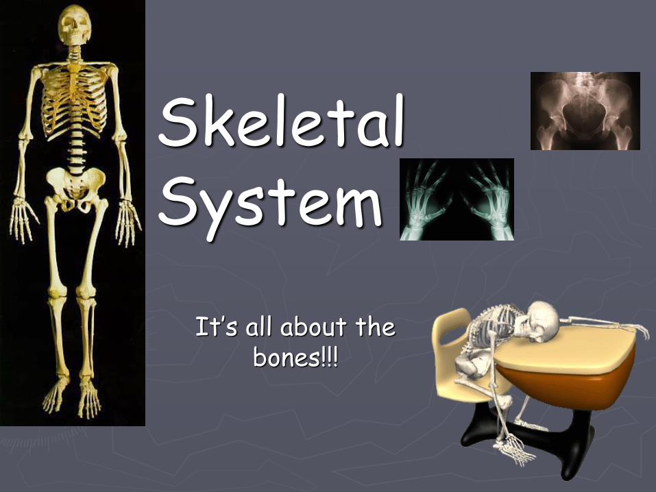
1. FYI 5 Cool Facts about the Skeletal System1. 20% of your body weight is bone
■ Do the math■ (Your body weight) X .20= the weight of
your bones
2. There are 30 bones in your skull3. You have 206 bones
■ More than ½ of these are in your hands and feet
4. Your largest bone is your femur and your smallest bone is in your Ear!
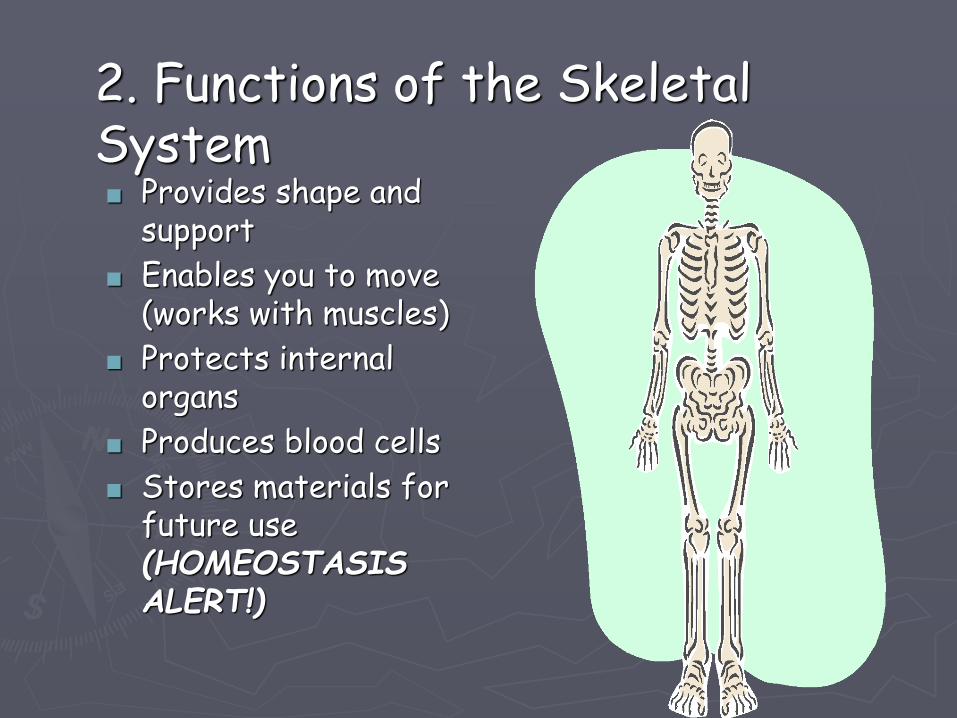
2. Functions of the Skeletal System■ Provides shape and
support
■ Enables you to move (works with muscles)
■ Protects internal organs
■ Produces blood cells
■ Stores materials for future use (HOMEOSTASIS ALERT!)
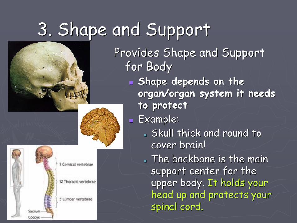
3. Shape and SupportProvides Shape and Support
for Body■ Shape depends on the
organ/organ system it needs to protect
■ Example:
■ Skull thick and round to cover brain!
■ The backbone is the main support center for the upper body. It holds your head up and protects your spinal cord.
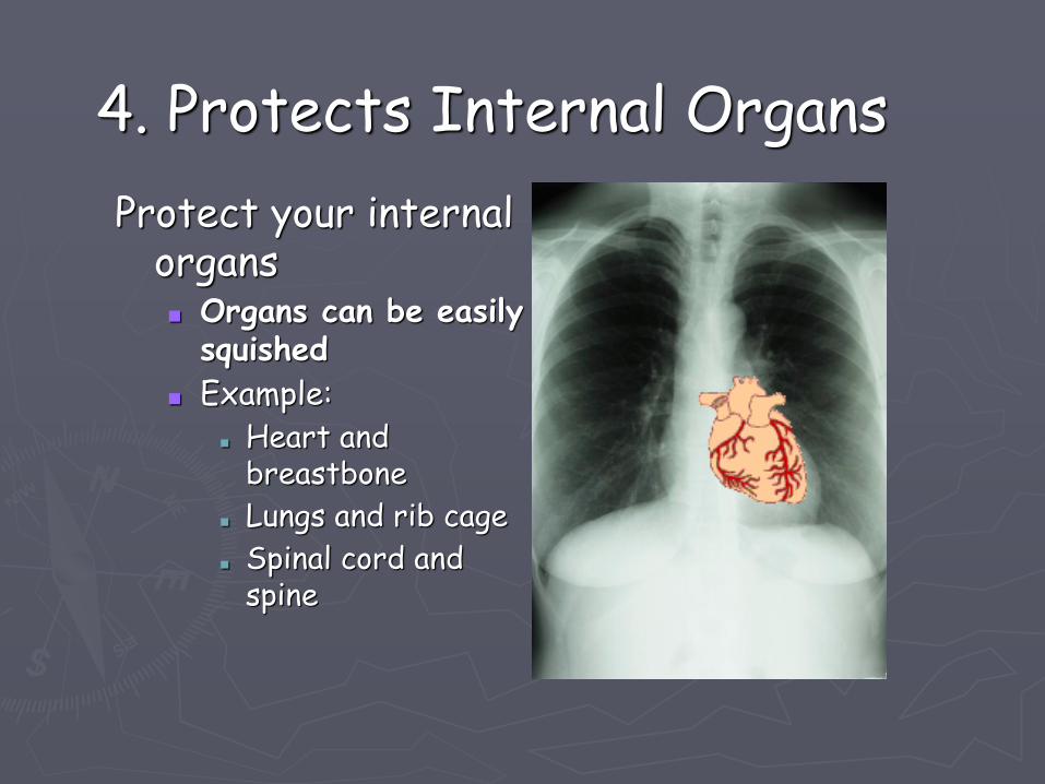
4. Protects Internal Organs
Protect your internal organs■ Organs can be easily
squished
■ Example:■ Heart and
breastbone
■ Lungs and rib cage
■ Spinal cord and spine
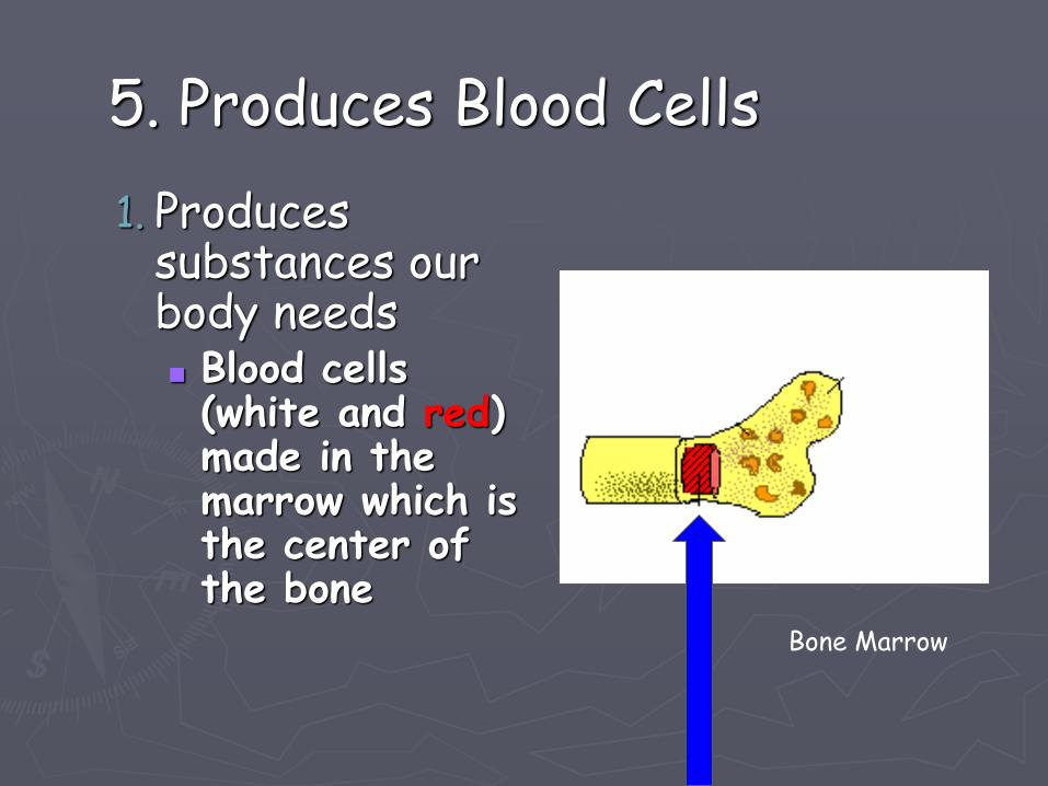
5. Produces Blood Cells
1. Produces substances our body needs■ Blood cells (white and red) made in the marrow which is the center of the bone
Bone Marrow
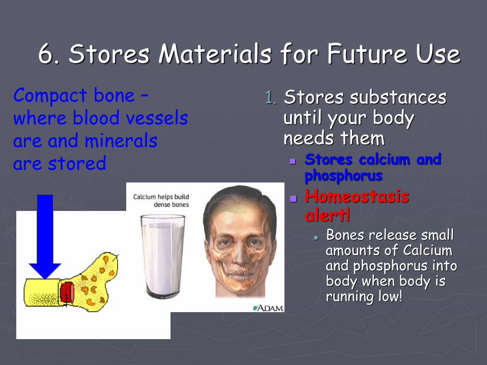
6. Stores Materials for Future Use
1. Stores substances until your body needs them■ Stores calcium and
phosphorus
■ Homeostasis alert!
■ Bones release small amounts of Calcium and phosphorus into body when body is running low!
Compact bone –where blood vessels are and minerals are stored
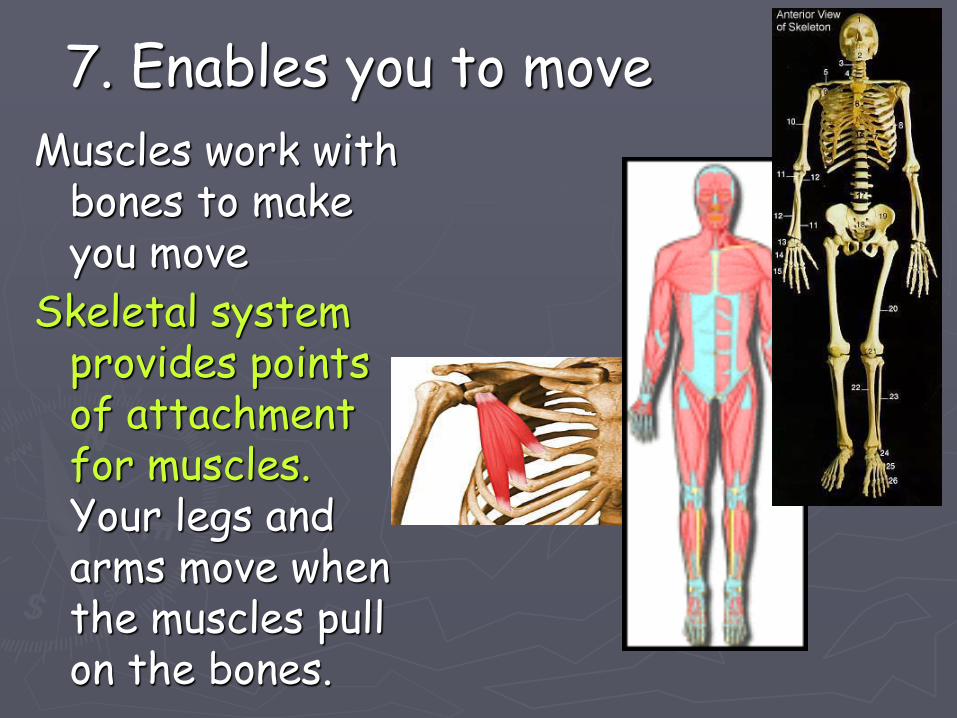
7. Enables you to move
Muscles work with bones to make you move
Skeletal system provides points of attachment for muscles.Your legs and arms move when the muscles pull on the bones.
11
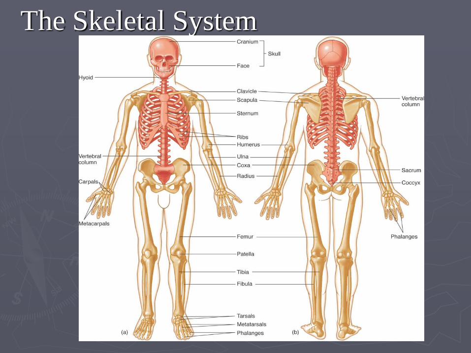
The Skeletal System
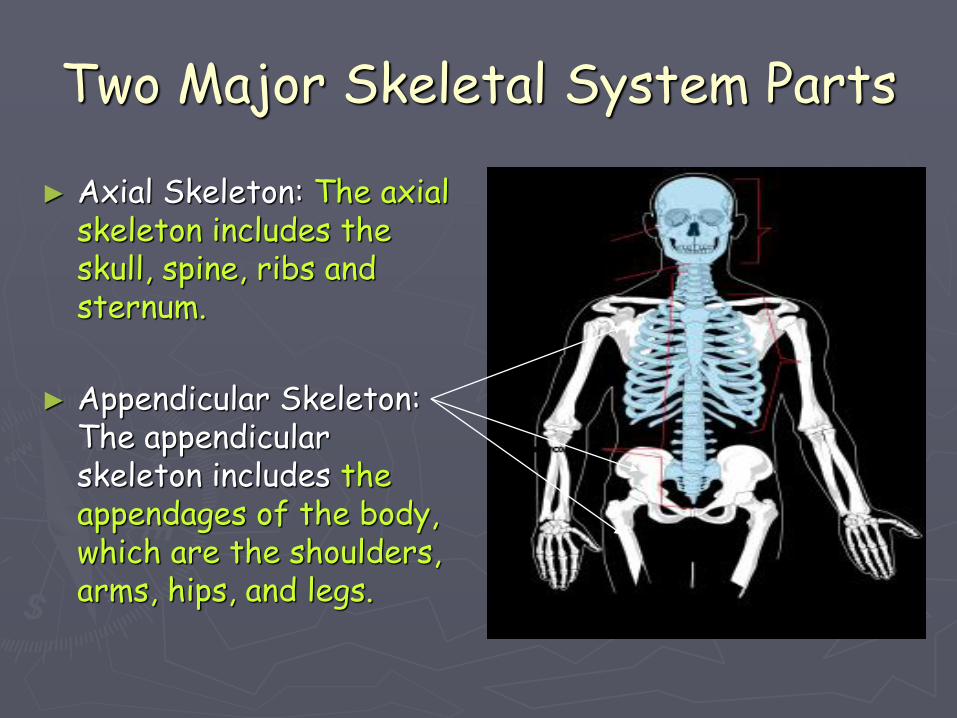
Two Major Skeletal System Parts
► Axial Skeleton: The axial skeleton includes the skull, spine, ribs and sternum.
► Appendicular Skeleton: The appendicular skeleton includes the appendages of the body, which are the shoulders, arms, hips, and legs.
13
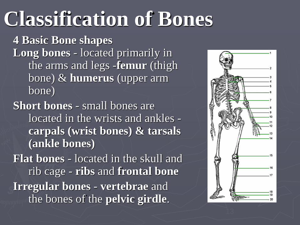
Classification of Bones4 Basic Bone shapes Long bones - located primarily in
the arms and legs -femur (thigh bone) & humerus (upper arm bone)
Short bones - small bones are located in the wrists and ankles -carpals (wrist bones) & tarsals (ankle bones)
Flat bones - located in the skull and rib cage - ribs and frontal bone
Irregular bones - vertebrae and the bones of the pelvic girdle.
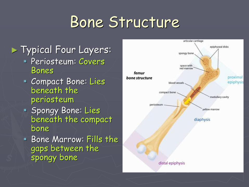
Bone Structure
►Typical Four Layers:▪ Periosteum: Covers
Bones▪ Compact Bone: Lies
beneath the periosteum
▪ Spongy Bone: Lies beneath the compact bone
▪ Bone Marrow: Fills the gaps between the spongy bone
15
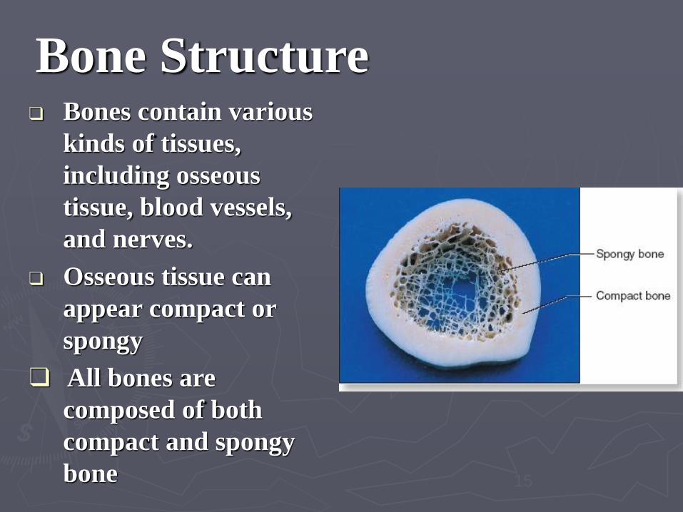
Bone Structure❑ Bones contain various
kinds of tissues,
including osseous
tissue, blood vessels,
and nerves.
❑ Osseous tissue can
appear compact or
spongy
❑ All bones are
composed of both
compact and spongy
bone
16
Bone Structure (cont.)
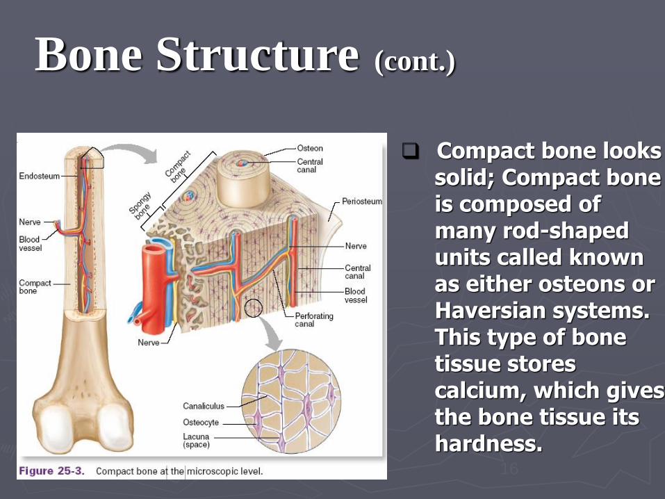
❑ Compact bone looks solid; Compact bone is composed of many rod-shaped units called known as either osteons or Haversian systems. This type of bone tissue stores calcium, which gives the bone tissue its hardness.
17
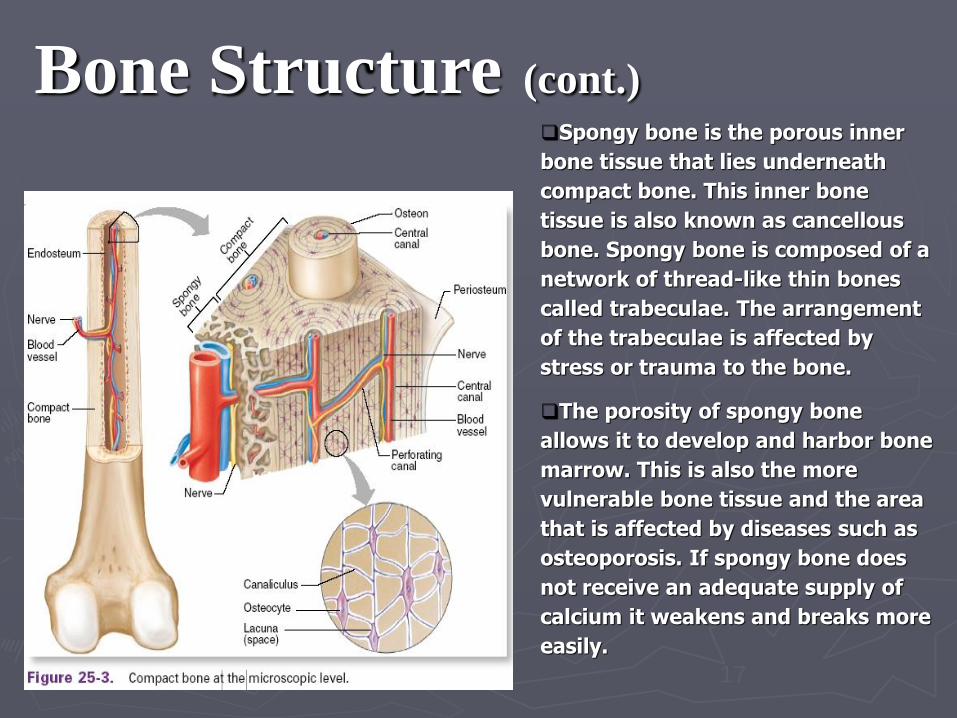
Bone Structure (cont.)❑Spongy bone is the porous inner
bone tissue that lies underneath
compact bone. This inner bone
tissue is also known as cancellous
bone. Spongy bone is composed of a
network of thread-like thin bones
called trabeculae. The arrangement
of the trabeculae is affected by
stress or trauma to the bone.
❑The porosity of spongy bone
allows it to develop and harbor bone
marrow. This is also the more
vulnerable bone tissue and the area
that is affected by diseases such as
osteoporosis. If spongy bone does
not receive an adequate supply of
calcium it weakens and breaks more
easily.
18
Functions of Bones
❑ Shape to body parts
❑ Support and protect soft structures in the body
❑ Body movement since skeletal muscles attach
to them
❑ Red bone marrow of bone produces new
blood cells
❑ Store calcium for the body
19
Bone Growth
❑ Ossification – process of bone growth
Intramembranous ossification –
❑ bones begin as tough, fibrous membrane
❑ bone-forming cells called osteoblasts turn the
membrane to bone (located in skull)
Endochondral ossification
❑ bones containing some cartilage between an epiphysis
and the diaphysis will continue to grow
❑ cells that form holes in bone are called osteoclasts
20
Apply Your Knowledge
Why is it important for the bones to
store calcium?
Every cell in the body needs
calcium so the body must have a
large supply readily available
22
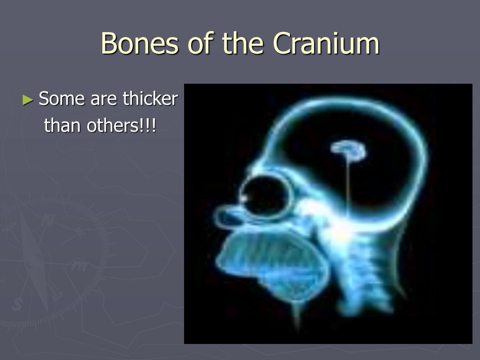
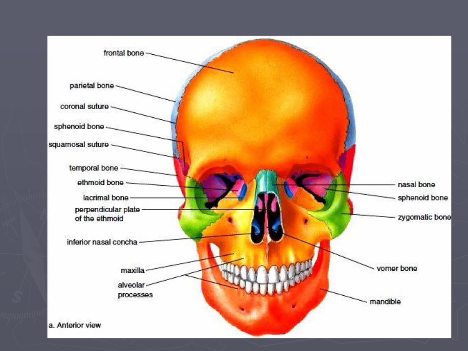
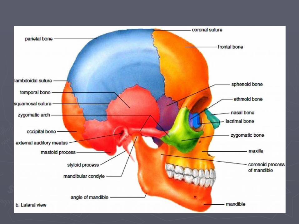
Bones of the Skull/faceTwo types:
❑ Cranial-form the top, sides, and back of the
skull
❑ Facial bones-form the face
“Soft spots" felt on an infant's skull are
actually fontanels.
▪ Tough membranes that connect the
incompletely developed bones.
23
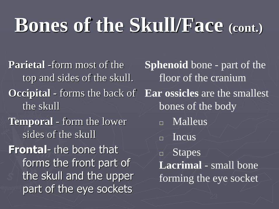
Bones of the Skull/Face (cont.)
Parietal -form most of the
top and sides of the skull.
Occipital - forms the back of
the skull
Temporal - form the lower
sides of the skull
Frontal- the bone that
forms the front part of the skull and the upper part of the eye sockets.
Sphenoid bone - part of the
floor of the cranium
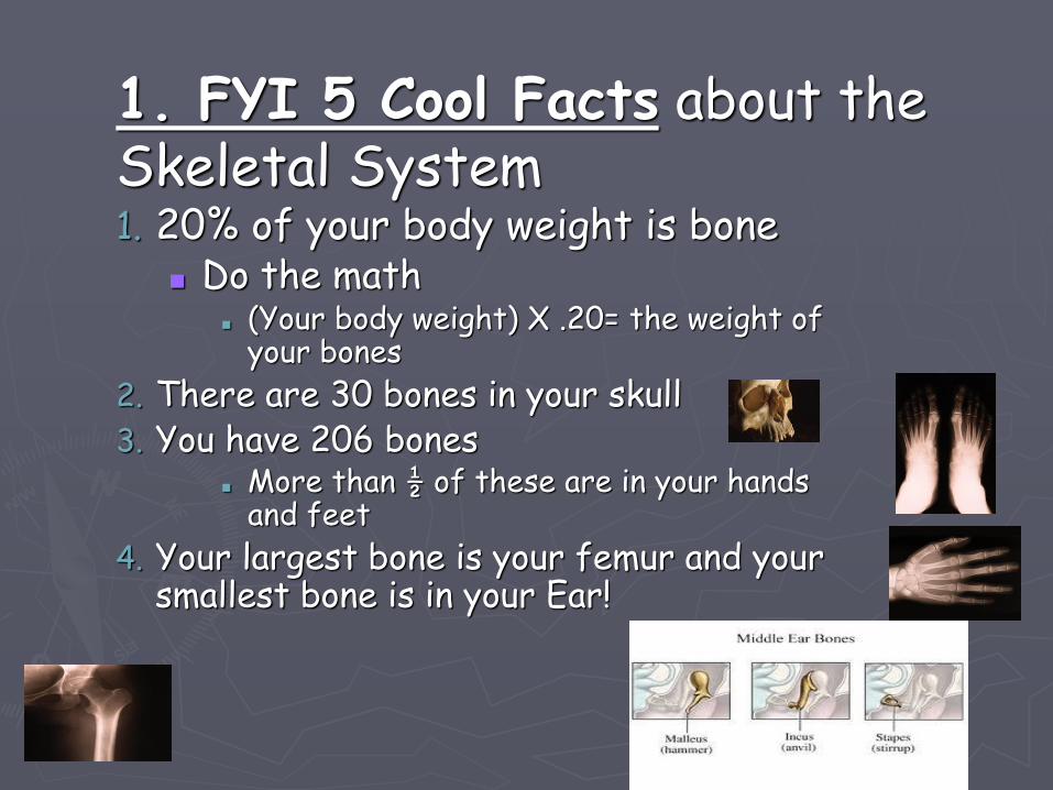
Ear ossicles are the smallest
bones of the body
□ Malleus
□ Incus
□ Stapes
Lacrimal - small bone
forming the eye socket
24
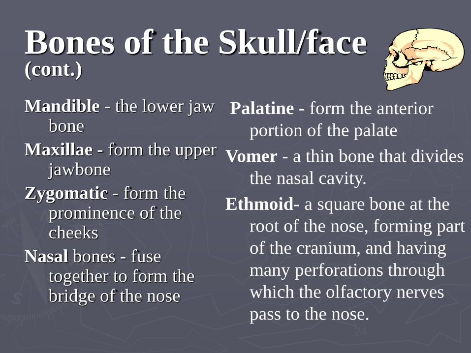
Bones of the Skull/face (cont.)
Mandible - the lower jaw bone
Maxillae - form the upper jawbone
Zygomatic - form the prominence of the cheeks
Nasal bones - fuse together to form the bridge of the nose
Palatine - form the anterior
portion of the palate
Vomer - a thin bone that divides
the nasal cavity.
Ethmoid- a square bone at the
root of the nose, forming part
of the cranium, and having
many perforations through
which the olfactory nerves
pass to the nose.
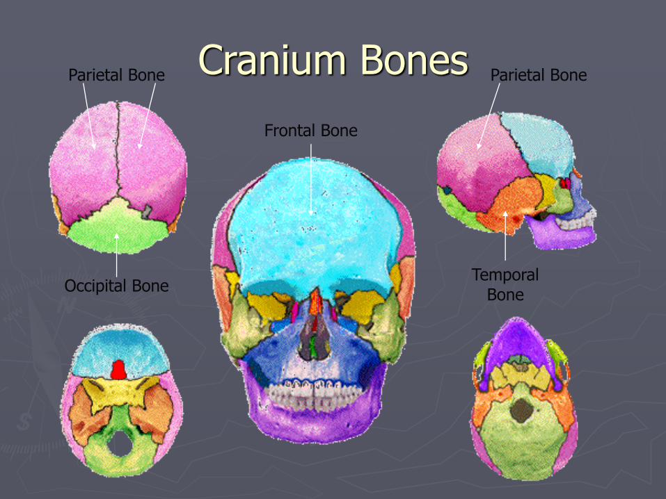
Cranium Bones
Frontal Bone
Occipital BoneTemporal
Bone
Parietal Bone Parietal Bone
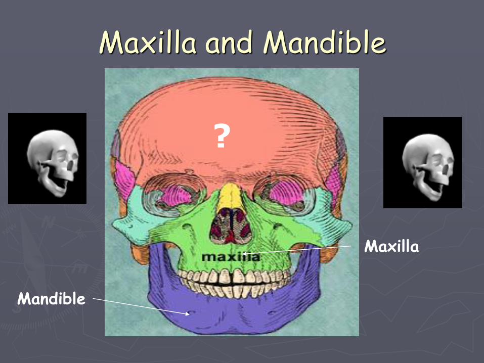
Maxilla and Mandible
Maxilla
Mandible
?
27
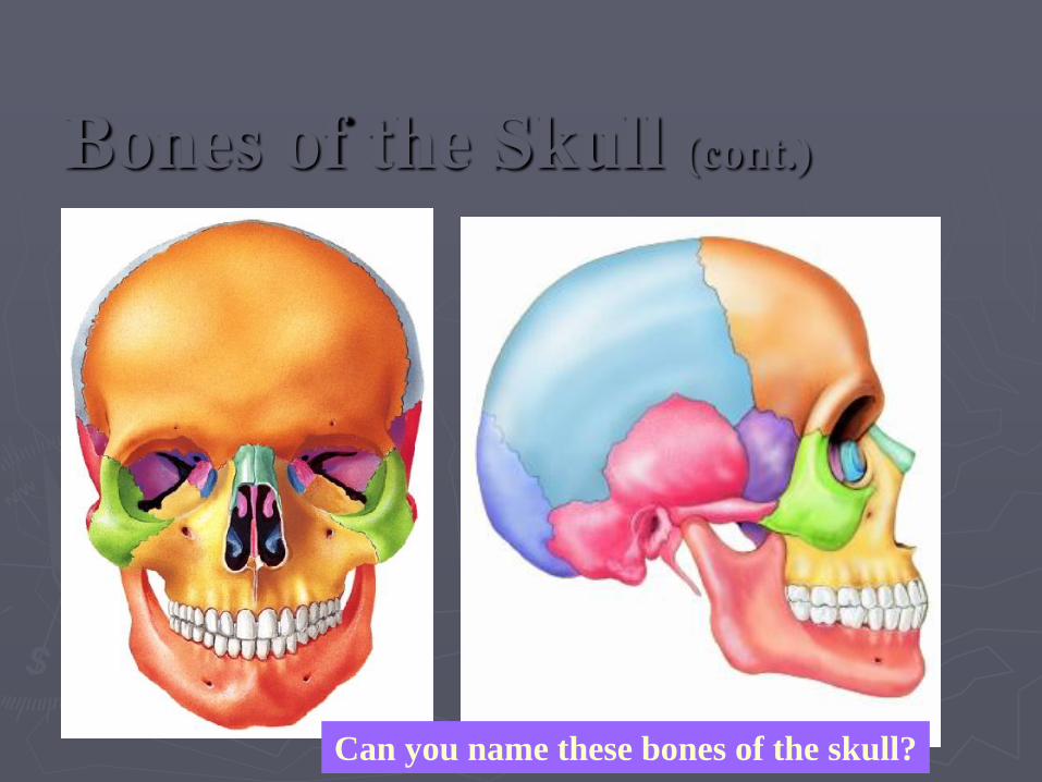
Bones of the Skull (cont.)
Can you name these bones of the skull?
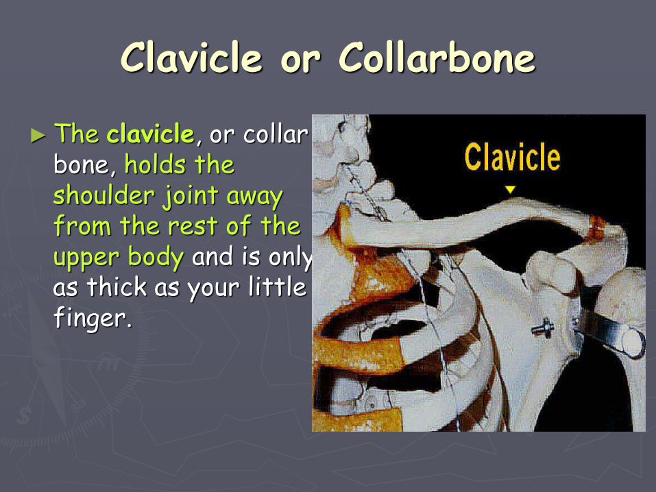
Clavicle or Collarbone
►The clavicle, or collar bone, holds the shoulder joint away from the rest of the upper body and is only as thick as your little finger.
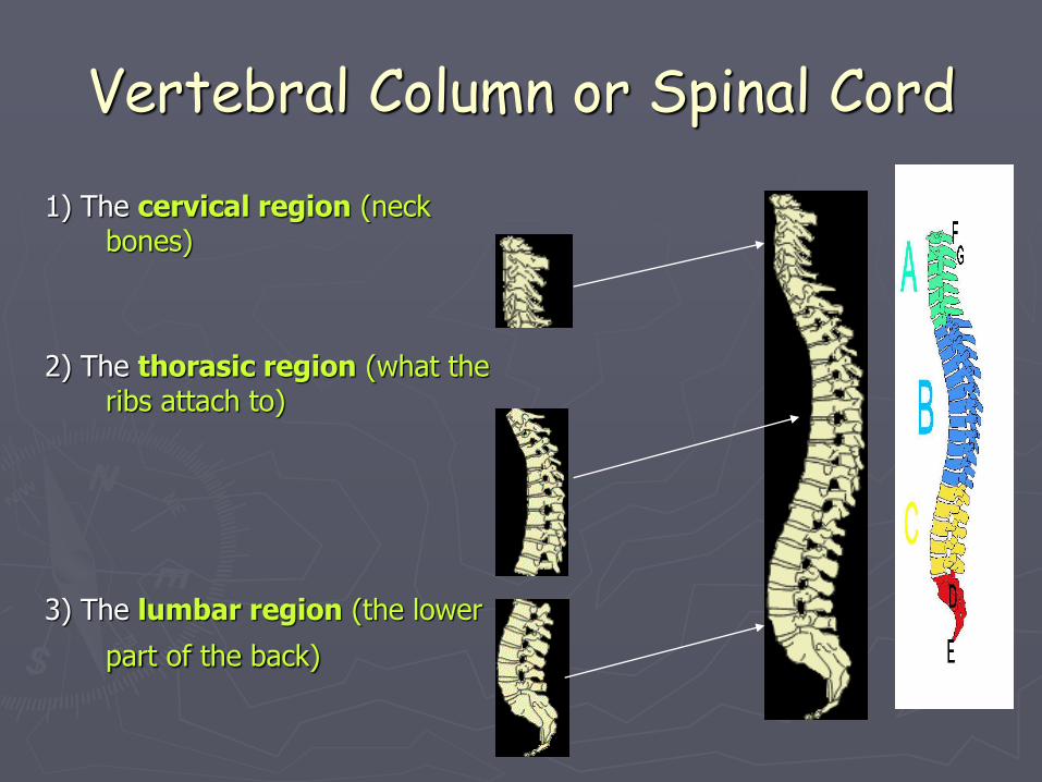
Vertebral Column or Spinal Cord
1) The cervical region (neck bones)
2) The thorasic region (what the ribs attach to)
3) The lumbar region (the lower
part of the back)
32
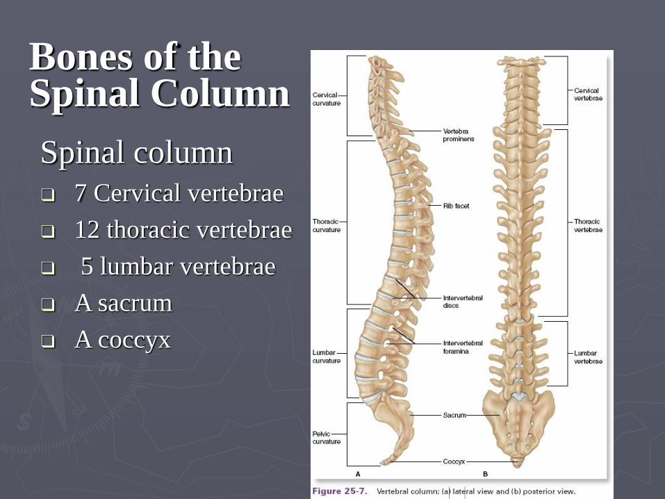
Bones of the Spinal Column
Spinal column
❑ 7 Cervical vertebrae
❑ 12 thoracic vertebrae
❑ 5 lumbar vertebrae
❑ A sacrum
❑ A coccyx
33
Bones of the Spinal Column (cont.)
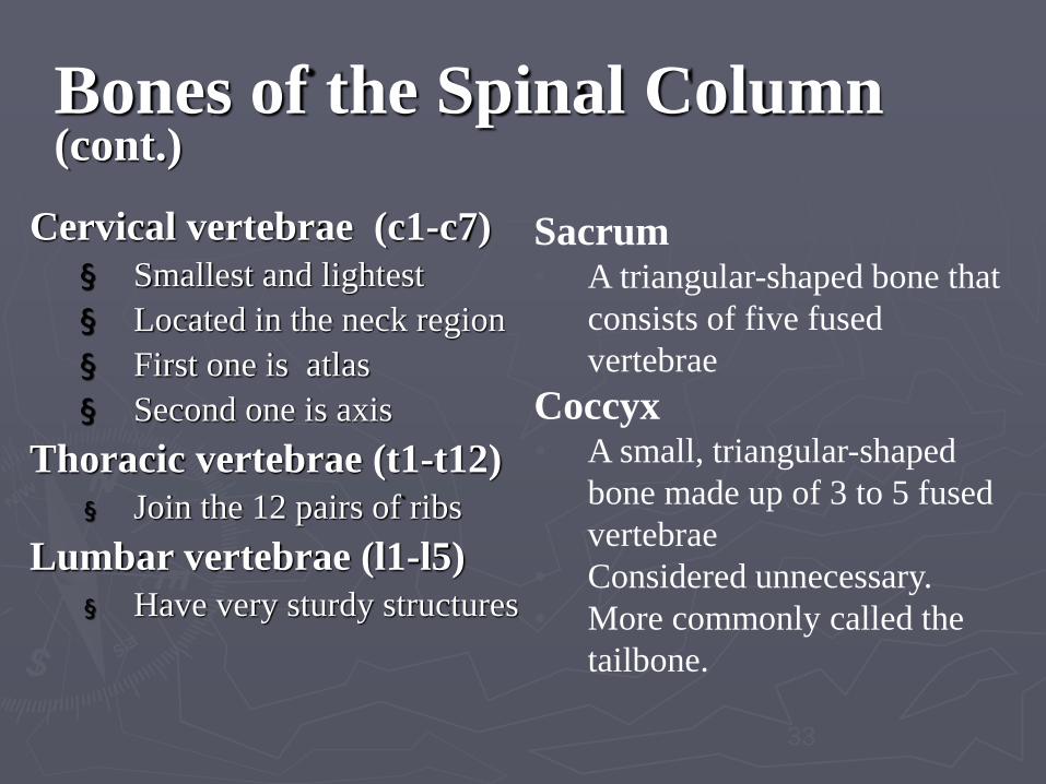
Cervical vertebrae (c1-c7)
§ Smallest and lightest
§ Located in the neck region
§ First one is atlas
§ Second one is axis
Thoracic vertebrae (t1-t12)
§ Join the 12 pairs of ribs
Lumbar vertebrae (l1-l5)
§ Have very sturdy structures
Sacrum • A triangular-shaped bone that
consists of five fused
vertebrae
Coccyx • A small, triangular-shaped
bone made up of 3 to 5 fused
vertebrae
• Considered unnecessary.
• More commonly called the
tailbone.
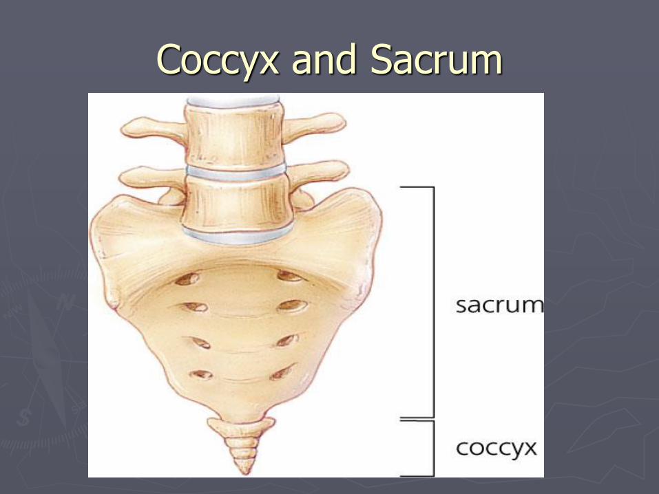
Coccyx and Sacrum

The vertebral body is the anterior part of the vertebrae. It is the weight-bearing component, and its size increases as the vertebral column descends (having to support increasing amounts of weight).The superior and inferior aspects of the vertebral body are lined with hyaline cartilage. Adjacent vertebral bodies are separated by a fibrocartilginous intervertebral disc.
The vertebral arch refers to the lateral and posterior parts of the vertebrae.
With the vertebral body, the vertebral arch forms an enclosed hole, called a vertebral foramen. The foramina of the all vertebrae line up to form the vertebral canal, which encloses the spinal cord.
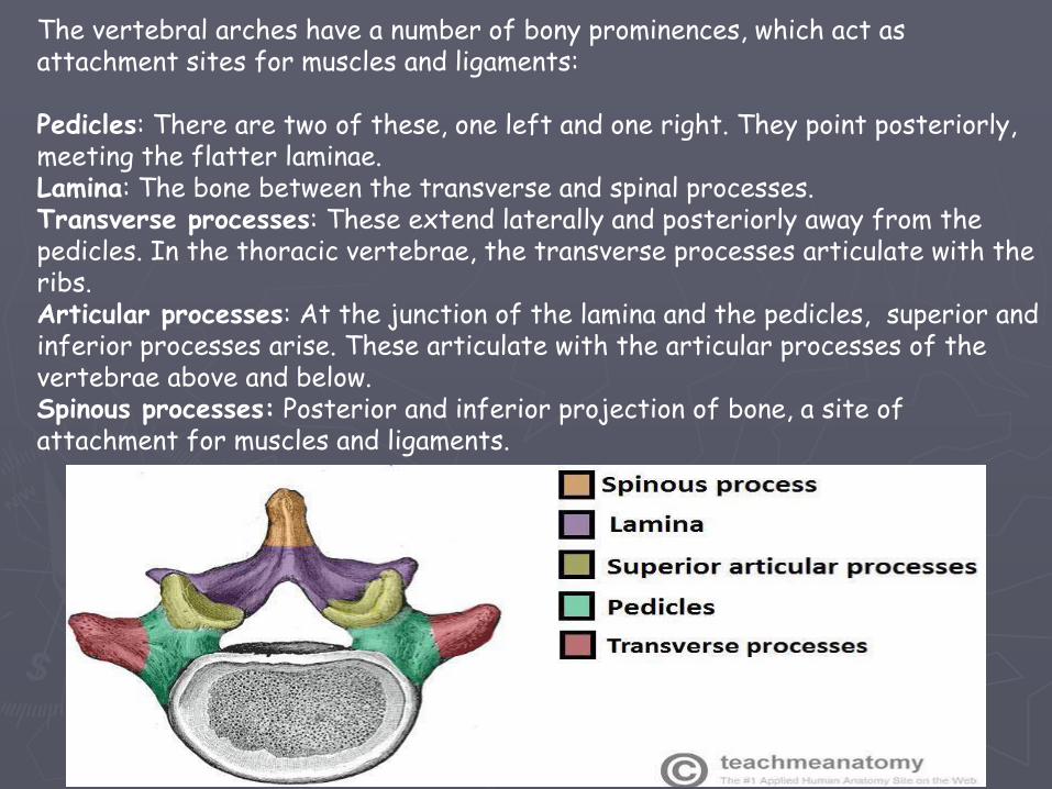
The vertebral arches have a number of bony prominences, which act as attachment sites for muscles and ligaments:
Pedicles: There are two of these, one left and one right. They point posteriorly, meeting the flatter laminae.Lamina: The bone between the transverse and spinal processes.Transverse processes: These extend laterally and posteriorly away from the pedicles. In the thoracic vertebrae, the transverse processes articulate with the ribs.Articular processes: At the junction of the lamina and the pedicles, superior and inferior processes arise. These articulate with the articular processes of the vertebrae above and below.Spinous processes: Posterior and inferior projection of bone, a site of attachment for muscles and ligaments.
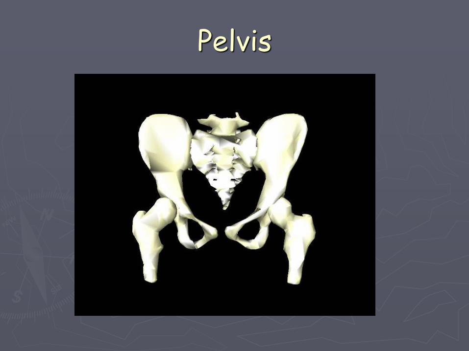
Pelvis
Bones of the Pelvic Girdle
Slide 5.37
• Composed of two coxal bones (hip bones)
•Composed of three pair of fused bones
• Ilium
• Ischium
• Pubis
• The total weight of the upper body rests on the pelvis
• Protects several organs
• Reproductive organs
• Urinary bladder
• Part of the large intestine
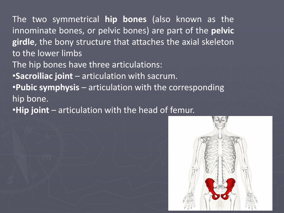
The two symmetrical hip bones (also known as theinnominate bones, or pelvic bones) are part of the pelvicgirdle, the bony structure that attaches the axial skeletonto the lower limbsThe hip bones have three articulations:•Sacroiliac joint – articulation with sacrum.•Pubic symphysis – articulation with the corresponding hip bone.•Hip joint – articulation with the head of femur.
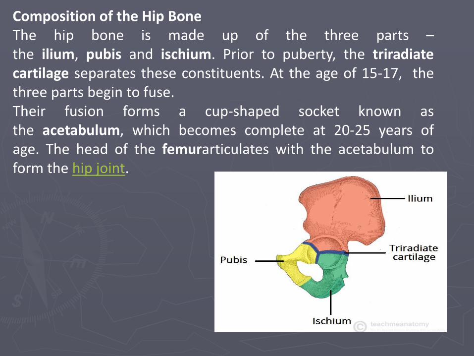
Composition of the Hip BoneThe hip bone is made up of the three parts –the ilium, pubis and ischium. Prior to puberty, the triradiatecartilage separates these constituents. At the age of 15-17, thethree parts begin to fuse.Their fusion forms a cup-shaped socket known asthe acetabulum, which becomes complete at 20-25 years ofage. The head of the femurarticulates with the acetabulum toform the hip joint.
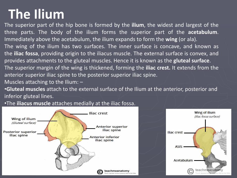
The superior part of the hip bone is formed by the ilium, the widest and largest of thethree parts. The body of the ilium forms the superior part of the acetabulum.Immediately above the acetabulum, the ilium expands to form the wing (or ala).The wing of the ilium has two surfaces. The inner surface is concave, and known asthe iliac fossa, providing origin to the iliacus muscle. The external surface is convex, andprovides attachments to the gluteal muscles. Hence it is known as the gluteal surface.The superior margin of the wing is thickened, forming the iliac crest. It extends from theanterior superior iliac spine to the posterior superior iliac spine.Muscles attaching to the Ilium: –•Gluteal muscles attach to the external surface of the Ilium at the anterior, posterior and inferior gluteal lines.•The iliacus muscle attaches medially at the iliac fossa.
The Ilium
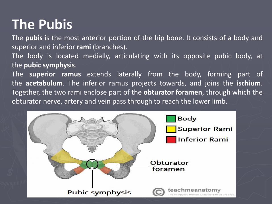
The PubisThe pubis is the most anterior portion of the hip bone. It consists of a body andsuperior and inferior rami (branches).The body is located medially, articulating with its opposite pubic body, atthe pubic symphysis.The superior ramus extends laterally from the body, forming part ofthe acetabulum. The inferior ramus projects towards, and joins the ischium.Together, the two rami enclose part of the obturator foramen, through which theobturator nerve, artery and vein pass through to reach the lower limb.
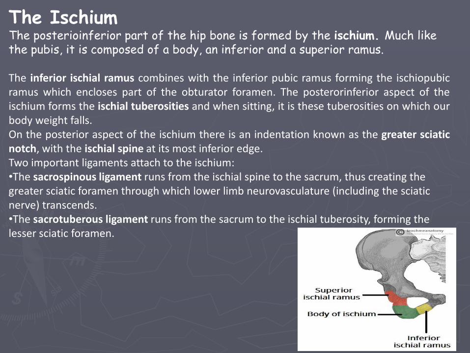
The IschiumThe posterioinferior part of the hip bone is formed by the ischium. Much like the pubis, it is composed of a body, an inferior and a superior ramus.
The inferior ischial ramus combines with the inferior pubic ramus forming the ischiopubicramus which encloses part of the obturator foramen. The posterorinferior aspect of theischium forms the ischial tuberosities and when sitting, it is these tuberosities on which ourbody weight falls.On the posterior aspect of the ischium there is an indentation known as the greater sciaticnotch, with the ischial spine at its most inferior edge.Two important ligaments attach to the ischium:•The sacrospinous ligament runs from the ischial spine to the sacrum, thus creating the greater sciatic foramen through which lower limb neurovasculature (including the sciatic nerve) transcends.•The sacrotuberous ligament runs from the sacrum to the ischial tuberosity, forming the lesser sciatic foramen.
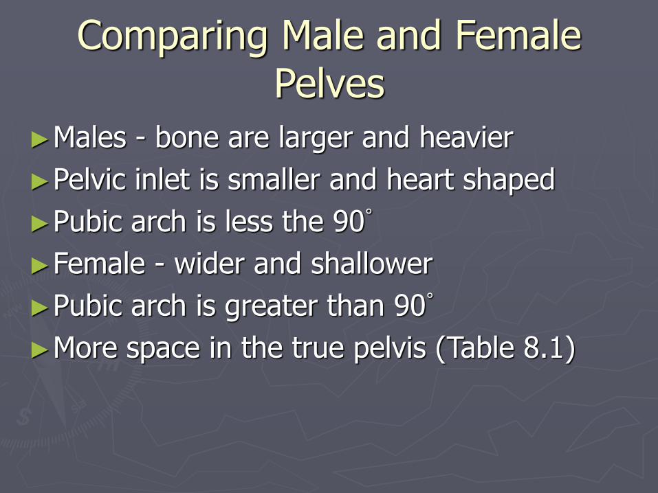
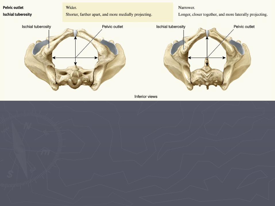
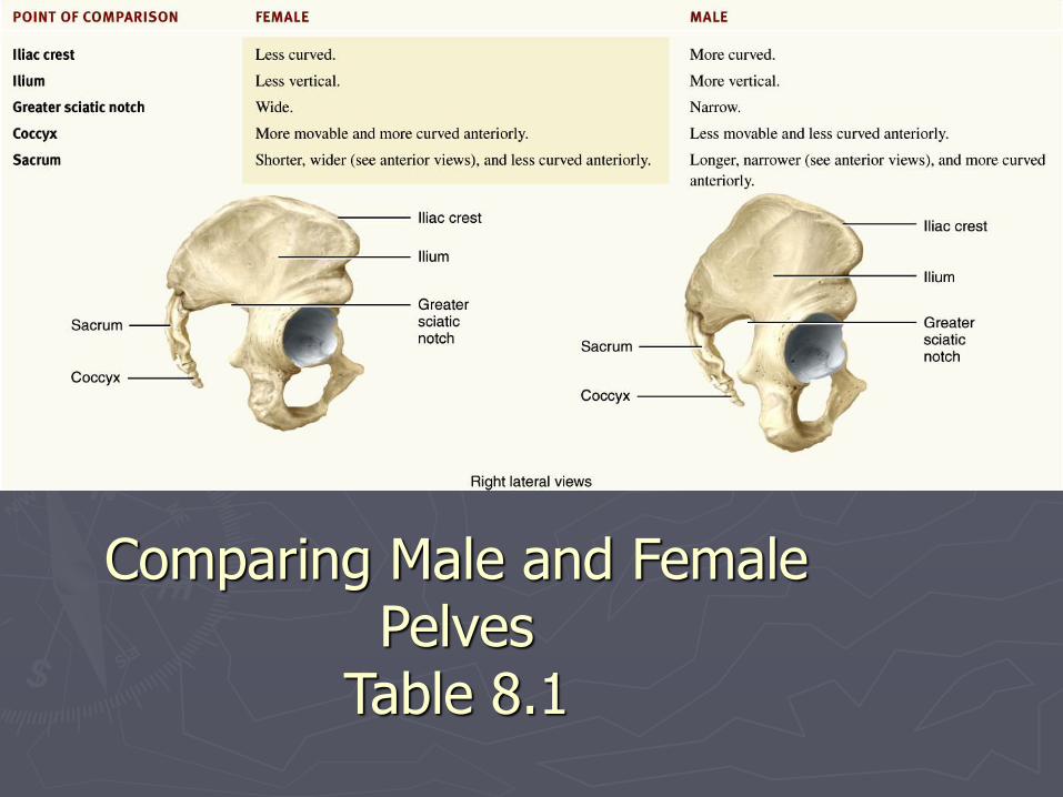
Comparing Male and Female Pelves
►Males - bone are larger and heavier
►Pelvic inlet is smaller and heart shaped
►Pubic arch is less the 90°
►Female - wider and shallower
►Pubic arch is greater than 90°
►More space in the true pelvis (Table 8.1)
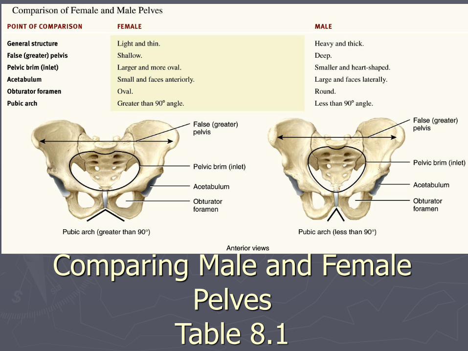
Comparing Male and Female Pelves
Table 8.1
Comparing Male and Female Pelves
Table 8.1