Embed Size (px)
Citation preview

Skeletal Paleopathology of Human Remains From Cemetery R37 at Harappa,
excavated in 1987 and 1988
© Nancy C Lovell
Professor Emerita of Anthropology University of Alberta
Edmonton AB Canada
This is an open access work distributed under the terms of the Creative Commons
Attribution-Noncommercial No-Derivative Works 3.0 License.
You may freely download, copy, and distribute this work provided the original author and source* are credited, but you may not alter, transform, or build upon this work.
This work may not be used for commercial purposes.
*To cite this work, please use the following bibliographic information: Author: Lovell, Nancy C. Title: Skeletal Paleopathology of Human Remains from Cemetery R37 at Harappa, excavated in 1987 and 1988 Year of Publication: 2014 Place of Publication: Edmonton, Canada Publisher: University of Alberta Education and Research Archive (ERA) Permalink to this work: http://hdl.handle.net/10402/era.39921
i
Abstract
Excavations at the archaeological site of Harappa, Pakistan in 1987 and 1988 uncovered the remains of at least 92 individuals (84 adults and 8 juveniles), although only 19 were complete skeletons in primary contexts. This report describes the frequencies and expressions of joint disease, trauma, congenital and developmental disorders, hematopoietic disorders, infection and inflammation, metabolic disorders, and neoplasia in these remains and in an additional adult skeleton, excavated in 1967 and displayed in the Harappa Museum. Fourteen of the 20 complete adult skeletons in primary contexts exhibited pathological lesions on bones. An additional 14 burial features of secondary deposits included remains with pathological lesions. The most common condition was joint disease, which affected 10 individuals, mainly in the spine, followed by trauma, which affected five individuals. Periosteal reactions on long bones, benign osteomas on the cranium, and two possible cases of anomalous development of the skeleton were also noted.
ii
Acknowledgements
This paper is dedicated to the memory of Kenneth Kennedy, my doctoral advisor at Cornell University. Kenneth was responsible for my introduction to South Asian archaeology and my participation in the Harappa Project. George F. Dales, my postdoctoral supervisor, was director of the University of California (Berkeley) Harappa Project from 1986 to 1992. After Dr. Dales’ death, direction of the project (now the Harappa Archaeological Research Project) was taken over by Drs. R.H. Meadow (Harvard University), J. Mark Kenoyer (University of Wisconsin-Madison) and Rita P. Wright (New York University). Excavations at Harappa were conducted in collaboration with the Department of Archaeology and Museums, Government of Pakistan. The University of California (Berkeley) Harappa Project, the Natural Sciences and Engineering and the Social Sciences and Humanities Research Councils of Canada, Cornell University, and the University of Alberta supported my research. I thank Gwen Robbins Schug for her helpful comments on an earlier version of this manuscript.
iii
Table of Contents
Introduction....................................................................................................................... 1 Materials and Methods..................................................................................................... 2 Pathological conditions..................................................................................................... 3
Joint Disease .................................................................................................................. 3 Trauma........................................................................................................................... 6 Congenital and Developmental Disorders .................................................................. 8 Hematopoietic Disorders .............................................................................................. 9 Infection and Inflammation ....................................................................................... 10 Metabolic Disorders.................................................................................................... 11 Neoplasia...................................................................................................................... 11
Discussion and Conclusions ........................................................................................... 11 Literature Cited .............................................................................................................. 16 Appendix: Details of Pathological Lesions in Adult Remains .................................... 21
iv
List of Figures
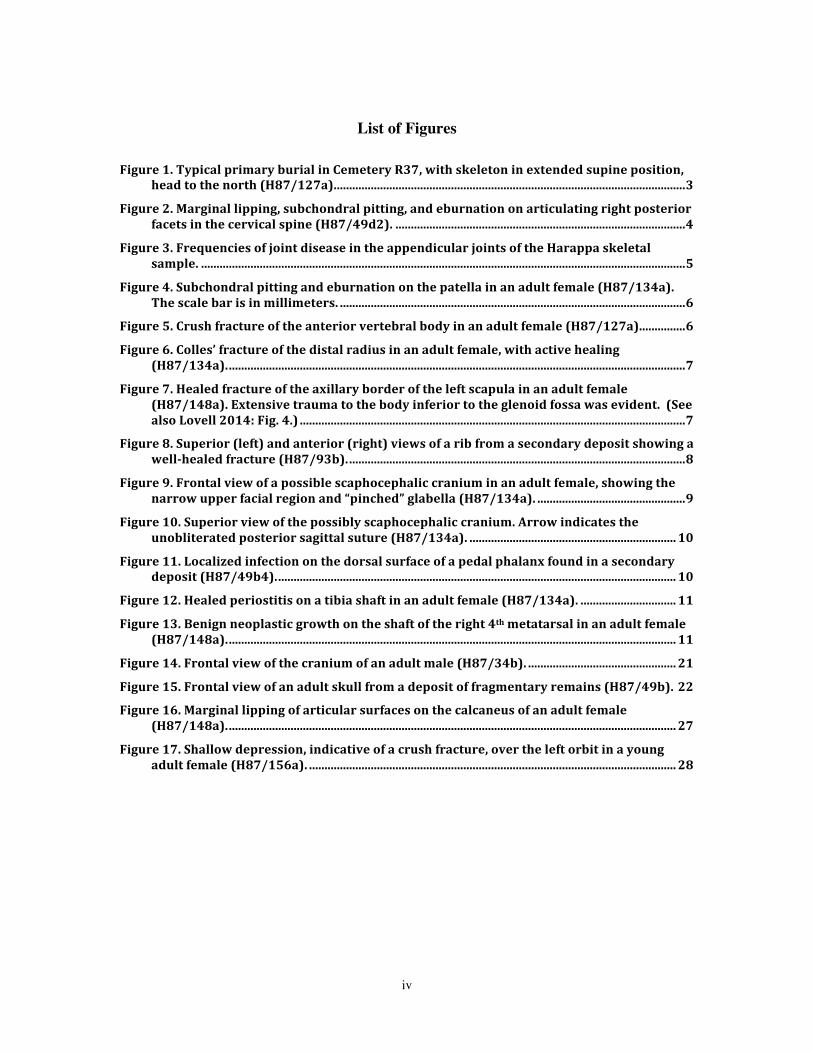
Figure 1. Typical primary burial in Cemetery R37, with skeleton in extended supine position, head to the north (H87/127a)..................................................................................................................3
Figure 2. Marginal lipping, subchondral pitting, and eburnation on articulating right posterior facets in the cervical spine (H87/49d2). ..............................................................................................4
Figure 3. Frequencies of joint disease in the appendicular joints of the Harappa skeletal sample. .............................................................................................................................................................5
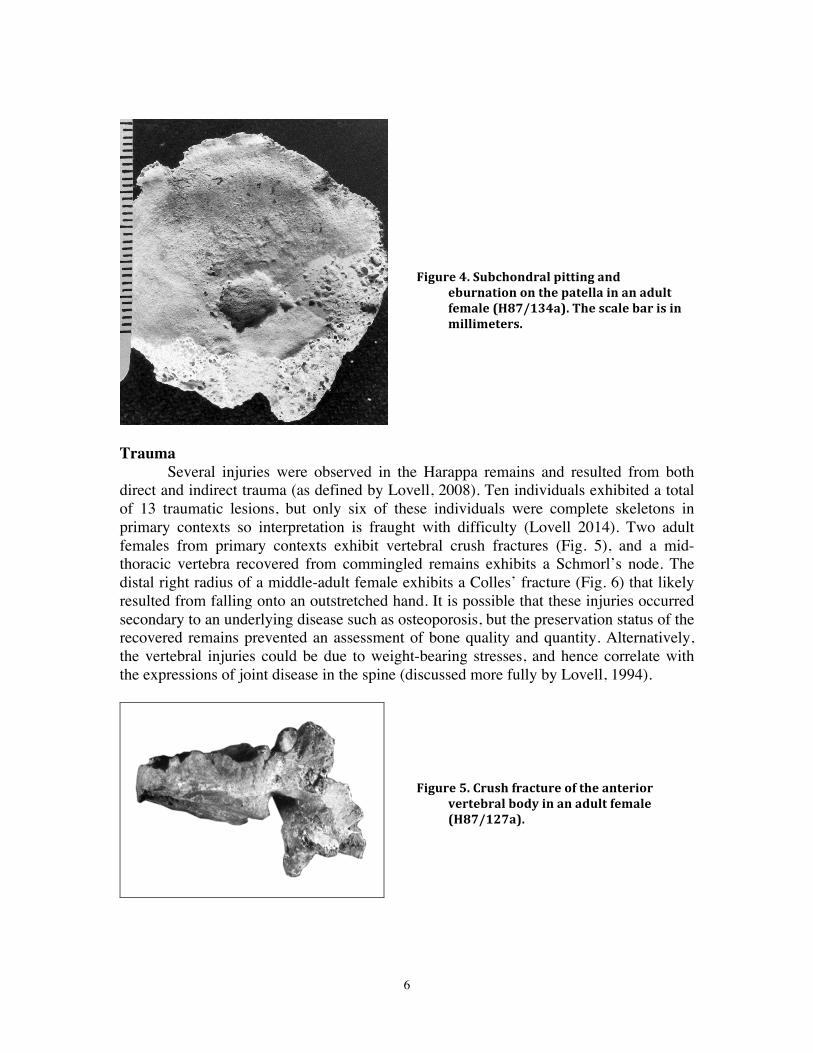
Figure 4. Subchondral pitting and eburnation on the patella in an adult female (H87/134a). The scale bar is in millimeters. ................................................................................................................6
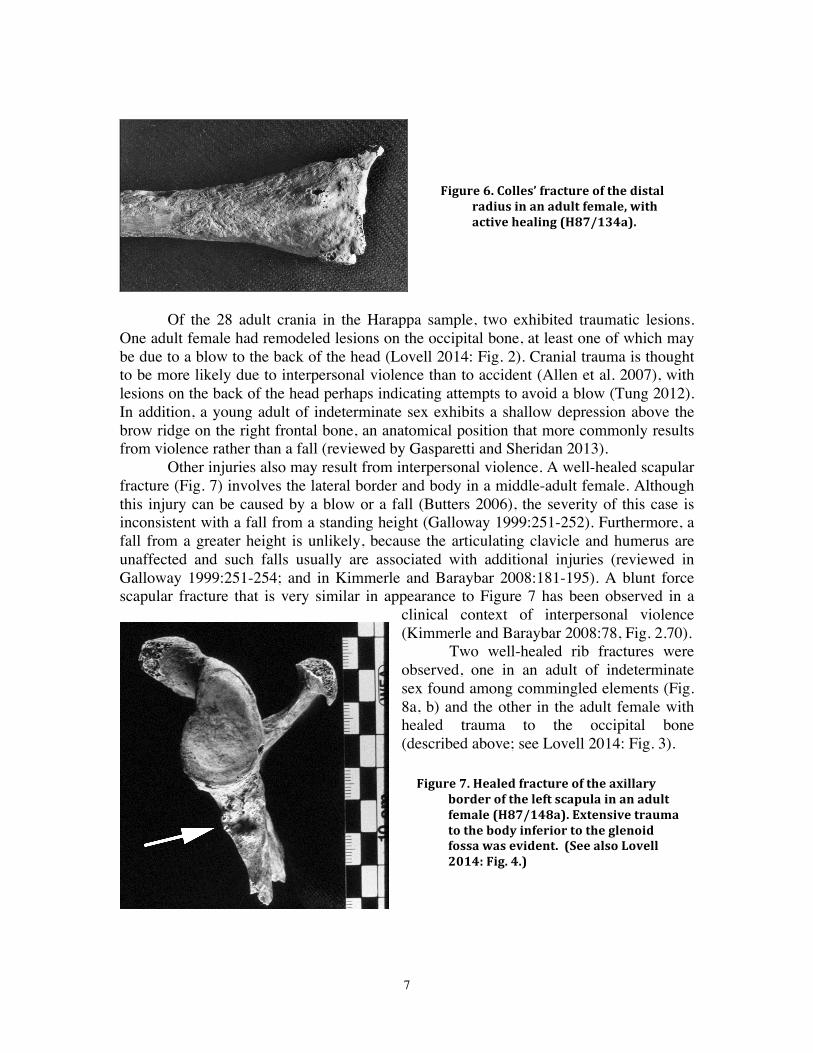
Figure 5. Crush fracture of the anterior vertebral body in an adult female (H87/127a)...............6
Figure 6. Colles’ fracture of the distal radius in an adult female, with active healing (H87/134a).....................................................................................................................................................7
Figure 7. Healed fracture of the axillary border of the left scapula in an adult female (H87/148a). Extensive trauma to the body inferior to the glenoid fossa was evident. (See also Lovell 2014: Fig. 4.) .............................................................................................................................7
Figure 8. Superior (left) and anterior (right) views of a rib from a secondary deposit showing a well-healed fracture (H87/93b)..............................................................................................................8
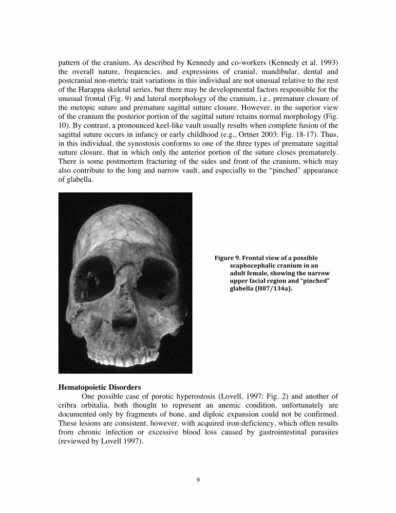
Figure 9. Frontal view of a possible scaphocephalic cranium in an adult female, showing the narrow upper facial region and “pinched” glabella (H87/134a). ................................................9
Figure 10. Superior view of the possibly scaphocephalic cranium. Arrow indicates the unobliterated posterior sagittal suture (H87/134a). ................................................................... 10
Figure 11. Localized infection on the dorsal surface of a pedal phalanx found in a secondary deposit (H87/49b4).................................................................................................................................. 10
Figure 12. Healed periostitis on a tibia shaft in an adult female (H87/134a). ............................... 11
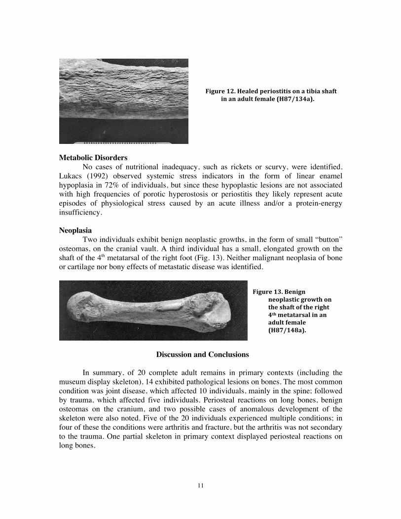
Figure 13. Benign neoplastic growth on the shaft of the right 4th metatarsal in an adult female (H87/148a).................................................................................................................................................. 11
Figure 14. Frontal view of the cranium of an adult male (H87/34b). ................................................ 21
Figure 15. Frontal view of an adult skull from a deposit of fragmentary remains (H87/49b). 22
Figure 16. Marginal lipping of articular surfaces on the calcaneus of an adult female (H87/148a).................................................................................................................................................. 27
Figure 17. Shallow depression, indicative of a crush fracture, over the left orbit in a young adult female (H87/156a). ....................................................................................................................... 28
1
Introduction
Harappa is the type-site of the Harappan, or Indus Valley, civilization that flourished some 4,000 - 5,000 years ago in the Indus River Valley and peripheral areas of what is now Pakistan and India, reaching from the Himalayan Mountains to the Arabian Sea. Although less well-known than the ancient Egyptian and Mesopotamian civilizations, the Indus Valley civilization was a major center of technology and commerce, characterized by its own writing system, a standardized system of weights and measures, technologies including bead-making, shell-working, ceramics production and metallurgy, and a richly symbolic iconography. Harappa flourished during the Urban Period from ca. 2600 – 1900 BC. At its largest, the site may have covered 150 hectares, with a population of about 22,500 to 30,000 (Wright 2010:107). The city consisted mainly of residential, administrative, and ritual buildings, and boasted architectural features related to public works, such as drainage systems and wells. In addition, two formal burial grounds have been identified at the site: Cemetery R37 (ca. 2400 – 2000 BCE) and Cemetery H (ca. 1900 – 1300 BCE). Although excavations at Harappa in recent decades have substantially increased the amount of information about the environment, architecture, and economy of the ancient city as well as the nature of craft specialization (Kenoyer 1991, 1998, 2003; Kenoyer and Meadow 2000; Meadow and Kenoyer 1997, 1999, 2005; Weber 2003; Wright 2010), the lives of the inhabitants of the city are comparatively poorly understood. The purpose of this report, then, is to add to the corpus of knowledge about the ancient Harappans themselves. The analysis presented here is based on published and unpublished data obtained from skeletal remains excavated from Cemetery R37 in 1987 and 1988 by the University of California (Berkeley) Harappa Project. Three colleagues (Kenneth A. R. Kennedy, John R. Lukacs, and Brian E. Hemphill) and I formed the biological anthropology team that was in the field for the Harappa excavations in 1987 and 1988. Preliminary reports of our work were submitted to the project directors and a summary was published by Dales and co-workers (1991), but the full report of the skeletal remains has not been published and awaits publication of the cemetery volume, which will include detailed analysis of cemetery stratigraphy, dating, and grave goods, by other participants in the project. Several aspects of the dental paleopathology of the Harappa remains have been published by Lukacs and co-workers (Lukacs and Pastor 1988; Lukacs and Hemphill 1990; Lukacs 1992), with those results discussed below in the context of the interpretation of the health of the Harappans more generally. My early attempts to reconstruct ancient diets at Harappa by stable isotope and trace element analyses were hampered by a lack of collagen preservation in bone and by other diagenetic problems (see Link and Lovell 1994), but Kenoyer and co-workers successfully analysed isotopes in tooth enamel in order to examine geographic origins and mobility among residents of Harappa. They report that there is substantial variation among the Harappa samples, suggesting multiple homelands for the inhabitants of the city (Kenoyer et al. 2013), which is consistent with the findings from dental metric and nonmetric traits (Hemphill and Lukacs 1991; Hemphill et al. 1991; Hemphill and Lukacs 1993). Although I have published descriptions of spinal arthritis (Lovell 1994), lesions
2
suggestive of chronic anemia (Lovell 1997), and trauma (Lovell 2014), this is the first full report of the skeletal paleopathology of the adult remains excavated in 1987 and 1988 and of a skeleton excavated by Dr. R. Mughal in 1966.
Materials and Methods
The 1987 and 1988 excavations of Cemetery R37 produced a minimum number (MNI) of 84 adults, of which only 19 were complete skeletons (skull and postcrania preserved) from primary contexts; three adult skeletons in primary contexts consisted of postcranial elements only, the skulls having been removed postmortem during subsequent grave digging by the Harappans. The incomplete remains of at least 61 other adults (including nine isolated crania and six separate mandibles), were recovered from debris contexts (Lovell 1994). Excavations also uncovered the incomplete and fragmentary remains of six juveniles from debris contexts, plus the remains of two juveniles that were found in primary contexts. One additional skeleton was analyzed for this report: the skeleton of a young adult male that was recovered from Cemetery R37 during excavations by Dr. R. Mughal in 1966 (Mughal 1968) and placed on display in the Harappa Museum. (Some of the skeletal elements had broken while on display, some were incorrectly placed, and others were discolored by consolidant applied decades previously; consequently, we added some elements from the 1987 and 1988 excavations and rearranged incorrectly placed elements in order to produce a more complete and accurate skeletal display.) Two disturbed burials and some additional skeletal elements were discovered in the cemetery area during digging for a new septic tank at Harappa camp in 1994 (R.H. Meadow, personal communication, May 10, 1994), but I have not examined those remains and therefore they are not included in this report. Primary burials in the cemetery are of skeletons in the extended supine position with the head to the north (Fig. 1), some interred in wooden coffins that were fully decomposed by the time of excavation. Most of the skeletons were accompanied by large numbers of pottery vessels, shell bangles, and beads of steatite, gold and semi-precious stones, suggesting that the individuals buried in Cemetery R-27 represent an upper stratum of Harappan society. In addition to the primary in situ burials, human bone also was recovered in secondary contexts in Cemetery R37 as a result of re-use of the cemetery by the Harappans and by the erosion of some skeletal elements into low-lying areas. A high water table and alkaline soils in some areas of the site resulted in poor preservation of some skeletal material. Age estimates were based on pubic symphysis and auricular surface morphology, cranial suture closure, and/or dental wear (Lovejoy et al. 1985; Meindl and Lovejoy 1985; Smith 1984), depending upon the state of preservation of the skeleton. Individuals were assigned to one of four age categories: young adult (20-30 years), middle adult (31-40 years), older adult (>40 years), and adult of indeterminate age, for the purposes of comparative paleopathological analysis. Sex was determined from pelvic and cranial morphology, using standard osteological criteria (Bass 1979; Ubelaker 1984).

3
Paleopathological diagnosis and interpretation followed the protocols established by Ortner and Putschar (1985). All skeletal elements and fragments were examined visually and with the aid of a 10x hand lens for macroscopic evidence of pathological lesions.
Pathological conditions
Joint Disease Joint disease is not common in the remains from Harappa, a finding that no doubt is influenced by the small sample size of individuals and the number of young and middle-aged adults, but it is the most common pathological condition observed. Joint disease appears most often in the spine, where it affects both the synovial posterior facet joints and the non-synovial joints between vertebral bodies (Lovell 1994). Not every articular surface was observable for pathological lesions, because of preservation issues and difficulty extracting skeletal elements (particularly porous cancellous bone) from a clay-like matrix in some locations of the cemetery, but a total of 3,084 joint margins and articular surfaces in the spine were examined for evidence of joint disease. Sometimes damaged spinal elements could not be identified more precisely than to cervical, thoracic, or lumbar segment and thus data were pooled for each segment presented as a percentage of observable surfaces in that segment, rather than as a percentage of individuals. Synovial facet joints were scored for presence and degree of expression of focal cartilage loss with subchondral bone reaction (pitting), bone proliferation around the margins of joints (vertebral osteophytosis, or marginal lipping), and eburnation, i.e., bone-on-bone contact that occurs after destruction of cartilage and which results in a
Figure 1. Typical primary burial in Cemetery R37, with skeleton in extended supine position, head to the north (H87/127a).
4
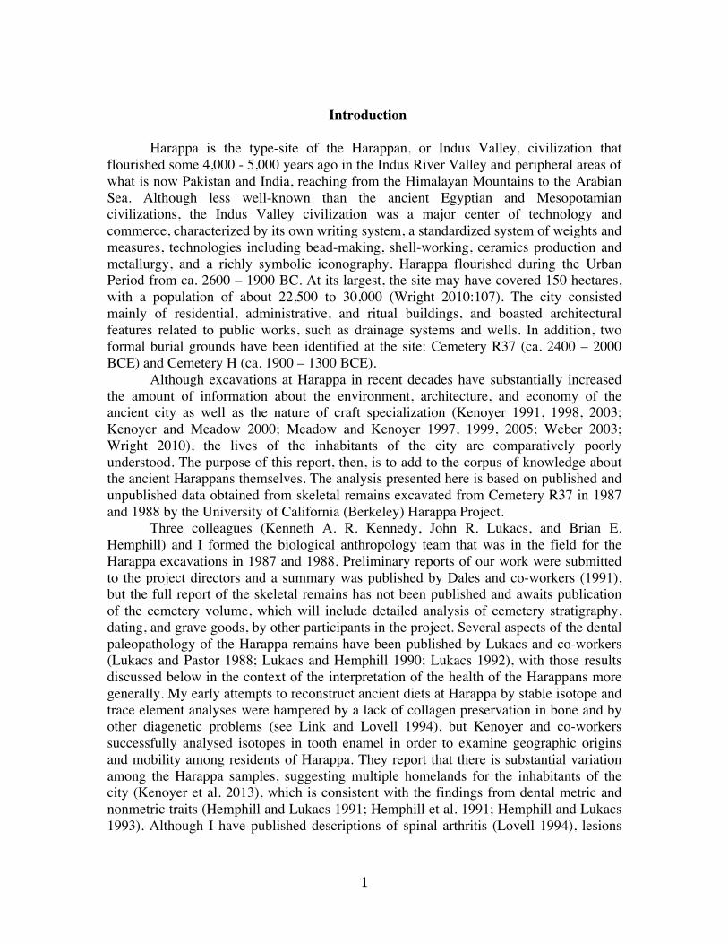
polishing, and often grooving, of the subchondral bone. The intervertebral fibrocartilaginous joints were scored for lipping around the margins of the vertebral bodies and erosive lesions on the vertebral body surfaces. Marginal lipping around vertebral bodies appears at consistent levels among the three vertebral segments: 32% of thoracic vertebral bodies, 36% of cervical vertebral bodies, and 37% of lumbar vertebral bodies were affected. This lipping is characterized by osteophyte formation around the margins of the fibrocartilaginous intervertebral discs. Typically, the condition results from anterior displacement of the disc, which places traction on the Sharpey's fibers that anchor the disc to the margins of the vertebral body; this traction stress leads to the development of osteophytes. Osteophytes begin to form several millimeters from the margin of the vertebral body, and grow in a horizontal direction before growing vertically. The formation of marginal osteophytes in the Harappa vertebrae is usually accompanied by erosive lesions (pitting) on the superior and/or inferior surfaces of the vertebral bodies. Pitting is most common on the vertebral body surfaces in the cervical spine (18%) of body surfaces affected, and is rare in other segments. The lumbar vertebrae exhibited the most cases of marginal lipping around the posterior facets (43%), followed by the cervical vertebrae (28%). Facets in the thoracic spine were rarely affected (4%). The cervical spine is affected most notably by the severity of alterations to the facet joints (Fig. 2), with the only cases of eburnation (three) and ankylosis (two). The ankylosis occurred in two males, aged approximately 20-25 years and 30-35 years, and cannot be attributed to congenital block vertebrae.
Figure 2. Marginal lipping, subchondral pitting, and eburnation on articulating right posterior facets in the cervical spine (H87/49d2).
5
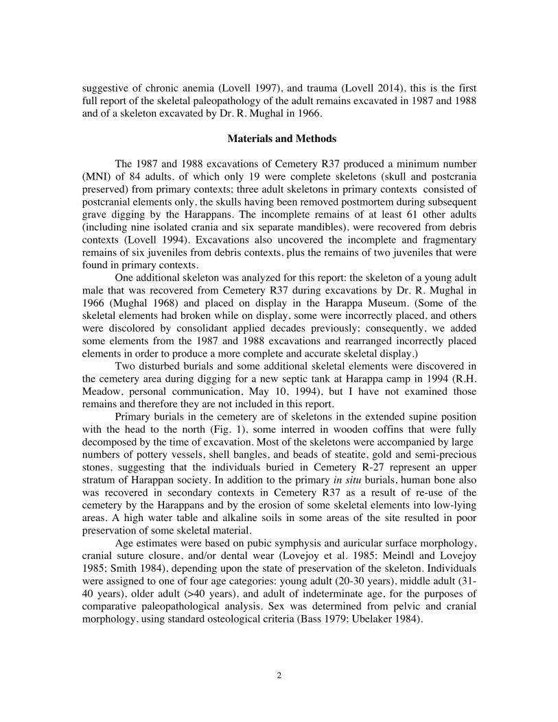
Deterioration of the integrity of synovial joints also affects the joints of the appendicular skeleton and the temporomandibular joint (TMJ) in the Harappa remains. A total of 1,994 appendicular joint margins and articular surfaces, in 156 appendicular joints, were examined for evidence of marginal lipping, pitting, and eburnation. Overall, the knee was the joint most affected by degenerative change (Fig. 3). A variety of clinical studies have shown that the knee is the appendicular joint most involved in arthritis and that the joints of the upper extremity are usually much less affected. However, most of the joint changes involve only slight marginal lipping and by this criterion all of the major joints of the appendicular skeleton were affected in at least one individual. Pitting of subchondral bone appears only in the knee, although the severity is slight. Eburnation is considered the most severe expression of degenerative joint disease and among the Harappans it affects the knee (Fig. 4), hand/wrist, and foot/ankle. Only the knee displays all three conditions. Only two of 17 individuals with observable components of the TMJ were affected by joint disease. One young adult with severe tooth wear exhibited slight lipping and eburnation at the TMJ, while a middle-adult female exhibited slight marginal lipping associated with excessive tooth wear, caries, and antemortem tooth loss. Other studies have noted
that TMJ degeneration is more frequent in middle-aged and older adults than in younger adults, but the relationship is not a strong linear one and is likely influenced by biomechanical factors associated with tooth wear and loss of the molar
teeth (Hodges 1991; Sheridan et al. 1991). The low frequency of TMJ arthritis in this sample may be related to a low prevalence of severe tooth wear (i.e., pulp cavity exposure) (Lukacs 1992).
Figure 3. Frequencies of joint disease in the appendicular joints of the Harappa skeletal sample.
6
Trauma Several injuries were observed in the Harappa remains and resulted from both
direct and indirect trauma (as defined by Lovell, 2008). Ten individuals exhibited a total of 13 traumatic lesions, but only six of these individuals were complete skeletons in primary contexts so interpretation is fraught with difficulty (Lovell 2014). Two adult females from primary contexts exhibit vertebral crush fractures (Fig. 5), and a mid-thoracic vertebra recovered from commingled remains exhibits a Schmorl’s node. The distal right radius of a middle-adult female exhibits a Colles’ fracture (Fig. 6) that likely resulted from falling onto an outstretched hand. It is possible that these injuries occurred secondary to an underlying disease such as osteoporosis, but the preservation status of the recovered remains prevented an assessment of bone quality and quantity. Alternatively, the vertebral injuries could be due to weight-bearing stresses, and hence correlate with the expressions of joint disease in the spine (discussed more fully by Lovell, 1994).
Figure 4. Subchondral pitting and eburnation on the patella in an adult female (H87/134a). The scale bar is in millimeters.
Figure 5. Crush fracture of the anterior vertebral body in an adult female (H87/127a).
7
Of the 28 adult crania in the Harappa sample, two exhibited traumatic lesions. One adult female had remodeled lesions on the occipital bone, at least one of which may be due to a blow to the back of the head (Lovell 2014: Fig. 2). Cranial trauma is thought to be more likely due to interpersonal violence than to accident (Allen et al. 2007), with lesions on the back of the head perhaps indicating attempts to avoid a blow (Tung 2012). In addition, a young adult of indeterminate sex exhibits a shallow depression above the brow ridge on the right frontal bone, an anatomical position that more commonly results from violence rather than a fall (reviewed by Gasparetti and Sheridan 2013). Other injuries also may result from interpersonal violence. A well-healed scapular fracture (Fig. 7) involves the lateral border and body in a middle-adult female. Although this injury can be caused by a blow or a fall (Butters 2006), the severity of this case is inconsistent with a fall from a standing height (Galloway 1999:251-252). Furthermore, a fall from a greater height is unlikely, because the articulating clavicle and humerus are unaffected and such falls usually are associated with additional injuries (reviewed in Galloway 1999:251-254; and in Kimmerle and Baraybar 2008:181-195). A blunt force scapular fracture that is very similar in appearance to Figure 7 has been observed in a
clinical context of interpersonal violence (Kimmerle and Baraybar 2008:78, Fig. 2.70). Two well-healed rib fractures were observed, one in an adult of indeterminate sex found among commingled elements (Fig. 8a, b) and the other in the adult female with healed trauma to the occipital bone (described above; see Lovell 2014: Fig. 3).
Figure 6. Colles’ fracture of the distal radius in an adult female, with active healing (H87/134a).
Figure 7. Healed fracture of the axillary border of the left scapula in an adult female (H87/148a). Extensive trauma to the body inferior to the glenoid fossa was evident. (See also Lovell 2014: Fig. 4.)

8
In a sample of approximately 200 observable phalanges, a fractured base of the left 1st distal pedal phalanx in a young adult male and a fracture of a middle manual phalangeal shaft in a middle-adult male might be attributed to punching and kicking (Jeanmonod et al. 2011, Laughame et al. 2013). The appearance of these fractures rules out crushing injuries, which are common in contemporary workplace accidents (Galloway 1999:156). A localized penetrating injury to one phalanx likely resulted from accident although interpersonal violence cannot be ruled out. Congenital and Developmental Disorders
Partial sacralization of the fifth lumbar vertebra, in which the transverse process of the last lumbar vertebra was fused to the sacrum on one side, was noted in one male. This condition is usually asymptomatic clinically and likely would not have caused any overt pain or affected the mobility of the individual. Its true prevalence in living populations is unknown since it is usually diagnosed by chance through radiographs aimed at identifying another condition, but its prevalence has been estimated to be between 5 and 11% (Nicholson et al. 1988; Tague 2011).
The only other case of a developmental disorder is that of a young adult female whose cranium exhibits asymmetry that may be a case of scaphocephaly. Scaphocephaly is defined as an abnormally long and narrow cranium, resulting from premature closure of the sagittal suture. (Barnes [2012:392] describes scaphocephaly as a result of sutural agenesis, that is, the failure of a suture to develop between adjacent cranial bones. However, since the embryonic development of the parietal bones is one of expansion from initial sites of primary ossification within a membrane, scaphocephaly would appear to result from premature fusion of the sagittal suture, not failure of the suture to develop.) Scaphocephaly is the most frequent of the craniosynostoses, conditions in which one or more of the fibrous sutures of the cranium fuse prematurely, thereby changing the growth
Figure 8. Superior (left) and anterior (right) views of a rib from a secondary deposit showing a well-healed fracture (H87/93b).
9
pattern of the cranium. As described by Kennedy and co-workers (Kennedy et al. 1993) the overall nature, frequencies, and expressions of cranial, mandibular, dental and postcranial non-metric trait variations in this individual are not unusual relative to the rest of the Harappa skeletal series, but there may be developmental factors responsible for the unusual frontal (Fig. 9) and lateral morphology of the cranium, i.e., premature closure of the metopic suture and premature sagittal suture closure. However, in the superior view of the cranium the posterior portion of the sagittal suture retains normal morphology (Fig. 10). By contrast, a pronounced keel-like vault usually results when complete fusion of the sagittal suture occurs in infancy or early childhood (e.g., Ortner 2003: Fig. 18-17). Thus, in this individual, the synostosis conforms to one of the three types of premature sagittal suture closure, that in which only the anterior portion of the suture closes prematurely. There is some postmortem fracturing of the sides and front of the cranium, which may also contribute to the long and narrow vault, and especially to the “pinched” appearance of glabella.
Hematopoietic Disorders One possible case of porotic hyperostosis (Lovell, 1997: Fig. 2) and another of
cribra orbitalia, both thought to represent an anemic condition, unfortunately are documented only by fragments of bone, and diploic expansion could not be confirmed. These lesions are consistent, however, with acquired iron-deficiency, which often results from chronic infection or excessive blood loss caused by gastrointestinal parasites (reviewed by Lovell 1997).
Figure 9. Frontal view of a possible scaphocephalic cranium in an adult female, showing the narrow upper facial region and “pinched” glabella (H87/134a).

10
Infection and Inflammation One individual exhibits a lesion indicative of a localized infection, secondary to a
penetrating wound on a pedal phalanx (Fig. 11). Other inflammatory lesions are generalized and affect the shafts of long bones: although postmortem damage hinders diagnosis, these non-specific lesions indicate a chronic inflammation of the periosteum (Fig. 12) that affected one or more long bones in five individuals, predominantly on the tibiae and fibulae.
Figure 10. Superior view of the possibly scaphocephalic cranium. Arrow indicates the unobliterated posterior sagittal suture (H87/134a).
Figure 11. Localized infection on the dorsal surface of a pedal phalanx found in a secondary deposit (H87/49b4).
11
Metabolic Disorders No cases of nutritional inadequacy, such as rickets or scurvy, were identified.
Lukacs (1992) observed systemic stress indicators in the form of linear enamel hypoplasia in 72% of individuals, but since these hypoplastic lesions are not associated with high frequencies of porotic hyperostosis or periostitis they likely represent acute episodes of physiological stress caused by an acute illness and/or a protein-energy insufficiency.
Neoplasia
Two individuals exhibit benign neoplastic growths, in the form of small “button” osteomas, on the cranial vault. A third individual has a small, elongated growth on the shaft of the 4th metatarsal of the right foot (Fig. 13). Neither malignant neoplasia of bone or cartilage nor bony effects of metastatic disease was identified.
Discussion and Conclusions
In summary, of 20 complete adult remains in primary contexts (including the museum display skeleton), 14 exhibited pathological lesions on bones. The most common condition was joint disease, which affected 10 individuals, mainly in the spine; followed by trauma, which affected five individuals. Periosteal reactions on long bones, benign osteomas on the cranium, and two possible cases of anomalous development of the skeleton were also noted. Five of the 20 individuals experienced multiple conditions; in four of these the conditions were arthritis and fracture, but the arthritis was not secondary to the trauma. One partial skeleton in primary context displayed periosteal reactions on long bones.
Figure 12. Healed periostitis on a tibia shaft in an adult female (H87/134a).
Figure 13. Benign neoplastic growth on the shaft of the right 4th metatarsal in an adult female (H87/148a).
12
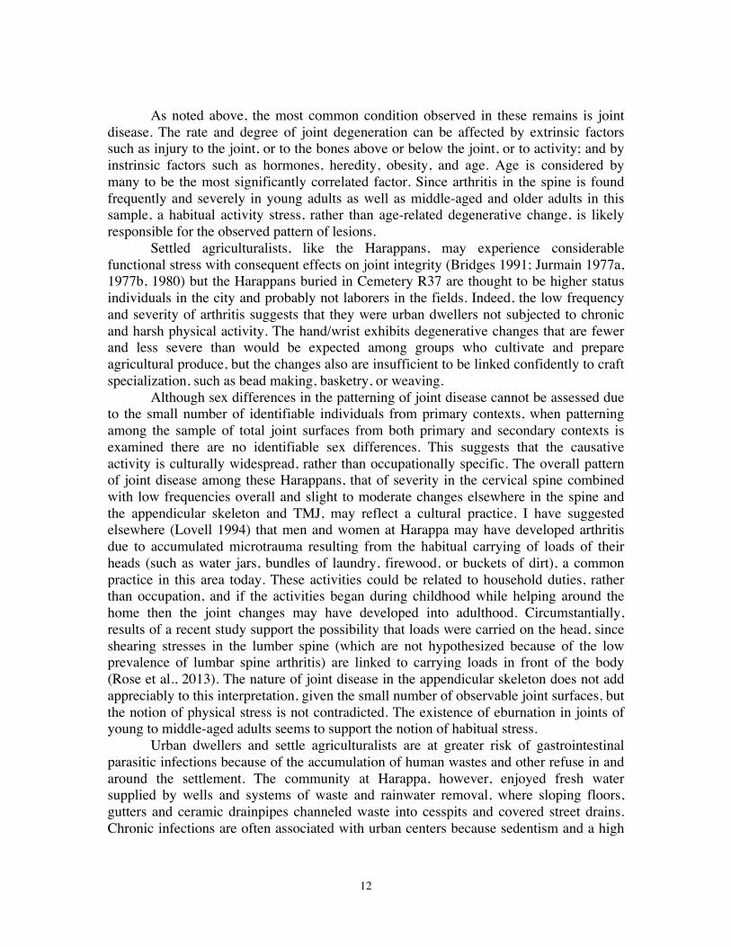
As noted above, the most common condition observed in these remains is joint disease. The rate and degree of joint degeneration can be affected by extrinsic factors such as injury to the joint, or to the bones above or below the joint, or to activity; and by instrinsic factors such as hormones, heredity, obesity, and age. Age is considered by many to be the most significantly correlated factor. Since arthritis in the spine is found frequently and severely in young adults as well as middle-aged and older adults in this sample, a habitual activity stress, rather than age-related degenerative change, is likely responsible for the observed pattern of lesions. Settled agriculturalists, like the Harappans, may experience considerable functional stress with consequent effects on joint integrity (Bridges 1991; Jurmain 1977a, 1977b, 1980) but the Harappans buried in Cemetery R37 are thought to be higher status individuals in the city and probably not laborers in the fields. Indeed, the low frequency and severity of arthritis suggests that they were urban dwellers not subjected to chronic and harsh physical activity. The hand/wrist exhibits degenerative changes that are fewer and less severe than would be expected among groups who cultivate and prepare agricultural produce, but the changes also are insufficient to be linked confidently to craft specialization, such as bead making, basketry, or weaving. Although sex differences in the patterning of joint disease cannot be assessed due to the small number of identifiable individuals from primary contexts, when patterning among the sample of total joint surfaces from both primary and secondary contexts is examined there are no identifiable sex differences. This suggests that the causative activity is culturally widespread, rather than occupationally specific. The overall pattern of joint disease among these Harappans, that of severity in the cervical spine combined with low frequencies overall and slight to moderate changes elsewhere in the spine and the appendicular skeleton and TMJ, may reflect a cultural practice. I have suggested elsewhere (Lovell 1994) that men and women at Harappa may have developed arthritis due to accumulated microtrauma resulting from the habitual carrying of loads of their heads (such as water jars, bundles of laundry, firewood, or buckets of dirt), a common practice in this area today. These activities could be related to household duties, rather than occupation, and if the activities began during childhood while helping around the home then the joint changes may have developed into adulthood. Circumstantially, results of a recent study support the possibility that loads were carried on the head, since shearing stresses in the lumber spine (which are not hypothesized because of the low prevalence of lumbar spine arthritis) are linked to carrying loads in front of the body (Rose et al., 2013). The nature of joint disease in the appendicular skeleton does not add appreciably to this interpretation, given the small number of observable joint surfaces, but the notion of physical stress is not contradicted. The existence of eburnation in joints of young to middle-aged adults seems to support the notion of habitual stress.
Urban dwellers and settle agriculturalists are at greater risk of gastrointestinal parasitic infections because of the accumulation of human wastes and other refuse in and around the settlement. The community at Harappa, however, enjoyed fresh water supplied by wells and systems of waste and rainwater removal, where sloping floors, gutters and ceramic drainpipes channeled waste into cesspits and covered street drains. Chronic infections are often associated with urban centers because sedentism and a high
13
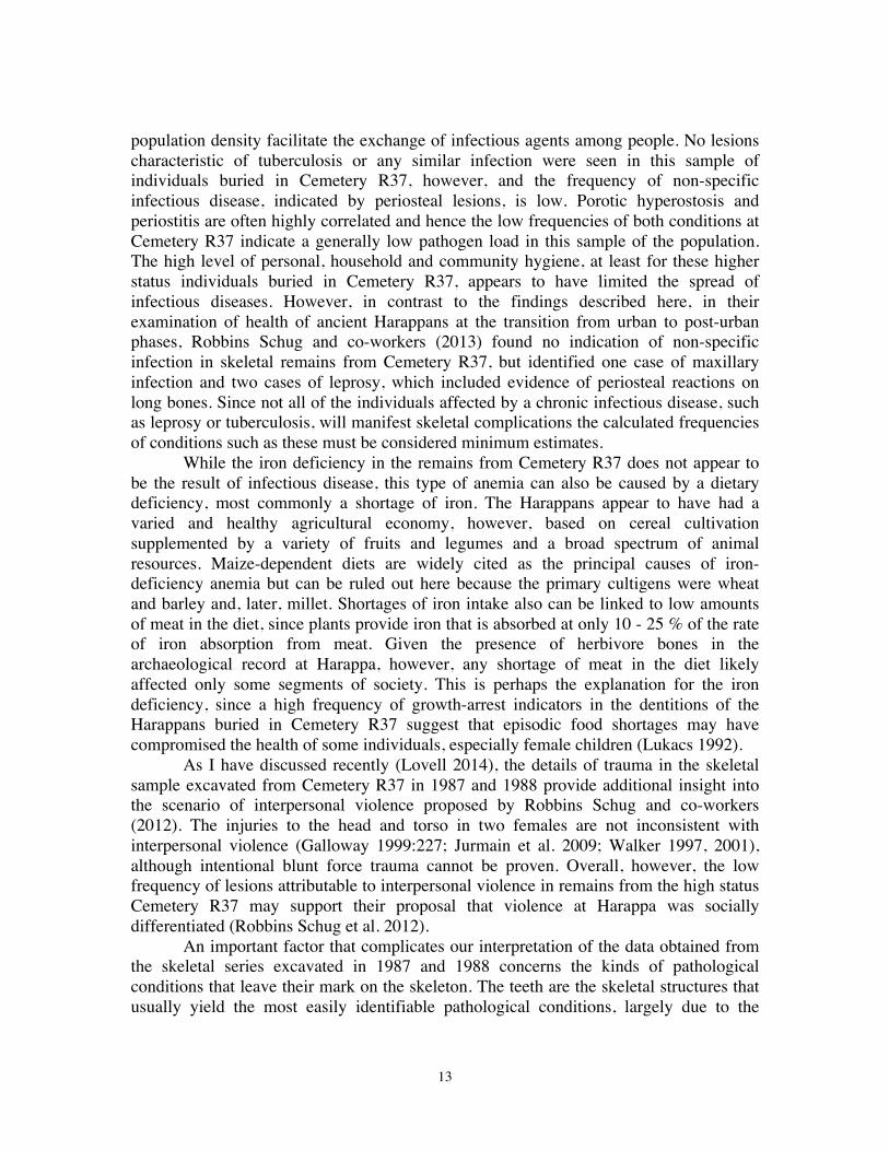
population density facilitate the exchange of infectious agents among people. No lesions characteristic of tuberculosis or any similar infection were seen in this sample of individuals buried in Cemetery R37, however, and the frequency of non-specific infectious disease, indicated by periosteal lesions, is low. Porotic hyperostosis and periostitis are often highly correlated and hence the low frequencies of both conditions at Cemetery R37 indicate a generally low pathogen load in this sample of the population. The high level of personal, household and community hygiene, at least for these higher status individuals buried in Cemetery R37, appears to have limited the spread of infectious diseases. However, in contrast to the findings described here, in their examination of health of ancient Harappans at the transition from urban to post-urban phases, Robbins Schug and co-workers (2013) found no indication of non-specific infection in skeletal remains from Cemetery R37, but identified one case of maxillary infection and two cases of leprosy, which included evidence of periosteal reactions on long bones. Since not all of the individuals affected by a chronic infectious disease, such as leprosy or tuberculosis, will manifest skeletal complications the calculated frequencies of conditions such as these must be considered minimum estimates. While the iron deficiency in the remains from Cemetery R37 does not appear to be the result of infectious disease, this type of anemia can also be caused by a dietary deficiency, most commonly a shortage of iron. The Harappans appear to have had a varied and healthy agricultural economy, however, based on cereal cultivation supplemented by a variety of fruits and legumes and a broad spectrum of animal resources. Maize-dependent diets are widely cited as the principal causes of iron-deficiency anemia but can be ruled out here because the primary cultigens were wheat and barley and, later, millet. Shortages of iron intake also can be linked to low amounts of meat in the diet, since plants provide iron that is absorbed at only 10 - 25 % of the rate of iron absorption from meat. Given the presence of herbivore bones in the archaeological record at Harappa, however, any shortage of meat in the diet likely affected only some segments of society. This is perhaps the explanation for the iron deficiency, since a high frequency of growth-arrest indicators in the dentitions of the Harappans buried in Cemetery R37 suggest that episodic food shortages may have compromised the health of some individuals, especially female children (Lukacs 1992). As I have discussed recently (Lovell 2014), the details of trauma in the skeletal sample excavated from Cemetery R37 in 1987 and 1988 provide additional insight into the scenario of interpersonal violence proposed by Robbins Schug and co-workers (2012). The injuries to the head and torso in two females are not inconsistent with interpersonal violence (Galloway 1999:227; Jurmain et al. 2009; Walker 1997, 2001), although intentional blunt force trauma cannot be proven. Overall, however, the low frequency of lesions attributable to interpersonal violence in remains from the high status Cemetery R37 may support their proposal that violence at Harappa was socially differentiated (Robbins Schug et al. 2012). An important factor that complicates our interpretation of the data obtained from the skeletal series excavated in 1987 and 1988 concerns the kinds of pathological conditions that leave their mark on the skeleton. The teeth are the skeletal structures that usually yield the most easily identifiable pathological conditions, largely due to the

14
resilience of these hard tissues in the burial environment. The types of conditions that can be diagnosed from the teeth include stresses experienced during childhood (the time of the formation of tooth crowns in the jaws) as well as the effects of tooth wear, caries, abscesses, periodontal disease, and tooth loss during the adult years. Although bones can preserve the evidence of childhood injuries and diseases, childhood conditions may be difficult to diagnose in adult skeletons because ongoing bone remodeling may erase the evidence. Bone reacts to pathological conditions by increasing or decreasing tissue formation or tissue resorption, or a combination of the two processes. The healing reaction to traumatic injury and the modifications to the skeleton resulting from various endocrine or developmental disorders produce lesions that can be identified as being outside the normal range of variation. Long-standing conditions that also produce identifiable bony reactions include arthritis, chronic infectious diseases, malnutrition, and secondary responses to soft tissue neoplasia, but many chronic diseases, such as cardiovascular disease, leave no mark on the skeleton. It is also the case that traumatic injury often affects only soft tissue, and so our reconstruction of trauma and interpersonal violence at Harappa may be woefully inadequate. One of the principal difficulties in interpreting the paleopathology data is that we excavated only a small portion of the cemetery, and the remains of perhaps more than 300 individuals (with possibly 60 complete burials in primary contexts) may lie in an unexcavated area that extends some 45 m to the west of our primary excavations, on the same north-south grid. This apparent extension of the cemetery was discovered in 1988 when testing was done to explore vegetation differences in a neighbouring farmer’s field: the test excavation uncovered two primary burials (H88/4a and 4b). Unfortunately, it may not be possible to investigate that portion of the cemetery because the plow zone indicates that the upper level of burials are probably destroyed, and not all of the area between the excavated portion of Cemetery R37 and the fields is amenable to future excavation. It is also the case that the sample of skeletal remains that we recovered in the 1987 and 1988 seasons is not the total complement of remains excavated historically from Cemetery R37. The cemetery was discovered in 1937; Sastri and Bose excavated 47 between 1937 and 1941, but only 15 were complete skeletons in primary contexts (Sastri 1965). Wheeler later excavated an additional 10 graves in 1946 (Wheeler 1947. The remains excavated between 1937 and 1946 are housed at the Anthropological Survey of India in Kolkata and are described by Gupta and co-workers (Gupta et al. 1962) and by Dutta (1983); the paleopathological conditions in those remains have been analysed by Robbins Schug and her co-workers (Robbins Schug et al. 2012, 2013). (Their results are discussed above where relevant for the material described in this report.) Mughal found an additional 11 burials in 1966 (Mughal 1968), but only one of the skeletons, the museum display skeleton, was available to us for examination at Harappa; the remaining skeletal material is believed to be stored in Karachi. It is also clear from the excavation of this cemetery that juveniles may have been treated differently from adults in death. Although not detailed in this report, the remains of eight juveniles were discovered in our 1987 and 1988 excavations, although only two were recovered from primary contexts. We might expect to see a higher frequency of
15
deaths of infants and children in an early urban settlement such as Harappa; the scarcity of juvenile remains in the cemetery could be due either to preservation problems or to mortuary practices. The excellent preservation of a perinatal skeleton (H88/194b) suggests, however, that the scarcity of juvenile remains may be attributable to mortuary practices that treated infants and children differently from adults. Although Cemetery R37 has been considered to be the burial ground of a higher status component of the Harappa population compared to individuals buried elsewhere at the site, no burials have been discovered at Harappa that exhibit a richness resembling that of the elite burials of the ancient Egyptian, Mayan, or other highly stratified societies. Thus, those buried in this cemetery may have belonged, for example, to a wealthy merchant class, rather than a ruling elite. We observed some variability in grave goods in Cemetery R37, and hence different social groups may have been sampled, but the difference was not easily defined. (A detailed analysis of the grave goods has not been published by the project archaeologists, so I have relied on my own excavation field notes and photographs.) Given the small portion of the cemetery that we excavated, however, a reconstruction of status variability from these burials is probably unwarranted. The Harappan elite may have been buried elsewhere and have not yet been discovered, or may have been accorded a different form of mortuary treatment, such as cremation. In addition to the small sample available for analysis, it is clear that the poor condition and fragmentary nature of much of the skeletal material from Harappa hinders our calculation of the prevalence of pathological lesions and their interpretation (see also Robbins Schug et al. 2013). These are factors that cannot be eliminated and stem largely from the condition of the sediments in the burial deposits as well as the re-use of the cemetery by the ancient Harappans. Although the Harappa Project team included conservators, attempts to desalinate, consolidate, and mend poorly preserved skeletal elements did not appreciably increase our sample of observable material.
16
Literature Cited
Allen, Terry, Shannon A. Novak, and Lawrence L. Bench. (2007) “Patterns of Injuries Accident or Abuse.” Violence Against Women 13, no. 8: 802–816. doi:10.1177/1077801207302040.
Barnes, Ethne. (2012) “Developmental Disorders in the Skeleton.” In A Companion to Paleopathology, edited by A. Grauer. Chichester: Wiley-Blackwell, pp 380–400.
Bass, William M. (1979) Human Osteology. 2nd edition. Columbia MO: Missouri Bridges, Patricia S. (1991) “Degenerative Joint Disease in Hunter–gatherers and
Agriculturalists from the Southeastern United States.” American Journal of Physical Anthropology 85, no. 4: 379–391. doi:10.1002/ajpa.1330850403.
Butters, K. P. (2006) “Fractures of the Scapula.” In Rockwood and Green’s Fractures in Adults, edited by R.W. Bucholz, J. D. Heckman, and C. Court-Brown. Philadelphia: Lippincott Williams and Wilkins, pp 1257–1282.
Dales, G. F., J. Mark Kenoyer, and the staff of the Harappa Project. (1991) “Summaries of Five Seasons of Research at Harappa (District Sahiwal, Punjab, Pakistan) 1986-1990.” In Harappa Excavations 1986-1990: A Multidisciplinary Approach to Third Millennium Urbanism, edited by Meadow, Richard H, Monographs in World Archaeology 3. Madison WI: Prehistory Press, pp 185–262.
Dutta, P. C. (1983) The Bronze Age Harappans. Calcutta: Anthropological Survey of India.
Galloway, A. (1999) Broken Bones: Anthropological Analysis of Blunt Force Trauma. Springfield IL: CC Thomas.
Gasperetti, Matthew Alexander, and Susan Guise Sheridan. (2013) “Cry Havoc: Interpersonal Violence at Early Bronze Age Bab edh-Dhra’.” American Anthropologist 115, no. 3: 388–410. doi:10.1111/aman.12024.
Gupta, P., P. C. Dutta, and A. Basu. (1962) Human Skeletal Remains from Harappa. Memoirs 9. Calcutta: Anthropological Survey of India.
Hemphill, B.E., and J.R. Lukacs. (1993) “Hegelian Logic and Harappan Civilization: An Investigation of Harappan Biological Affinities in the Light of Recent Biological and Archaeological Research.” In South Asian Archaeology 1991, edited by Gail, A.J. and Mevissen G.J.R. Stuttgart: Franz Steiner Verlag, pp 101–120.
Hemphill, B.E., J.R. Lukacs, and K.A.R. Kennedy. (1991) “Biological Adaptations and Affinities of Bronze Age Harappans.” In Harappa Excavations 1986-1990: A Multidisciplinary Approach to Third Millennium Urbanism, edited by Meadow, Richard H. Monographs in World Archaeology 3. Madison WI: Prehistory Press, pp 137-182.
17
Hodges, Denise C. (1991) “Temporomandibular Joint Osteoarthritis in a British Skeletal Population.” American Journal of Physical Anthropology 85, no. 4: 367–377. doi:10.1002/ajpa.1330850402.
Jeanmonod, Rebecca K., Donald Jeanmonod, Sara Damewood, Cheryl Perry, Marwan Powers, and Vicky Lazansky. (2011) “Punch Injuries: Insights into Intentional Closed Fist Injuries.” Western Journal of Emergency Medicine 12, no. 1: 6–10.
Jurmain, Robert D. (1977a) “Part One: Paleoepidemiology of Degenerative Knee Disease.” Medical Anthropology 1, no. 1: 1–23. doi:10.1080/01459740.1977.9965814.
Jurmain, Robert D. (1977b) “Stress and the Etiology of Osteoarthritis.” American Journal of Physical Anthropology 46, no. 2: 353–365. doi:10.1002/ajpa.1330460214.
Jurmain, Robert D. (1980) “The Pattern of Involvement of Appendicular Degenerative Joint Disease.” American Journal of Physical Anthropology 53, no. 1: 143–150. doi:10.1002/ajpa.1330530119.
Jurmain, Robert, Eric J. Bartelink, Alan Leventhal, Viviana Bellifemine, Irina Nechayev, Melynda Atwood, and Diane DiGiuseppe. (2009) “Paleoepidemiological Patterns of Interpersonal Aggression in a Prehistoric Central California Population from CA-ALA-329.” American Journal of Physical Anthropology 139, no. 4: 462–473. doi:10.1002/ajpa.21002.
Kennedy, K. A. R., Nancy C. Lovell, J. R. Lukacs, and B. E. Hemphill. (1993) “Scaphocephaly in a Prehistoric Skeleton from Harappa, Pakistan.” Anthropologischer Anzeiger 51, no. 1: 1–29.
Kenoyer, J. Mark (1991) “The Indus Valley Tradition of Pakistan and Western India.” Journal of World Prehistory 5: 331–85.
Kenoyer, J. Mark. (1998) Ancient Cities of the Indus Civilization. Karachi: Oxford University Press.
Kenoyer, J. Mark (2003) “Uncovering the Keys to the Lost Indus Cities.” Scientific American July: 67–75.
Kenoyer, J. Mark, and Meadow, Richard H. (2000) “The Ravi Phase: a New Cultural Manifestation at Harappa.” In South Asian Archaeology 1997, edited by M Taddei and G De Marco, 1: 55–76. Rome/Naples: IsIAO/IUO
Kenoyer, J. Mark, T. Douglas Price, and James H. Burton. (2013) “A New Approach to Tracking Connections Between the Indus Valley and Mesopotamia: Initial Results of Strontium Isotope Analyses from Harappa and Ur.” Journal of Archaeological Science 40, no. 5: 2286–2297. doi:10.1016/j.jas.2012.12.040.
Kimmerle, EH, and JP Baraybar. (2008) Skeletal Trauma: Identification of Injuries Resulting from Human Right Abuse and Armed Conflict. Boca Raton, FL: CRC Press.

18
Laugharne, Edward, Dhruvar Bhavsar, and Vaikunthan Rajaratnam. (2013) “The Distribution of Hand Fractures: a British Perspective.” European Journal of Plastic Surgery 36, no. 6: 367–370. doi:10.1007/s00238-012-0775-2.
Link, D. W., and Nancy C Lovell. (1994) “Characterization of Postmortem Change in Archaeological Bone from Harappa, Pakistan.” In From Sumer to Meluhha: Contributions to the Archaeology of South and West Asia in Memory of George F Dales, Jr., edited by J. Mark Kenoyer, Wisconsin Archaeological Reports 3. Madison WI: University of Wisconsin, pp 151-171.
Lovejoy, C. Owen, Richard S. Meindl, Thomas R. Pryzbeck, and Robert P. Mensforth. (1985) “Chronological Metamorphosis of the Auricular Surface of the Ilium: A New Method for the Determination of Adult Skeletal Age at Death.” American Journal of Physical Anthropology 68, no. 1: 15–28. doi:10.1002/ajpa.1330680103.
Lovell, Nancy C. (1994) “Spinal Arthritis and Physical Stress at Bronze Age Harappa.” American Journal of Physical Anthropology 93, no. 2: 149–164. doi:10.1002/ajpa.1330930202.
Lovell, Nancy C. (1997) “Anemia in the Ancient Indus Valley.” International Journal of Osteoarchaeology 7, no. 2: 115-123.
Lovell, Nancy C. (2008) “Analysis and Interpretation of Skeletal Trauma.” In Biological Anthropology of the Human Skeleton, edited by MA Katzenberg and Shelley Rae Saunders, 341–386. New York, NY: John Wiley and Sons, 2008.
Lovell, Nancy C. (2014) “Additional Data on Trauma at Harappa.” International Journal of Paleopathology 6: 1–4. doi:10.1016/j.ijpp.2014.01.002.
Lukacs, J.R., and B.E. Hemphill. (1990) “Traumatic Injuries of Prehistoric Teeth: New Evidence from Baluchistan and Punjab Provinces, Pakistan.” Anthropologischer Anzeiger 48, no. 4: 351–363.
Lukacs, John R. (1992) “Dental Paleopathology and Agricultural Intensification in South Asia: New Evidence from Bronze Age Harappa.” American Journal of Physical Anthropology 87, no. 2: 133–150. doi:10.1002/ajpa.1330870202.
Lukacs, John R., and Robert F. Pastor. (1988) “Activity-induced Patterns of Dental Abrasion in Prehistoric Pakistan: Evidence from Mehrgarh and Harappa.” American Journal of Physical Anthropology 76, no. 3: 377–398. doi:10.1002/ajpa.1330760310.
Meadow, Richard H, and J. Mark Kenoyer. (1997) “Excavations at Harappa 1994-1995: New Perspectives on the Indus Script, Craft Activities, and City Organization.” In South Asian Archaeology 1995, edited by B Allchin and F R Allchin, New Delhi: Oxford and IBH, pp 139–172.
Meadow, Richard H, and J. Mark Kenoyer. (1999) “Harappa Excavations 1998-1999: New Evidence for the Development and Manifestation of the Harappan

19
Phenomenon.” In South Asian Archaeology 1999, edited by EM Raven. Groningen: Egbert Forsten, pp 85–109.
Meadow, Richard H, and J. Mark Kenoyer. (2005) “Excavations at Harappa 2000-2001: New Insights on Chronology and City Organization.” In South Asian Archaeology 2001, edited by C Jarrige and V Lefèvre, Paris: Editions Recherche sur les Civilisations, pp 207–224.
Meindl, Richard S., and C. Owen Lovejoy. (1985) “Ectocranial Suture Closure: A Revised Method for the Determination of Skeletal Age at Death Based on the Lateral-anterior Sutures.” American Journal of Physical Anthropology 68, no. 1: 57–66. doi:10.1002/ajpa.1330680106.
Mughal, R. (1968) “Harappa 1966 (Cemetery R37).” Pakistan Archaeology 5: 63–68. Nicholson, A. A., G. M. Roberts, and L. A. Williams. (1988) “The Measured Height of
the Lumbosacral Disc in Patients with and Without Transitional Vertebrae.” British Journal of Radiology 61, no. 726: 454–455. doi:10.1259/0007-1285-61-726-454.
Ortner, D. J., and W. G. J. Putschar. (1985) Identification of Pathological Conditions in Human Skeletal Remains. Washington DC: Smithsonian Institution Press.
Ortner, Donald J. (2003) Identification of Pathological Conditions in Human Skeletal Remains. 2nd edition. San Diego, CA: Academic Press.
Robbins Schug, Gwen, K. Elaine Blevins, Brett Cox, Kelsey Gray, and V. Mushrif-Tripathy. (2013) “Infection, Disease, and Biosocial Processes at the End of the Indus Civilization.” PLoS ONE 8, no. 12: e84814. doi:10.1371/journal.pone.0084814.
Robbins Schug, Gwen, Kelsey Gray, V. Mushrif-Tripathy, and A. R. Sankhyan. (2012) “A Peaceful Realm? Trauma and Social Differentiation at Harappa.” International Journal of Paleopathology, 2, no. 2/3: 136–147. doi:10.1016/j.ijpp.2012.09.012.
Rose, J. D., Mendel, E., and Marras, W. S. (2013) “Carrying and Spine Loading.” Ergonomics 56, no. 11: 1722-732. doi10.1080/00140139.2013.835870.
Sastri, K. N. (1965) New Light on the Indus Civilization. Vol. 2. Delhi: Atma Ram and Sons.
Sheridan, Susan G., Diane M. Mittler, Dennis P. Van Gerven, and Herbert H. Covert. (1991) “Biomechanical Association of Dental and Temporomandibular Pathology in a Medieval Nubian Population.” American Journal of Physical Anthropology 85, no. 2: 201–205. doi:10.1002/ajpa.1330850208.
Smith, B. Holly. (1984) “Patterns of Molar Wear in Hunter–gatherers and Agriculturalists.” American Journal of Physical Anthropology 63, no. 1: 39–56. doi:10.1002/ajpa.1330630107.

20
Tague, Robert G. (2011) “Sacralization Is Not Associated with Elongated Cervical Costal Process and Cervical Rib.” Clinical Anatomy 24, no. 2: 209–17. doi:10.1002/ca.21087.
Tung, TA. (2012) “Violence Against Women: Differential Treatment of Local Vs Foreign Females in the Heartland of the Wari Empire, Peru.” In The Bioarchaeology of Violence, edited by Debra L. Martin, RP Harrod, and VR Perez. Gainesville, Fla.: The University Press of Florida, pp 180–98.
Ubelaker, Douglas H. (1984) Human Skeletal Remains: Excavation, Analysis, Interpretation. Taraxacum.
Walker, Phillip L. (1997) “Wife Beating, Boxing, and Broken Noses: Skeletal Evidence for the Cultural Patterning of Violence.” In Troubled Times: Violence and Warfare in the Past, edited by Debra L. Martin and David W. Frayer. Boca Raton, FL: Gordon and Breach, pp 145–79.
Walker, Phillip L. (2001) “A Bioarchaeological Perspective on the History of Violence.” Annual Review of Anthropology 30: 573–96.
Weber, Steven A. (2003) “Archaeobotany at Harappa: Indications for Change.” In Indus Ethnobiology: New Perspectives from the Field, edited by Steven A. Weber and William R. Belcher. USA: Lexington Books, pp 175–98.
Wheeler, R. E. M. (1947) “Harappa 1946: The Defences and Cemetery R 37.” Ancient India 3: 58–130.
Wright, Rita P. (2010) The Ancient Indus : Urbanism, Economy, and Society. New York : Cambridge University Press.
21
Appendix: Details of Pathological Lesions in Adult Remains
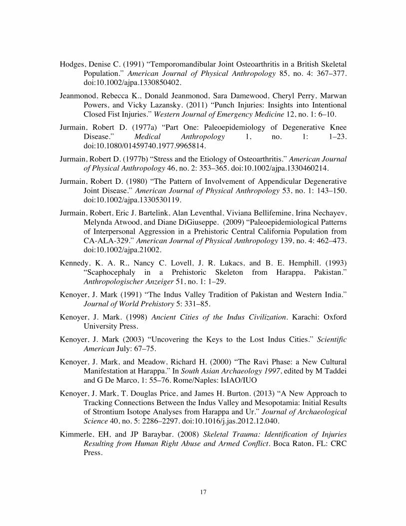
H87/34b
This feature represents the upper body including cranium (Fig. 14) of an adult male, aged approximately 30 to 35 years of age, in a primary context. The only pathological condition observed is slight osteoarthritis: porosity and marginal lipping of the vertebral ends of two ribs, affecting the head of one and the tubercle of the other.
Figure 14. Frontal view of the cranium of an adult male (H87/34b).
22
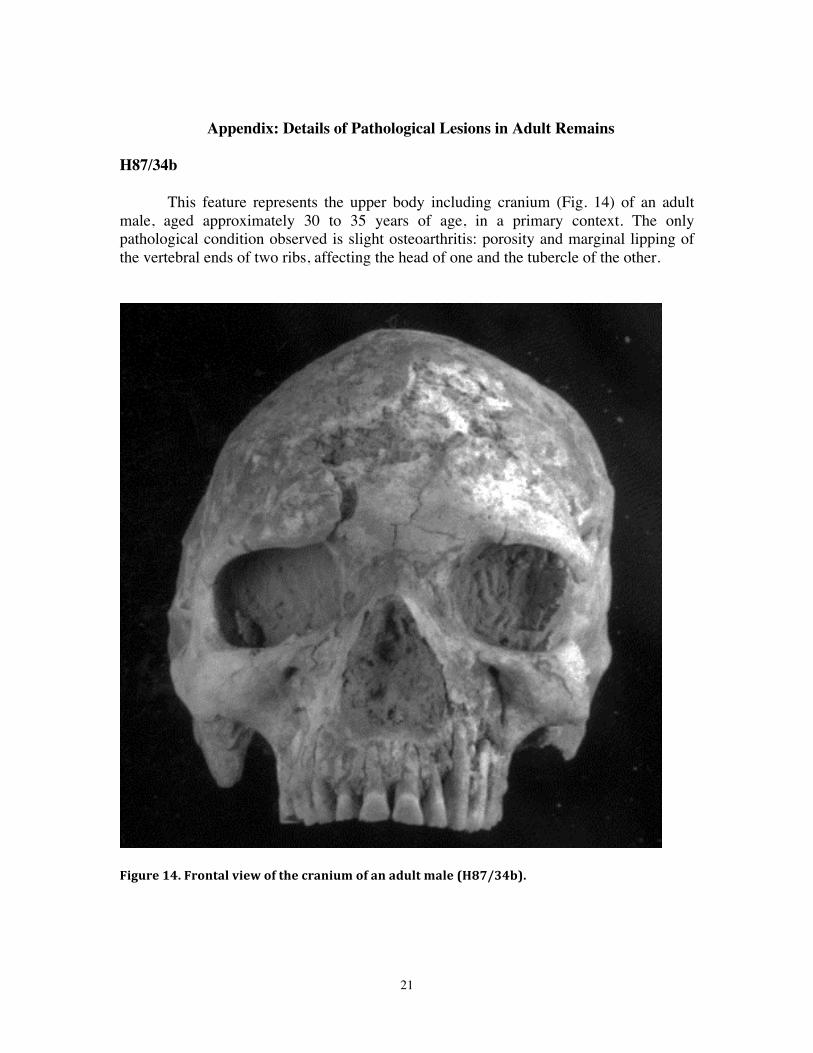
H87/49b
These are miscellaneous fragmentary remains, including a skull (Fig. 15). The second proximal phalanx of the right foot displays an oval proliferative lesion, indicative of localized inflammation, on the dorsal-medial surface of the proximal half of the shaft. The lesion is approximately 11 mm long and 6 mm wide, and has raised margins (see Fig. 11).
Figure 15. Frontal view of an adult skull from a deposit of fragmentary remains (H87/49b).
23
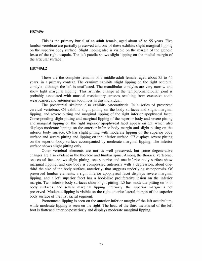
H87/49c
This is the primary burial of an adult female, aged about 45 to 55 years. Five lumbar vertebrae are partially preserved and one of these exhibits slight marginal lipping on the superior body surface. Slight lipping also is visible on the margin of the glenoid fossa of the right scapula. The left patella shows slight lipping on the medial margin of the articular surface. H87/49d.2
These are the complete remains of a middle-adult female, aged about 35 to 45 years, in a primary context. The cranium exhibits slight lipping on the right occipital condyle, although the left is unaffected. The mandibular condyles are very narrow and show light marginal lipping. This arthritic change at the temporomandibular joint is probably associated with unusual masticatory stresses resulting from excessive tooth wear, caries, and antemortem tooth loss in this individual. The postcranial skeleton also exhibits osteoarthritis. In a series of preserved cervical vertebrae, C4 exhibits slight pitting on the body surfaces and slight marginal lipping, and severe pitting and marginal lipping of the right inferior apophyseal facet. Corresponding slight pitting and marginal lipping of the superior body and severe pitting and marginal lipping on the right superior apophyseal facet appear on C5, which also displays moderate lipping on the anterior inferior body margin and slight pitting on the inferior body surface. C6 has slight pitting with moderate lipping on the superior body surface and severe pitting and lipping on the inferior surface. C7 displays severe pitting on the superior body surface accompanied by moderate marginal lipping. The inferior surface shows slight pitting only. Other vertebral elements are not as well preserved, but some degenerative changes are also evident in the thoracic and lumbar spine. Among the thoracic vertebrae, one costal facet shows slight pitting, one superior and one inferior body surface show marginal lipping, and one body is compressed anteriorly with a depression, about one-third the size of the body surface, anteriorly, that suggests underlying osteoporosis. Of preserved lumbar elements, a right inferior apophyseal facet displays severe marginal lipping, and a left superior facet has a hook-like proliferative lesion on the inferior margin. Two inferior body surfaces show slight pitting. L5 has moderate pitting on both body surfaces, and severe marginal lipping inferiorly; the superior margin is not preserved. Moderate lipping is visible on the right anterior-lateral margin of the superior body surface of the first sacral segment. Pronounced lipping is seen on the anterior-inferior margin of the left acetabulum, while moderate lipping is seen on the right. The head of the third metatarsal of the left foot is flattened anterior-posteriorly and displays moderate marginal lipping.

24
H87/49f
This is a collection of highly fragmentary miscellaneous human and nonhuman bone. One small cranial fragment displays “pin-prick” porosity that may indicate porotic hyperostosis (Lovell 1997: Fig. 2). H87/49g
This is a primary burial of a middle-adult female. Superior and inferior lumbar apophyseal articular facets show moderate lipping. H87/49h
This feature is a complete primary burial of a male aged about 30 years. Only the vertebrae exhibit pathological lesions: one lumbar body surface shows moderate lipping on the anterior margin, and C3, C4, and C5 also show arthritic change. C3 and C4 are ankylosed on the left side at the articular facets, and there is moderate lipping too on the right side, with incipient fusion. The left inferior articular facet of C4 has an irregular surface and lipping. This is matched on the left superior facet on C5. The right superior articular facet of C5 also shows slight lipping. H87/79a
This is a collection of miscellaneous fragments. One incomplete thoracic vertebra exhibits osteophyte development on the left inferior margin of the body, immediately adjacent to the inferior costal facet. H87/90a
These are small fragments of bone in a commingled context. One mid-thoracic vertebra shows slight osteoarthritic porosity at the costal facets, and shallow Schmorl’s nodes inferiorly. H87/93b
This feature consists only of small fragments of bone. One right rib exhibits a well-healed fracture of the shaft, with altered curvature (see Fig. 8a, b). H87/127a
This is a primary burial of the complete skeleton of a middle-adult female. The vertebral elements exhibit slight to severe degrees of osteoarthritis, on both body margins and apophyseal facets. The dens facet on C1 shows moderate lipping. There is slight lipping on the margins of the inferior body surface of C2 and on the superior and inferior
25
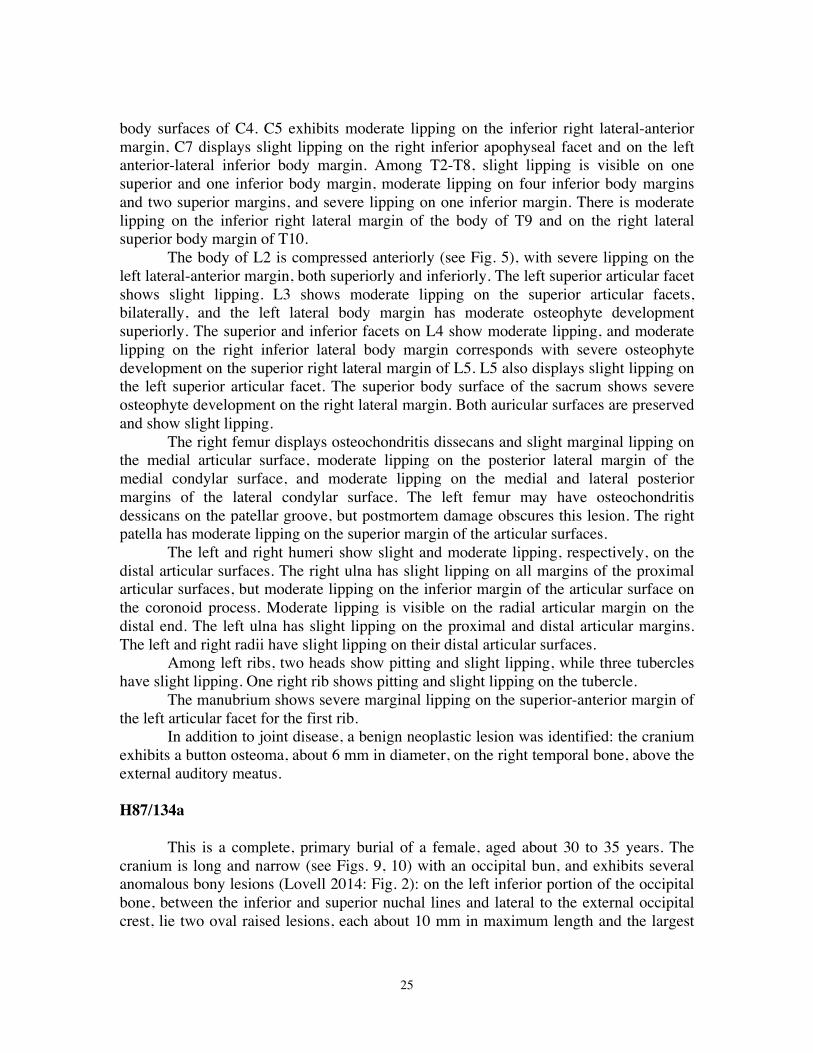
body surfaces of C4. C5 exhibits moderate lipping on the inferior right lateral-anterior margin, C7 displays slight lipping on the right inferior apophyseal facet and on the left anterior-lateral inferior body margin. Among T2-T8, slight lipping is visible on one superior and one inferior body margin, moderate lipping on four inferior body margins and two superior margins, and severe lipping on one inferior margin. There is moderate lipping on the inferior right lateral margin of the body of T9 and on the right lateral superior body margin of T10. The body of L2 is compressed anteriorly (see Fig. 5), with severe lipping on the left lateral-anterior margin, both superiorly and inferiorly. The left superior articular facet shows slight lipping. L3 shows moderate lipping on the superior articular facets, bilaterally, and the left lateral body margin has moderate osteophyte development superiorly. The superior and inferior facets on L4 show moderate lipping, and moderate lipping on the right inferior lateral body margin corresponds with severe osteophyte development on the superior right lateral margin of L5. L5 also displays slight lipping on the left superior articular facet. The superior body surface of the sacrum shows severe osteophyte development on the right lateral margin. Both auricular surfaces are preserved and show slight lipping. The right femur displays osteochondritis dissecans and slight marginal lipping on the medial articular surface, moderate lipping on the posterior lateral margin of the medial condylar surface, and moderate lipping on the medial and lateral posterior margins of the lateral condylar surface. The left femur may have osteochondritis dessicans on the patellar groove, but postmortem damage obscures this lesion. The right patella has moderate lipping on the superior margin of the articular surfaces. The left and right humeri show slight and moderate lipping, respectively, on the distal articular surfaces. The right ulna has slight lipping on all margins of the proximal articular surfaces, but moderate lipping on the inferior margin of the articular surface on the coronoid process. Moderate lipping is visible on the radial articular margin on the distal end. The left ulna has slight lipping on the proximal and distal articular margins. The left and right radii have slight lipping on their distal articular surfaces. Among left ribs, two heads show pitting and slight lipping, while three tubercles have slight lipping. One right rib shows pitting and slight lipping on the tubercle. The manubrium shows severe marginal lipping on the superior-anterior margin of the left articular facet for the first rib. In addition to joint disease, a benign neoplastic lesion was identified: the cranium exhibits a button osteoma, about 6 mm in diameter, on the right temporal bone, above the external auditory meatus. H87/134a
This is a complete, primary burial of a female, aged about 30 to 35 years. The cranium is long and narrow (see Figs. 9, 10) with an occipital bun, and exhibits several anomalous bony lesions (Lovell 2014: Fig. 2): on the left inferior portion of the occipital bone, between the inferior and superior nuchal lines and lateral to the external occipital crest, lie two oval raised lesions, each about 10 mm in maximum length and the largest
26
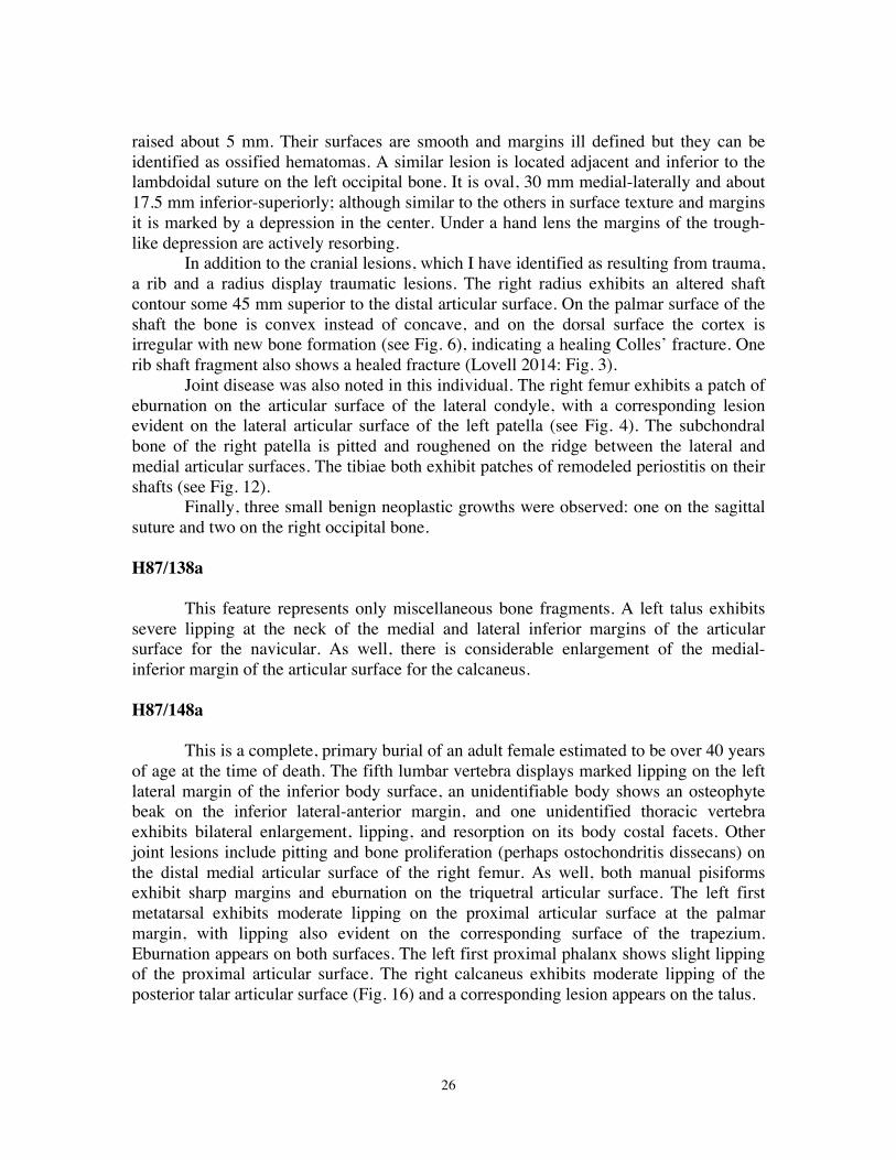
raised about 5 mm. Their surfaces are smooth and margins ill defined but they can be identified as ossified hematomas. A similar lesion is located adjacent and inferior to the lambdoidal suture on the left occipital bone. It is oval, 30 mm medial-laterally and about 17.5 mm inferior-superiorly; although similar to the others in surface texture and margins it is marked by a depression in the center. Under a hand lens the margins of the trough-like depression are actively resorbing. In addition to the cranial lesions, which I have identified as resulting from trauma, a rib and a radius display traumatic lesions. The right radius exhibits an altered shaft contour some 45 mm superior to the distal articular surface. On the palmar surface of the shaft the bone is convex instead of concave, and on the dorsal surface the cortex is irregular with new bone formation (see Fig. 6), indicating a healing Colles’ fracture. One rib shaft fragment also shows a healed fracture (Lovell 2014: Fig. 3). Joint disease was also noted in this individual. The right femur exhibits a patch of eburnation on the articular surface of the lateral condyle, with a corresponding lesion evident on the lateral articular surface of the left patella (see Fig. 4). The subchondral bone of the right patella is pitted and roughened on the ridge between the lateral and medial articular surfaces. The tibiae both exhibit patches of remodeled periostitis on their shafts (see Fig. 12). Finally, three small benign neoplastic growths were observed: one on the sagittal suture and two on the right occipital bone. H87/138a
This feature represents only miscellaneous bone fragments. A left talus exhibits severe lipping at the neck of the medial and lateral inferior margins of the articular surface for the navicular. As well, there is considerable enlargement of the medial-inferior margin of the articular surface for the calcaneus. H87/148a
This is a complete, primary burial of an adult female estimated to be over 40 years of age at the time of death. The fifth lumbar vertebra displays marked lipping on the left lateral margin of the inferior body surface, an unidentifiable body shows an osteophyte beak on the inferior lateral-anterior margin, and one unidentified thoracic vertebra exhibits bilateral enlargement, lipping, and resorption on its body costal facets. Other joint lesions include pitting and bone proliferation (perhaps ostochondritis dissecans) on the distal medial articular surface of the right femur. As well, both manual pisiforms exhibit sharp margins and eburnation on the triquetral articular surface. The left first metatarsal exhibits moderate lipping on the proximal articular surface at the palmar margin, with lipping also evident on the corresponding surface of the trapezium. Eburnation appears on both surfaces. The left first proximal phalanx shows slight lipping of the proximal articular surface. The right calcaneus exhibits moderate lipping of the posterior talar articular surface (Fig. 16) and a corresponding lesion appears on the talus.

27
The left scapula has a healed fracture of the axillary border, just inferior to the glenoid fossa (see Fig. 7), and of the body (and see Lovell 2014: Fig. 4). The callus is well remodeled but an irregular contour and angular deformity of the border are evident. The glenoid articular surface does not show any subsequent osteoarthritis. The right 4th metatarsal has a rectangular ivory bone growth on the dorsal-lateral margin, approximately 10 mm in the proximal-distal axis and 3 mm in the dorso-plantar plane (see Fig. 13). Since this is an unlikely location for a trauma-induced ossified haematoma, I have classified it as a benign osteoma. H87/156a
This is a complete, primary burial of a female, aged approximately 25 to 30 years at the time of death. The remains were slightly scattered, but are of one individual. A circular depressed feature on the frontal bone, above the left orbit, may represent a crush fracture resulting from a blow to the head (Fig. 17).
Figure 16. Marginal lipping of articular surfaces on the calcaneus of an adult female (H87/148a).

28
H88/4a
This is the primary extended burial of a female, aged approximately 20 to 25 years. The only pathological condition observed is marginal lipping of the articular surface for C1 on the odontoid process of C2. H88/4b
This feature represents a male, aged approximately 35 to 40 years; the skeleton is flexed at the waist, with the head resting on the knees and the joints articulated. This may represent a secondary burial prior to decomposition of the articulating soft tissue, or a primary burial in a grave pit that was not dug to size for this individual. The vertebrae exhibit slight lipping on the anterior margin of the superior body surfaces of T6, T7, T10, and T11, and on the inferior body surfaces of T4, T6, T9, and T10. L3 and L4 display moderate lipping on the anterior margins of the superior and inferior body surfaces. L5 shows slight lipping on the anterior margin of the superior body surface. There is slight lipping on the glenoid margin of the right scapula, but the corresponding humerus shows no pathological lesions. The left femur shows a small (5 mm diameter) lesion of osteochondritis dissecans on the distal lateral condyle, dorsal surface. The pisiform of the left hand shows slight lipping.
Figure 17. Shallow depression, indicative of a crush fracture, over the left orbit in a young adult female (H87/156a).
29
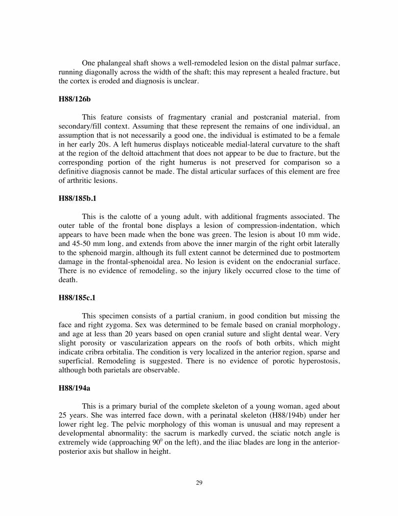
One phalangeal shaft shows a well-remodeled lesion on the distal palmar surface, running diagonally across the width of the shaft; this may represent a healed fracture, but the cortex is eroded and diagnosis is unclear. H88/126b
This feature consists of fragmentary cranial and postcranial material, from secondary/fill context. Assuming that these represent the remains of one individual, an assumption that is not necessarily a good one, the individual is estimated to be a female in her early 20s. A left humerus displays noticeable medial-lateral curvature to the shaft at the region of the deltoid attachment that does not appear to be due to fracture, but the corresponding portion of the right humerus is not preserved for comparison so a definitive diagnosis cannot be made. The distal articular surfaces of this element are free of arthritic lesions. H88/185b.1
This is the calotte of a young adult, with additional fragments associated. The outer table of the frontal bone displays a lesion of compression-indentation, which appears to have been made when the bone was green. The lesion is about 10 mm wide, and 45-50 mm long, and extends from above the inner margin of the right orbit laterally to the sphenoid margin, although its full extent cannot be determined due to postmortem damage in the frontal-sphenoidal area. No lesion is evident on the endocranial surface. There is no evidence of remodeling, so the injury likely occurred close to the time of death. H88/185c.1
This specimen consists of a partial cranium, in good condition but missing the face and right zygoma. Sex was determined to be female based on cranial morphology, and age at less than 20 years based on open cranial suture and slight dental wear. Very slight porosity or vascularization appears on the roofs of both orbits, which might indicate cribra orbitalia. The condition is very localized in the anterior region, sparse and superficial. Remodeling is suggested. There is no evidence of porotic hyperostosis, although both parietals are observable. H88/194a
This is a primary burial of the complete skeleton of a young woman, aged about 25 years. She was interred face down, with a perinatal skeleton (H88/194b) under her lower right leg. The pelvic morphology of this woman is unusual and may represent a developmental abnormality: the sacrum is markedly curved, the sciatic notch angle is extremely wide (approaching 900 on the left), and the iliac blades are long in the anterior-posterior axis but shallow in height.
30
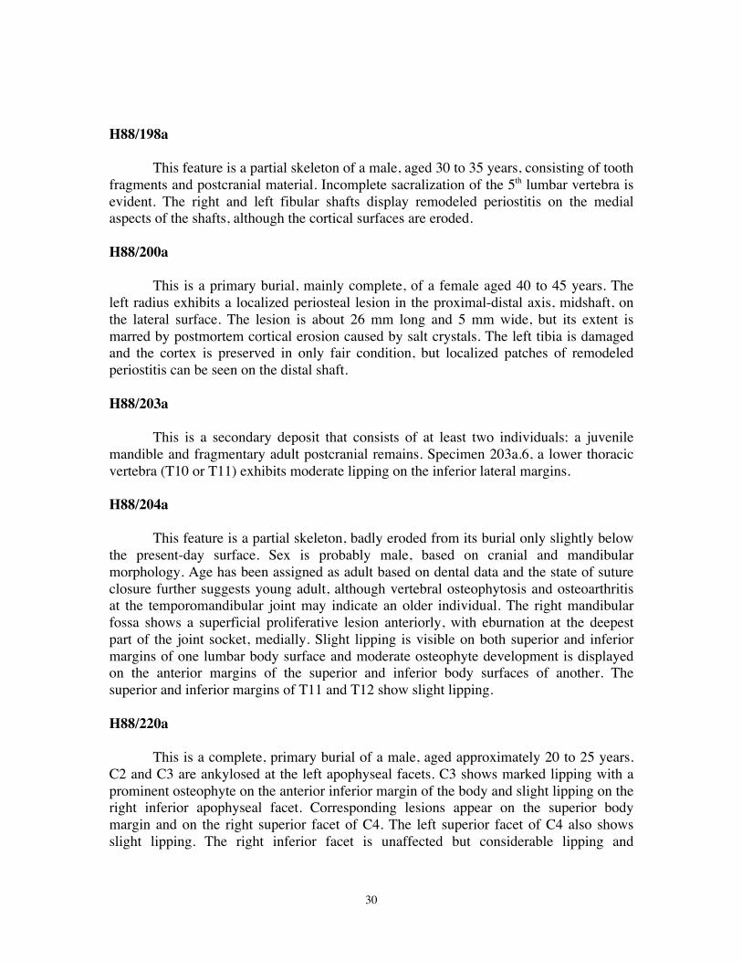
H88/198a
This feature is a partial skeleton of a male, aged 30 to 35 years, consisting of tooth fragments and postcranial material. Incomplete sacralization of the 5th lumbar vertebra is evident. The right and left fibular shafts display remodeled periostitis on the medial aspects of the shafts, although the cortical surfaces are eroded. H88/200a
This is a primary burial, mainly complete, of a female aged 40 to 45 years. The left radius exhibits a localized periosteal lesion in the proximal-distal axis, midshaft, on the lateral surface. The lesion is about 26 mm long and 5 mm wide, but its extent is marred by postmortem cortical erosion caused by salt crystals. The left tibia is damaged and the cortex is preserved in only fair condition, but localized patches of remodeled periostitis can be seen on the distal shaft. H88/203a
This is a secondary deposit that consists of at least two individuals: a juvenile mandible and fragmentary adult postcranial remains. Specimen 203a.6, a lower thoracic vertebra (T10 or T11) exhibits moderate lipping on the inferior lateral margins. H88/204a
This feature is a partial skeleton, badly eroded from its burial only slightly below the present-day surface. Sex is probably male, based on cranial and mandibular morphology. Age has been assigned as adult based on dental data and the state of suture closure further suggests young adult, although vertebral osteophytosis and osteoarthritis at the temporomandibular joint may indicate an older individual. The right mandibular fossa shows a superficial proliferative lesion anteriorly, with eburnation at the deepest part of the joint socket, medially. Slight lipping is visible on both superior and inferior margins of one lumbar body surface and moderate osteophyte development is displayed on the anterior margins of the superior and inferior body surfaces of another. The superior and inferior margins of T11 and T12 show slight lipping. H88/220a
This is a complete, primary burial of a male, aged approximately 20 to 25 years. C2 and C3 are ankylosed at the left apophyseal facets. C3 shows marked lipping with a prominent osteophyte on the anterior inferior margin of the body and slight lipping on the right inferior apophyseal facet. Corresponding lesions appear on the superior body margin and on the right superior facet of C4. The left superior facet of C4 also shows slight lipping. The right inferior facet is unaffected but considerable lipping and
31
eburnation appears on the left inferior facet. The anterior margin of the inferior surface of the C4 body shows considerable lipping, which is matched on the superior margin of the body of C5. C5 also exhibits considerable lipping on the inferior body margin. The left superior facet shows considerable lipping corresponding to that on the inferior apophyseal facet of C4. Of cervical vertebral fragments, two partial bodies show moderate lipping. There is slight lipping on the right superior articular facet of L2, and some resorption and proliferation on the right inferior facet. All four articular facets of L3 are preserved; the right superior and inferior facets show moderate lipping and proliferative lesions mid-surface. On L4, slight proliferative lesions are visible on the superior articular facets and a resorptive lesion is seen on the right inferior facet. The fifth lumbar vertebra exhibits slight lipping on the anterior superior margin of the body. Unidentifiable vertebral fragments include three articular facets, probably lumbar, that show slight resorption and lipping. The right and left patellae display pitting on the medial articular surfaces. Salt granules probably exacerbate the extent of these lesions. A sesamoid of the right great toe shows lipping and eburnation. The third medial phalanx of the right foot exhibits lipping at the base, as does the first distal phalanx. The left first distal phalanx displays a healed fracture with extensive post-traumatic lipping at the base. H88/232a
This is a partial skeleton in primary context. Only the lower half of the skeleton remains, the upper part being cut into by a later pit. The only pathological condition observable is well-remodeled periostitis on the shafts of the right tibia and left and right fibulae. Museum Skeleton (1966)
This is the complete skeleton of a male with an estimated age between 25 and 35 years, which rests in a display case in the Harappa Museum. It was excavated from Cemetery R37 in 1966 by Dr. R. Mughal (Mughal, 1968). The right fibula shows localized thickening, characteristic of remodeled periostitis, in the midshaft region.